PRODUCT
Summary
TNF alpha antibody recognizes tumor necrosis factor alpha protein, a cytokine with a predicted molecular weight of 26 kDa. TNF alpha is mainly secreted by macrophages, but is also produced by various immune cells such as neutrophils, T helper cells, and NK cells. Dysregulation of TNF alpha expression is associated with various diseases including cancers and autoimmune disorders.
APPLICATION
Application Note
*Optimal dilutions/concentrations should be determined by the researcher.
Application |
Recommended Dilution |
1:500-1:3000 |
1:100-1:1000 |
1:100-1:1000 |
Assay dependent |
Not tested in other applications.
Calculated MW
Positive Control
NCI-H929 , C8D30 , PC-12 , Rat-2 , Raji , TNF-transfected 293T
Predict Reactivity
Cat, Dog, Pig, Chimpanzee, Rhesus Monkey(>80% identity)
PROPERTIES
Form
Liquid
Buffer
PBS, 1% BSA, 20% Glycerol
Preservative
0.025% ProClin 300
Storage
Store as concentrated solution. Centrifuge briefly prior to opening vial. For short-term storage (1-2 weeks), store at 4ºC. For long-term storage, aliquot and store at -20ºC or below. Avoid multiple freeze-thaw cycles.
Concentration
0.28 mg/ml (Please refer to the vial label for the specific concentration.)
Antigen Species
Human
Immunogen
Full length human TNF alpha Recombinant protein.
Purification
Purified by antigen-affinity chromatography.
Conjugation
Unconjugated
RRID
AB_2885286
Note
For laboratory research use only. Not for any clinical, therapeutic, or diagnostic use in humans or animals. Not for animal or human consumption.
Purchasers shall not, and agree not to enable third parties to, analyze, copy, reverse engineer or otherwise attempt to determine the structure or sequence of the product.
TARGET
Synonyms
tumor necrosis factor , DIF , TNF-alpha , TNFA , TNFSF2 , TNLG1F
Cellular Localization
Cell membrane; Single-pass type II membrane protein , Tumor necrosis factor , soluble form: Secreted
Background
This gene encodes a multifunctional proinflammatory cytokine that belongs to the tumor necrosis factor (TNF) superfamily. This cytokine is mainly secreted by macrophages. It can bind to, and thus functions through its receptors TNFRSF1A/TNFR1 and TNFRSF1B/TNFBR. This cytokine is involved in the regulation of a wide spectrum of biological processes including cell proliferation, differentiation, apoptosis, lipid metabolism, and coagulation. This cytokine has been implicated in a variety of diseases, including autoimmune diseases, insulin resistance, and cancer. Knockout studies in mice also suggested the neuroprotective function of this cytokine. [provided by RefSeq]
Database
Research Area
DATA IMAGES
|
GTX110520 WB Image
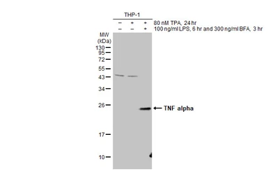
Untreated (–) and treated (+) THP-1 whole cell extracts (30 μg) were separated by 12% SDS-PAGE, and the membrane was blotted with TNF alpha antibody (GTX110520) diluted at 1:500. The HRP-conjugated anti-rabbit IgG antibody (GTX213110-01) was used to detect the primary antibody.
|
|
GTX110520 WB Image
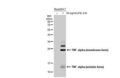
Untreated (–) and treated (+) Raw264.7 whole cell extracts (30 μg) were separated by 12% SDS-PAGE, and the membrane was blotted with TNF alpha antibody (GTX110520) diluted at 1:1000. The HRP-conjugated anti-rabbit IgG antibody (GTX213110-01) was used to detect the primary antibody.
|
|
GTX110520 WB Image
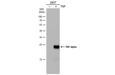
Non-transfected (–) and transfected (+) 293T whole cell extracts (30 μg) were separated by 12% SDS-PAGE, and the membrane was blotted with TNF alpha antibody (GTX110520) diluted at 1:1000. The HRP-conjugated anti-rabbit IgG antibody (GTX213110-01) was used to detect the primary antibody.
|
|
GTX110520 ICC/IF Image
TNF alpha antibody detects TNF alpha protein at membrane by immunofluorescent analysis. Sample: jurkat cells were fixed in 4% paraformaldehyde at RT for 15 min. Green: TNF alpha protein stained by TNF alpha antibody (GTX110520) diluted at 1:500. Blue: Hoechst 33342 staining.
|
|
GTX110520 WB Image
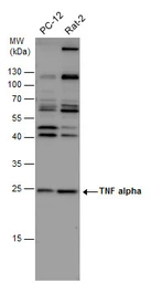
TNF alpha antibody detects TNF alpha protein by western blot analysis. Various whole cell extracts (30 μg) were separated by 12% SDS-PAGE, and the membrane was blotted with TNF alpha antibody (GTX110520) diluted at 1:500. The HRP-conjugated anti-rabbit IgG antibody (GTX213110-01) was used to detect the primary antibody.
|
|
GTX110520 ICC/IF Image
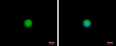
TNF alpha antibody detects TNF alpha protein at cytoplasm by immunofluorescent analysis. Sample: Raw264.7 cells were fixed in 4% paraformaldehyde at RT for 15 min. Green: TNF alpha protein stained by TNF alpha antibody (GTX110520) diluted at 1:500. Blue: Hoechst 33342 staining.
|
|
GTX110520 WB Image
TNF alpha antibody detects TNF alpha protein by western blot analysis. Whole cell extracts (30 μg) was separated by 12% SDS-PAGE, and the membrane was blotted with TNF alpha antibody (GTX110520) diluted at 1:500. The HRP-conjugated anti-rabbit IgG antibody (GTX213110-01) was used to detect the primary antibody.
|
|
GTX110520 WB Image
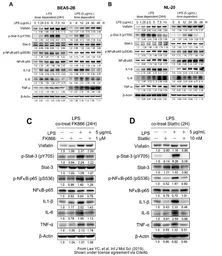
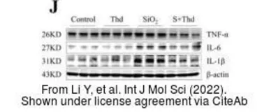
The data was published in the 2022 in Int J Mol Sci. PMID: 35628464
|
|
GTX110520 WB Image
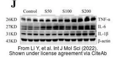
The data was published in the 2022 in Int J Mol Sci. PMID: 35628464
|
|
GTX110520 WB Image
The data was published in the journal Cardiovasc Diabetol in 2014. PMID: 24666525
|
|
GTX110520 WB Image
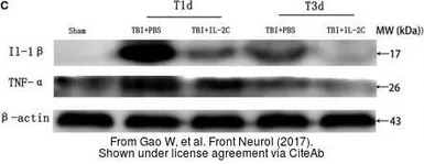
The data was published in the journal Front Neurol in 2017.PMID: 28713327
|
|
GTX110520 WB Image
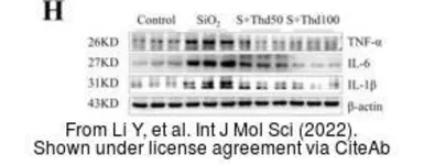
The data was published in the 2022 in Int J Mol Sci. PMID: 35628464
|
|
GTX110520 WB Image
The data was published in the journal Int J Mol Sci in 2019. PMID: 30987270
|
|
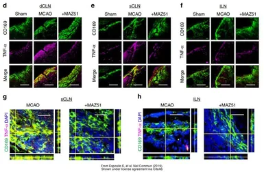
GTX110520 IHC-Fr Image
The data was published in the journal Nat Commun in 2019.PMID: 31757960
|
|
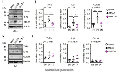
GTX110520 WB Image
The data was published in the journal Nat Commun in 2019.PMID: 31757960
|
|
GTX110520 IHC-P Image
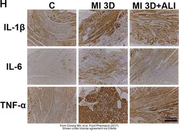
The data was published in the journal Front Pharmacol in 2017.PMID: 29184499
|
|
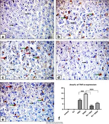
GTX110520 IHC-P Image
The data was published in the 2022 in PLoS One. PMID: 35303036
|
REFERENCE
REVIEW
TNF alpha antibody
Cat. No. GTX110520
Rating
|
|
( Average 4 based on 1 users reviews)
|
Application
Western Blot(WB)
|
|
( Average 4 based on 1 users reviews)
|
Date :
Anonymous submitted on 22-Feb-2021
Application Tested :
WB
Sample Species :
Ms
Sample :
colonic mucosa
Amount used :
25 μg
Blocking :
5% fat dry milk in TBST, 25°C, 1Hr
Primary Antibody :
1:1000, 4°C, 16Hr