Tyrosine Hydroxylase antibody
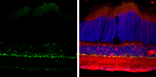
Tyrosine Hydroxylase antibody detects Tyrosine Hydroxylase protein by immunohistochemical analysis.
Sample: Frozen sectioned adult mouse retina.
Green: Tyrosine Hydroxylase protein stained by Tyrosine Hydroxylase antibody (GTX113016) diluted at 1:250.
Red: beta Tubulin 3/ TUJ1, stained by beta Tubulin 3/ TUJ1 antibody [GT11710] (GTX631836) diluted at 1:250.
Blue: Fluoroshield with DAPI (GTX30920).
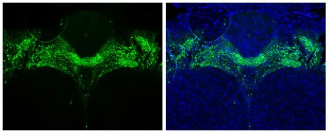
Tyrosine Hydroxylase antibody detects Tyrosine Hydroxylase protein in midbrain dopaminergic neurons by immunohistochemical analysis.Sample: Paraffin-embedded mouse brain.Green: Tyrosine Hydroxylase stained by Tyrosine Hydroxylase antibody (GTX113016) diluted at 1:1000.Blue: Fluoroshield with DAPI (GTX30920).Antigen Retrieval: Citrate buffer, pH 6.0, 15 min

Rat tissue extract (50 μg) was separated by 7.5% SDS-PAGE, and the membrane was blotted with Tyrosine Hydroxylase antibody (GTX113016) diluted at 1:2000.
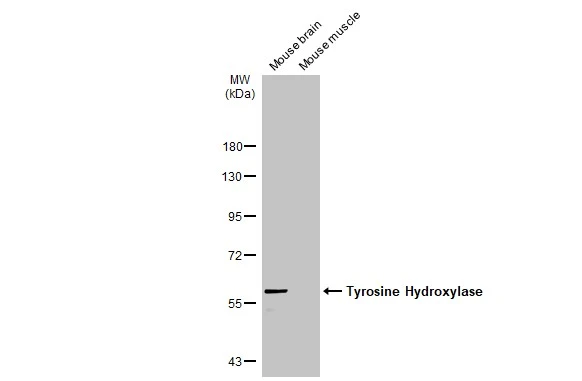
Various tissue extracts (50 μg) were separated by 7.5% SDS-PAGE, and the membrane was blotted with Tyrosine Hydroxylase antibody (GTX113016) diluted at 1:500. The HRP-conjugated anti-rabbit IgG antibody (GTX213110-01) was used to detect the primary antibody.
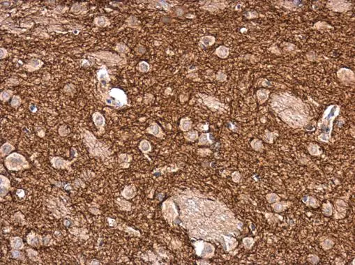
Tyrosine Hydroxylase antibody detects Tyrosine Hydroxylase protein at cytoplasm in rat brain by immunohistochemical analysis.
Sample: Paraffin-embedded rat brain.
Tyrosine Hydroxylase antibody (GTX113016) diluted at 1:2500.
Antigen Retrieval: Citrate buffer, pH 6.0, 15 min
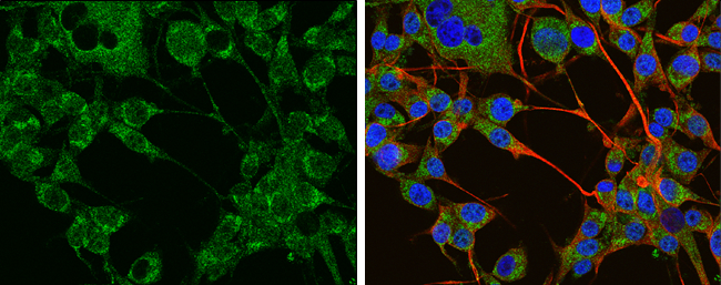
Tyrosine Hydroxylase antibody detects Tyrosine Hydroxylase protein at cytoplasm by immunofluorescent analysis.
Sample: U-87 MG cells were fixed in 4% paraformaldehyde at RT for 15 min.
Green: Tyrosine Hydroxylase protein stained by Tyrosine Hydroxylase antibody (GTX113016) diluted at 1:400.
Red: beta Tubulin 3/ TUJ1 protein stained by beta Tubulin 3/ TUJ1 antibody (GTX631836) diluted at 1:200.
Blue: Hoechst 33342 staining.
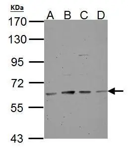
Tyrosine Hydroxylase antibody detects TH protein by western blot analysis.
A. 30 μg NT2D1 whole cell lysate/extract
B. 30 μg PC-3 whole cell lysate/extract
C. 30 μg U87-MG whole cell lysate/extract
D. 30 μg SK-N-SH whole cell lysate/extract
7.5% SDS-PAGE
Tyrosine Hydroxylase antibody (GTX113016) dilution: 1:500
The HRP-conjugated anti-rabbit IgG antibody (GTX213110-01) was used to detect the primary antibody.
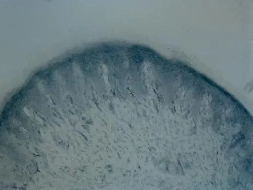
Immunohistochemical analysis of Rat hindlimb pad skin tissue (paraformaldehyde-fixed frozen sections), using Tyrosine Hydroxylase(GTX113016) antibody at 1:100 dilution.
Antigen Retrieval: Citrate buffer, pH 6.0, 15 min
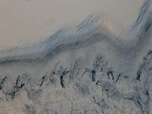
Immunohistochemical analysis of Rat hindlimb pad skin tissue (paraformaldehyde-fixed frozen sections), using Tyrosine Hydroxylase(GTX113016) antibody at 1:100 dilution.
Antigen Retrieval: Citrate buffer, pH 6.0, 15 min
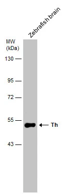
Zebrafish tissue extract (30 μg) was separated by 7.5% SDS-PAGE, and the membrane was blotted with Tyrosine Hydroxylase antibody (GTX113016) diluted at 1:500.
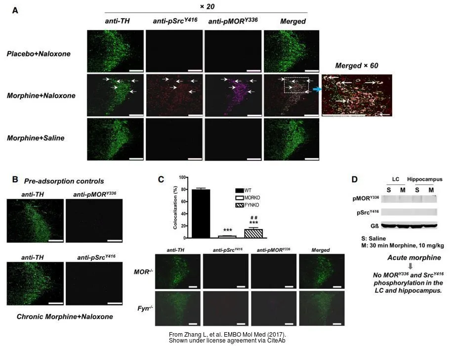
The data was published in the journal EMBO Mol Med in 2017. PMID: 28818835
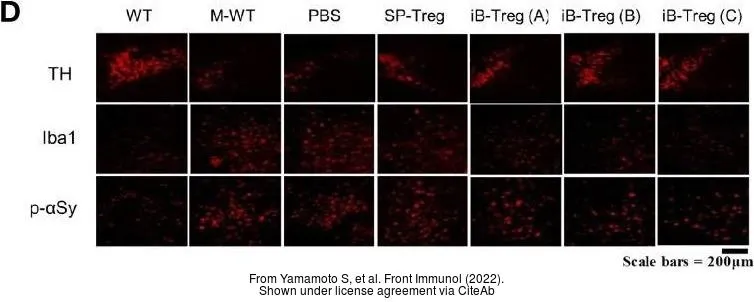
The data was published in the 2022 in Front Immunol. PMID: 35911740
-
HostRabbit
-
ClonalityPolyclonal
-
IsotypeIgG
-
ApplicationsWB ICC/IF IHC-P IHC-Fr
-
ReactivityHuman, Mouse, Rat, Zebrafish, Vole, Calf