APPLICATION
Application Note
*Optimal dilutions/concentrations should be determined by the researcher.
Application |
Recommended Dilution |
1:500-1:3000 |
1:100-1:1000 |
Not tested in other applications.
Calculated MW
Product Note
Specificity: Based on sequence homology, this antibody may cross react with UBE2B protein.
Predict Reactivity
Mouse, Rat, Zebrafish, Chicken, Xenopus laevis, Rhesus Monkey(>80% identity)
PROPERTIES
Form
Liquid
Buffer
PBS, 20% Glycerol
Preservative
0.01% Thimerosal
Storage
Store as concentrated solution. Centrifuge briefly prior to opening vial. For short-term storage (1-2 weeks), store at 4ºC. For long-term storage, aliquot and store at -20ºC or below. Avoid multiple freeze-thaw cycles.
Concentration
0.93 mg/ml (Please refer to the vial label for the specific concentration.)
Antigen Species
Human
Immunogen
Full length human UBE2A Recombinant protein.
Purification
Purified by antigen-affinity chromatography.
Conjugation
Unconjugated
RRID
AB_1241237
Note
For laboratory research use only. Not for any clinical, therapeutic, or diagnostic use in humans or animals. Not for animal or human consumption.
Purchasers shall not, and agree not to enable third parties to, analyze, copy, reverse engineer or otherwise attempt to determine the structure or sequence of the product.
TARGET
Synonyms
ubiquitin conjugating enzyme E2 A , HHR6A , MRXS30 , MRXSN , RAD6A , UBC2
Background
The modification of proteins with ubiquitin is an important cellular mechanism for targeting abnormal or short-lived proteins for degradation. Ubiquitination involves at least three classes of enzymes: ubiquitin-activating enzymes, or E1s, ubiquitin-conjugating enzymes, or E2s, and ubiquitin-protein ligases, or E3s. This gene encodes a member of the E2 ubiquitin-conjugating enzyme family. This enzyme is required for post-replicative DNA damage repair. Multiple alternatively spliced transcript variants have been found for this gene and they encode distinct isoforms. [provided by RefSeq]
Database
Research Area
DATA IMAGES
|
GTX100426 ICC/IF Image
UBE2A antibody detects UBE2A protein at cytoplasm and nucleus by immunofluorescent analysis. Sample: A375 cells were fixed in 4% paraformaldehyde at RT for 15 min. Green: UBE2A protein stained by UBE2A antibody (GTX100426) diluted at 1:500. Blue: Hoechst 33342 staining. Scale bar = 10 μm.
|
|
GTX100426 WB Image
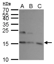
UBE2A antibody detects UBE2A protein by Western blot analysis. A. 30 μg K562 whole cell lysate/extract B. 30 μg THP-1 whole cell lysate/extract C. 30 μg HL-60 whole cell lysate/extract 15 % SDS-PAGE UBE2A antibody (GTX100426) dilution: 1:1000
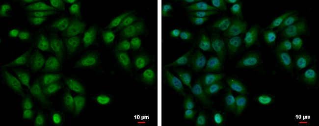
|
REFERENCE
There are currently no references for UBE2A antibody (GTX100426). Be the first to share your publications with this product.
REVIEW
There are currently no reviews for UBE2A antibody (GTX100426). Be the first to share your experience with this product.