UBE2D1 antibody
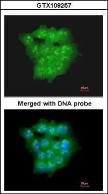
Immunofluorescence analysis of paraformaldehyde-fixed A431, using UBE2D1(GTX109257) antibody at 1:200 dilution.
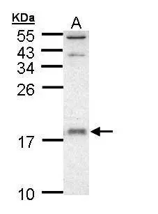
Sample (30 ug of whole cell lysate)
A: A431
10% SDS PAGE
GTX109257 diluted at 1:1000
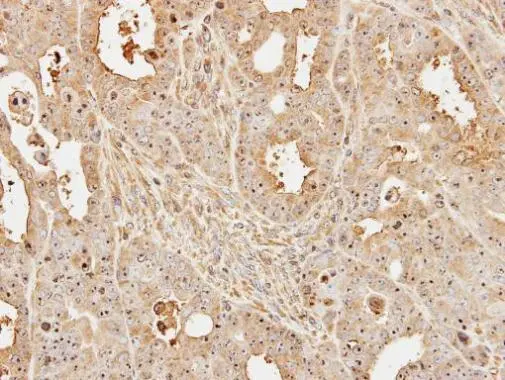
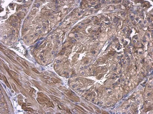
Immunohistochemical analysis of paraffin-embedded TOV-112D xenograft, using UBE2D1(GTX109257) antibody at 1:500 dilution.
Antigen Retrieval: Trilogy™ (EDTA based, pH 8.0) buffer, 15min
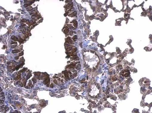
UBE2D1 antibody detects UBE2D1 protein at cytosol on mouse lung by immunohistochemical analysis.
Sample: Paraffin-embedded mouse lung.
UBE2D1 antibody (GTX109257) dilution: 1:500.
Antigen Retrieval: Trilogy™ (EDTA based, pH 8.0) buffer, 15min
UBE2D1 antibody detects UBE2D1 protein at cytosol on mouse prostate by immunohistochemical analysis.
Sample: Paraffin-embedded mouse prostate.
UBE2D1 antibody (GTX109257) dilution: 1:500.
Antigen Retrieval: Trilogy™ (EDTA based, pH 8.0) buffer, 15min
-
HostRabbit
-
ClonalityPolyclonal
-
IsotypeIgG
-
ApplicationsWB ICC/IF IHC-P
-
ReactivityHuman, Mouse