UGDH antibody


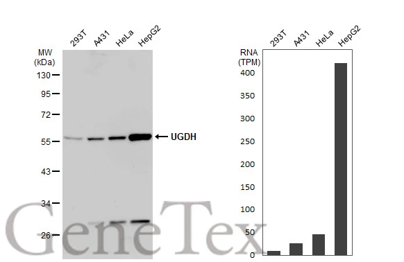
Various whole cell extracts (30 μg) were separated by 10% SDS-PAGE, and the membrane was blotted with UGDH antibody (GTX104993) diluted at 1:10000. The HRP-conjugated anti-rabbit IgG antibody (GTX213110-01) was used to detect the primary antibody. Corresponding RNA expression data for the same cell lines are based on Human Protein Atlas program.
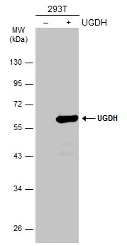
Non-transfected (–) and transfected (+) 293T whole cell extracts (30 μg) were separated by 10% SDS-PAGE, and the membrane was blotted with UGDH antibody (GTX104993) diluted at 1:5000. The HRP-conjugated anti-rabbit IgG antibody (GTX213110-01) was used to detect the primary antibody.
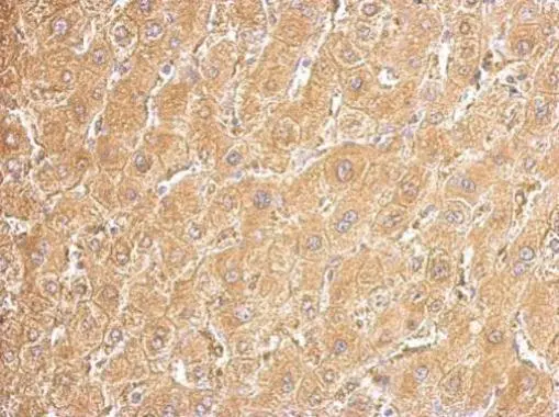
UGDH antibody detects UGDH protein at cytosol on human hepatoma by immunohistochemical analysis.
Sample: Paraffin-embedded hepatoma tissue.
UGDH antibody (GTX104993) dilution: 1:500.
Antigen Retrieval: Trilogy™ (EDTA based, pH 8.0) buffer, 15min
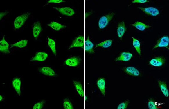
UGDH antibody detects UGDH protein at nucleus by immunofluorescent analysis.Sample: HeLa cells were fixed in 4% paraformaldehyde at RT for 15 min.Green: UGDH stained by UGDH antibody (GTX104993) diluted at 1:500.
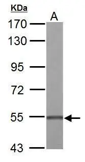
UGDH antibody detects UGDH protein by Western blot analysis.
A. 50 μg Rat liver lysate/extract
7.5 % SDS-PAGE
UGDH antibody (GTX104993) dilution: 1:2000
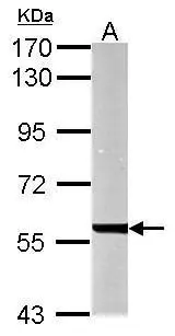
Sample (50 ug of whole cell lysate)
A: mouse liver
7.5% SDS PAGE
GTX104993 diluted at 1:10000
-
HostRabbit
-
ClonalityPolyclonal
-
IsotypeIgG
-
ApplicationsWB ICC/IF IHC-P
-
ReactivityHuman, Mouse, Rat