VAPB antibody


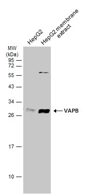
HepG2 whole cell and membrane extracts (30 μg) were separated by 12% SDS-PAGE, and the membrane was blotted with VAPB antibody (GTX131631) diluted at 1:1000. The HRP-conjugated anti-rabbit IgG antibody (GTX213110-01) was used to detect the primary antibody.
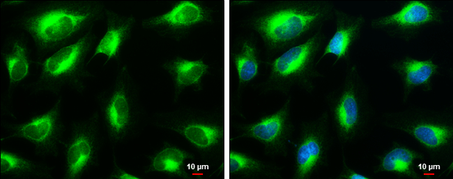
VAPB antibody detects VAPB protein at endoplasmic reticulum by immunofluorescent analysis.Sample: HeLa cells were fixed in 4% paraformaldehyde at RT for 15 min.Green: VAPB stained by VAPB antibody (GTX131631) diluted at 1:1000.Blue: Hoechst 33342 staining.Scale bar= 10μm.
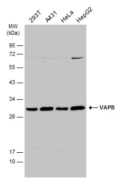
Various whole cell extracts (30 μg) were separated by 12% SDS-PAGE, and the membrane was blotted with VAPB antibody (GTX131631) diluted at 1:1000. The HRP-conjugated anti-rabbit IgG antibody (GTX213110-01) was used to detect the primary antibody.
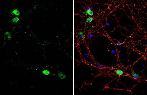
VAPB antibody detects VAPB protein by immunofluorescent analysis.Sample: DIV9 rat E18 primary cortical neuron cells were fixed in 4% paraformaldehyde at RT for 15 min.Green: VAPB stained by VAPB antibody (GTX131631) diluted at 1:500.Red: Tau, stained by Tau antibody [GT287] (GTX634809) diluted at 1:500.Blue: Fluoroshield with DAPI (GTX30920).
-
HostRabbit
-
ClonalityPolyclonal
-
IsotypeIgG
-
ApplicationsWB ICC/IF IP
-
ReactivityHuman, Rat