VASP antibody
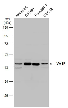
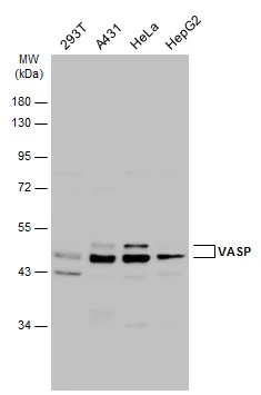
Various whole cell extracts (30 μg) were separated by 10% SDS-PAGE, and the membrane was blotted with VASP antibody (GTX132983) diluted at 1:1000. The HRP-conjugated anti-rabbit IgG antibody (GTX213110-01) was used to detect the primary antibody.
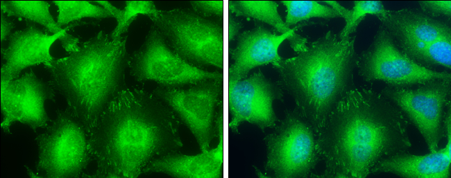
VASP antibody detects VASP protein at focal adhesion by immunofluorescent analysis.
Sample: HeLa cells were fixed in 4% paraformaldehyde at RT for 15 min.
Green: VASP protein stained by VASP antibody (GTX132983) diluted at 1:500.
Blue: Hoechst 33342 staining.
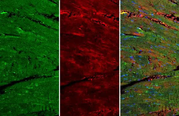
VASP antibody detects VASP protein at cell membrane and cytoplasm by immunohistochemical analysis.Sample: Paraffin-embedded mouse heart.Green: VASP stained by VASP antibody (GTX132983) diluted at 1:250.Red: beta Actin, a cytoskeleton marker, stained by beta Actin antibody [GT5512] (GTX629630) diluted at 1:500.Blue: Fluoroshield with DAPI (GTX30920).Antigen Retrieval: Citrate buffer, pH 6.0, 15 min
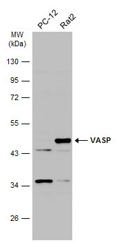
Various whole cell extracts (30 μg) were separated by 10% SDS-PAGE, and the membrane was blotted with VASP antibody (GTX132983) diluted at 1:1000. The HRP-conjugated anti-rabbit IgG antibody (GTX213110-01) was used to detect the primary antibody.
Various whole cell extracts (30 μg) were separated by 10% SDS-PAGE, and the membrane was blotted with VASP antibody (GTX132983) diluted at 1:1000. The HRP-conjugated anti-rabbit IgG antibody (GTX213110-01) was used to detect the primary antibody.
-
HostRabbit
-
ClonalityPolyclonal
-
IsotypeIgG
-
ApplicationsWB ICC/IF IHC-P
-
ReactivityHuman, Mouse, Rat