Villin antibody


*The competitor is not affiliated with GeneTex and does not endorse this product.
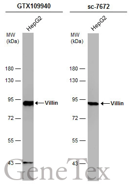
Whole cell extract (30 μg) was separated by 7.5% SDS-PAGE, and the membranes were blotted with Villin antibody (GTX109940) diluted at 1:5000 and competitor's antibody (sc-7672) diluted at 1:100. The HRP-conjugated anti-rabbit IgG antibody (GTX213110-01) was used to detect the primary antibody.

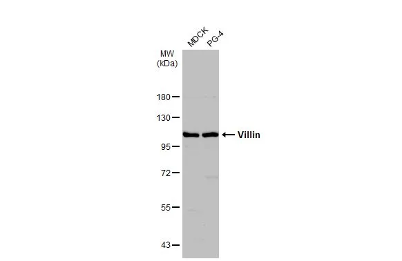
Various whole cell extracts (30 μg) were separated by 7.5% SDS-PAGE, and the membrane was blotted with Villin antibody (GTX109940) diluted at 1:2000. The HRP-conjugated anti-rabbit IgG antibody (GTX213110-01) was used to detect the primary antibody. Corresponding RNA expression data for the same cell lines are based on Human Protein Atlas program.
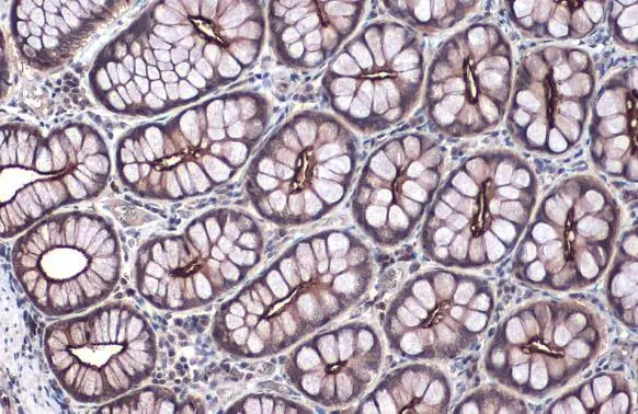
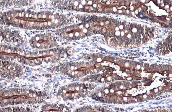
Villin antibody detects Villin protein at cytoplasm by immunohistochemical analysis.Sample: Paraffin-embedded mouse duodenum.Villin stained by Villin antibody (GTX109940) diluted at 1:500.Antigen Retrieval: Citrate buffer, pH 6.0, 15 min

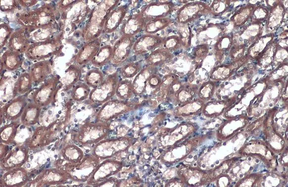
Villin antibody detects Villin protein at cytoplasm by immunohistochemical analysis.Sample: Paraffin-embedded mouse kidney.Villin stained by Villin antibody (GTX109940) diluted at 1:500.Antigen Retrieval: Citrate buffer, pH 6.0, 15 min
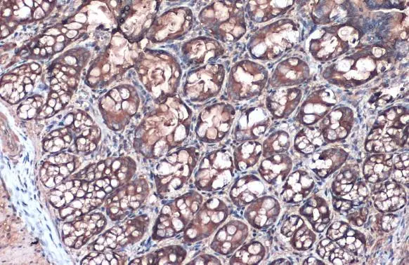
Villin antibody detects Villin protein at cytoplasm by immunohistochemical analysis.Sample: Paraffin-embedded rat colon.Villin stained by Villin antibody (GTX109940) diluted at 1:500.Antigen Retrieval: Citrate buffer, pH 6.0, 15 min
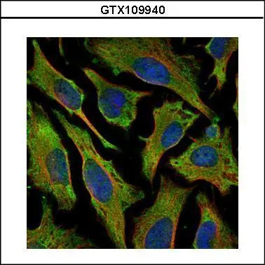
Confocal immunofluorescence analysis (Olympus FV10i) of methanol-fixed HeLa, using Villin(GTX109940) antibody (Green) at 1:500 dilution. Alpha-tubulin filaments were labeled with GTX11304 (Red) at 1:2000.
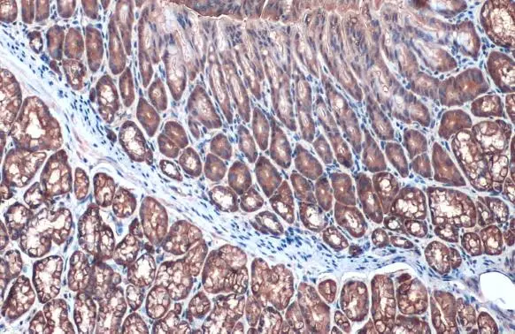
Villin antibody detects Villin protein at cytoplasm by immunohistochemical analysis.Sample: Paraffin-embedded rat kidney.Villin stained by Villin antibody (GTX109940) diluted at 1:500.Antigen Retrieval: Citrate buffer, pH 6.0, 15 min
Villin antibody detects VIL1 protein by western blot analysis.
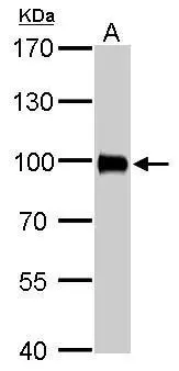
A. 50 μg rat kidney lysate/extract
7.5% SDS-PAGE
Villin antibody (GTX109940) dilution: 1:2000
The HRP-conjugated anti-rabbit IgG antibody (GTX213110-01) was used to detect the primary antibody.
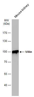
Mouse tissue extract (50 μg) was separated by 7.5% SDS-PAGE, and the membrane was blotted with Villin antibody (GTX109940) diluted at 1:1000. The HRP-conjugated anti-rabbit IgG antibody (GTX213110-01) was used to detect the primary antibody.
Villin antibody detects Villin protein at cytoplasm by immunohistochemical analysis.Sample: Paraffin-embedded mouse duodenum.Villin stained by Villin antibody (GTX109940) diluted at 1:500.Antigen Retrieval: Citrate buffer, pH 6.0, 15 min
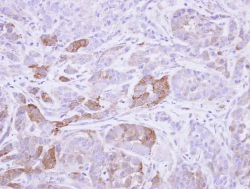
Immunohistochemical analysis of paraffin-embedded A549 xenograft, using Villin (GTX109940) antibody at 1:500 dilution.
Antigen Retrieval: Trilogy™ (EDTA based, pH 8.0) buffer, 15min
Various whole cell extracts (30 μg) were separated by 7.5% SDS-PAGE, and the membrane was blotted with Villin antibody (GTX109940) diluted at 1:500. The HRP-conjugated anti-rabbit IgG antibody (GTX213110-01) was used to detect the primary antibody.
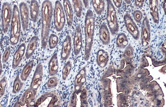
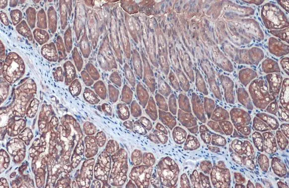
Villin antibody detects VIL1 protein at cell membrane by immunohistochemical analysis.Sample: Paraffin-embedded cat colon.VIL1 stained by Villin antibody (GTX109940) diluted at 1:500.Antigen Retrieval: Citrate buffer, pH 6.0, 15 min
Villin antibody detects VIL1 protein at cell membrane by immunohistochemical analysis.Sample: Paraffin-embedded dog colon.VIL1 stained by Villin antibody (GTX109940) diluted at 1:500.Antigen Retrieval: Citrate buffer, pH 6.0, 15 min
Villin antibody detects VIL1 protein at cell membrane by immunohistochemical analysis.Sample: Paraffin-embedded dog intestine.VIL1 stained by Villin antibody (GTX109940) diluted at 1:500.Antigen Retrieval: Citrate buffer, pH 6.0, 15 min
-
HostRabbit
-
ClonalityPolyclonal
-
IsotypeIgG
-
ApplicationsWB ICC/IF IHC-P IHC-Fr
-
ReactivityHuman, Mouse, Rat, Cat, Dog