WIPI1 antibody
Cat. No. GTX129279
Cat. No. GTX129279

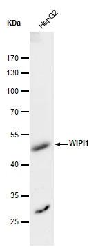
GTX129279 WB Image
WIPI1 antibody detects WIPI1 protein by western blot analysis. Whole cell extracts (30 μg) was separated by 10 % SDS-PAGE, and blotted with WIPI1 antibody (GTX129279) diluted by 1:500
1 / 2
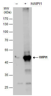
GTX129279 WB Image
WIPI1 antibody detects WIPI1 protein by western blot analysis. Non-transfected (-) and -transfected (+, ) 293T whole cell extracts (30 μg) were separated by 10% SDS-PAGE, and the membrane was blotted with WIPI1 antibody (GTX129279) diluted by 1:20000.
2 / 2
-
HostRabbit
-
ClonalityPolyclonal
-
IsotypeIgG
-
ApplicationsWB
-
ReactivityHuman