XBP1 antibody

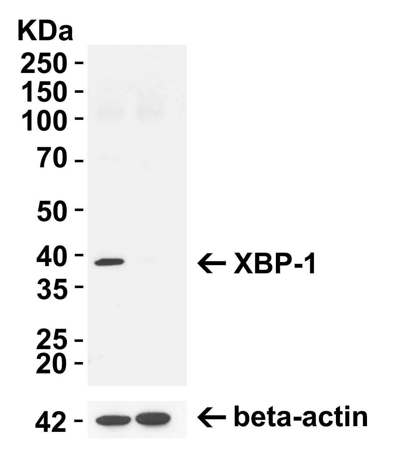
WB analysis of 293T cells with/without XBP1 KO treatment using GTX31293 XBP1 antibody.
Left lane : 293T cells
Right lane : 293T cells with XBP1 KO treatment
Loading : 10μg
Dilution : 1μg/ml
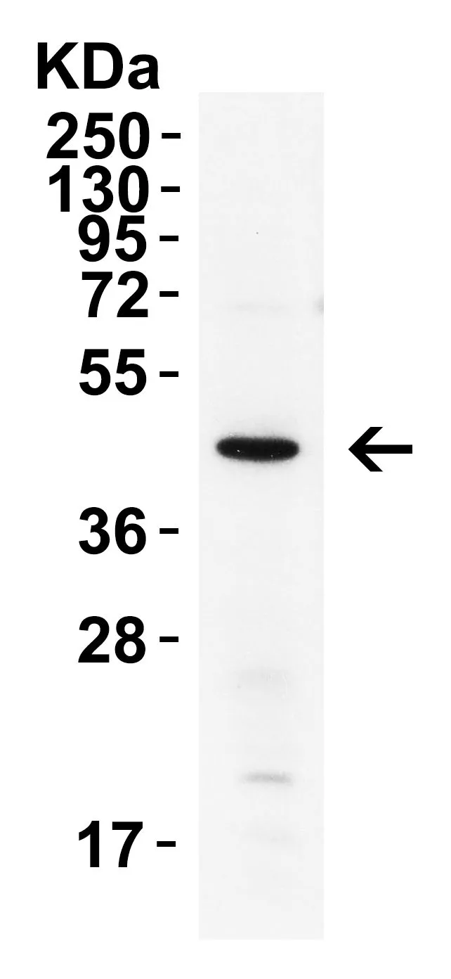
WB analysis of mouse pancreas tissue lysate using GTX31293 XBP1 antibody.
Loading : 15μg
Dilution : 4μg/ml
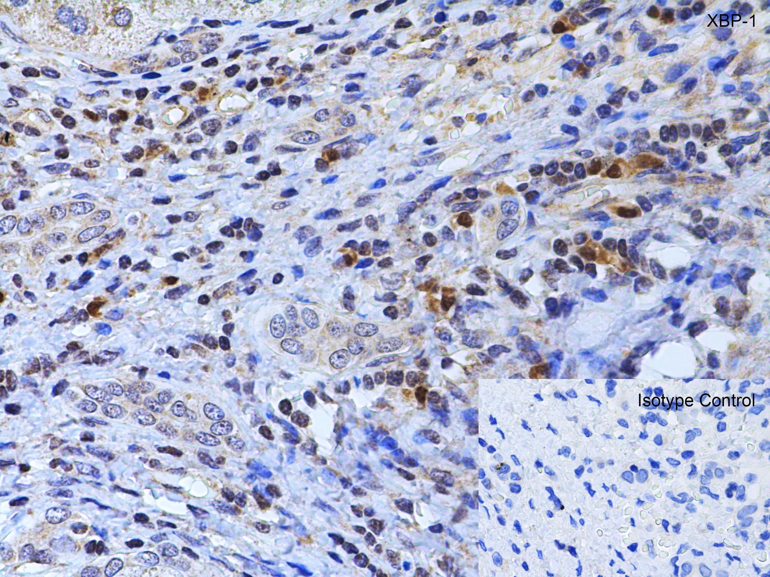
IHC-P analysis of mouse testis tissue using GTX31293 XBP1 antibody.
Antigen retrieval : Heat mediation with citrate buffer(pH6).
Dilution : 2μg/ml
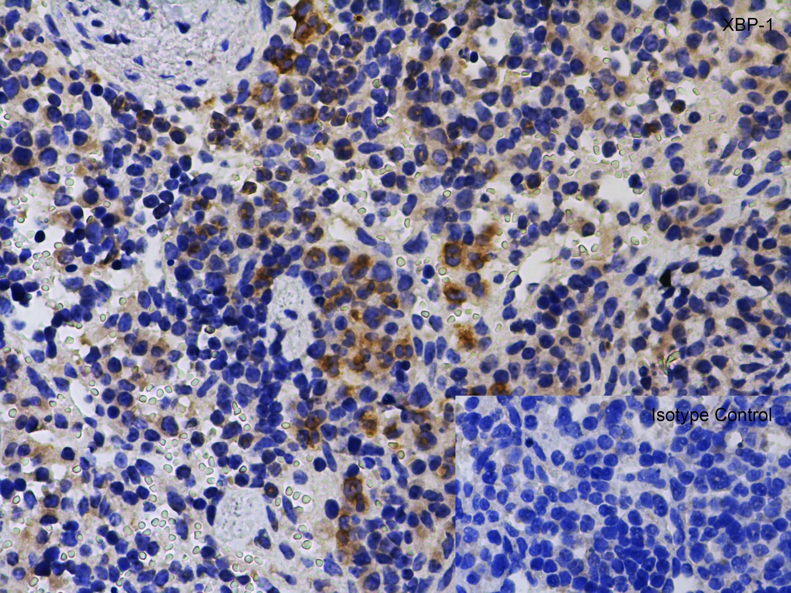
IHC-P analysis of mouse spleen tissue using GTX31293 XBP1 antibody.
Antigen retrieval : Heat mediation with citrate buffer(pH6).
Dilution : 1μg/ml
IHC-P analysis of rat spleen tissue using GTX31293 XBP1 antibody.
Antigen retrieval : Heat mediation with citrate buffer(pH6).
Dilution : 1μg/ml
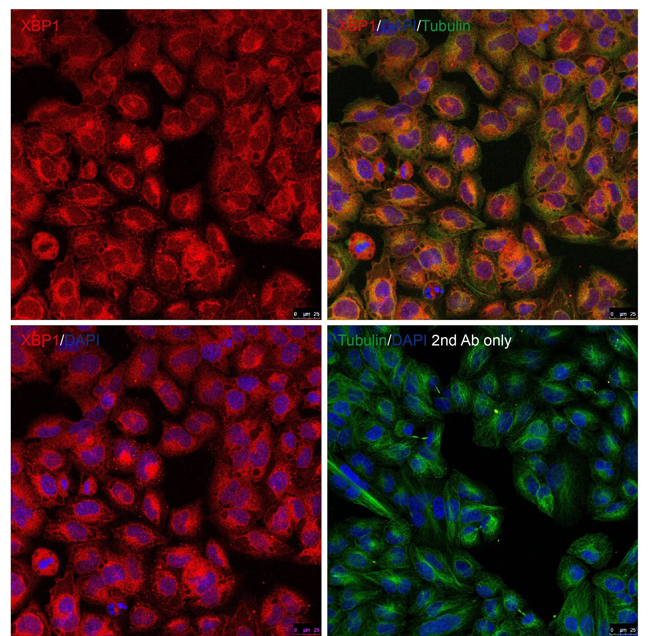
ICC/IF analysis of methanol-fixed HeLa cells using GTX31293 XBP1 antibody.
Red : Primary antibody.
Green : Alpha tubulin
Blue : DAPI
Dilution : 10 μg/mL
WB analysis of rat liver tissue lysate using GTX31293 XBP1 antibody in the present and absent of blocking peptide.
Dilution : 1 μg/mL
Loading : 15 μg
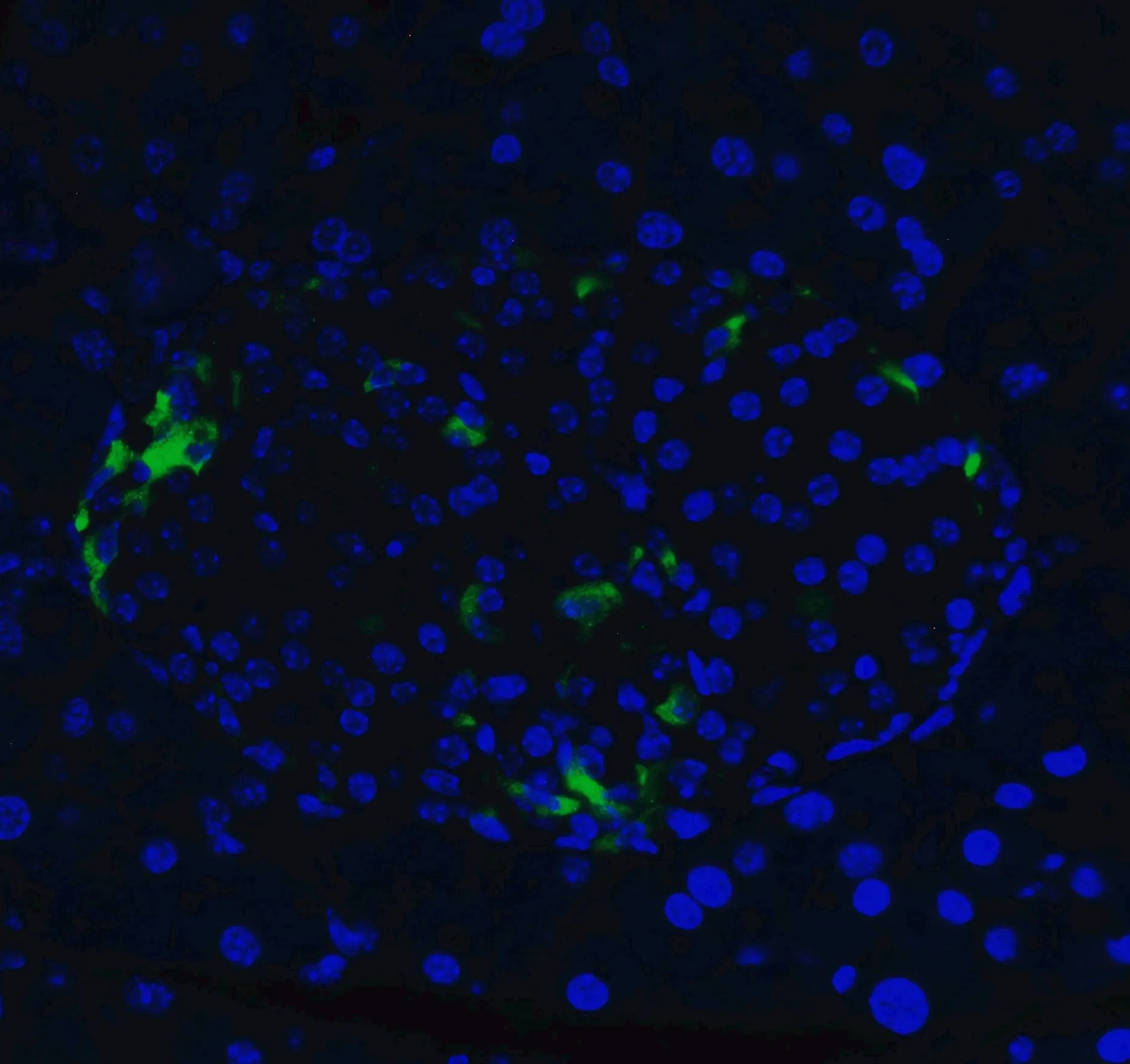
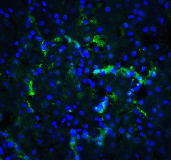
IHC-P analysis of 4% paraformaldehyde-fixed mouse pancreas tissue using GTX31293 XBP1 antibody.
Dilution : 20 μg/ml
Green : Primary antibody
Blue : DAPI
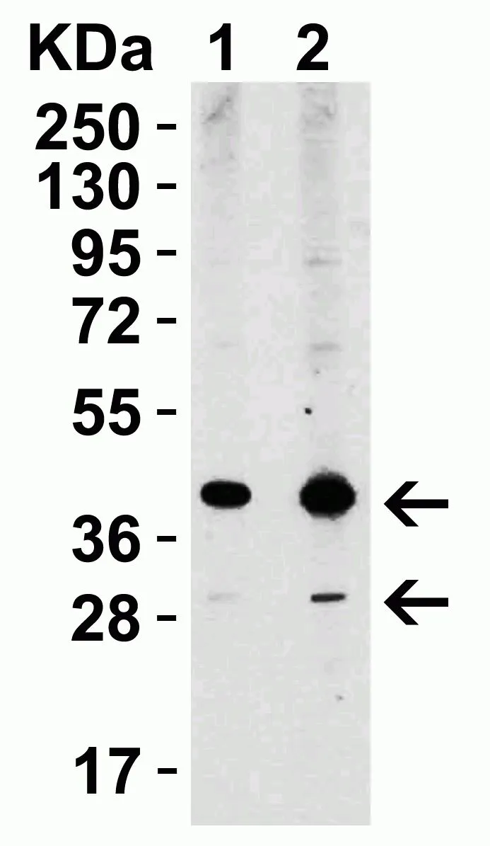
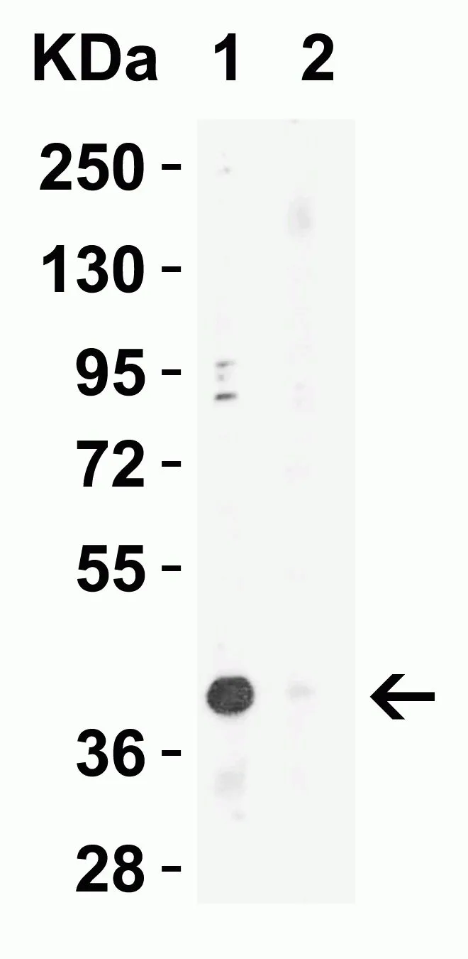
WB analysis of HepG2 cell lysate using GTX31293 XBP1 antibody.
Dilution : 1 μg/mL (Lane 1) / 2 μg/mL (Lane 2)
Loading : 15 μg
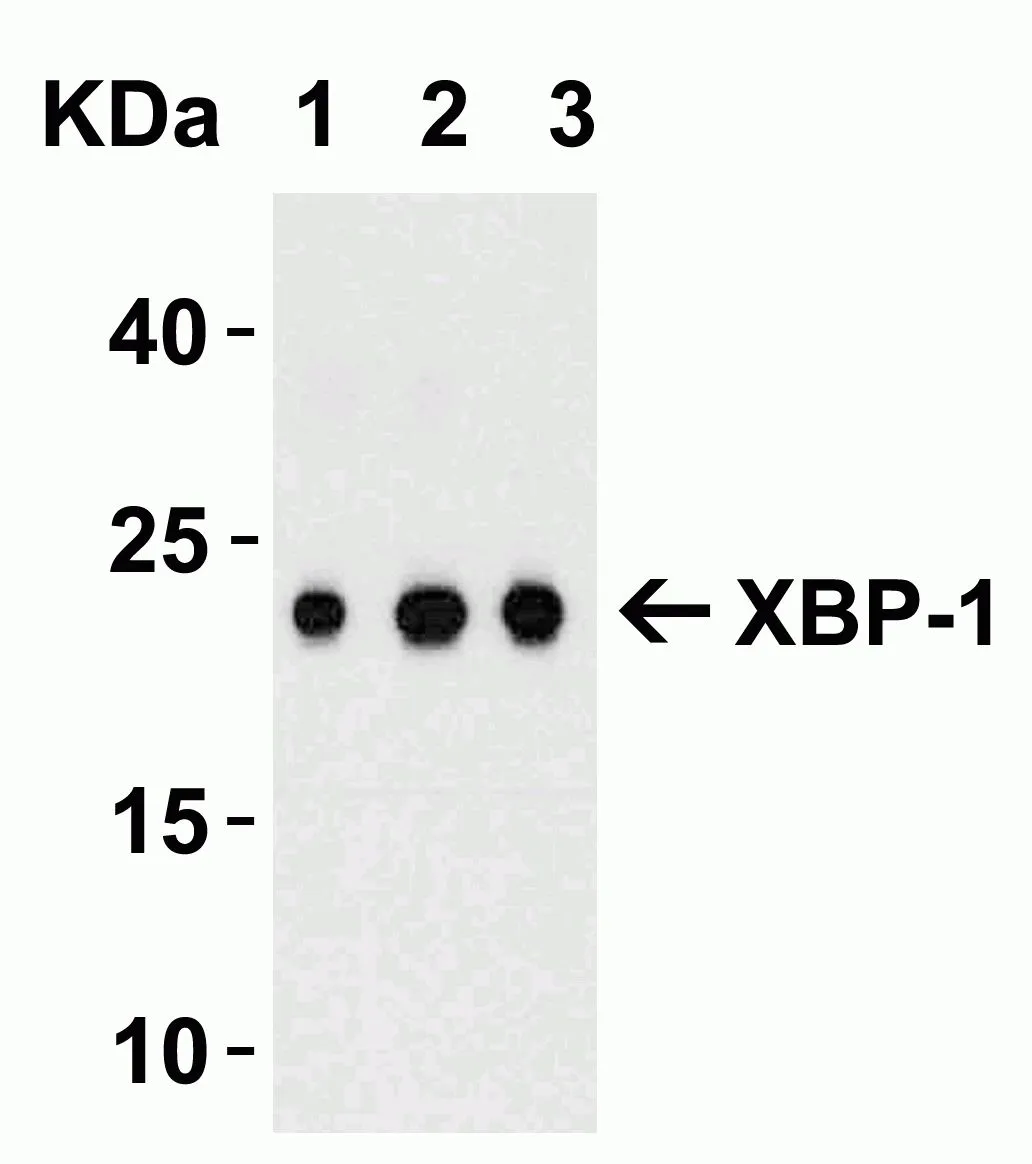
WB analysis of 100 ng of human XBP-1 recombinant protein lysate using GTX31293 XBP1 antibody.
Dilution : 0.5 μg/mL (Lane 1) / 1 μg/mL (Lane 2) / 2 μg/mL (Lane 3)
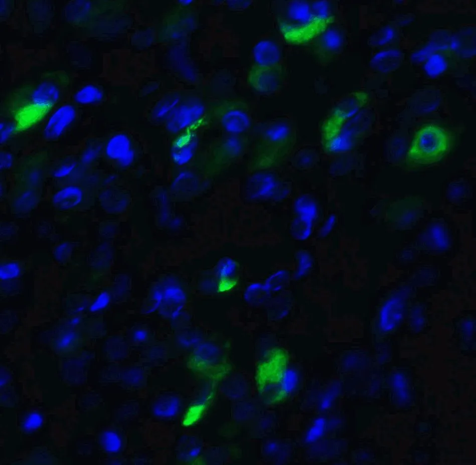
IHC-P analysis of 4% paraformaldehyde-fixed human pancreas tissue using GTX31293 XBP1 antibody.
Dilution : 20 μg/ml
Green : Primary antibody
Blue : DAPI
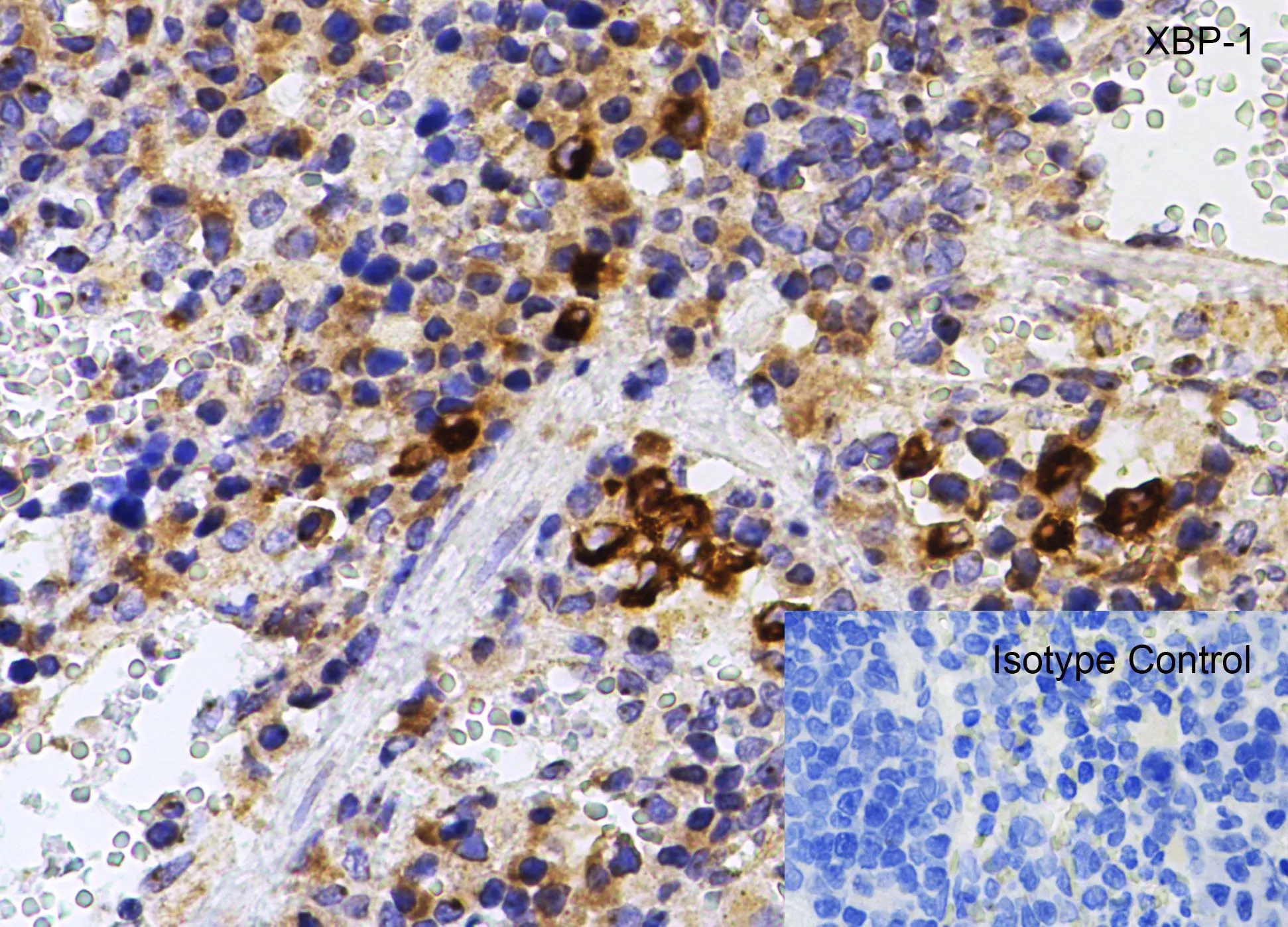
IHC-P analysis of human liver tissue using GTX31293 XBP1 antibody.
Working concentration : 20 μg/ml
-
HostRabbit
-
ClonalityPolyclonal
-
IsotypeIgG
-
ApplicationsWB ICC/IF IHC-P ELISA
-
ReactivityHuman, Mouse, Rat