alpha Synuclein antibody


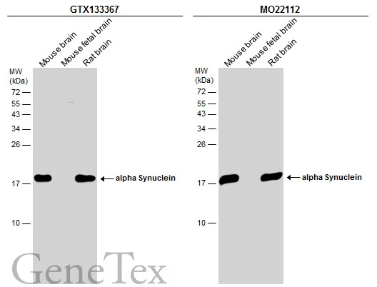
Various tissue extracts (50 μg) were separated by 15% SDS-PAGE, and the membranes were blotted with alpha Synuclein antibody (GTX133367) diluted at 1:2000 and competitor's antibody (MO22112) diluted at 1:2000. The HRP-conjugated anti-rabbit IgG antibody (GTX213110-01) was used to detect the primary antibody.
*The competitor is not affiliated with GeneTex and does not endorse this product.

Various whole cell extracts (30 μg) were separated by 12% SDS-PAGE, and the membrane was blotted with alpha Synuclein antibody (GTX133367) diluted at 1:1000. The HRP-conjugated anti-rabbit IgG antibody (GTX213110-01) was used to detect the primary antibody.
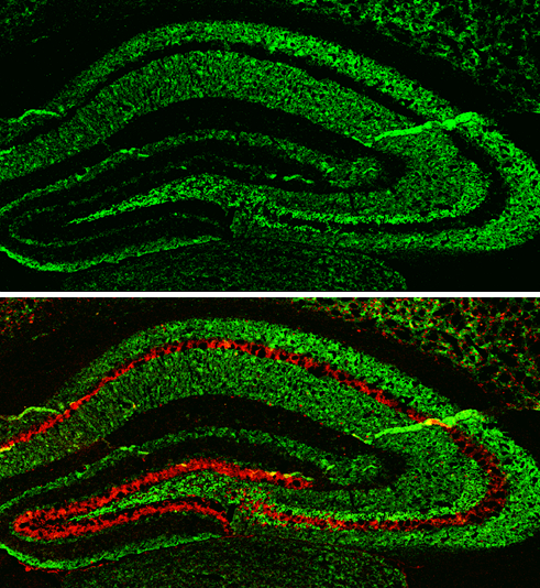
alpha Synuclein antibody detects alpha Synuclein protein expression by immunohistochemical analysis.
Sample: Frozen-sectioned adult mouse hippocampus.
Green: alpha Synuclein protein stained by alpha Synuclein antibody (GTX133367) diluted at 1:250.
Red: NeuN, stained by NeuN antibody [2Q158] (GTX30773) diluted at 1:500.
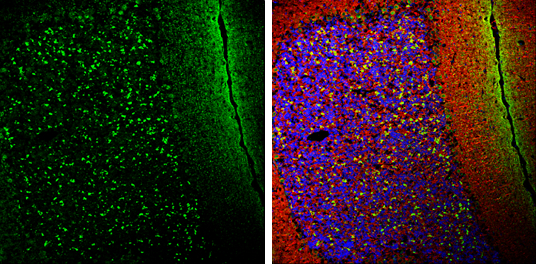
alpha Synuclein antibody detects alpha Synuclein protein expression by immunohistochemical analysis.
Sample: Frozen-sectioned adult mouse cerebellum.
Green: alpha Synuclein protein stained by alpha Synuclein antibody (GTX133367) diluted at 1:250.
Red: beta Tubulin 3/ TUJ1, stained by beta Tubulin 3/ TUJ1 antibody [GT11710] (GTX631836) diluted at 1:500.
Blue: Fluoroshield with DAPI (GTX30920).
-
HostRabbit
-
ClonalityPolyclonal
-
IsotypeIgG
-
ApplicationsWB IHC-P IHC-Fr
-
ReactivityHuman, Mouse, Rat