alpha Tubulin antibody

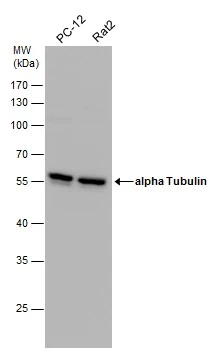
Various whole cell extracts (30 μg) were separated by 10% SDS-PAGE, and the membrane was blotted with alpha Tubulin antibody (GTX112141) diluted at 1:20000. The HRP-conjugated anti-rabbit IgG antibody (GTX213110-01) was used to detect the primary antibody.
Various whole cell extracts were separated by 10% SDS-PAGE, and the membrane was blotted with alpha Tubulin antibody (GTX112141) diluted at 1:10000. The HRP-conjugated anti-rabbit IgG antibody (GTX213110-01) was used to detect the primary antibody.
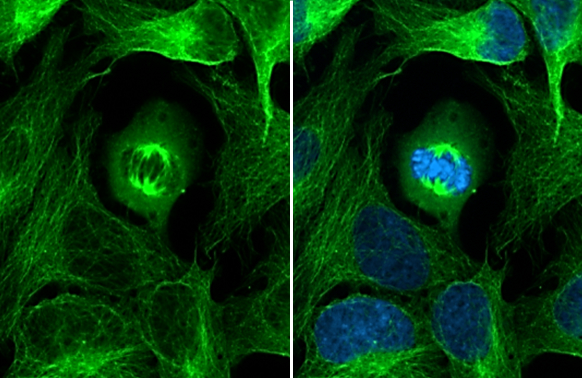
alpha Tubulin antibody detects alpha Tubulin protein at cytoskeleton by immunofluorescent analysis.Sample: HeLa cells were fixed in 4% paraformaldehyde at RT for 15 min.Green: alpha Tubulin stained by alpha Tubulin antibody (GTX112141) diluted at 1:500.Blue: Fluoroshield with DAPI (GTX30920).
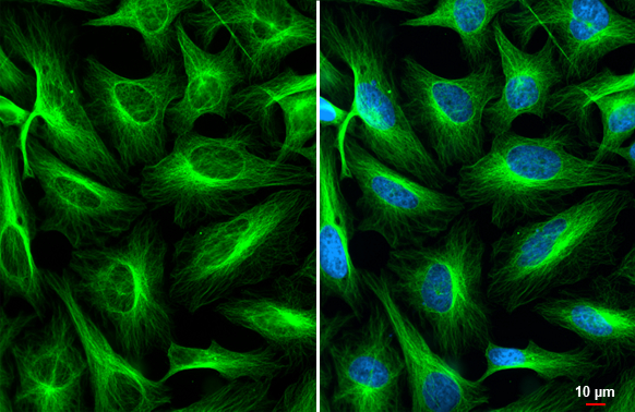
alpha Tubulin antibody detects alpha Tubulin protein at cytoskeleton by immunofluorescent analysis.Sample: HeLa cells were fixed in 4% paraformaldehyde at RT for 15 min.Green: alpha Tubulin stained by alpha Tubulin antibody (GTX112141) diluted at 1:500.Blue: Fluoroshield with DAPI (GTX30920).
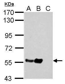
Sample (30 μg of cell lysate)
A: HeLa
B: HeLa cytosol fraction
C: HeLa nucleus fraction
10% SDS PAGE
GTX112141 diluted at 1:10000
The HRP-conjugated anti-rabbit IgG antibody (GTX213110-01) was used to detect the primary antibody.
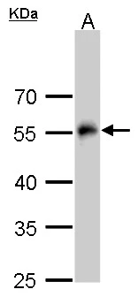
alpha Tubulin antibody detects alpha Tubulin protein by western blot analysis.
A. 30 μg Drosophila lysate/extract
10% SDS-PAGE
alpha Tubulin antibody (GTX112141) dilution: 1:2000
The HRP-conjugated anti-rabbit IgG antibody (GTX213110-01) was used to detect the primary antibody.
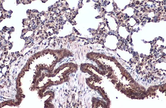
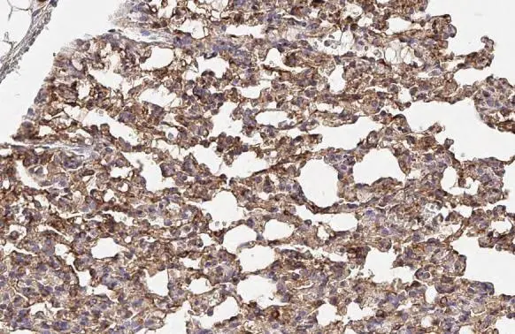
alpha Tubulin antibody detects alpha Tubulin protein at cell membrane and cytoplasm by immunohistochemical analysis.Sample: Paraffin-embedded mouse lung.alpha Tubulin stained by alpha Tubulin antibody (GTX112141) diluted at 1:500.Antigen Retrieval: Citrate buffer, pH 6.0, 15 min
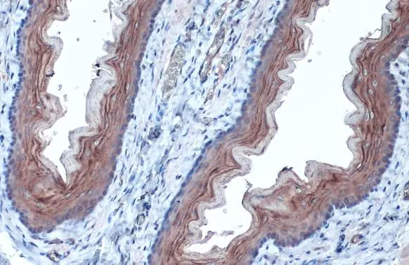
alpha Tubulin antibody detects alpha Tubulin protein at cell membrane and cytoplasm by immunohistochemical analysis.Sample: Paraffin-embedded rat esophagus.alpha Tubulin stained by alpha Tubulin antibody (GTX112141) diluted at 1:1000.Antigen Retrieval: Citrate buffer, pH 6.0, 15 min
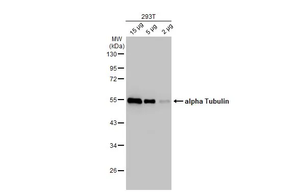
Various whole cell extracts (30 μg) were separated by 10% SDS-PAGE, and the membrane was blotted with alpha Tubulin antibody (GTX112141) diluted at 1:10000. The HRP-conjugated anti-rabbit IgG antibody (GTX213110-01) was used to detect the primary antibody.
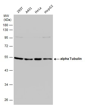
Various whole cell extracts (30 μg) were separated by 10% SDS-PAGE, and the membrane was blotted with alpha Tubulin antibody (GTX112141) diluted at 1:10000. The HRP-conjugated anti-rabbit IgG antibody (GTX213110-01) was used to detect the primary antibody.
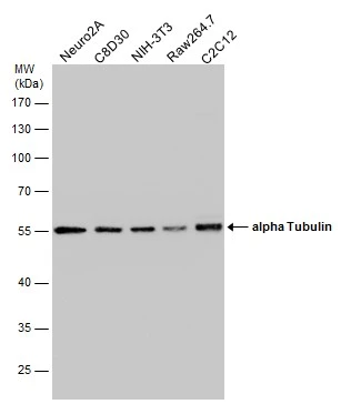
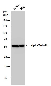
Various whole cell extracts (30 μg) were separated by 10% SDS-PAGE, and the membrane was blotted with alpha Tubulin antibody (GTX112141) diluted at 1:20000. The HRP-conjugated anti-rabbit IgG antibody (GTX213110-01) was used to detect the primary antibody.
alpha Tubulin antibody detects alpha Tubulin protein at cytoplasm and nucleus by immunohistochemical analysis.Sample: Paraffin-embedded rat lung.alpha Tubulin stained by alpha Tubulin antibody (GTX112141) diluted at 1:500.Antigen Retrieval: Citrate buffer, pH 6.0, 15 min
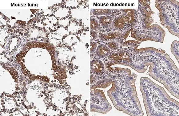
alpha Tubulin antibody detects alpha Tubulin protein by immunohistochemical analysis.Sample: Paraffin-embedded mouse tissues.alpha Tubulin stained by alpha Tubulin antibody (GTX112141) diluted at 1:500.Antigen Retrieval: Citrate buffer, pH 6.0, 15 min
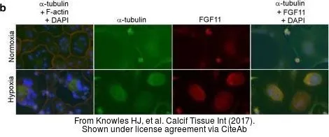
The data was published in the journal Calcif Tissue Int in 2017. PMID: 28097375
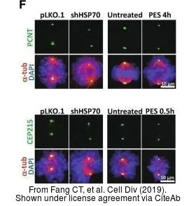
The data was published in the journal Cell Div in 2019. PMID: 31110557
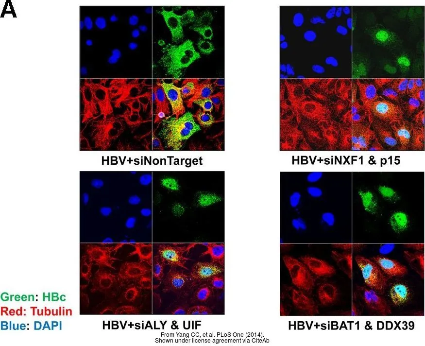
The data was published in the journal PLoS One in 2014. PMID: 25360769
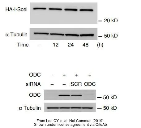
The data was published in the journal Nat Commun in 2019. PMID: 30622262
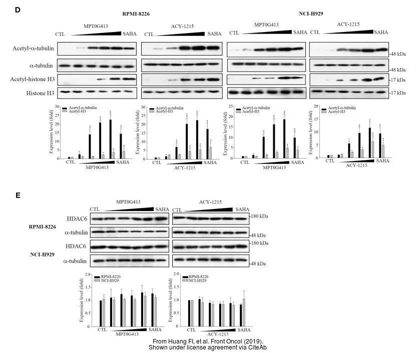
The data was published in the journal Front Oncol in 2019. PMID: 31024851
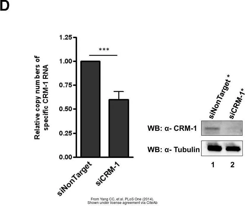
The data was published in the journal PLoS One in 2014. PMID: 25360769
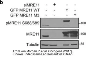
The data was published in the journal Oncogene in 2017. PMID: 28436950
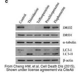
The data was published in the journal Cell Death Dis in 2015. PMID: 25950483
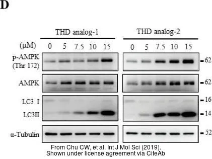
The data was published in the journal Int J Mol Sci in 2019. PMID: 30678307
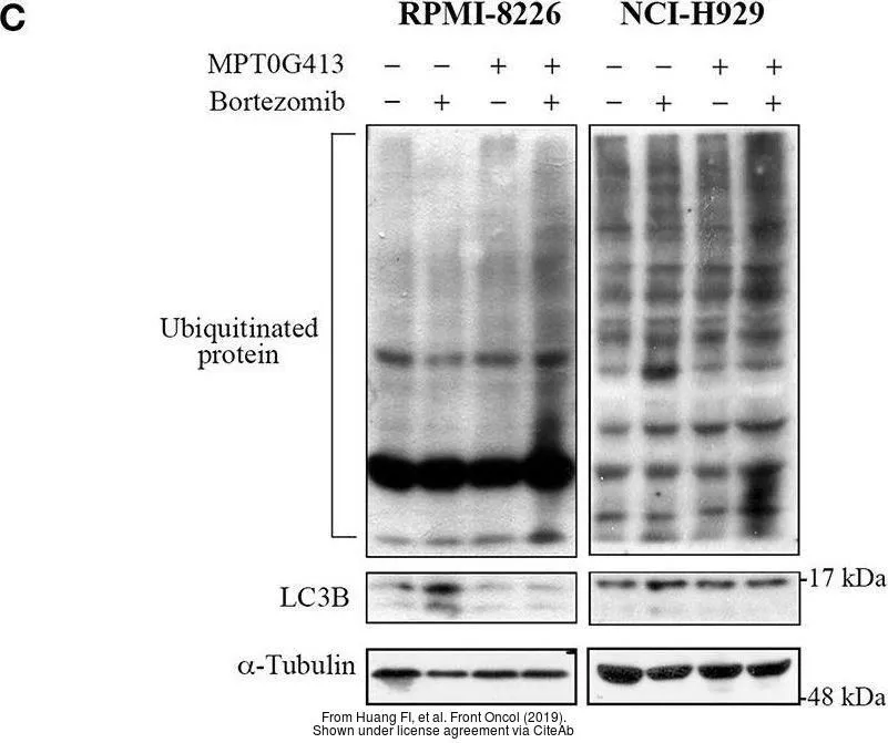
The data was published in the journal Front Oncol in 2019. PMID: 31024851
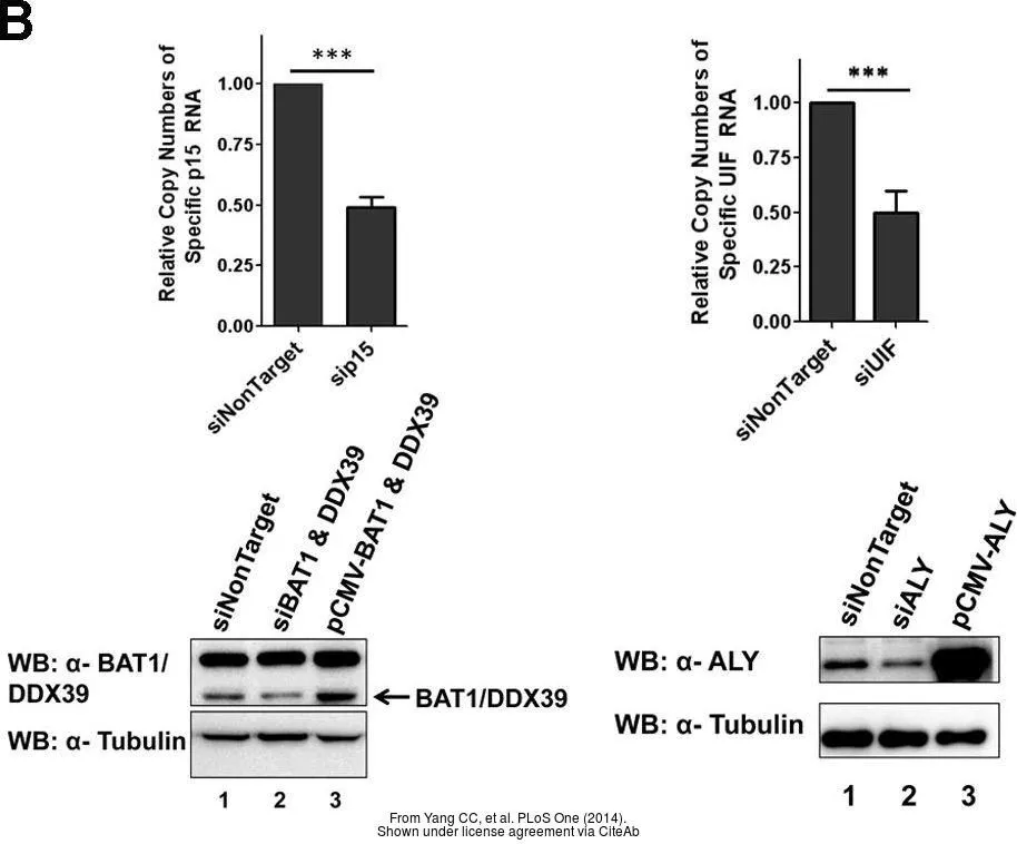
The data was published in the journal PLoS One in 2014. PMID: 25360769
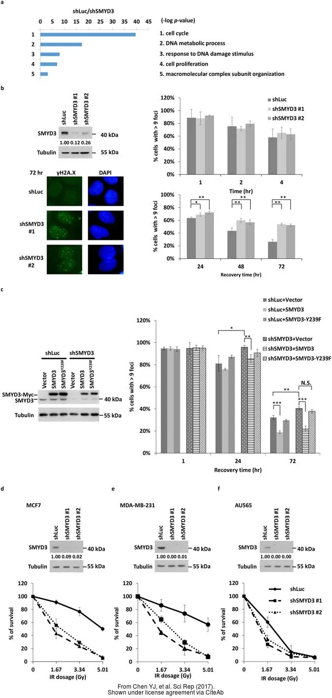
The data was published in the journal Sci Rep in 2017. PMID: 28630472
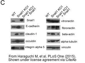
The data was published in the journal PLoS One in 2015. PMID: 26161782
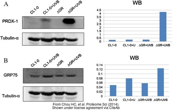
The data was published in the journal Proteome Sci in 2014. PMID: 24405781
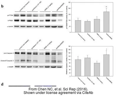
The data was published in the journal Sci Rep in 2016. PMID: 26838546
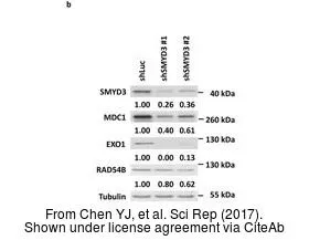
The data was published in the journal Sci Rep in 2017. PMID: 28630472
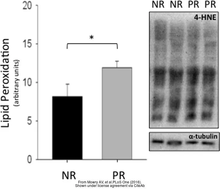
The data was published in the journal PLoS One in 2016. PMID: 27537547
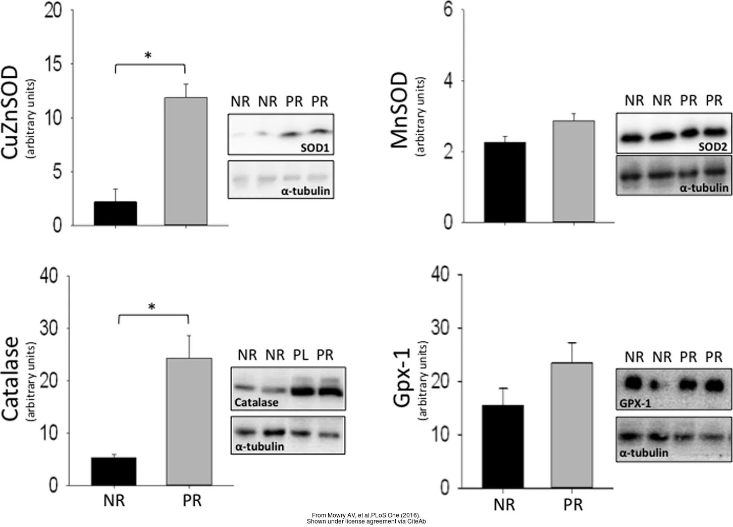
The data was published in the journal PLoS One in 2016. PMID: 27537547
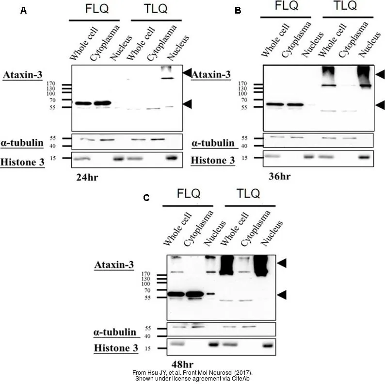
The data was published in the journal Front Mol Neurosci in 2017.PMID: 28676741
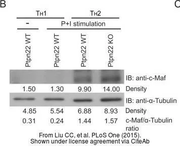
The data was published in the journal PLoS One in 2015.PMID: 25993510
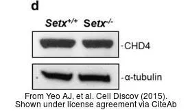
The data was published in the journal Cell Discov in 2015.PMID: 27462424
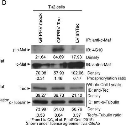
The data was published in the journal PLoS One in 2015.PMID: 25993510
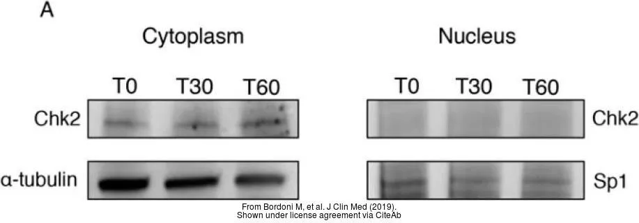
The data was published in the 2019 in J Clin Med. PMID: 31121901
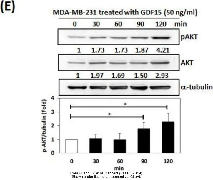
The data was published in the 2019 in Cancers (Basel). PMID: 31861872
-
HostRabbit
-
ClonalityPolyclonal
-
IsotypeIgG
-
ApplicationsWB ICC/IF IHC-P IHC-Fr
-
ReactivityHuman, Mouse, Rat, Drosophila, Chicken, Caenorhabditis elegans, Mosquito, Tetrahymena