beta Actin antibody


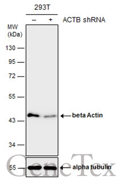
Non-transfected (–) and transfected (+) 293T whole cell extracts (10 μg) were separated by 10% SDS-PAGE, and the membrane was blotted with beta Actin antibody (GTX110564) diluted at 1:15000. The HRP-conjugated anti-rabbit IgG antibody (GTX213110-01) was used to detect the primary antibody.
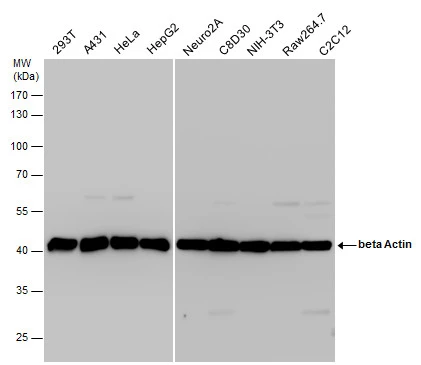
beta Actin antibody detects beta Actin protein by western blot analysis. Various whole cell extracts (30 μg) were separated by 10% SDS-PAGE, and the membrane was blotted with beta Actin antibody (GTX110564) diluted at a dilution of 1:20000. The HRP-conjugated anti-rabbit IgG antibody (GTX213110-01) was used to detect the primary antibody.
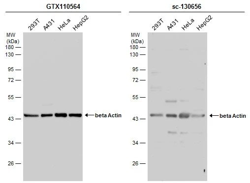
*The competitor is not affiliated with GeneTex and does not endorse this product.
Various whole cell extracts (30 μg) were separated by 10% SDS-PAGE, and the membranes were blotted with beta Actin antibody (GTX110564) diluted at 1:10000 and competitor's antibody (sc-130656) diluted at 1:100. The HRP-conjugated anti-rabbit IgG antibody (GTX213110-01) was used to detect the primary antibody.
Various whole cell extracts were separated by 10% SDS-PAGE, and the membrane was blotted with beta Actin antibody (GTX110564) diluted at 1:10000. The HRP-conjugated anti-rabbit IgG antibody (GTX213110-01) was used to detect the primary antibody.

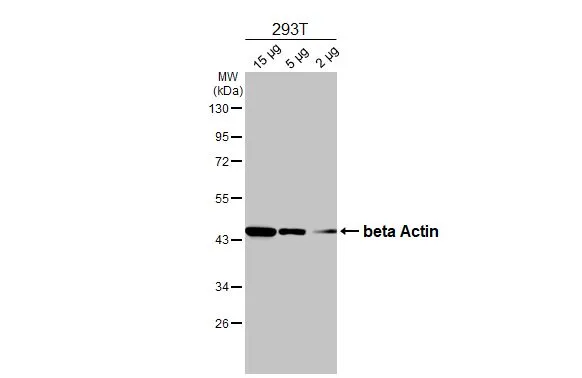
Various whole cell extracts (30 μg) were separated by 10% SDS-PAGE, and the membrane was blotted with beta Actin antibody (GTX110564) diluted at 1:10000. The HRP-conjugated anti-rabbit IgG antibody (GTX213110-01) was used to detect the primary antibody.
beta Actin antibody detects beta Actin protein at cytoplasm in mouse cervix by immunohistochemical analysis.
Sample: Paraffin-embedded mouse cervix.
beta Actin antibody (GTX110564) diluted at 1:500.
Antigen Retrieval: Citrate buffer, pH 6.0, 15 min
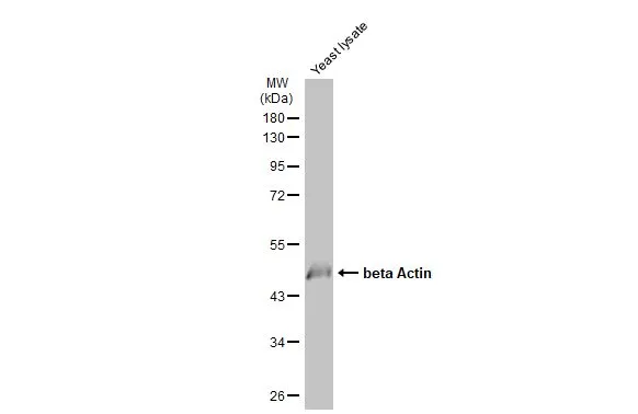
Yeast lysate (10 μg) was separated by 10% SDS-PAGE, and the membrane was blotted with beta Actin antibody (GTX110564) diluted at 1:1000. The HRP-conjugated anti-rabbit IgG antibody (GTX213110-01) was used to detect the primary antibody.
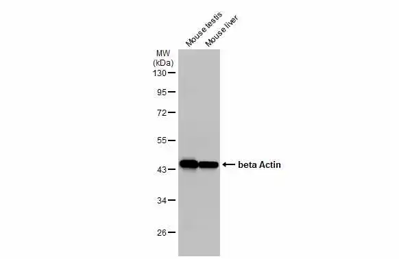
Various tissue extracts (50 μg) were separated by 10% SDS-PAGE, and the membrane was blotted with beta Actin antibody (GTX110564) diluted at 1:1000. The HRP-conjugated anti-rabbit IgG antibody (GTX213110-01) was used to detect the primary antibody.
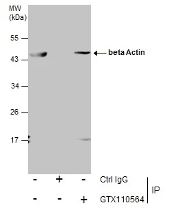
Immunoprecipitation of beta Actin protein from 293T whole cell extracts using 5 μg of beta Actin antibody (GTX110564).
Western blot analysis was performed using beta Actin antibody (GTX110564).
EasyBlot anti-Rabbit IgG (GTX221666-01) was used as a secondary reagent.
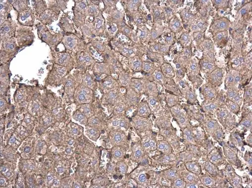
beta Actin antibody detects beta Actin protein at cell membrane and cytoplasm in rat liver by immunohistochemical analysis.
Sample: Paraffin-embedded rat liver.
beta Actin antibody (GTX110564) diluted at 1:500.
Antigen Retrieval: Citrate buffer, pH 6.0, 15 min
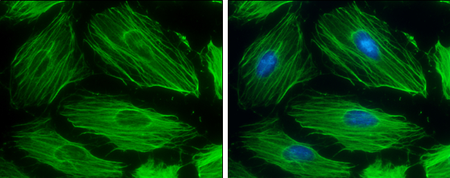
beta Actin antibody detects beta Actin protein at cytoskeleton by immunofluorescent analysis.
Sample: HeLa cells were fixed in 0.5% Triton X-100 for 1 min, then ice-cold methanol for 5 min.
Green: beta Actin protein stained by beta Actin antibody (GTX110564) diluted at 1:500.
Blue: Hoechst 33342 staining.
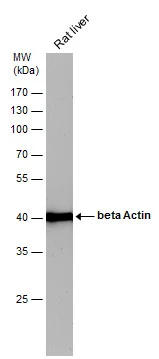
beta Actin antibody detects beta Actin protein by western blot analysis. Rat tissue extracts (50 μg) was separated by 10% SDS-PAGE, and the membrane was blotted with beta Actin antibody (GTX110564) diluted by 1:20000. The HRP-conjugated anti-rabbit IgG antibody (GTX213110-01) was used to detect the primary antibody.
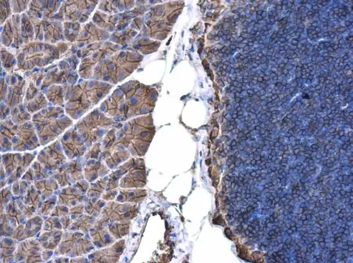
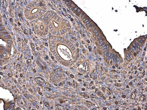
beta Actin antibody detects beta Actin protein at cell membrane and cytoplasm in mouse pancreas by immunohistochemical analysis.
Sample: Paraffin-embedded mouse pancreas.
beta Actin antibody (GTX110564) diluted at 1:500.
Antigen Retrieval: Citrate buffer, pH 6.0, 15 min
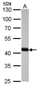
beta Actin antibody detects ACTB protein by western blot analysis.
A. 30 μg drosophila lysate/extract
10% SDS-PAGE
beta Actin antibody (GTX110564) dilution: 1:10000
The HRP-conjugated anti-rabbit IgG antibody (GTX213110-01) was used to detect the primary antibody.
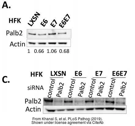
The data was published in the journal PLoS Pathog in 2019. PMID: 30818369
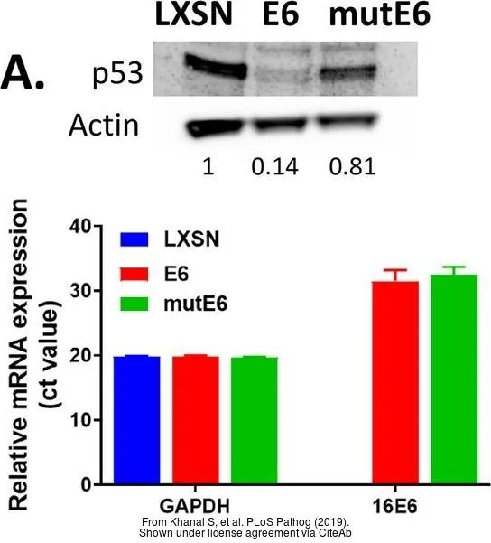
The data was published in the journal PLoS Pathog in 2019. PMID: 30818369
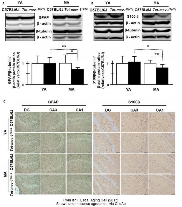
The data was published in the journal Aging Cell in 2017. PMID: 27623715
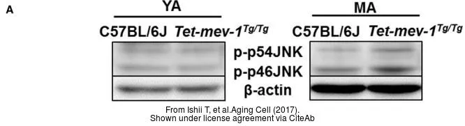
The data was published in the journal Aging Cell in 2017. PMID: 27623715
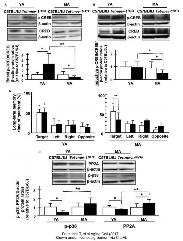
The data was published in the journal Aging Cell in 2017. PMID: 27623715
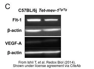
The data was published in the journal Redox Biol in 2014. PMID: 24936442
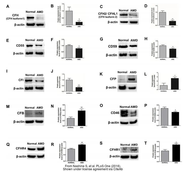
The data was published in the journal PLoS One in 2016.PMID: 27486856
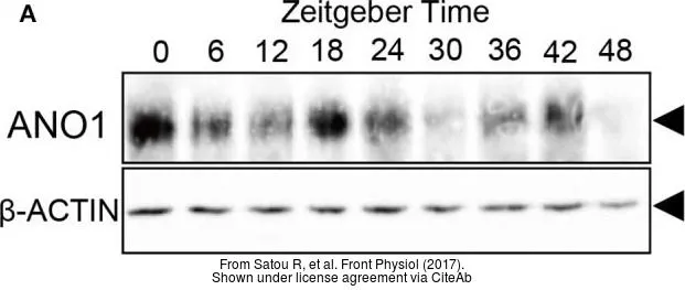
The data was published in the journal Front Physiol in 2017.PMID: 28588500
-
HostRabbit
-
ClonalityPolyclonal
-
IsotypeIgG
-
ApplicationsWB ICC/IF IHC-P IP IHC
-
ReactivityHuman, Mouse, Rat, Yeast, Drosophila