beta Tubulin 2 antibody
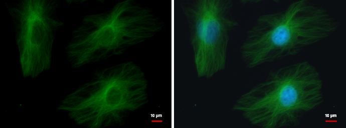
TUBB2A antibody detects TUBB2A protein at cytoskeleton by immunofluorescent analysis.
Sample: HeLa cells were fixed in 4% paraformaldehyde/PBS for 15 min.
Green: TUBB2A protein stained by TUBB2A antibody (GTX100117) diluted at 1:500.
Blue: Hoechst 33342 staining.
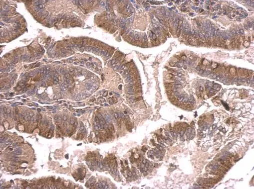
beta Tubulin 2 antibody detects beta Tubulin 2 protein at cytosol on mouse duodenum by immunohistochemical analysis.
Sample: Paraffin-embedded mouse duodenum.
beta Tubulin 2 antibody (GTX100117) dilution: 1:500.
Antigen Retrieval: Trilogy™ (EDTA based, pH 8.0) buffer, 15min
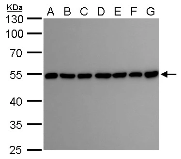
beta Tubulin 2 antibody detects beta Tubulin 2 protein by Western blot analysis.
A. 30 μg Neuro2A whole cell lysate/extract
B. 30 μg GL261 whole cell lysate/extract
C. 30 μg C8D30 whole cell lysate/extract
D. 30 μg NIH-3T3 whole cell lysate/extract
E. 30 μg BCL-1 whole cell lysate/extract
F. 30 μg Raw 264.7 whole cell lysate/extract
G. 30 μg C2Cl2 whole cell lysate/extract
10 % SDS-PAGE
beta Tubulin 2 antibody (GTX100117) dilution: 1:5000
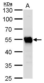
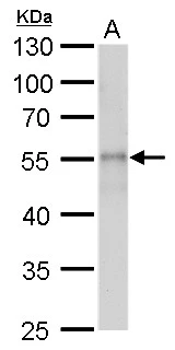
beta Tubulin 2 antibody detects beta Tubulin 2 protein by Western blot analysis.
A. 50 μg rat brain lysate/extract
10 % SDS-PAGE
beta Tubulin 2 antibody (GTX100117) dilution: 1:5000
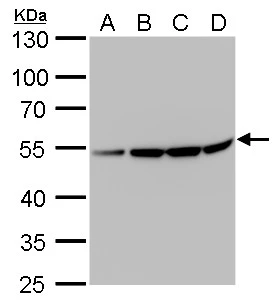
beta Tubulin 2 antibody detects beta Tubulin 2 protein by Western blot analysis.
A. 30 μg U87-MG whole cell lysate/extract
B. 30 μg SK-N-SH whole cell lysate/extract
C. 30 μg IMR32 whole cell lysate/extract
D. 30 μg SK-N-AS whole cell lysate/extract
10 % SDS-PAGE
beta Tubulin 2 antibody (GTX100117) dilution: 1:10000
beta Tubulin 2 antibody detects beta Tubulin 2 protein by Western blot analysis.
A. 30 μg drosophila lysate/extract
10 % SDS-PAGE
beta Tubulin 2 antibody (GTX100117) dilution: 1:2000
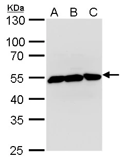
beta Tubulin 2 antibody detects beta Tubulin 2 protein by Western blot analysis.
A. 30 μg Jurkat whole cell lysate/extract
B. 30 μg Raji whole cell lysate/extract
C. 30 μg NCI-H929 whole cell lysate/extract
10 % SDS-PAGE
beta Tubulin 2 antibody (GTX100117) dilution: 1:5000
-
HostRabbit
-
ClonalityPolyclonal
-
IsotypeIgG
-
ApplicationsWB ICC/IF IHC-P
-
ReactivityHuman, Mouse, Rat, Drosophila