APPLICATION
Application Note
*Optimal dilutions/concentrations should be determined by the researcher.
Application |
Recommended Dilution |
1:1000-1:10000 |
1:100-1:1000 |
Not tested in other applications.
Calculated MW
Positive Control
MCF-7 (500nM PMA treatment for 0.5hr) , MCF-7 (500nM PMA treatment for 1hr)
Predict Reactivity
Mouse, Sheep, Bovine, Cat, Pig, Chimpanzee, Rhesus Monkey(>80% identity)
PROPERTIES
Form
Liquid
Buffer
PBS, 20% Glycerol
Preservative
0.01% Thimerosal
Storage
Store as concentrated solution. Centrifuge briefly prior to opening vial. For short-term storage (1-2 weeks), store at 4ºC. For long-term storage, aliquot and store at -20ºC or below. Avoid multiple freeze-thaw cycles.
Concentration
1 mg/ml (Please refer to the vial label for the specific concentration.)
Antigen Species
Human
Immunogen
Recombinant protein encompassing a sequence within the center region of human c-Fos. The exact sequence is proprietary.
Purification
Purified by antigen-affinity chromatography.
Conjugation
Unconjugated
RRID
AB_2885225
Note
For laboratory research use only. Not for any clinical, therapeutic, or diagnostic use in humans or animals. Not for animal or human consumption.
Purchasers shall not, and agree not to enable third parties to, analyze, copy, reverse engineer or otherwise attempt to determine the structure or sequence of the product.
TARGET
Synonyms
Fos proto-oncogene, AP-1 transcription factor subunit , AP-1 , C-FOS , p55
Cellular Localization
Nucleus
Background
The Fos gene family consists of 4 members: FOS, FOSB, FOSL1, and FOSL2. These genes encode leucine zipper proteins that can dimerize with proteins of the JUN family, thereby forming the transcription factor complex AP-1. As such, the FOS proteins have been implicated as regulators of cell proliferation, differentiation, and transformation. In some cases, expression of the FOS gene has also been associated with apoptotic cell death. [provided by RefSeq]
Database
Research Area
DATA IMAGES
|
GTX101196 WB Image
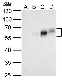
c-Fos antibody detects FOS protein by Western blot analysis. A. 30 μg MCF-7 whole cell lysate/extract (serum starvation) B. 30 μg MCF-7 whole cell lysate/extract (500nM PMA treatment for 0.5hr) C. 30 μg MCF-7 whole cell lysate/extract (500nM PMA treatment for 1hr) D. 30 μg MCF-7 whole cell lysate/extract (500nM PMA treatment for 2hr) 10 % SDS-PAGE c-Fos antibody (GTX101196) dilution: 1:5000
|
|
GTX101196 ICC/IF Image
c-Fos antibody detects c-Fos protein at cytoplasm and nucleus by immunofluorescent analysis. Sample: HeLa cells were fixed in 4% paraformaldehyde at RT for 15 min. Green: c-Fos protein stained by c-Fos antibody (GTX101196) diluted at 1:500. Red: phalloidin, a cytoskeleton marker, diluted at 1:50. Scale bar = 10 μm.
|
|
GTX101196 ICC/IF Image
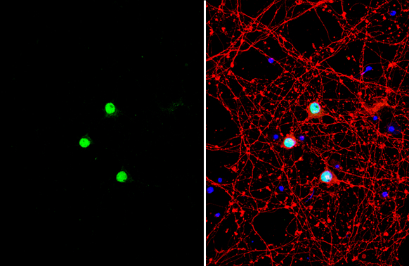
c-Fos antibody detects c-Fos protein by immunofluorescent analysis.Sample: DIV9 rat E18 primary cortical neuron cells were fixed in 4% paraformaldehyde at RT for 15 min.Green: c-Fos stained by c-Fos antibody (GTX101196) diluted at 1:1500.Red: beta Tubulin 3/ Tuj1, stained by beta Tubulin 3/ Tuj1 antibody [GT11710] (GTX631836) diluted at 1:500.Blue: Fluoroshield with DAPI (GTX30920).
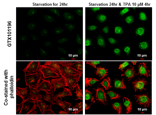
|
REFERENCE
There are currently no references for c-Fos antibody (GTX101196). Be the first to share your publications with this product.
REVIEW
There are currently no reviews for c-Fos antibody (GTX101196). Be the first to share your experience with this product.