c-Jun (phospho Ser73) antibody


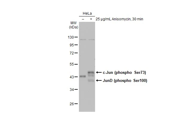
Untreated (–) and treated (+) HeLa whole cell extracts (30 μg) were separated by 10% SDS-PAGE, and the membrane was blotted with c-Jun (phospho Ser73) antibody (GTX133873) diluted at 1:500. The HRP-conjugated anti-rabbit IgG antibody (GTX213110-01) was used to detect the primary antibody.
c-Jun (phospho Ser73) antibody detects c-Jun (phospho Ser73) protein at nucleus by immunohistochemical analysis.Sample: Paraffin-embedded mouse colon.c-Jun (phospho Ser73) stained by c-Jun (phospho Ser73) antibody (GTX133873) diluted at 1:500.Antigen Retrieval: Citrate buffer, pH 6.0, 15 min
*The competitor is not affiliated with GeneTex and does not endorse this product.
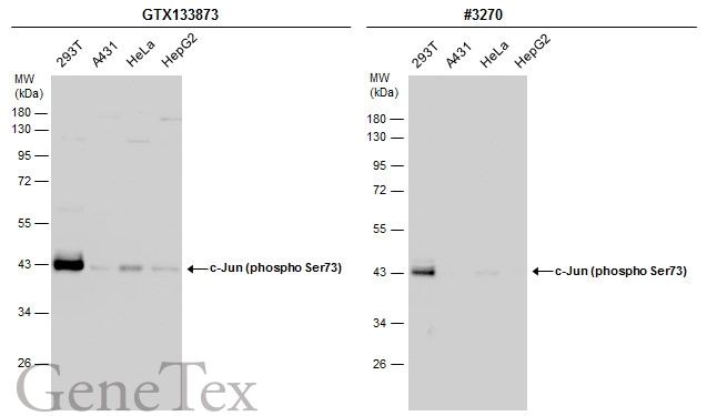
Various whole cell extracts (30 μg) were separated by 10% SDS-PAGE, and the membranes were blotted with c-Jun (phospho Ser73) antibody (GTX133873) diluted at 1:500 and competitor's antibody (CST#3270) diluted at 1:500. The HRP-conjugated anti-rabbit IgG antibody (GTX213110-01) was used to detect the primary antibody.
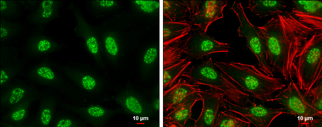
c-Jun (phospho Ser73) antibody detects c-Jun (phospho Ser73) protein at nucleus by immunofluorescent analysis.Sample: HeLa cells were fixed in 4% paraformaldehyde at RT for 15 min.Green: c-Jun (phospho Ser73) stained by c-Jun (phospho Ser73) antibody (GTX133873) diluted at 1:500.Red: phalloidin, a cytoskeleton marker, diluted at 1:100.Scale bar= 10μm.
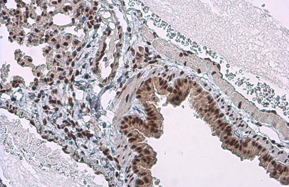
c-Jun (phospho Ser73) antibody detects c-Jun (phospho Ser73) protein at nucleus by immunohistochemical analysis.Sample: Paraffin-embedded mouse lung.c-Jun (phospho Ser73) stained by c-Jun (phospho Ser73) antibody (GTX133873) diluted at 1:500.Antigen Retrieval: Citrate buffer, pH 6.0, 15 min
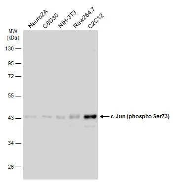
Various whole cell extracts (30 μg) were separated by 10% SDS-PAGE, and the membrane was blotted with c-Jun (phospho Ser73) antibody (GTX133873) diluted at 1:500. The HRP-conjugated anti-rabbit IgG antibody (GTX213110-01) was used to detect the primary antibody.
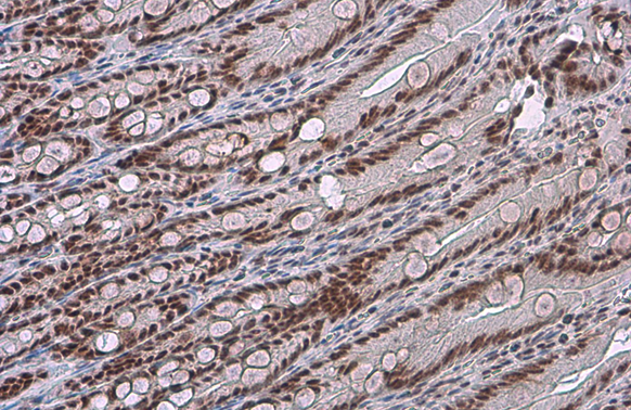
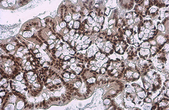
c-Jun (phospho Ser73) antibody detects c-Jun (phospho Ser73) protein at nucleus by immunohistochemical analysis.Sample: Paraffin-embedded rat duodenum.c-Jun (phospho Ser73) stained by c-Jun (phospho Ser73) antibody (GTX133873) diluted at 1:500.Antigen Retrieval: Citrate buffer, pH 6.0, 15 min
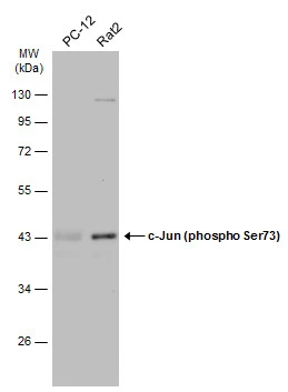
Various whole cell extracts (30 μg) were separated by 10% SDS-PAGE, and the membrane was blotted with c-Jun (phospho Ser73) antibody (GTX133873) diluted at 1:500. The HRP-conjugated anti-rabbit IgG antibody (GTX213110-01) was used to detect the primary antibody.
-
HostRabbit
-
ClonalityPolyclonal
-
IsotypeIgG
-
ApplicationsWB ICC/IF IHC-P
-
ReactivityHuman, Mouse, Rat