eRF1 antibody
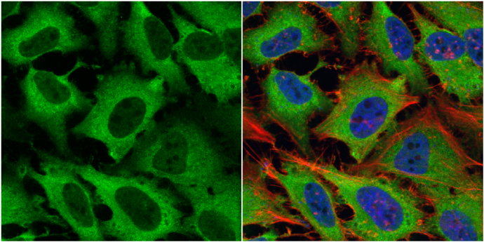
eRF1 antibody detects eRF1 protein at cytoplasm by immunofluorescent analysis.
Sample: HeLa cells were fixed in 4% paraformaldehyde at RT for 15 min.
Green: eRF1 protein stained by eRF1 antibody (GTX108271) diluted at 1:500.
Red: phalloidin, a cytoskeleton marker, stained by phalloidin (invitrogen, A12380) diluted at 1:200.
Blue: Hoechst 33342 staining.
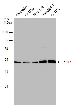
Various whole cell extracts (30 μg) were separated by 10% SDS-PAGE, and the membrane was blotted with eRF1 antibody (GTX108271) diluted at 1:500.
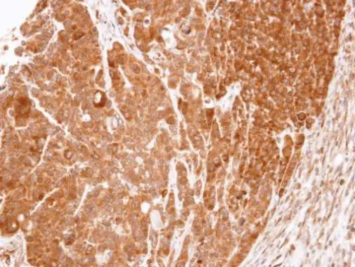
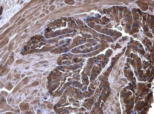
Immunohistochemical analysis of paraffin-embedded SW480 xenograft, using eRF1(GTX108271) antibody at 1:100 dilution.
Antigen Retrieval: Trilogy™ (EDTA based, pH 8.0) buffer, 15min
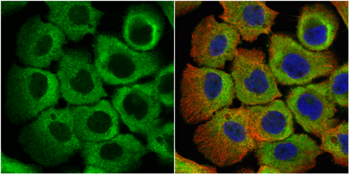
eRF1 antibody detects eRF1 protein at cytoplasm by immunofluorescent analysis.
Sample: A431 cells were fixed in 4% paraformaldehyde at RT for 15 min.
Green: eRF1 protein stained by eRF1 antibody (GTX108271) diluted at 1:500.
Red: alpha Tubulin, a cytoskeleton marker, stained by alpha Tubulin antibody [GT114] (GTX628802) diluted at 1:1000.
Blue: Hoechst 33342 staining.

eRF1 antibody detects eRF1 protein at cytoplasm on mouse testis by immunohistochemical analysis.
Sample: Paraffin-embedded mouse testis.
eRF1 antibody (GTX108271) diluted at 1:500.
Antigen Retrieval: Trilogy™ (EDTA based, pH 8.0) buffer, 15min
eRF1 antibody detects eRF1 protein at cytoplasm on mouse testis by immunohistochemical analysis.
Sample: Paraffin-embedded mouse testis.
eRF1 antibody (GTX108271) diluted at 1:500.
Antigen Retrieval: Trilogy™ (EDTA based, pH 8.0) buffer, 15min
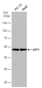
Various whole cell extracts (30 μg) were separated by 10% SDS-PAGE, and the membrane was blotted with eRF1 antibody (GTX108271) diluted at 1:500.
-
HostRabbit
-
ClonalityPolyclonal
-
IsotypeIgG
-
ApplicationsWB ICC/IF IHC-P
-
ReactivityHuman, Mouse, Rat