eRF1 antibody
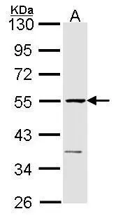
Sample (30 ug of whole cell lysate)
A: Raji
10% SDS PAGE
eRF1 antibody
GTX108296 diluted at 1:1000
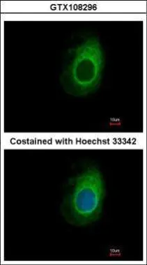
Immunofluorescence analysis of methanol-fixed HeLa, using eRF1(GTX108296) antibody at 1:500 dilution.
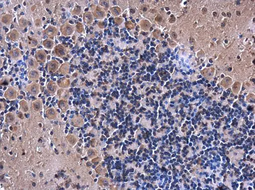
eRF1 antibody detects eRF1 protein at cytoplasm in mouse brain by immunohistochemical analysis.
Sample: Paraffin-embedded mouse brain.
eRF1 antibody (GTX108296) diluted at 1:500.
Antigen Retrieval: Citrate buffer, pH 6.0, 15 min
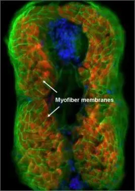
eRF1 antibody detects eRF1 protein on myofiber membranes by immunohistochemical analysis Sample: Agarose-embedded zebrafish embryo eRF1 antibody (GTX108296) dilution: 1:200. Image provided with permission courtesy of Dr. T. Schilling at UC, Irvine.
-
HostRabbit
-
ClonalityPolyclonal
-
IsotypeIgG
-
ApplicationsWB ICC/IF IHC-P IHC
-
ReactivityHuman, Mouse, Zebrafish