GeneTex
  • Country / Location Selection

United States (US)

nNOS antibody

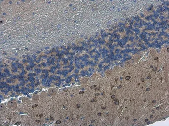
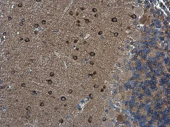
Anti-nNOS antibody used in IHC (Paraffin sections) (IHC-P). GTX133407
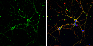
Anti-nNOS antibody used in Immunocytochemistry/ Immunofluorescence (ICC/IF). GTX133407
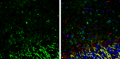
Anti-nNOS antibody used in IHC (Frozen sections) (IHC-Fr). GTX133407
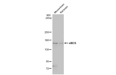
Anti-nNOS antibody used in Western Blot (WB). GTX133407

Cat. No. GTX133407

Host

Rabbit

Clonality

Polyclonal

Isotype

IgG

Application

WB, ICC/IF, IHC-P, IHC-Fr, IHC-Wm

Reactivity

Mouse, Rat, Zebrafish
Package
100 μl ($399),
25 μl ($169)

APPLICATION

Application Note

*Optimal dilutions/concentrations should be determined by the researcher.
Application Recommended Dilution
WB 1:500-1:3000
ICC/IF 1:100-1:1000
IHC-P 1:100-1:1000
IHC-Fr 1:100-1:1000
IHC-Wm Assay dependent
Not tested in other applications.

Calculated MW

161 kDa. ( Note )

Predict Reactivity

Human, Rabbit, Dog, Japanese Medaka(>80% identity)

PROPERTIES

Form

Liquid

Buffer

PBS, 20% Glycerol

Preservative

0.025% ProClin 300

Storage

Store as concentrated solution. Centrifuge briefly prior to opening vial. For short-term storage (1-2 weeks), store at 4ºC. For long-term storage, aliquot and store at -20ºC or below. Avoid multiple freeze-thaw cycles.

Concentration

0.79 mg/ml (Please refer to the vial label for the specific concentration.)

Antigen Species

Human

Immunogen

Carrier-protein conjugated synthetic peptide encompassing a sequence within the C-terminus region of human nNOS. The exact sequence is proprietary.

Purification

Purified by antigen-affinity chromatography.

Conjugation

Unconjugated

RRID

AB_2886970

Note

For laboratory research use only. Not for any clinical, therapeutic, or diagnostic use in humans or animals. Not for animal or human consumption.

Purchasers shall not, and agree not to enable third parties to, analyze, copy, reverse engineer or otherwise attempt to determine the structure or sequence of the product.

TARGET

Synonyms

nitric oxide synthase 1 , IHPS1 , N-NOS , NC-NOS , NOS , bNOS , nNOS

Cellular Localization

Cell membrane , sarcolemma; Peripheral membrane protein , Cell projection , dendritic spine

Background

Nitric oxide (NO) is a messenger molecule with diverse functions throughout the body. In the brain and peripheral nervous system, NO displays many properties of a neurotransmitter; it is implicated in neurotoxicity associated with stroke and neurodegenerative diseases, neural regulation of smooth muscle, including peristalsis, and penile erection. NO is also responsible for endothelium-derived relaxing factor activity regulating blood pressure. In macrophages, NO mediates tumoricidal and bactericidal actions, as indicated by the fact that inhibitors of NO synthase (NOS) block these effects. Neuronal NOS and macrophage NOS (MIM 163730) are distinct isoforms (Lowenstein et al., 1992 [PubMed 1379716]). Both the neuronal and the macrophage forms are unusual among oxidative enzymes in requiring several electron donors: FAD (see MIM 610595), flavin mononucleotide (FMN), NADPH, and tetrahydrobiopterin.[supplied by OMIM]

Database

Research Area

DATA IMAGES

Anti-nNOS antibody used in IHC (Paraffin sections) (IHC-P). GTX133407

GTX133407 IHC-P Image

nNOS antibody detects nNOS protein at cytoplasm in rat brain by immunohistochemical analysis.
Sample: Paraffin-embedded rat brain.
nNOS antibody (GTX133407) diluted at 1:500.

Antigen Retrieval: Citrate buffer, pH 6.0, 10 min

Anti-nNOS antibody used in IHC (Paraffin sections) (IHC-P). GTX133407

GTX133407 IHC-P Image

nNOS antibody detects nNOS protein at cytoplasm in mouse brain by immunohistochemical analysis.
Sample: Paraffin-embedded mouse brain.
nNOS antibody (GTX133407) diluted at 1:500.

Antigen Retrieval: Citrate buffer, pH 6.0, 10 min

Anti-nNOS antibody used in Immunocytochemistry/ Immunofluorescence (ICC/IF). GTX133407

GTX133407 ICC/IF Image

nNOS antibody detects nNOS protein by immunofluorescent analysis.
Sample: DIV9 rat E18 primary cortical neuron cells were fixed in 4% paraformaldehyde at RT for 15 min.
Green: nNOS stained by nNOS antibody (GTX133407) diluted at 1:500.
Red: beta Tubulin 3/ Tuj1, stained by beta Tubulin 3/ Tuj1 antibody [GT1338] (GTX631831) diluted at 1:500.
Blue: Fluoroshield with DAPI (GTX30920).

Anti-nNOS antibody used in IHC (Frozen sections) (IHC-Fr). GTX133407

GTX133407 IHC-Fr Image

nNOS antibody detects nNOS protein expression by immunohistochemical analysis.
Sample: Frozen-sectioned adult mouse cerebellum.
Green: nNOS protein stained by nNOS antibody (GTX133407) diluted at 1:250.
Red: NF-H, stained by NF-H antibody [GT114] (GTX634289) diluted at 1:500.
Blue: Fluoroshield with DAPI (GTX30920).

Antigen Retrieval: Citrate buffer, pH 6.0, 10 min

Anti-nNOS antibody used in Western Blot (WB). GTX133407

GTX133407 WB Image

Various tissue extracts (50 μg) were separated by 5% SDS-PAGE, and the membrane was blotted with nNOS antibody (GTX133407) diluted at 1:1000. The HRP-conjugated anti-rabbit IgG antibody (GTX213110-01) was used to detect the primary antibody.

REFERENCE

REVIEW

There are currently no reviews for nNOS antibody (GTX133407). Be the first to share your experience with this product.
SDS
PBS.pdf
Glycerol.pdf
Proclin.pdf
Package List Price ($)
$ 399
$ 169