p21 Cip1 antibody

Cat. No. GTX112898
Cat. No.  GTX112898
Anti-p21 Cip1 antibody used in Western Blot (WB). GTX112898
GTX112898 WB Image

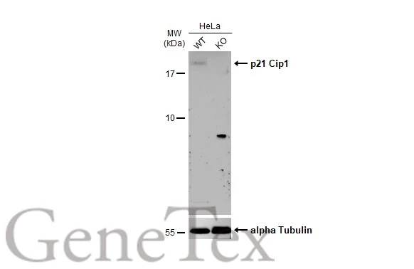
Wild-type (WT) and p21 Cip1 knockout (KO) HeLa cell extracts (30 μg) were separated by 15% SDS-PAGE, and the membrane was blotted with p21 Cip1 antibody (GTX112898) diluted at 1:500. The HRP-conjugated anti-rabbit IgG antibody (GTX213110-01) was used to detect the primary antibody, and the signal was developed with Trident ECL plus-Enhanced.
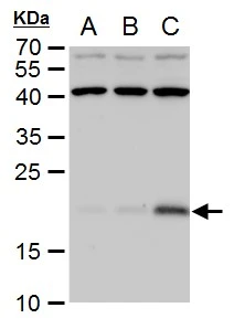
1 / 10
Anti-p21 Cip1 antibody used in IHC (Paraffin sections) (IHC-P). GTX112898
GTX112898 IHC-P Image

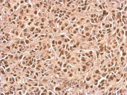
p21 Cip1 antibody detects CDKN1A protein at nucleus on SkHep1xenograft by immunohistochemical analysis.
Sample: Paraffin-embedded SkHep1xenograft.
p21 Cip1 antibody (GTX112898) dilution: 1:500.

Antigen Retrieval: Trilogy™ (EDTA based, pH 8.0) buffer, 15min

2 / 10
Anti-p21 Cip1 antibody used in Western Blot (WB). GTX112898
GTX112898 WB Image

Sample (30 ug of whole cell lysate)
A: HCT116
B: HCT116 treated with 30uM cisplatin for 24hr
12% SDS PAGE
GTX112898 diluted at 1:1000

3 / 10
Anti-p21 Cip1 antibody used in Immunocytochemistry/ Immunofluorescence (ICC/IF). GTX112898
GTX112898 ICC/IF Image

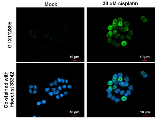
p21 Cip1 antibody detects p21 Cip1 protein at nucleus by immunofluorescent analysis.
Samples: HeLa cells mock (left) and treated with 30 μM Cisplatin for 24 hrs (right) were fixed in 4% paraformaldehyde at RT for 15 min.
Green: p21 Cip1 protein stained by p21 Cip1 antibody (GTX112898) diluted at 1:500.
Blue: Hoechst 33342 staining.
Scale bar = 10 μm.

4 / 10
Anti-p21 Cip1 antibody used in IHC (Paraffin sections) (IHC-P). GTX112898
GTX112898 IHC-P Image

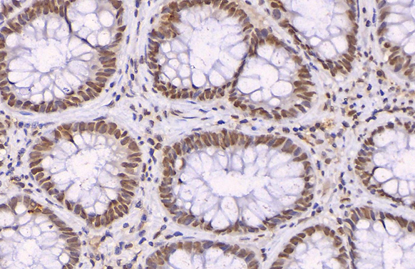
p21 Cip1 antibody detects p21 Cip1 protein at nucleus by immunohistochemical analysis (Autostainer Formulated).
Sample: Paraffin-embedded human normal colon tissue.
p21 Cip1 stained by p21 Cip1 antibody (GTX112898) diluted at 1:500.
Antigen Retrieval: EDTA buffer, 20 min

5 / 10
Anti-p21 Cip1 antibody used in Western Blot (WB). GTX112898
GTX112898 WB Image

The data was published in the journal Front Genet in 2020. PMID: 32158470

6 / 10
Anti-p21 Cip1 antibody used in Western Blot (WB). GTX112898
GTX112898 WB Image

The data was published in the journal Front Genet in 2020. PMID: 32158470

7 / 10
Anti-p21 Cip1 antibody used in Western Blot (WB). GTX112898
GTX112898 WB Image

The data was published in the journal Front Physiol in 2018.PMID: 30333755

8 / 10
Anti-p21 Cip1 antibody used in Western Blot (WB). GTX112898
GTX112898 WB Image

The data was published in the journal Cell Death Dis in 2018.PMID: 29511169

9 / 10
Anti-p21 Cip1 antibody used in Western Blot (WB). GTX112898
GTX112898 WB Image

The data was published in the journal Cell Death Dis in 2018.PMID: 29511169

10 / 10
Anti-p21 Cip1 antibody used in Western Blot (WB). GTX112898
Anti-p21 Cip1 antibody used in IHC (Paraffin sections) (IHC-P). GTX112898
Anti-p21 Cip1 antibody used in Western Blot (WB). GTX112898
Anti-p21 Cip1 antibody used in Immunocytochemistry/ Immunofluorescence (ICC/IF). GTX112898
Anti-p21 Cip1 antibody used in IHC (Paraffin sections) (IHC-P). GTX112898
Anti-p21 Cip1 antibody used in Western Blot (WB). GTX112898
  • Host
    Rabbit
  • Clonality
    Polyclonal
  • Isotype
    IgG
  • Applications
    WB ICC/IF IHC-P
  • Reactivity
    Human, Chicken
See all p21 Cip1 products