p21 Cip1 antibody


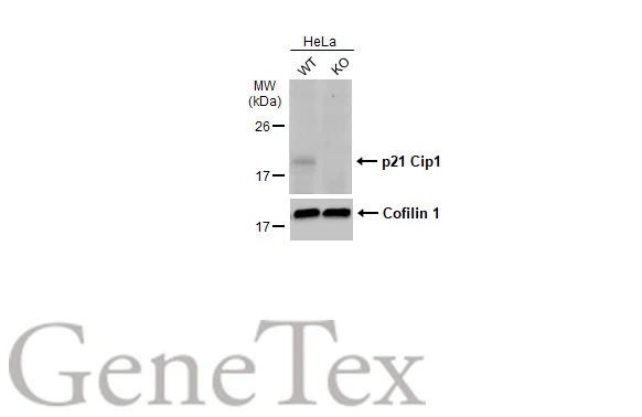
Wild-type (WT) and p21 Cip1 knockout (KO) HeLa cell extracts (30 μg) were separated by 15% SDS-PAGE, and the membrane was blotted with p21 Cip1 antibody (GTX135142) diluted at 1:1000. The HRP-conjugated anti-rabbit IgG antibody (GTX213110-01) was used to detect the primary antibody, and the signal was developed with Trident ECL plus-Enhanced.
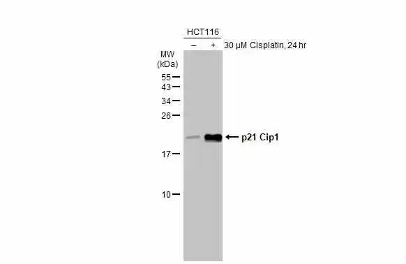
Untreated (–) and treated (+) HCT116 whole cell extract (30 μg) were separated by 15% SDS-PAGE, and the membrane was blotted with p21 Cip1 antibody (GTX135142) diluted at 1:1000. The HRP-conjugated anti-rabbit IgG antibody (GTX213110-01) was used to detect the primary antibody.
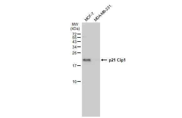
Various whole cell extracts (30 μg) were separated by 15% SDS-PAGE, and the membrane was blotted with p21 Cip1 antibody (GTX135142) diluted at 1:1000. The HRP-conjugated anti-rabbit IgG antibody (GTX213110-01) was used to detect the primary antibody.
-
HostRabbit
-
ClonalityPolyclonal
-
IsotypeIgG
-
ApplicationsWB
-
ReactivityHuman