p27 Kip1 antibody

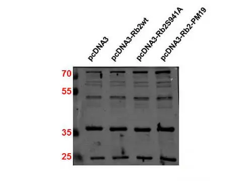
WB analysis of non-transfected and transfected 293T whole cell lysate using GTX26547 p27 Kip1 antibody.
Loading : 30 μg
Dilution : 1:2000
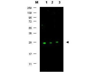
WB analysis of MCF-7 whole cell lysate using GTX26547 p27 Kip1 antibody.
Dilution : 1:500

Western blot using GeneTex affinity purified anti-p27 antibody (GTX26547) shows detection of p27 protein in MCF7 whole cell lysate (lanes 1-3) (arrowhead). Separation was achieved using a 4-20% gradient gel. Blocking occurred using 5% BLOTTO. Primary antibody was diluted 1:500 in 1% BLOTTO. The membrane was washed and reacted with a 1:10,000 dilution of Dylight™ 800 conjugated Goat anti Rabbit IgG. Molecular weight estimation was made by comparison to prestained MW markers indicated at the left (lane M). Other detection systems will yield similar results.
-
HostRabbit
-
ClonalityPolyclonal
-
IsotypeIgG
-
ApplicationsWB IP ELISA
-
ReactivityHuman