p53 antibody


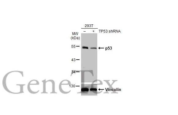
Non-transfected (–) and transfected (+) 293T whole cell extracts (30 μg) were separated by 10% SDS-PAGE, and the membrane was blotted with p53 antibody (GTX102965) diluted at 1:1000. The HRP-conjugated anti-rabbit IgG antibody (GTX213110-01) was used to detect the primary antibody.
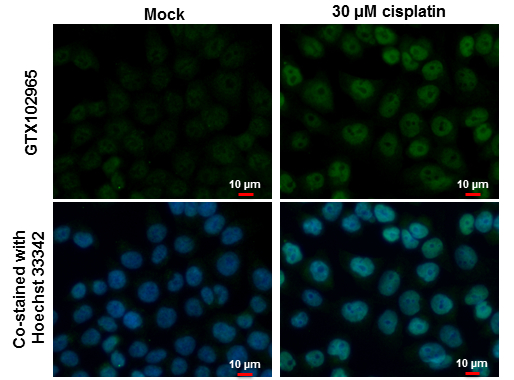
p53 antibody detects p53 protein at nucleus by immunofluorescent analysis.
Samples: HCT 116 cells mock (left) and treated with 30 μM Cisplatin for 24 hrs (right) were fixed in 4% paraformaldehyde at RT for 15 min.
Green: p53 protein stained by p53 antibody (GTX102965) diluted at 1:500.
Blue: Hoechst 33342 staining.
Scale bar = 10 μm.
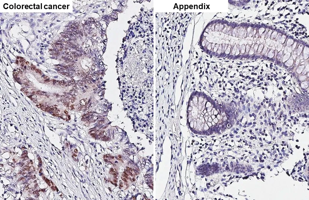
p53 antibody detects p53 protein by immunohistochemical analysis.Sample: Paraffin-embedded human tissues.p53 stained by p53 antibody (GTX102965) diluted at 1:500.Antigen Retrieval: Citrate buffer, pH 6.0, 15 min
Immunoprecipitation of p53 protein. HCT116 lysates with 30uM cisplatin treatment for 24 hours were subjected to immunoprecipitation using (B) normal rabbit IgG or (C) 2.5 ug of anti-p53 antibody (GTX102965). (A) Input, 20ug of HCT116 lysates. The precipitated protein was detected by GTX102965 diluted at 1:10000. EasyBlot anti-Rabbit IgG Kit (GTX225856-01) was used in Western blot.
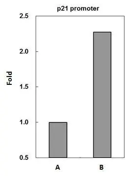
p53 antibody immunoprecipitates p53 protein-DNA complex in ChIP experiments. ChIP Sample: HCT116 whole cell lysate/extract treated with CPT 500nM for 6hr
A. 5 μg preimmune rabbit IgG
B. 5 μg of p53 antibody (GTX102965)
The precipitated DNA was detected by PCR with primer set targeting to p21 promoter.
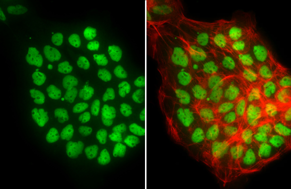
p53 antibody detects p53 protein at nucleus by immunofluorescent analysis.Sample: A431 cells were fixed in 4% paraformaldehyde at RT for 15 min.Green: p53 stained by p53 antibody (GTX102965) diluted at 1:500.Red: phalloidin, a cytoskeleton marker, diluted at 1:200.
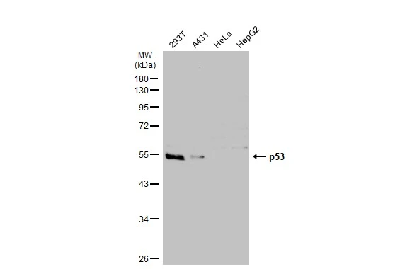
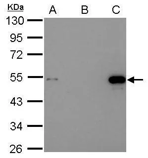
Various whole cell extracts (30 μg) were separated by 10% SDS-PAGE, and the membrane was blotted with p53 antibody (GTX102965) diluted at 1:1000. The HRP-conjugated anti-rabbit IgG antibody (GTX213110-01) was used to detect the primary antibody.
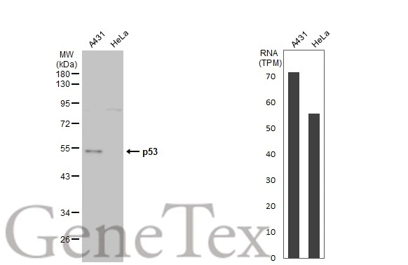
Various whole cell extracts (30 μg) were separated by 10% SDS-PAGE, and the membrane was blotted with p53 antibody (GTX102965) diluted at 1:1000. The HRP-conjugated anti-rabbit IgG antibody (GTX213110-01) was used to detect the primary antibody. Corresponding RNA expression data for the same cell lines are based on Human Protein Atlas program.
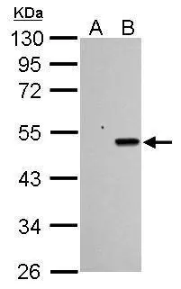
Sample (30 μg of whole cell lysate)
A: HCT116 cells with mock treatment for 24 hr
B: HCT116 cells with 30 μM cisplatin treatment for 24 hr
10% SDS PAGE
GTX102965 diluted at 1:1000
The HRP-conjugated anti-rabbit IgG antibody (GTX213110-01) was used to detect the primary antibody.
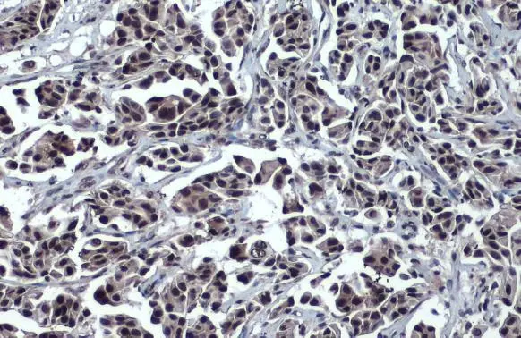
p53 antibody detects p53 protein at nucleus by immunohistochemical analysis.Sample: Paraffin-embedded human breast carcinoma.p53 stained by p53 antibody (GTX102965) diluted at 1:500.Antigen Retrieval: Citrate buffer, pH 6.0, 15 min
-
HostRabbit
-
ClonalityPolyclonal
-
IsotypeIgG
-
ApplicationsWB ICC/IF IHC-P IP ChIP assay
-
ReactivityHuman, Zebrafish