GeneTex
  • Country / Location Selection

United States (US)

GFP antibody

Anti-GFP antibody used in IHC (Whole mount) (IHC-Wm). GTX113617
Anti-GFP antibody used in Western Blot (WB). GTX113617
Anti-GFP antibody used in Immunoprecipitation (IP). GTX113617
Anti-GFP antibody used in Immunocytochemistry/ Immunofluorescence (ICC/IF). GTX113617
Anti-GFP antibody used in Western Blot (WB). GTX113617
Anti-GFP antibody used in Western Blot and Immunoprecipitation (WB IP). GTX113617
Anti-GFP antibody used in Western Blot (WB). GTX113617
Anti-GFP antibody used in IHC (Paraffin sections) (IHC-P). GTX113617
Anti-GFP antibody used in Western Blot (WB). GTX113617
Anti-GFP antibody used in Western Blot and Immunoprecipitation (WB IP). GTX113617
Anti-GFP antibody used in Western Blot (WB). GTX113617
Anti-GFP antibody used in IHC (Frozen sections) (IHC-Fr). GTX113617
Anti-GFP antibody used in Western Blot (WB). GTX113617
Anti-GFP antibody used in Western Blot and Immunoprecipitation (WB IP). GTX113617

Cat. No. GTX113617

Host

Rabbit

Clonality

Polyclonal

Isotype

IgG

Application

WB, ICC/IF, IHC-P, IHC-Fr, IHC-Wm, IP, Dot, ELISA, IHC

Reactivity

Species independent
Package
100 μl ($399),
25 μl ($169)
PRODUCT

Summary

GFP antibody recognizes the green fluorescent protein (GFP), a fluorophore in widespread use in biomedical research. GFP fluorescence allows real-time study of physiological processes involving fusion proteins or GFP-expressing cells. This antibody also detects other GFP variants, including EGFP and YFP, but not red fluorescent protein (RFP).

APPLICATION

Application Note

*Optimal dilutions/concentrations should be determined by the researcher.
Application Recommended Dilution
WB 1:5000-1:20000
ICC/IF 1:1000-1:5000
IHC-P Assay dependent
IHC-Fr Assay dependent
IHC-Wm 1:100-1:500
IP 1:100-1:500
Dot Assay dependent
ELISA Assay dependent
IHC Assay dependent
Not tested in other applications.

Calculated MW

29 kDa. ( Note )

Positive Control

293T expressing GFP-tagged protein

Product Note

This antibody reacts with GFP variants including GFP, EGFP, and YFP.

PROPERTIES

Form

Liquid

Buffer

PBS, 1% BSA, 20% Glycerol

Preservative

0.025% ProClin 300

Storage

Store as concentrated solution. Centrifuge briefly prior to opening vial. For short-term storage (1-2 weeks), store at 4ºC. For long-term storage, aliquot and store at -20ºC or below. Avoid multiple freeze-thaw cycles.

Concentration

0.17 mg/ml (Please refer to the vial label for the specific concentration.)

Immunogen

Full length GFP recombinant protein

Purification

Purified by antigen-affinity chromatography.

Conjugation

Unconjugated

RRID

AB_1950371

Note

For laboratory research use only. Not for any clinical, therapeutic, or diagnostic use in humans or animals. Not for animal or human consumption.

Purchasers shall not, and agree not to enable third parties to, analyze, copy, reverse engineer or otherwise attempt to determine the structure or sequence of the product.

TARGET

Synonyms

Green Fluorescent Protein , GFP , eGFP , enhanced green fluorescent protein

Research Area

DATA IMAGES

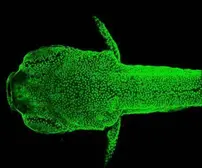
Anti-GFP antibody used in IHC (Whole mount) (IHC-Wm). GTX113617

GTX113617 IHC-Wm Image

Immunohistochemical analysis (whole mount) of transgenic zebrafish embryo (fixed with ice-cold acetone), using GFP(GTX113617) antibody at 1:200 dilution.

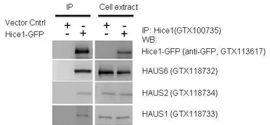
Anti-GFP antibody used in Western Blot (WB). GTX113617

GTX113617 WB Image

IP-WB assay to show that Hice1 (GTX113617) co-immunoprecipitated with other Augmin components HAUS6 (GTX118732), HAUS2 (GTX118734) and HAUS1 (GTX118733) in U2OS cells. The HRP-conjugated anti-rabbit IgG antibody (GTX213110-01) was used to detect the primary antibody.

Anti-GFP antibody used in Immunoprecipitation (IP). GTX113617

GTX113617 IP Image

GFP was immunoprecipitated from bacterial GFP-expressing lysate using 1 ug of anti-GFP antibody (GTX113617).
The precipitated GFP was detected by GTX113617 diluted at 1:15000.

Anti-GFP antibody used in Immunocytochemistry/ Immunofluorescence (ICC/IF). GTX113617

GTX113617 ICC/IF Image

Immunofluorescence analysis of GFP-transfected HeLa. A:GFP is expressed in the tranfected cell. B: The cell expressing GFP can be detected using rabbit anti-GFP ab ( GTX113617, 1:5000) followed by Alexa Fluor 594 (1:500) goat anti-rabbit IgG. C: Merged with DNA probe, the lower cell with no GFP expressed as a negtive control.

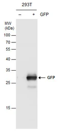
Anti-GFP antibody used in Western Blot (WB). GTX113617

GTX113617 WB Image

GFP antibody detects GFP protein by Western blot analysis. Non-transfected (-) and GFP-transfected (+) 293T whole cell extracts (30 μg) were separated by 12% SDS-PAGE, and the membrane was blotted with GFP antibody (GTX113617) diluted at 1:10000.

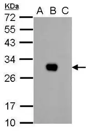
Anti-GFP antibody used in Western Blot (WB). GTX113617

GTX113617 WB Image

GFP antibody detects GFP protein by western blot analysis.
A. 1 μg 293T whole cell extract
B. 1 μg 293T whole cell extract expressing GFP-tagged protein
C. 1 μg 293T whole cell extract expressing RFP-tagged protein
12% SDS-PAGE
GFP antibody (GTX113617) dilution: 1:10000
The HRP-conjugated anti-rabbit IgG antibody (GTX213110-01) was used to detect the primary antibody.

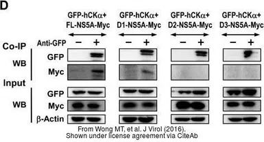
Anti-GFP antibody used in Western Blot and Immunoprecipitation (WB IP). GTX113617

GTX113617 WB IP Image

The data was published in the journal J Virol in 2016. PMID: 27489281

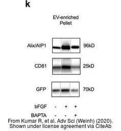
Anti-GFP antibody used in Western Blot (WB). GTX113617

GTX113617 WB Image

The data was published in the journal Adv Sci (Weinh) in 2020. PMID: 32195080

Anti-GFP antibody used in Western Blot (WB). GTX113617

GTX113617 WB Image

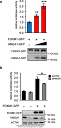
The data was published in the journal J Exp Clin Cancer Res in 2019. PMID: 31311575

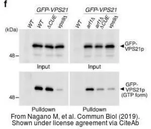
Anti-GFP antibody used in Western Blot (WB). GTX113617

GTX113617 WB Image

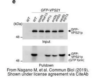
The data was published in the journal Commun Biol in 2019. PMID: 31754649

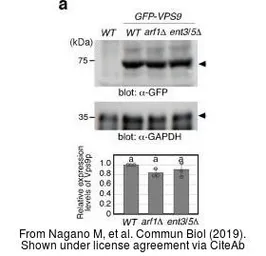
Anti-GFP antibody used in Western Blot (WB). GTX113617

GTX113617 WB Image

The data was published in the journal Commun Biol in 2019. PMID: 31754649

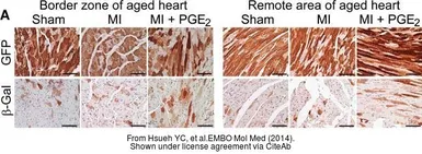
Anti-GFP antibody used in IHC (Paraffin sections) (IHC-P). GTX113617

GTX113617 IHC-P Image

The data was published in the journal EMBO Mol Med in 2014. PMID: 24448489

Anti-GFP antibody used in Western Blot (WB). GTX113617

GTX113617 WB Image

The data was published in the journal Commun Biol in 2019. PMID: 31754649

Anti-GFP antibody used in Western Blot (WB). GTX113617

GTX113617 WB Image

The data was published in the journal J Exp Clin Cancer Res in 2019. PMID: 31311575

Anti-GFP antibody used in Western Blot and Immunoprecipitation (WB IP). GTX113617

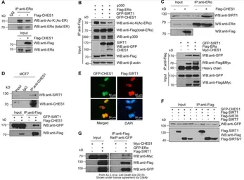
GTX113617 WB IP Image

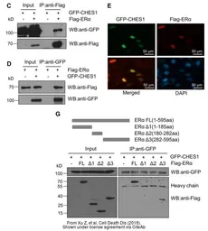
The data was published in the journal Cell Death Dis in 2018.PMID: 29752474

Anti-GFP antibody used in Western Blot (WB). GTX113617

GTX113617 WB Image

The data was published in the journal Cell Death Dis in 2018.PMID: 29752474

Anti-GFP antibody used in Western Blot (WB). GTX113617

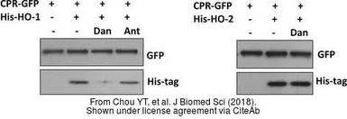
GTX113617 WB Image

The data was published in the journal J Biomed Sci in 2018.PMID: 29361943

Anti-GFP antibody used in IHC (Frozen sections) (IHC-Fr). GTX113617

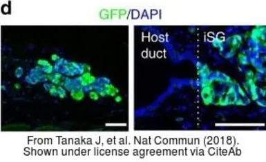
GTX113617 IHC-Fr Image

The data was published in the journal Nat Commun in 2018.PMID: 30310071

Anti-GFP antibody used in Western Blot (WB). GTX113617

GTX113617 WB Image

The data was published in the journal Cell Death Dis in 2018.PMID: 29752474

Anti-GFP antibody used in Western Blot and Immunoprecipitation (WB IP). GTX113617

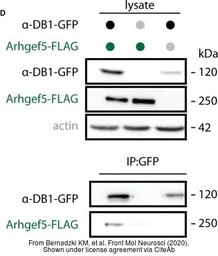
GTX113617 WB IP Image

The data was published in the 2020 in Front Mol Neurosci. PMID: 32587503

REFERENCE

REVIEW

GFP antibody Cat. No. GTX113617

Rating ( Average 4.9 based on 8 users reviews)
Application
Western Blot(WB) ( Average 4.8 based on 5 users reviews)
IHC (Paraffin sections)(IHC-P) ( Average 5 based on 1 users reviews)
IHC (Whole mount)(IHC-Wm) ( Average 5 based on 1 users reviews)
Immunohistochemistry(IHC) ( Average 5 based on 1 users reviews)
Date : Anonymous submitted on 08-Mar-2022
Score :
Application Tested : WB
Sample : Transgenic mouse treated with TAM
Amount used : 20μg
Blocking : 5% gelatin in T-TBS, 20°C, 0.5Hr
Primary Antibody : 1:1500, 4°C, 16Hr
Figure Note : Intestinal specific GFP expression was examined in transgenic mouse treated with TAM.
Date : Anonymous submitted on 31-Aug-2021
Score :
Application Tested : IHC-P
Sample : mFLE-EGFP mouse testis, 2-dpp, paraffin-section 5μm
Blocking : 5% donkey-serum in TNB, 0.3% Triton X-100, 25°C, 1Hr
Primary Antibody : 1:1000, 4°C, 12Hr
Figure Note : The 2-dpp testis tissue section of mFLE-EGFP transgenic mouse which present fetal Leydig cell (fLC) was stained by GFP antibody and HSD-3b1 antibody. GFP only expressed in fLC.
Date : Anonymous submitted on 18-Oct-2018
Score :
Application Tested : WB
Sample : BY4741 (budding yeast) expressing GFP(S65T)-tagged Vps21p (small GTPase Rab5 homologue)
Amount used : 5 μg
Blocking : 1% nonfat milk in PBS-0.05% Tween-20, 25°C, 0.5Hr
Primary Antibody : 1:10000, 4°C, 5Hr
Date : Anonymous submitted on 01-Jun-2018
Score :
Application Tested : IHC-Wm
Sample : Drosophila eye
Fixation : 4% PFA, 4°C, 0.17Hr
Permeabilization /Antigen retrieval : 0.3% PBST, 10 minHr
Primary Antibody : 1:500, 4°C, 16Hr
Date : Anonymous submitted on 22-Feb-2018
Score :
Application Tested : IHC
Sample : Drosophila Retinal tissue
Blocking : 10% NGS in 0.3% PBST (triton-X-100), 25°C, 1Hr
Fixation : 4% PFA
Primary Antibody : 1:200, 4°C, 16Hr
Figure Note : Green: GTX113617 GFP antibody: photoreceptor cell R8, Red: GTX630189 mcherry antibody: medulla
Date : Anonymous submitted on 09-Oct-2014
Score :
Application Tested : WB
Sample : GFP recombinant protein transfected 293T cells
Amount used : 20μg
Blocking : 5% milk, 25°C, 1Hr
Primary Antibody : 1:5000, 4°C, 16Hr
Date : Anonymous submitted on 09-Oct-2014
Score :
Application Tested : WB
Sample : GFP-p53 transfected U2OS cells
Amount used : 15μg
Blocking : 5% milk in 0.1%TBST, 25°C, 0.5Hr
Primary Antibody : 1:5000, 25°C, 1Hr
Lane Description : A: U2OS cells transfected with GFP-p53, B: U2OS cells transfected with control plasmid (no GFP-tag)
Date : Anonymous submitted on 12-Apr-2012
Score :
Application Tested : WB
Sample : Protoplast from Rosette leaves
Amount used : 30μg
Blocking : 5% milk in PBST, 4°C, 16Hr
Primary Antibody : 1:10000, 25°C, 1Hr
Application Reference
SDS
PBS.pdf
Glycerol.pdf
Proclin.pdf
Package List Price ($)
$ 399
$ 169