DAG1 antibody


Various tissue extracts (50 μg) were separated by 7.5% SDS-PAGE, and the membrane was blotted with DAG1 antibody (GTX105038) diluted at 1:500. The HRP-conjugated anti-rabbit IgG antibody (GTX213110-01) was used to detect the primary antibody, and the signal was developed with Trident ECL plus-Enhanced.
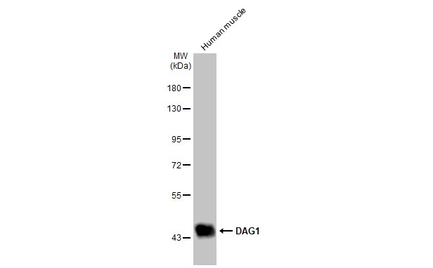
Human tissue extract (30 μg) was separated by 7.5% SDS-PAGE, and the membrane was blotted with DAG1 antibody (GTX105038) diluted at 1:1000. The HRP-conjugated anti-rabbit IgG antibody (GTX213110-01) was used to detect the primary antibody.
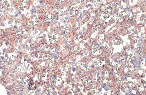
DAG1 antibody detects DAG1 protein at cell membrane and cytoplasm by immunohistochemical analysis.Sample: Paraffin-embedded mouse placenta.DAG1 stained by DAG1 antibody (GTX105038) diluted at 1:500.Antigen Retrieval: Citrate buffer, pH 6.0, 15 min
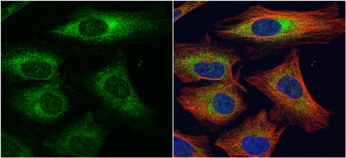
alpha Dystroglycan antibody detects alpha Dystroglycan protein at cytoplasm by immunofluorescent analysis.
Sample: HeLa cells were fixed in 4% paraformaldehyde at RT for 15 min.
Green: alpha Dystroglycan protein stained by alpha Dystroglycan antibody (GTX105038) diluted at 1:1000.
Red: alpha Tubulin, a cytoskeleton marker, stained by alpha Tubulin antibody [B-5-1-2] (GTX11304) diluted at 1:10000.
Blue: Hoechst 33342 staining.
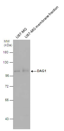
DAG1 antibody detects DAG1 protein by western blot analysis. U87-MG whole cell extracts and membrane extracts (30 μg) were separated by 7.5% SDS-PAGE, and the membrane was blotted with DAG1 antibody (GTX105038) diluted at 1:500. The HRP-conjugated anti-rabbit IgG antibody (GTX213110-01) was used to detect the primary antibody.
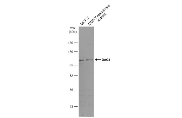
MCF-7 whole cell and membrane extracts (30 μg) were separated by 7.5% SDS-PAGE, and the membrane was blotted with DAG1 antibody (GTX105038) diluted at 1:500. The HRP-conjugated anti-rabbit IgG antibody (GTX213110-01) was used to detect the primary antibody.
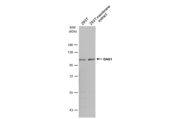
293T whole cell and membrane extracts (30 μg) were separated by 7.5% SDS-PAGE, and the membrane was blotted with DAG1 antibody (GTX105038) diluted at 1:500. The HRP-conjugated anti-rabbit IgG antibody (GTX213110-01) was used to detect the primary antibody.
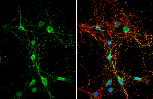
DAG1 antibody detects DAG1 protein by immunofluorescent analysis.Sample: DIV10 rat E18 primary hippocampal neuron cells were fixed in 4% paraformaldehyde at RT for 15 min.Green: DAG1 stained by DAG1 antibody (GTX105038) diluted at 1:500.Red: Tau, stained by Tau antibody [GT287] (GTX634809) diluted at 1:500.Blue: Fluoroshield with DAPI (GTX30920).
-
HostRabbit
-
ClonalityPolyclonal
-
IsotypeIgG
-
ApplicationsWB ICC/IF IHC-P
-
ReactivityHuman, Mouse, Rat, Horse