HMGB1 antibody


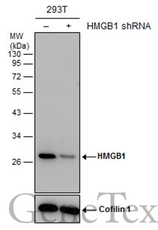
Non-transfected (–) and transfected (+) 293T whole cell extracts (30 μg) were separated by 12% SDS-PAGE, and the membrane was blotted with HMGB1 antibody (GTX101277) diluted at 1:5000. The HRP-conjugated anti-rabbit IgG antibody (GTX213110-01) was used to detect the primary antibody.
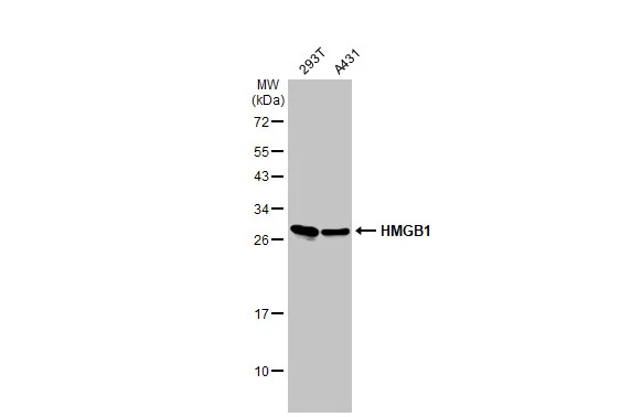
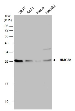
Various whole cell extracts (30 μg) were separated by 12% SDS-PAGE, and the membrane was blotted with HMGB1 antibody (GTX101277) diluted at 1:3000. The HRP-conjugated anti-rabbit IgG antibody (GTX213110-01) was used to detect the primary antibody.
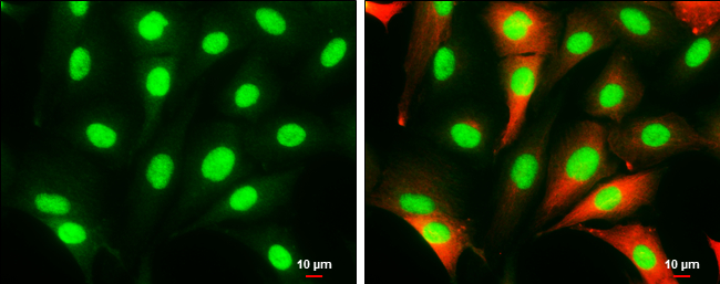
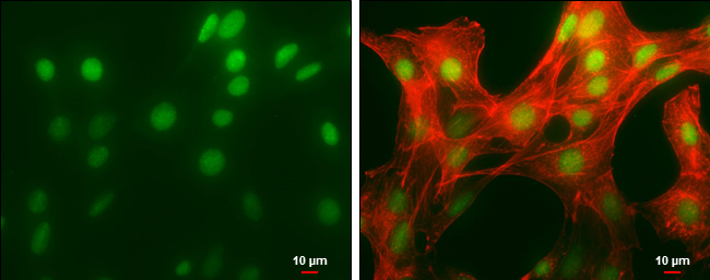
HMGB1 antibody detects HMGB1 protein at cytoplasm and nucleus by immunofluorescent analysis.
Sample: SK-N-SH cells were fixed in 4% paraformaldehyde at RT for 15 min.
Green: HMGB1 protein stained by HMGB1 antibody (GTX101277) diluted at 1:1000.
Red: beta Tubulin 3/ Tuj1 stained by beta Tubulin 3/ Tuj1 antibody [GT11710] (GTX631836) diluted at 1:500.
Scale bar = 10 μm.
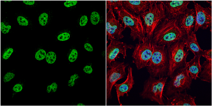
HMGB1 antibody detects HMGB1 protein at nucleus by immunofluorescent analysis.
Sample: HeLa cells were fixed in 4% paraformaldehyde at RT for 15 min.
Green: HMGB1 protein stained by HMGB1 antibody (GTX101277) diluted at 1:1000.
Red: phalloidin, a cytoskeleton marker, stained by phalloidin (invitrogen, A12380) diluted at 1:200.
Blue: Hoechst 33342 staining.
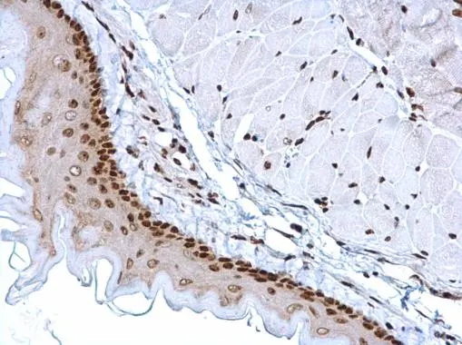
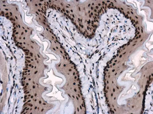
HMGB1 antibody detects HMGB1 protein at nucleus on mouse esophagus by immunohistochemical analysis.
Sample: Paraffin-embedded mouse esophagus.
HMGB1 antibody (GTX101277) dilution: 1:1000.
Antigen Retrieval: Trilogy™ (EDTA based, pH 8.0) buffer, 15min
HMGB1 antibody detects HMGB1 protein at nucleus by immunofluorescent analysis.
Sample: NIH/3T3 cells were fixed in 4% paraformaldehyde at RT for 15 min.
Green: HMGB1 protein stained by HMGB1 antibody (GTX101277) diluted at 1:500.
Red: phalloidin, a cytoskeleton marker, diluted at 1:50.
Scale bar = 10 μm.
Various whole cell extracts (30 μg) were separated by 12% SDS-PAGE, and the membrane was blotted with HMGB1 antibody (GTX101277) diluted at 1:3000. The HRP-conjugated anti-rabbit IgG antibody (GTX213110-01) was used to detect the primary antibody.
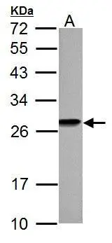
HMGB1 antibody detects HMGB1 protein by western blot analysis.
A. 30 μg PC-12 whole cell lysate/extract
12% SDS-PAGE
HMGB1 antibody (GTX101277) dilution: 1:3000
The HRP-conjugated anti-rabbit IgG antibody (GTX213110-01) was used to detect the primary antibody.
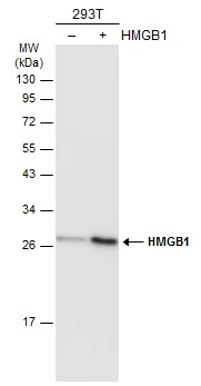
Non-transfected (–) and transfected (+) 293T whole cell extracts (30 μg) were separated by 12% SDS-PAGE, and the membrane was blotted with HMGB1 antibody (GTX101277) diluted at 1:5000. The HRP-conjugated anti-rabbit IgG antibody (GTX213110-01) was used to detect the primary antibody.
HMGB1 antibody detects HMGB1 protein at nucleus in mouse esophagus by immunohistochemical analysis.
Sample: Paraffin-embedded mouse esophagus.
HMGB1 antibody (GTX101277) diluted at 1:1000.
Antigen Retrieval: Citrate buffer, pH 6.0, 15 min
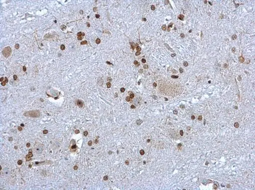
HMGB1 antibody detects HMGB1 protein at nucleus on rat brain stem by immunohistochemical analysis.
Sample: Paraffin-embedded rat brain stem.
HMGB1 antibody (GTX101277) dilution: 1:1000.
Antigen Retrieval: Trilogy™ (EDTA based, pH 8.0) buffer, 15min
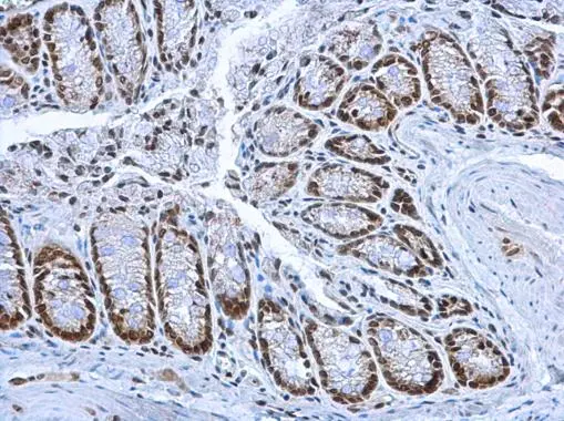
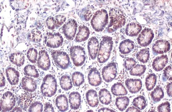
HMGB1 antibody detects HMGB1 protein at nucleus on mouse colon by immunohistochemical analysis.
Sample: Paraffin-embedded mouse colon.
HMGB1 antibody (GTX101277) dilution: 1:1000.
Antigen Retrieval: Trilogy™ (EDTA based, pH 8.0) buffer, 15min
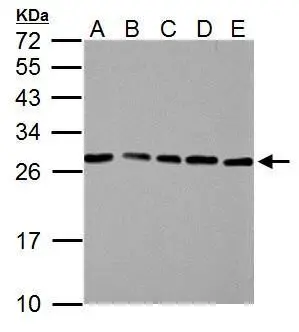
HMGB1 antibody detects HMGB1 protein by western blot analysis.
A. 30 μg NIH-3T3 whole cell lysate/extract
B. 30 μg JC whole cell lysate/extract
C. 30 μg BCL-1 whole cell lysate/extract
D. 30 μg C2C12 whole cell lysate/extract
E. 30 μg Raw264.7 whole cell lysate/extract
12% SDS-PAGE
HMGB1 antibody (GTX101277) dilution: 1:3000
The HRP-conjugated anti-rabbit IgG antibody (GTX213110-01) was used to detect the primary antibody.
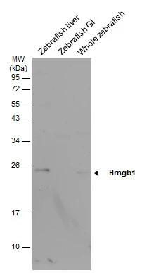
Various tissue extracts (30 μg) were separated by 12% SDS-PAGE, and the membrane was blotted with HMGB1 antibody (GTX101277) diluted at 1:1000. The HRP-conjugated anti-rabbit IgG antibody (GTX213110-01) was used to detect the primary antibody. (GI: gastrointestinal)
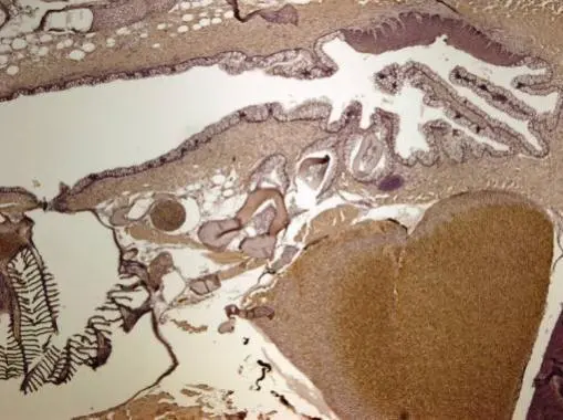
Immunohistochemical analysis of paraffin-embedded zebrafish tissue, using HMGB1 antibody (GTX101277) at 1:300 dilution.
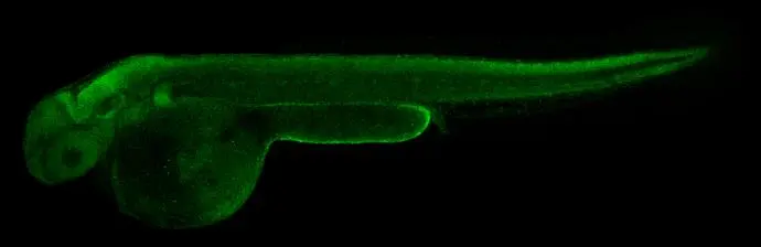
HMGB1 antibody detects Hmgb1 protein on whole mount zebrafish by immunohistochemical analysis.Sample: Paraformaldehyde-fixed 2 days-post-fertilization zebrafish embryo.Green: Hmgb1 stained by HMGB1 antibody (GTX101277) diluted at 1:100.Antigen Retrieval: Tris-HCl buffer, pH 9.0, 20 min at 70ºC
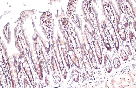
HMGB1 antibody detects HMGB1 protein at nucleus by immunohistochemical analysis.
Sample: Paraffin-embedded rat colon.
HMGB1 stained by HMGB1 antibody (GTX101277) diluted at 1:500.
Antigen Retrieval: Citrate buffer, pH 6.0, 15 min
HMGB1 antibody detects HMGB1 protein at nucleus by immunohistochemical analysis.
Sample: Paraffin-embedded rat colon.
HMGB1 stained by HMGB1 antibody (GTX101277) diluted at 1:500.
Antigen Retrieval: Citrate buffer, pH 6.0, 15 min
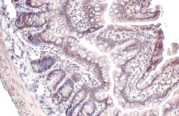
HMGB1 antibody detects HMGB1 protein at nucleus by immunohistochemical analysis.
Sample: Paraffin-embedded mouse intestine.
HMGB1 stained by HMGB1 antibody (GTX101277) diluted at 1:500.
Antigen Retrieval: Citrate buffer, pH 6.0, 15 min
-
HostRabbit
-
ClonalityPolyclonal
-
IsotypeIgG
-
ApplicationsWB ICC/IF IHC-P IHC-Wm IHC IHC (Free Floating)
-
ReactivityHuman, Mouse, Rat, Zebrafish, Pig