Normal Tissue Gallery- GTX04969

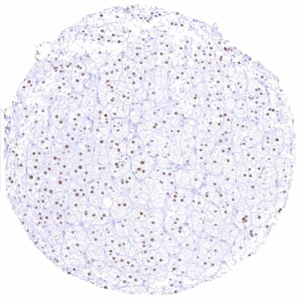
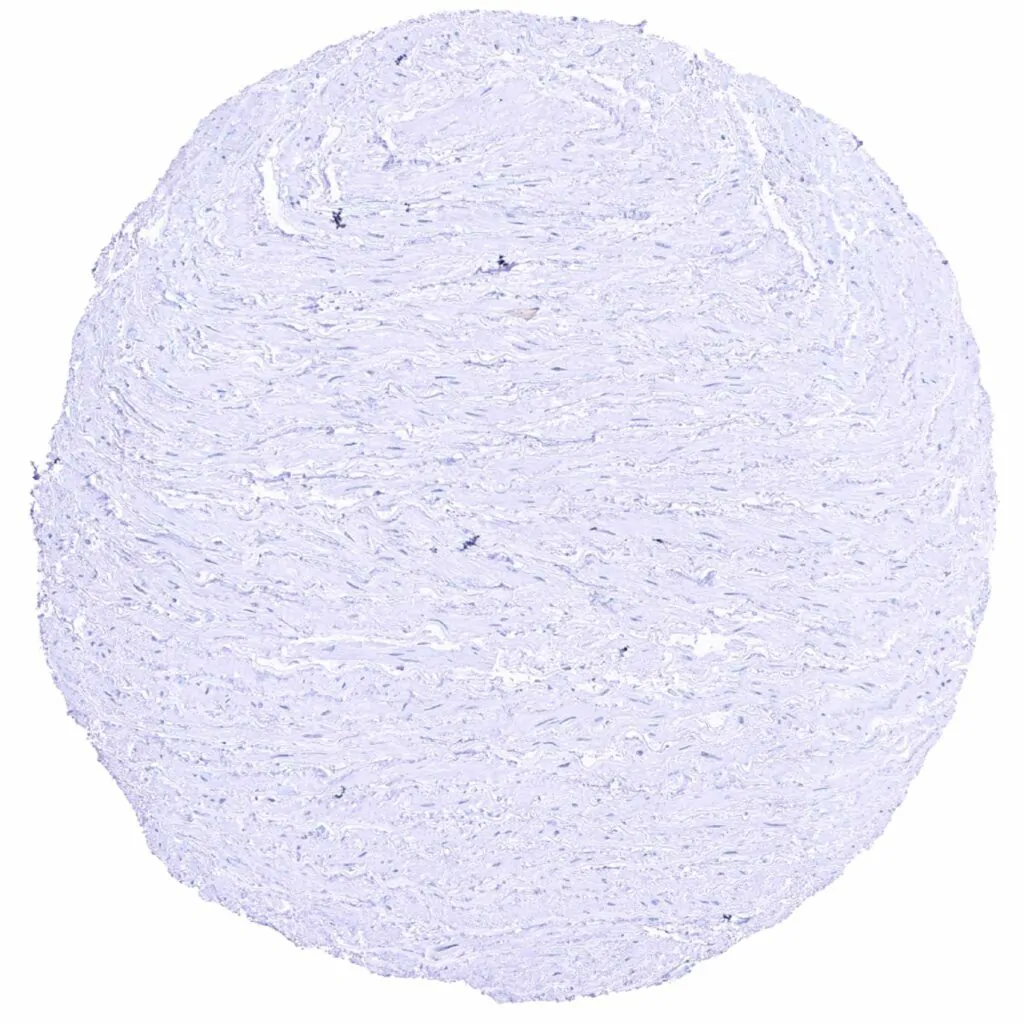
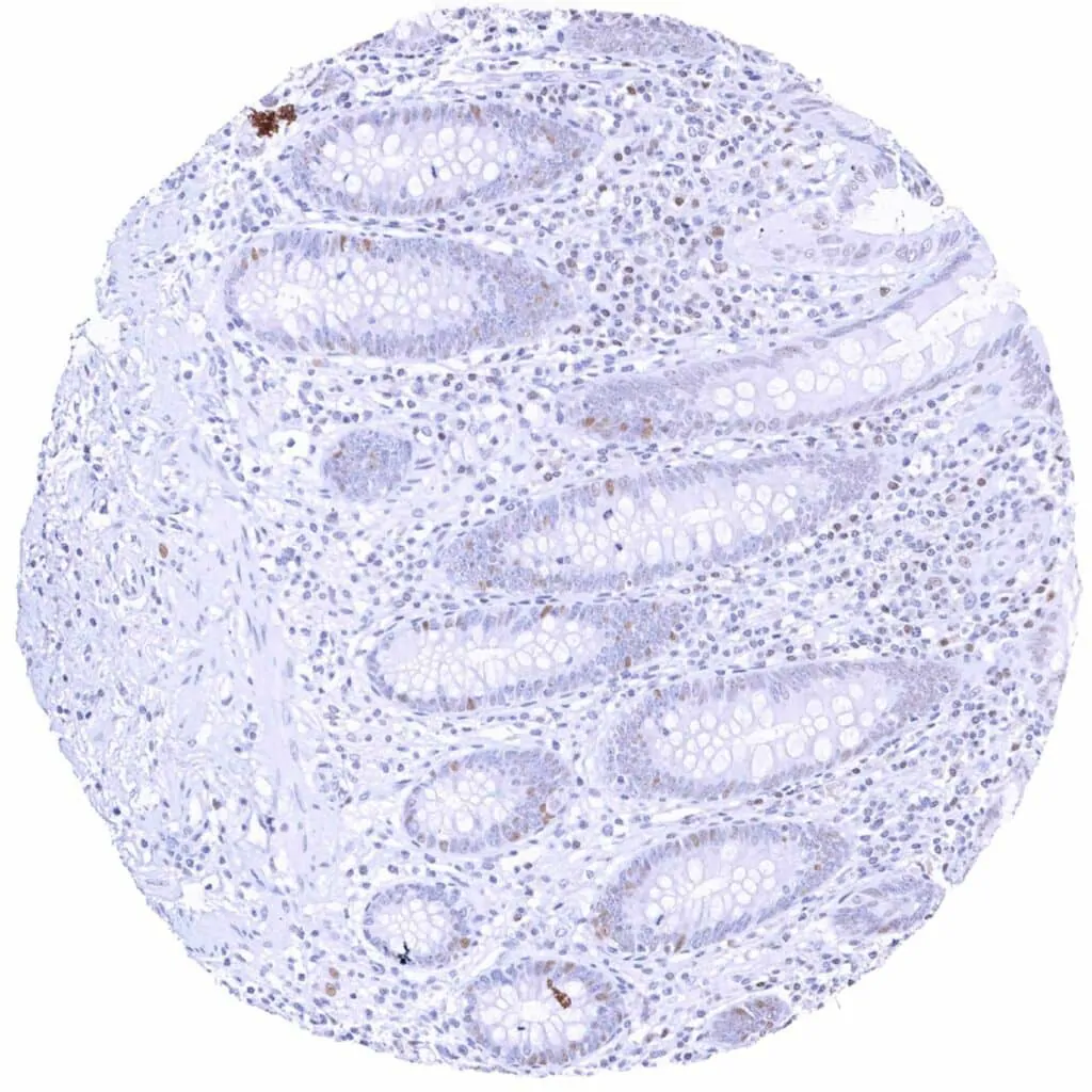
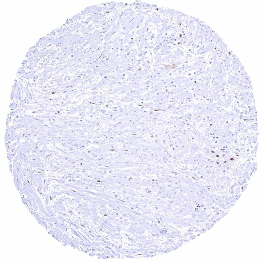
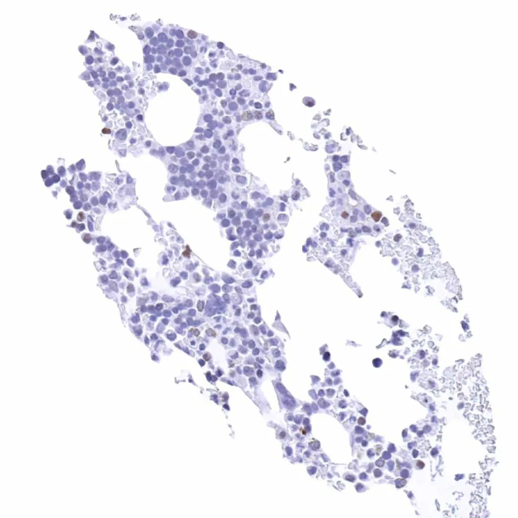
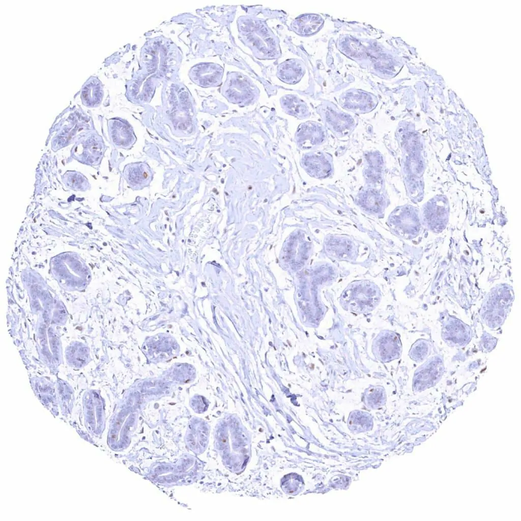
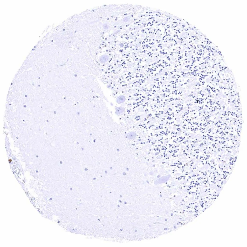
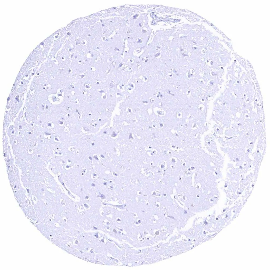
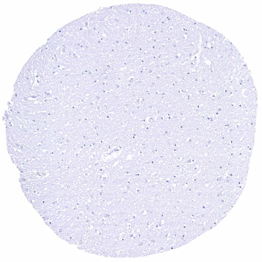
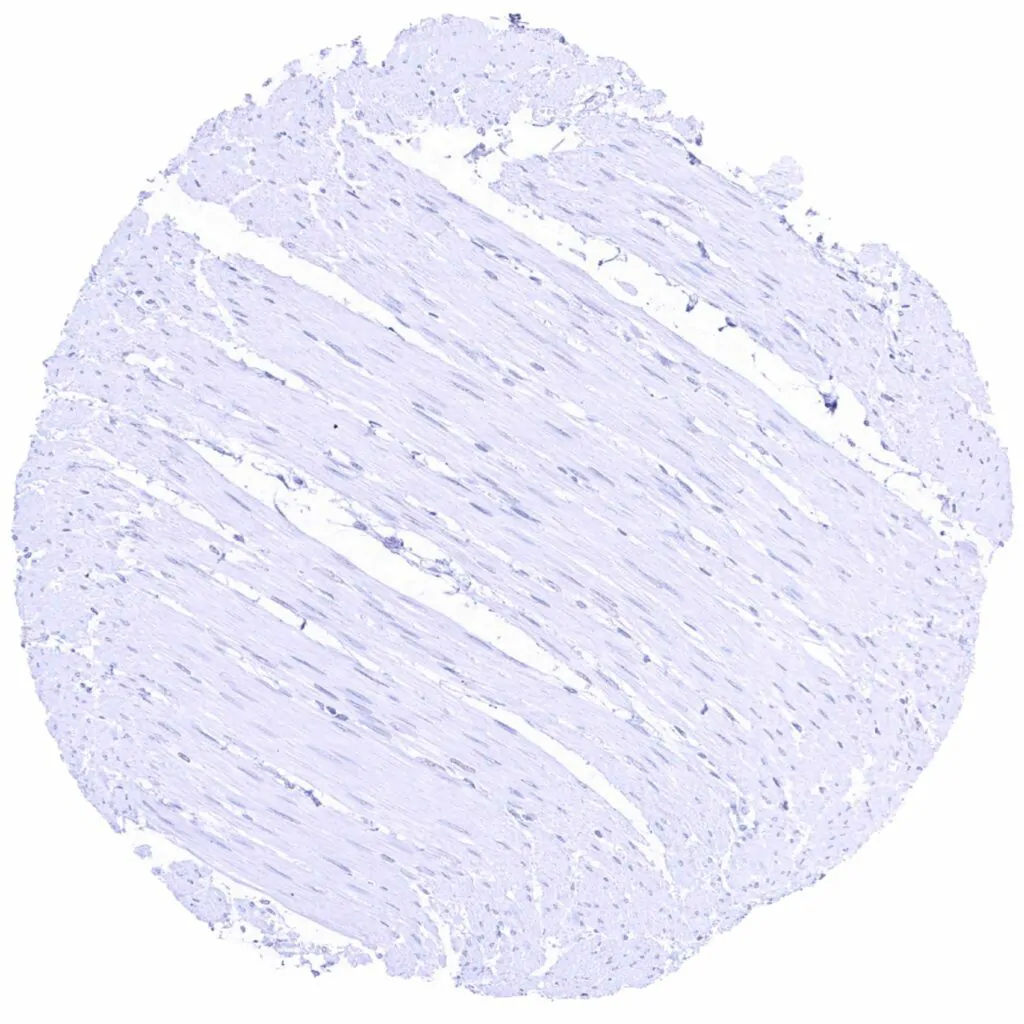
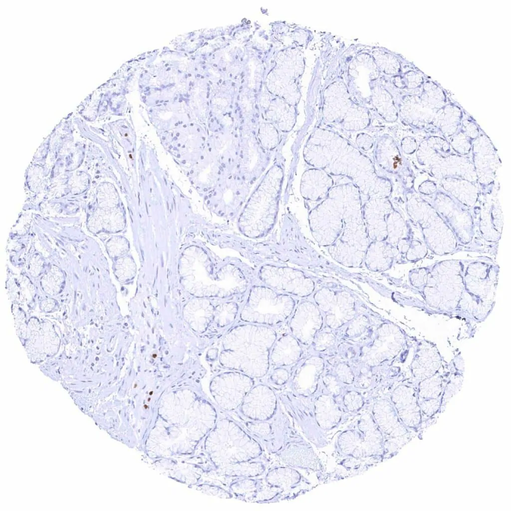
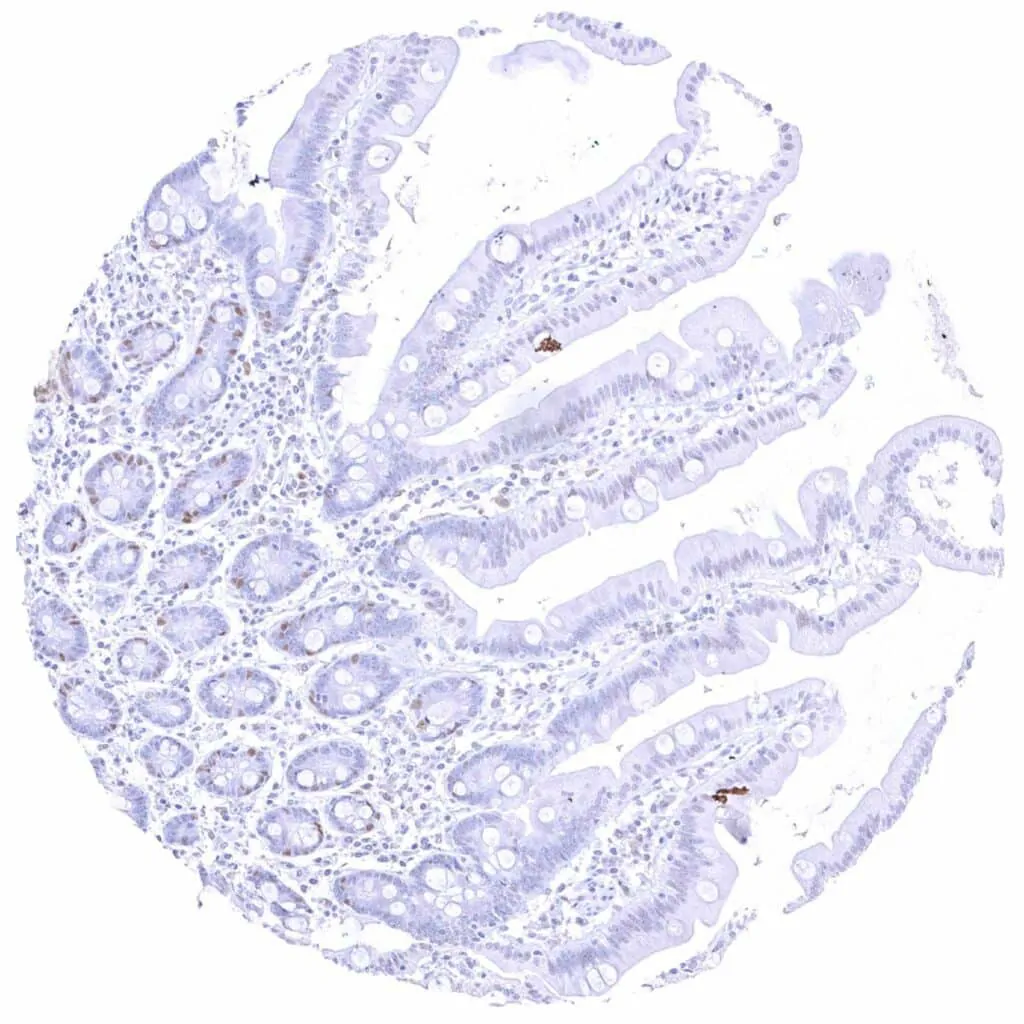
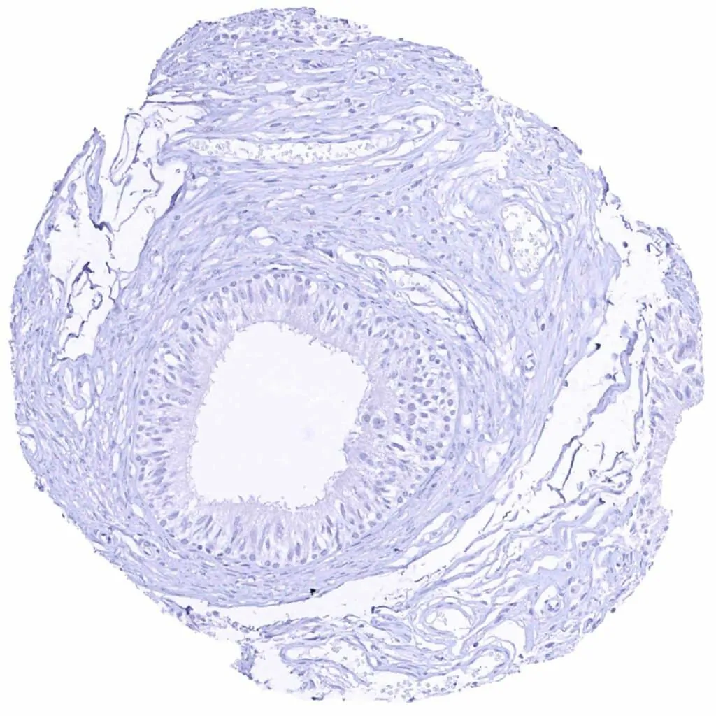
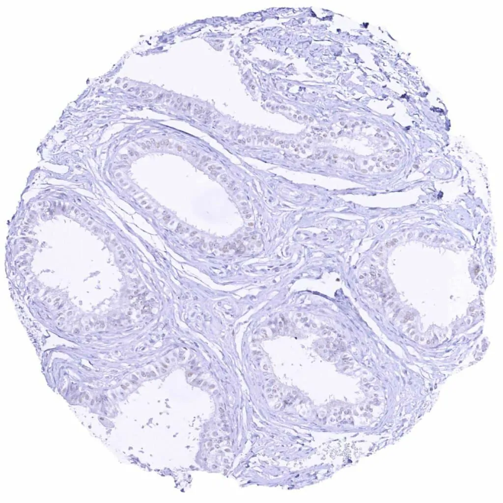
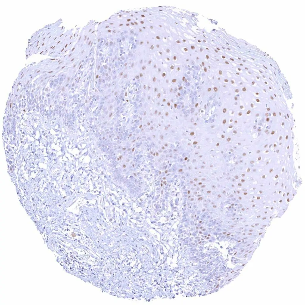
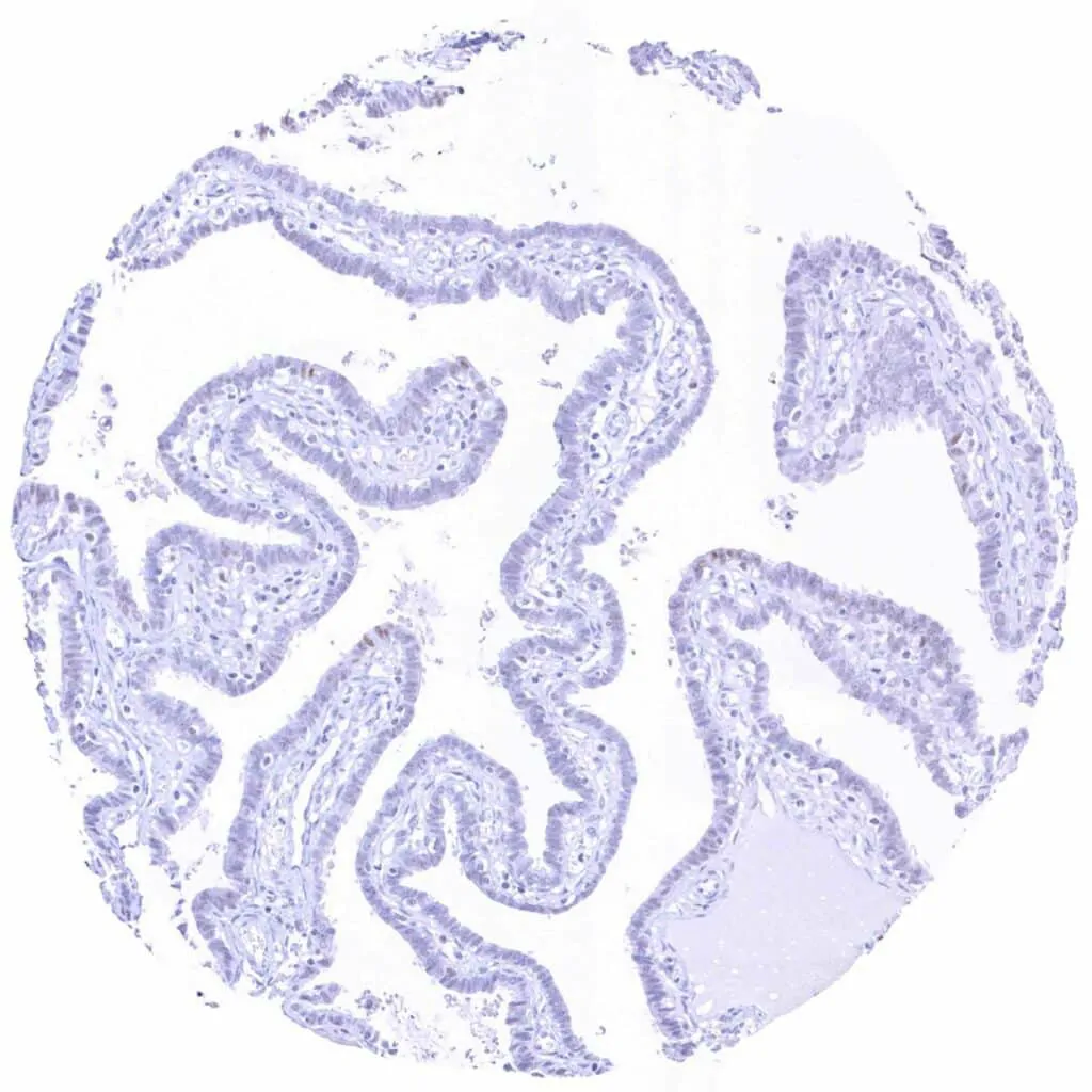
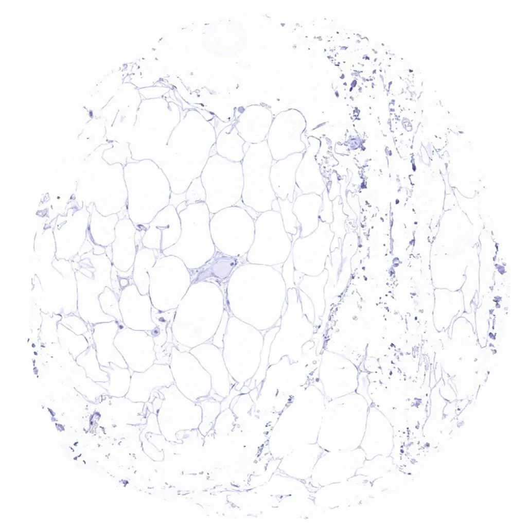
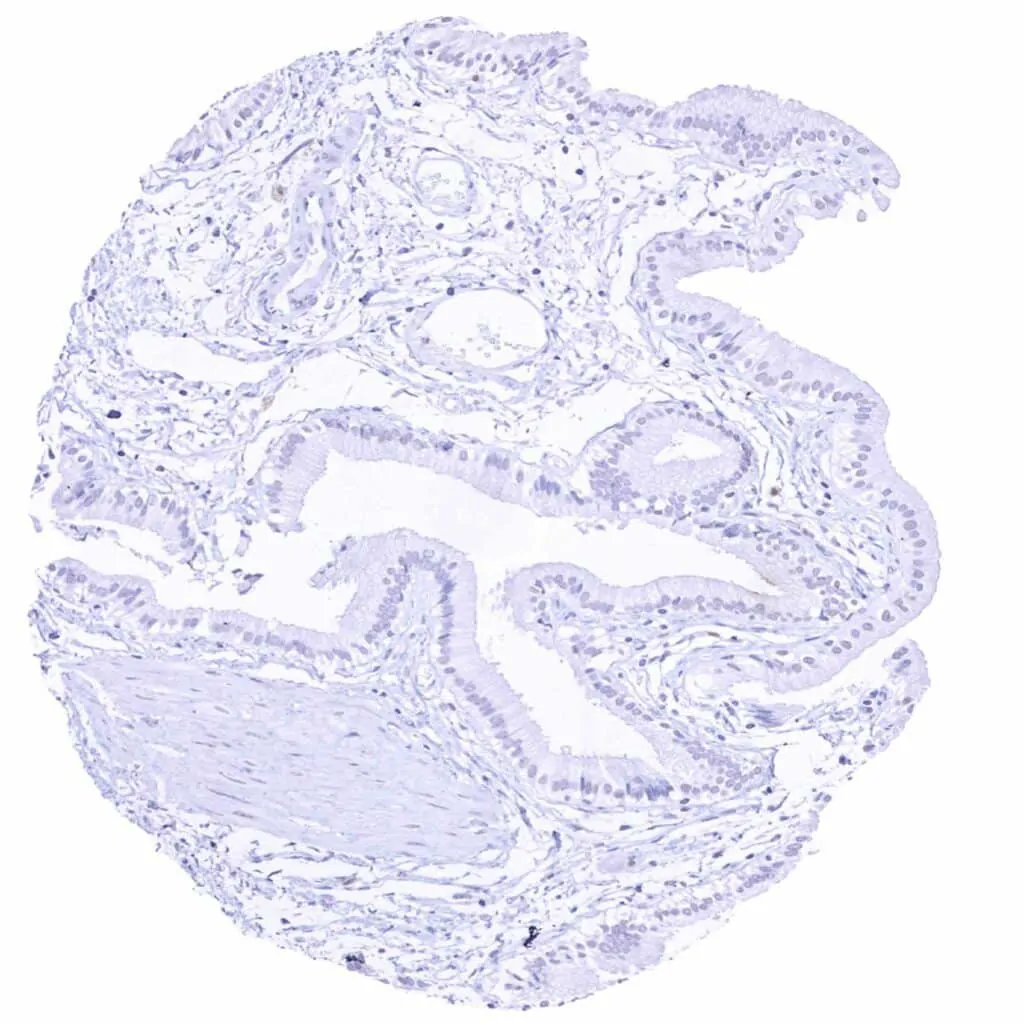
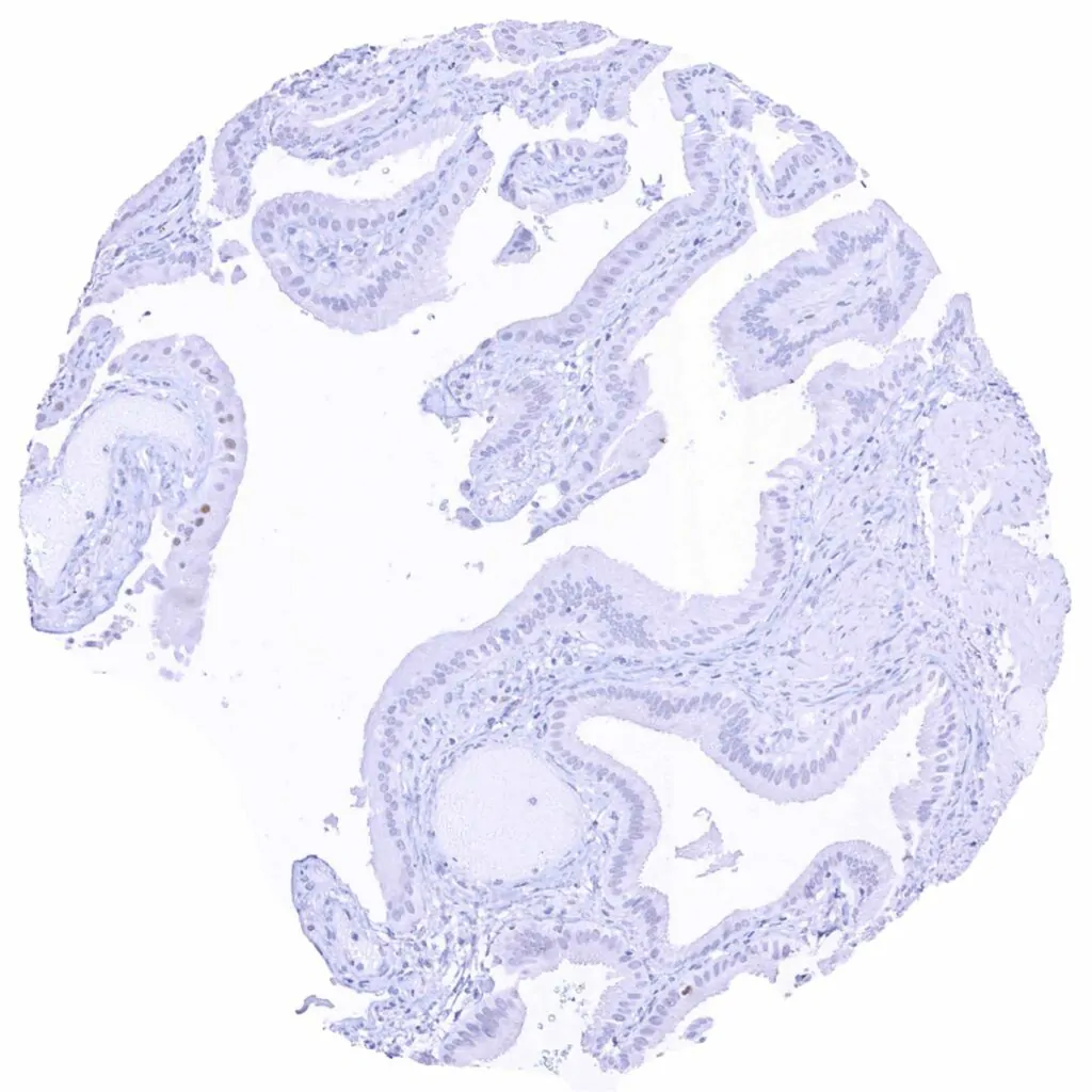
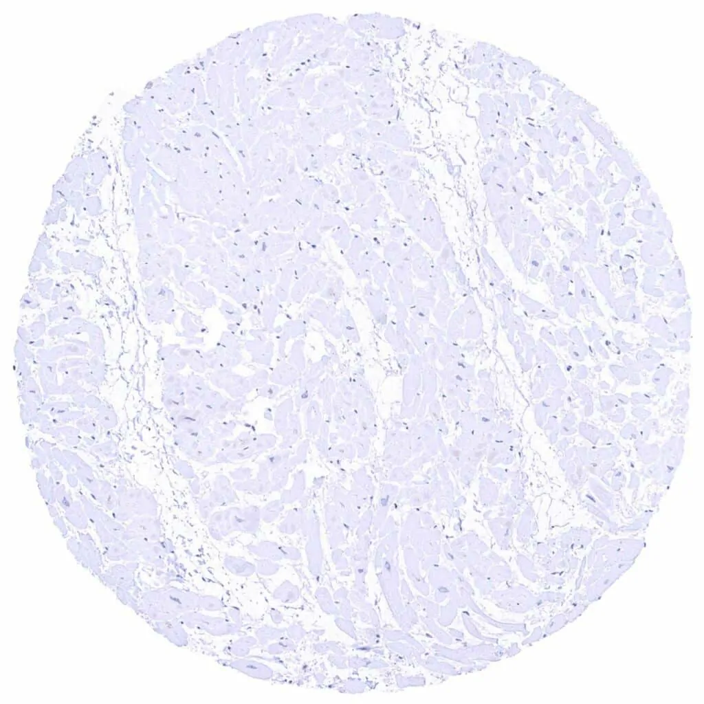
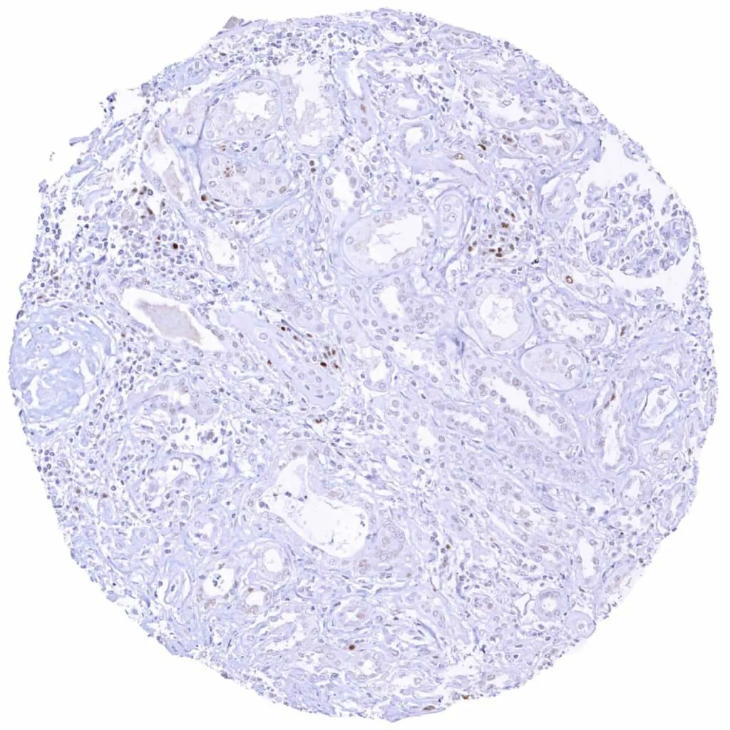
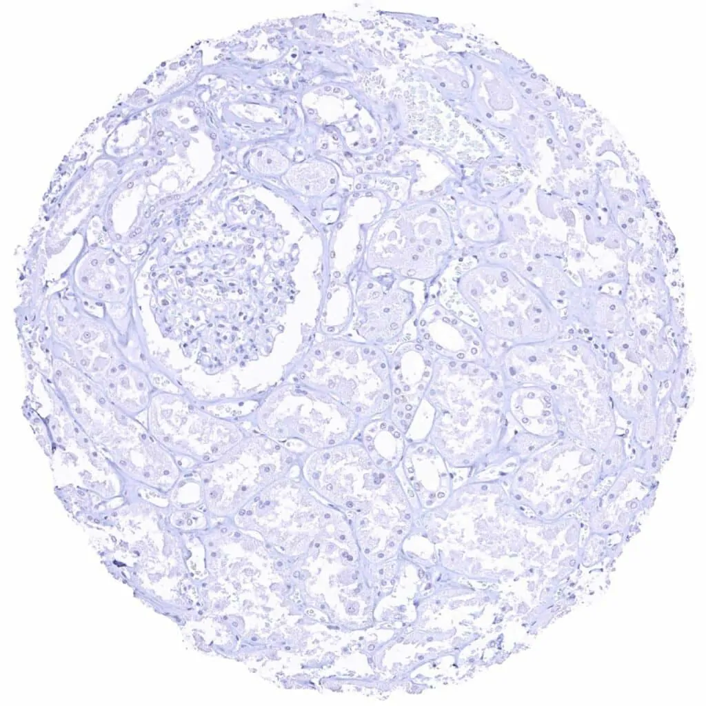
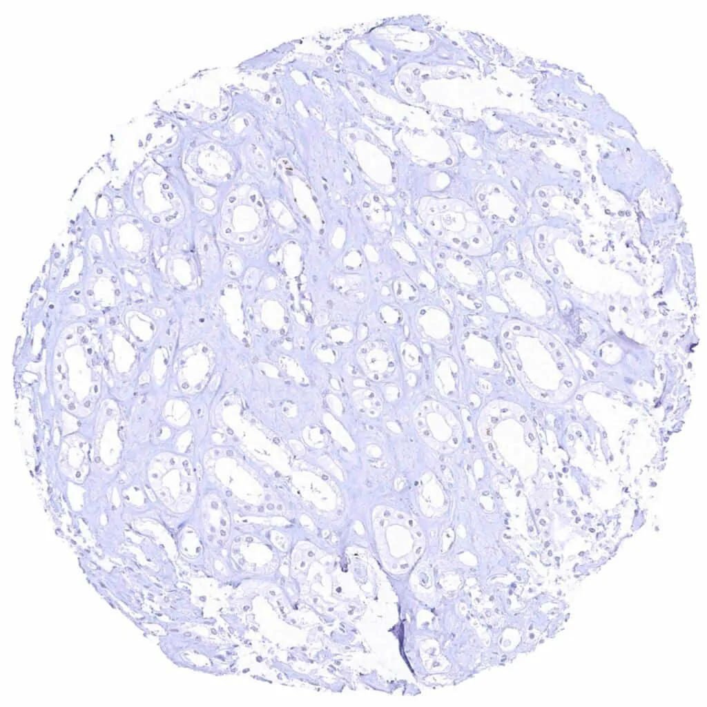
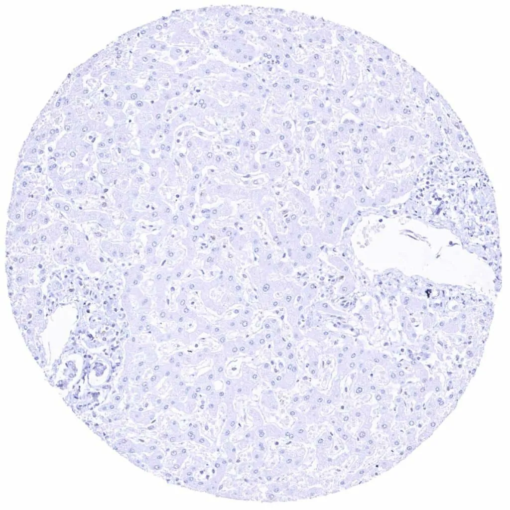
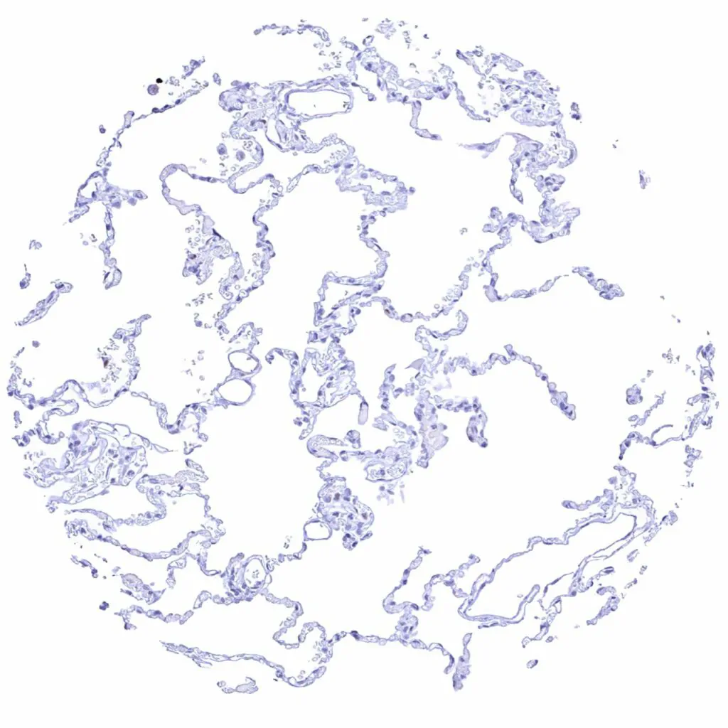
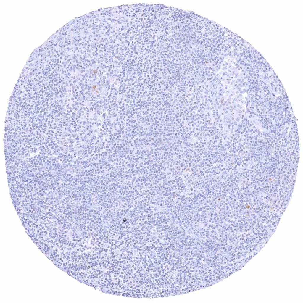
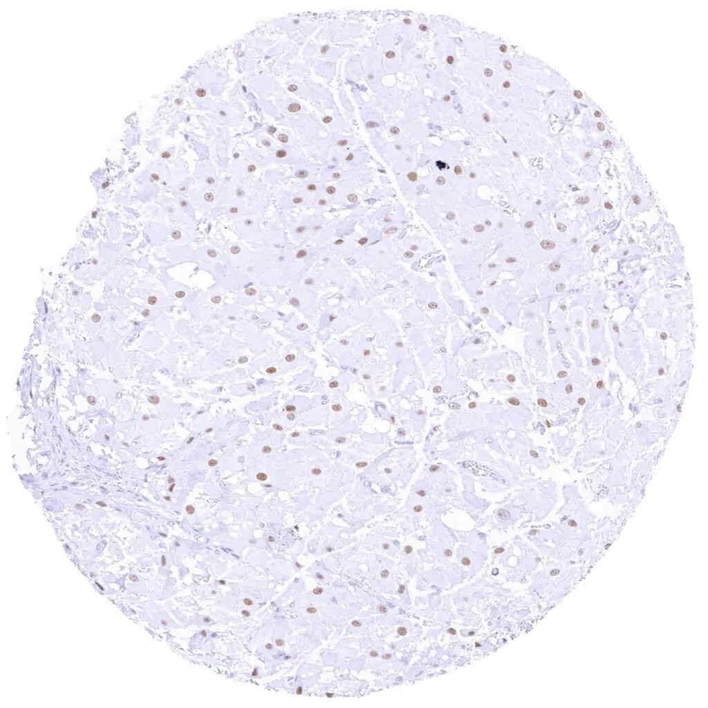
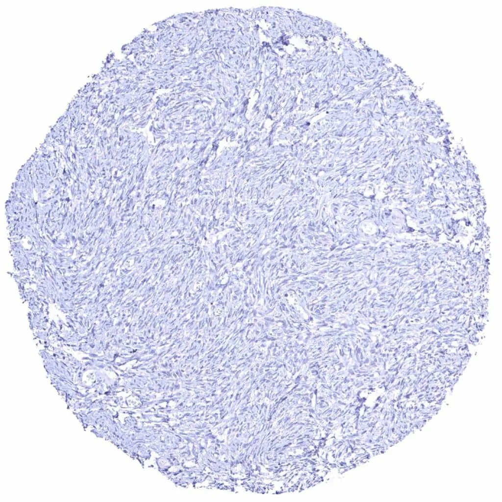
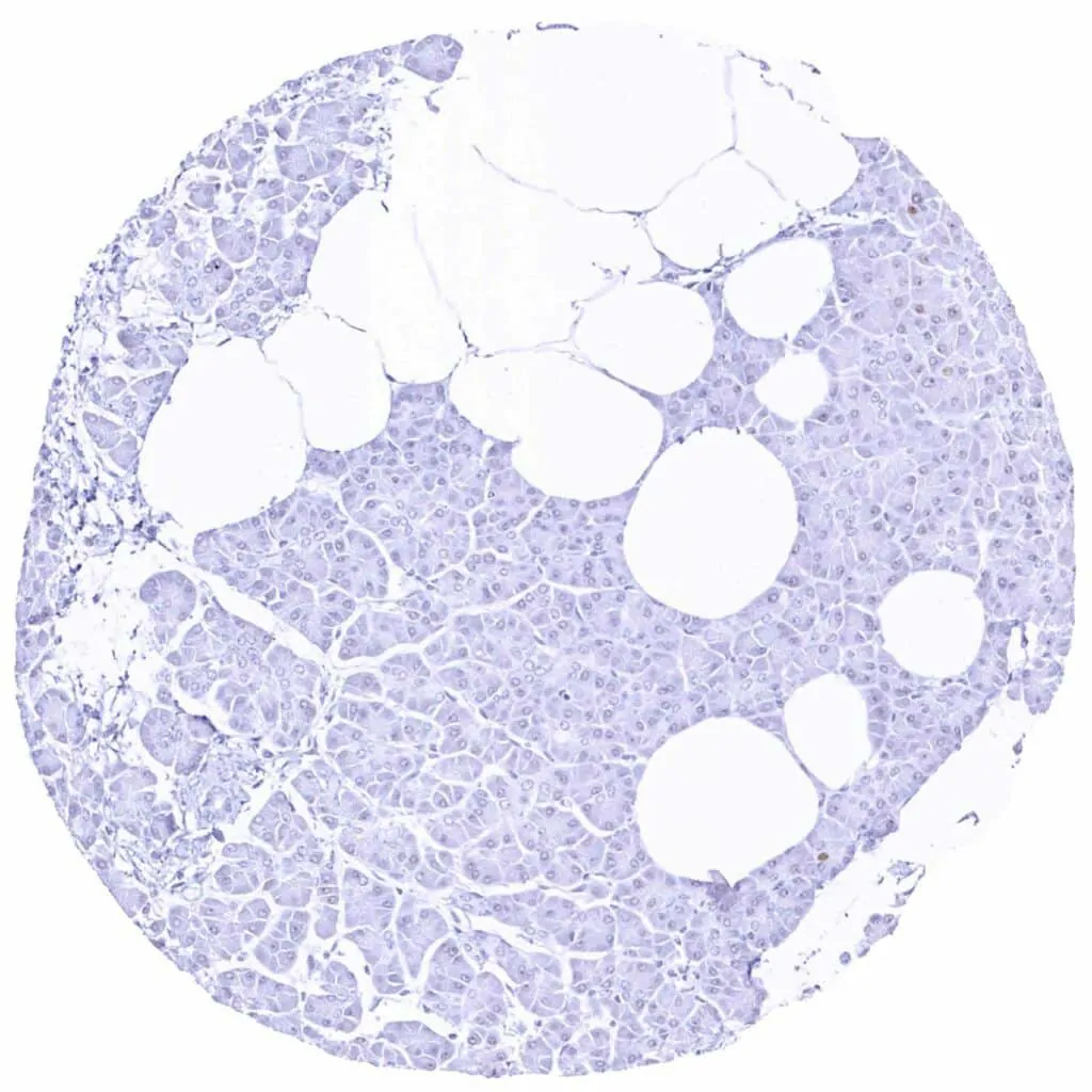
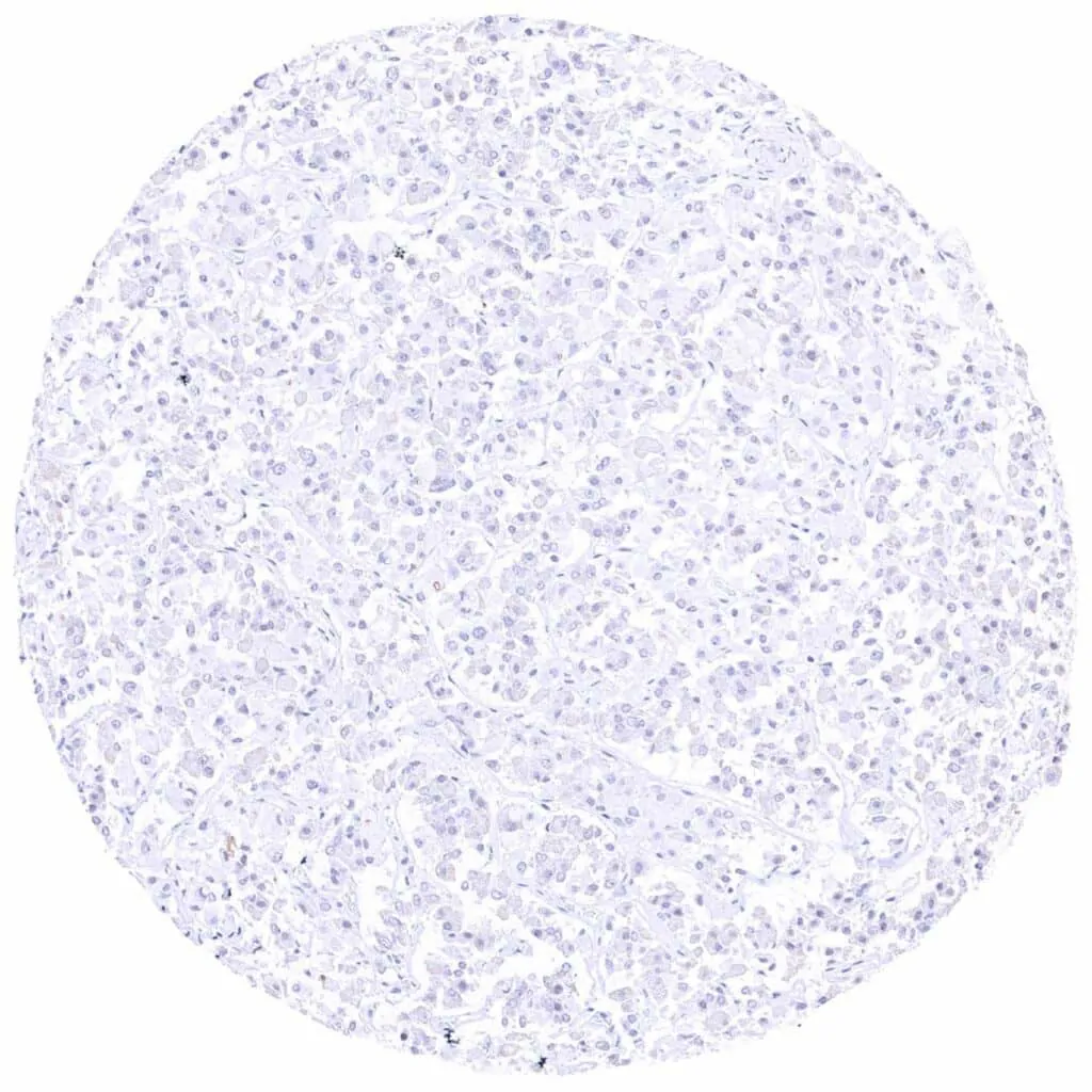
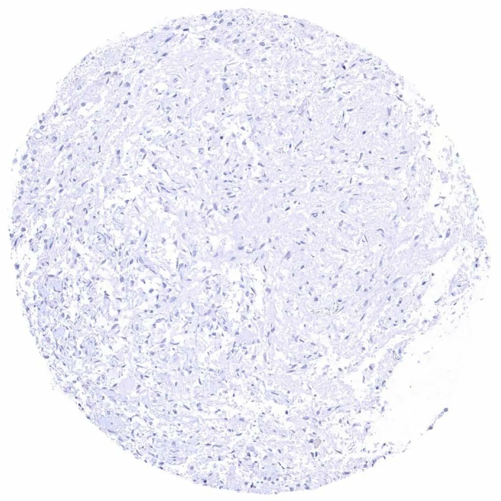
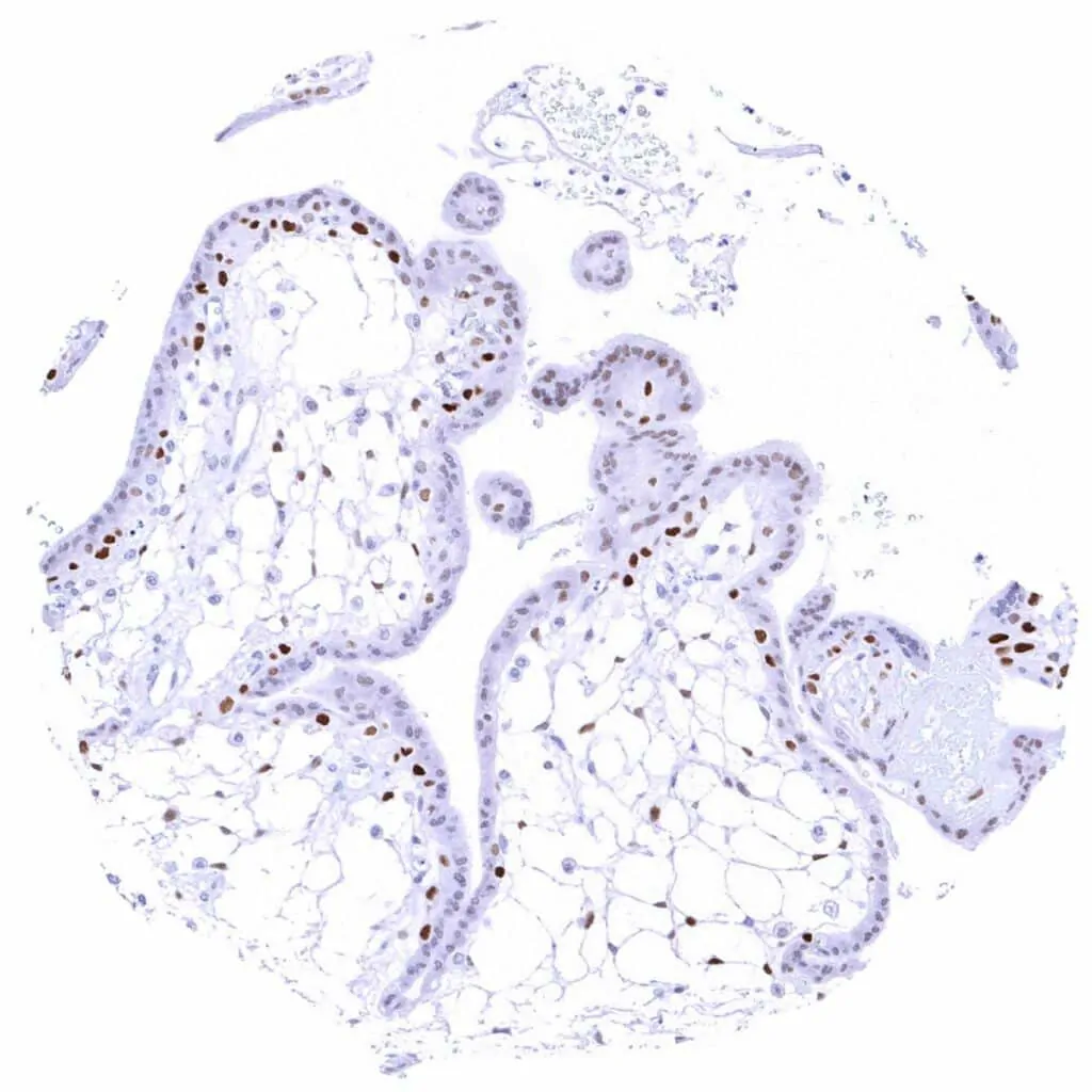
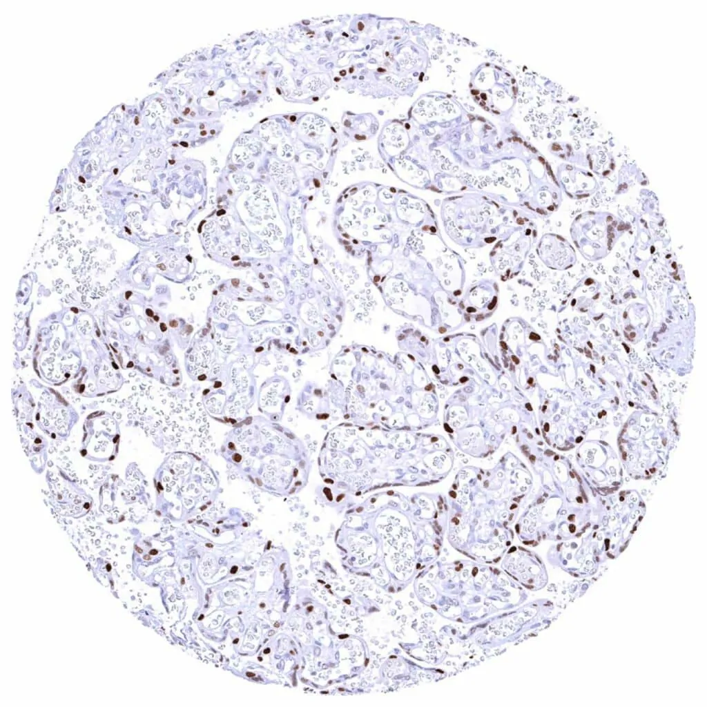
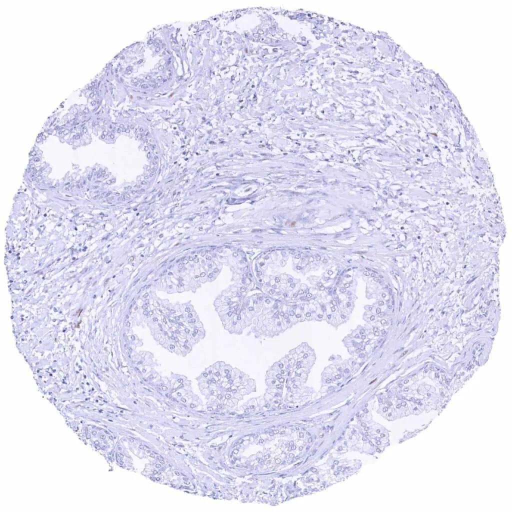
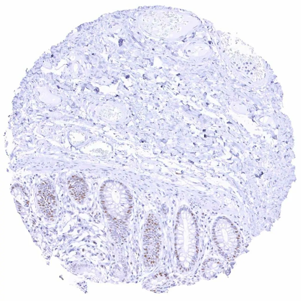
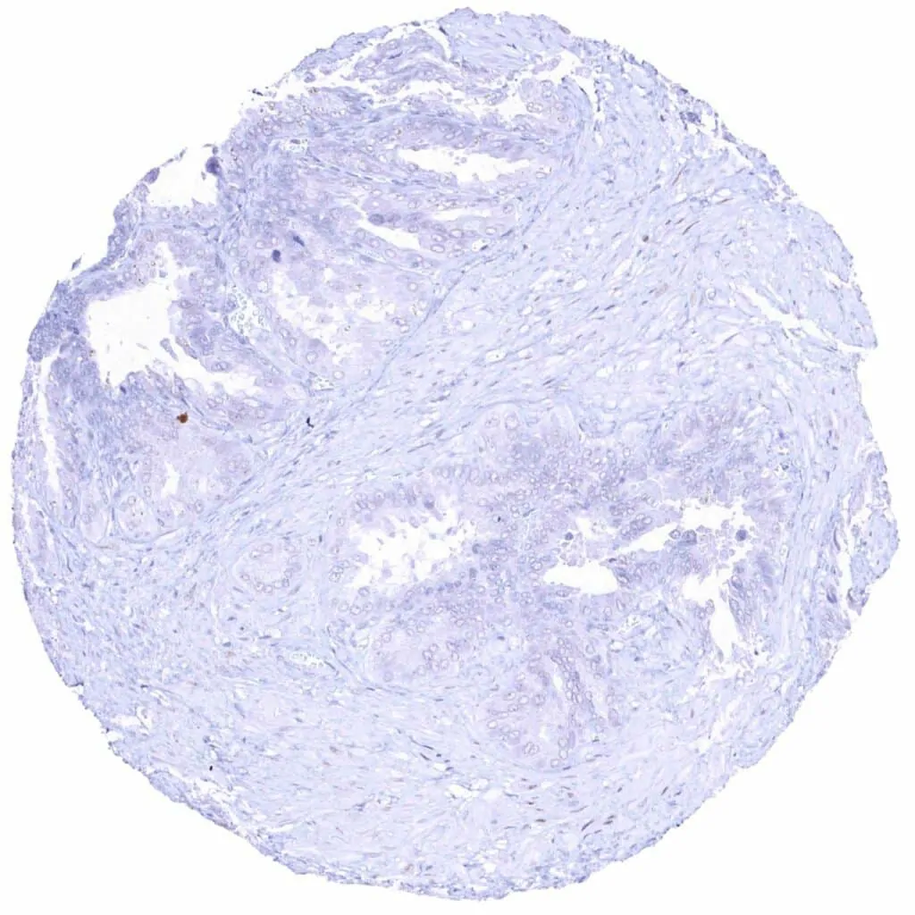
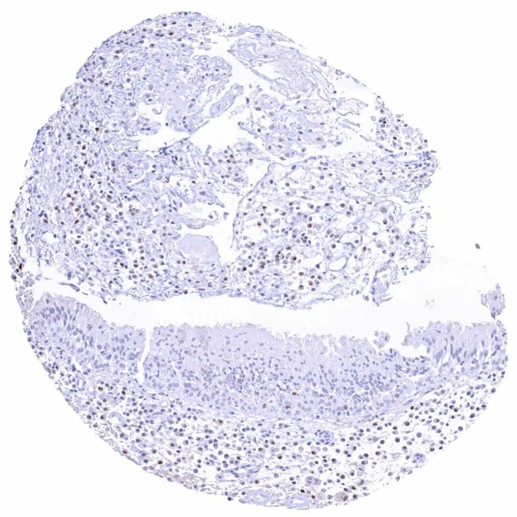
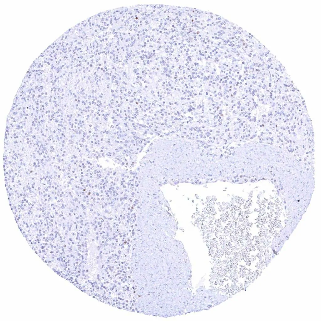
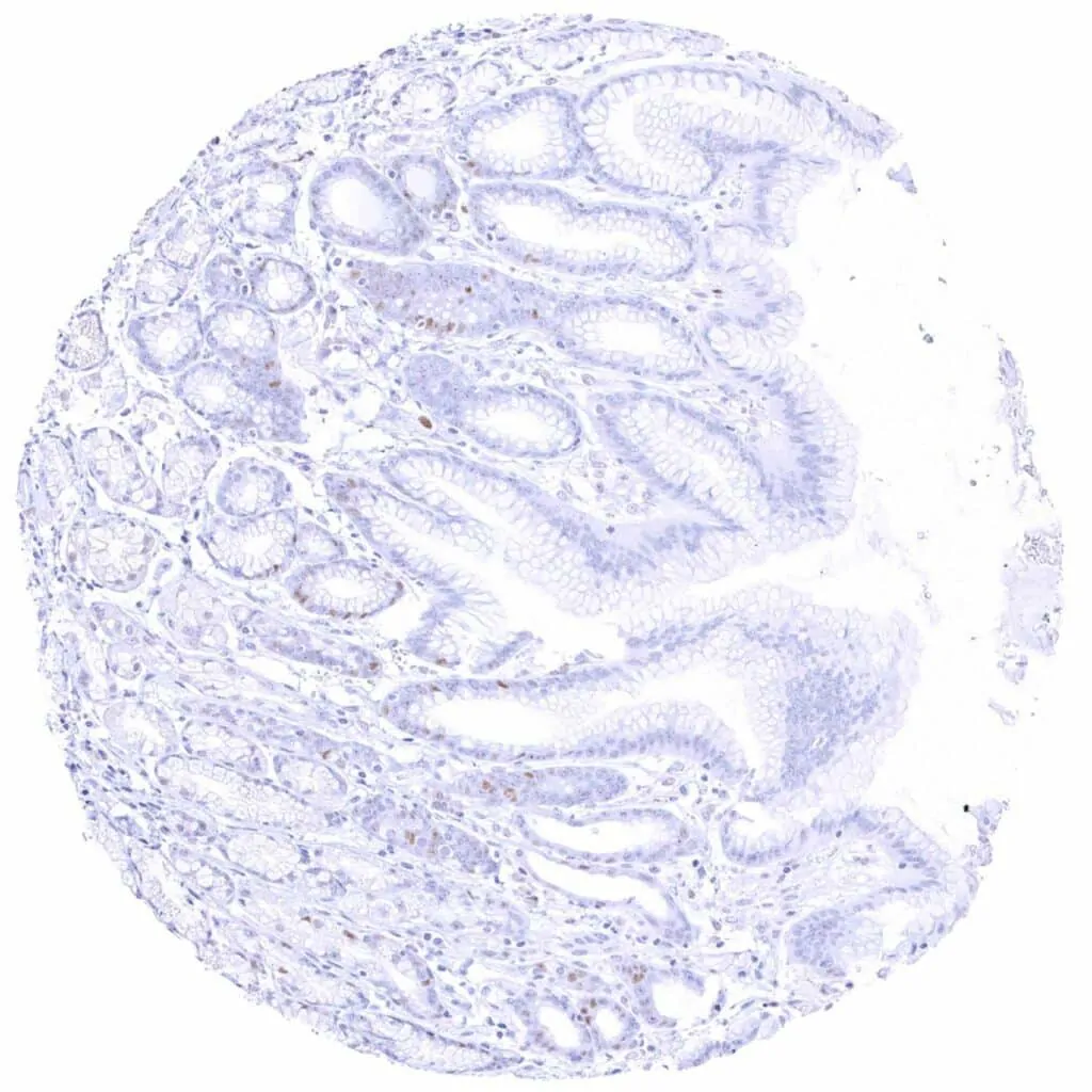
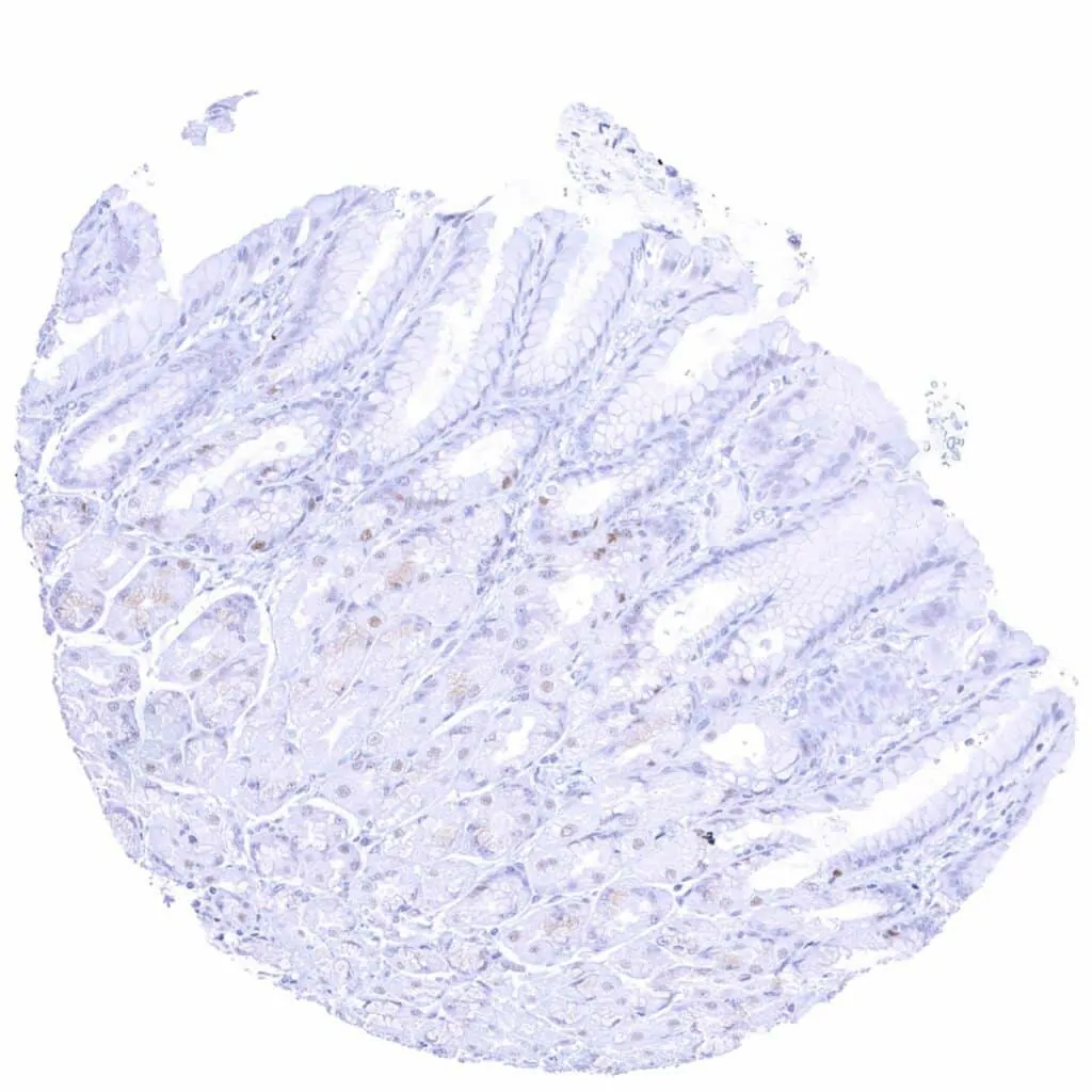
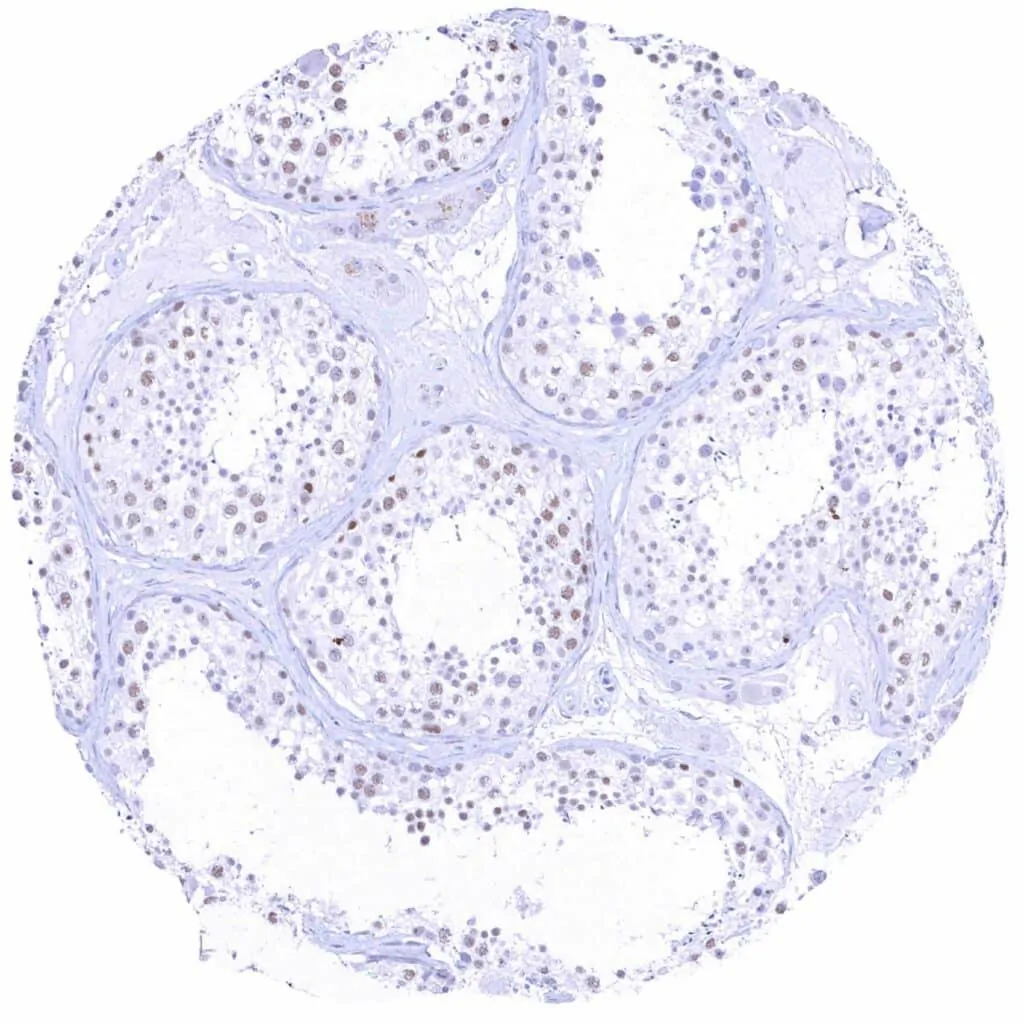
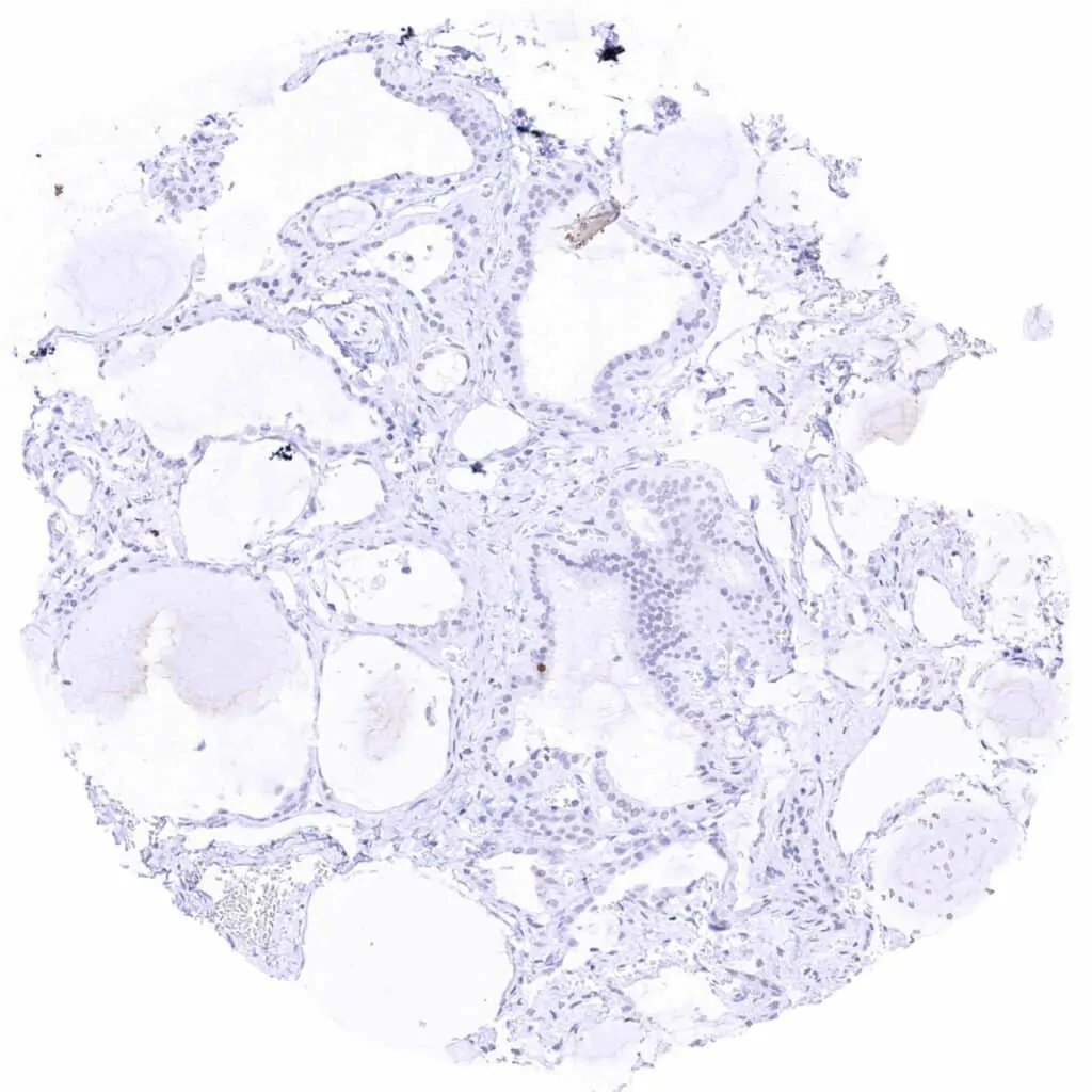
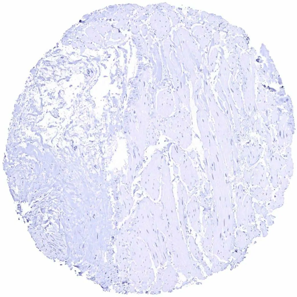
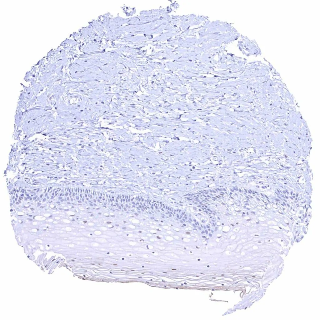
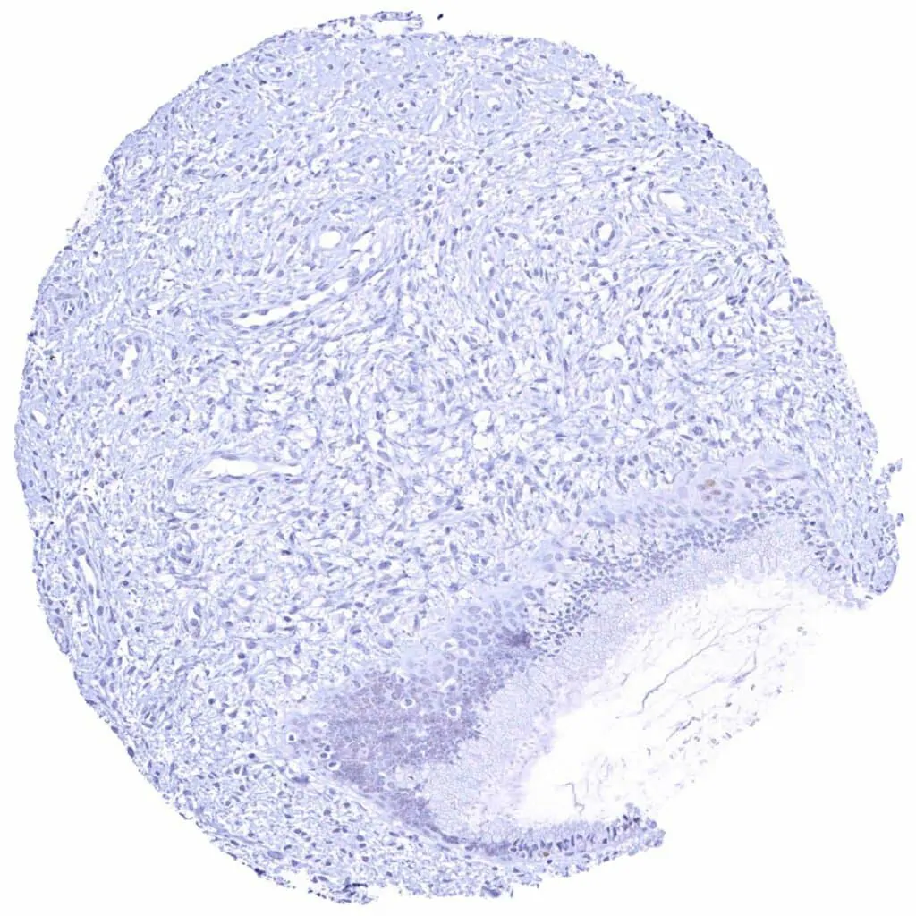
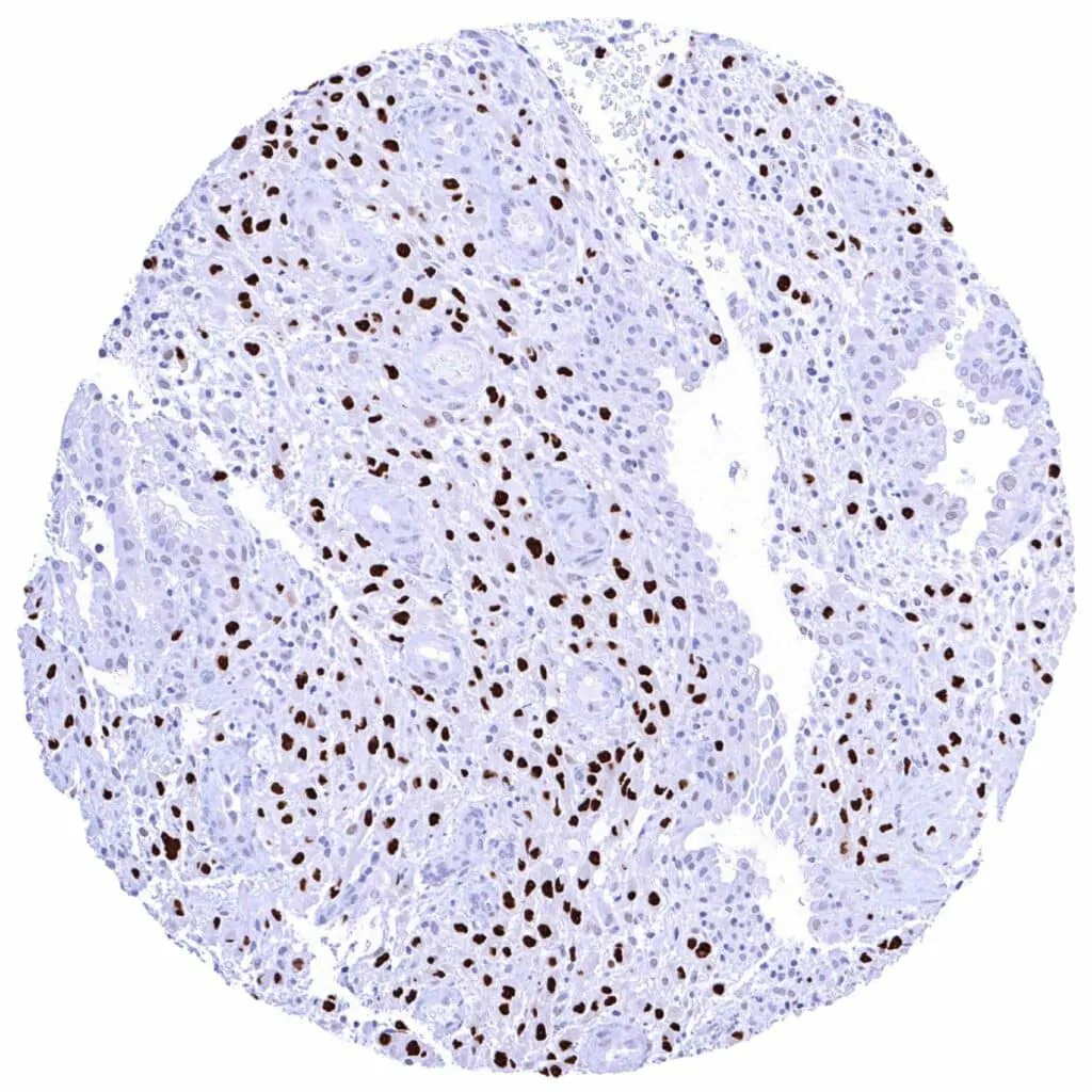
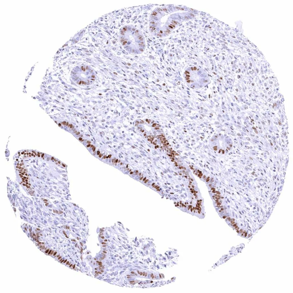
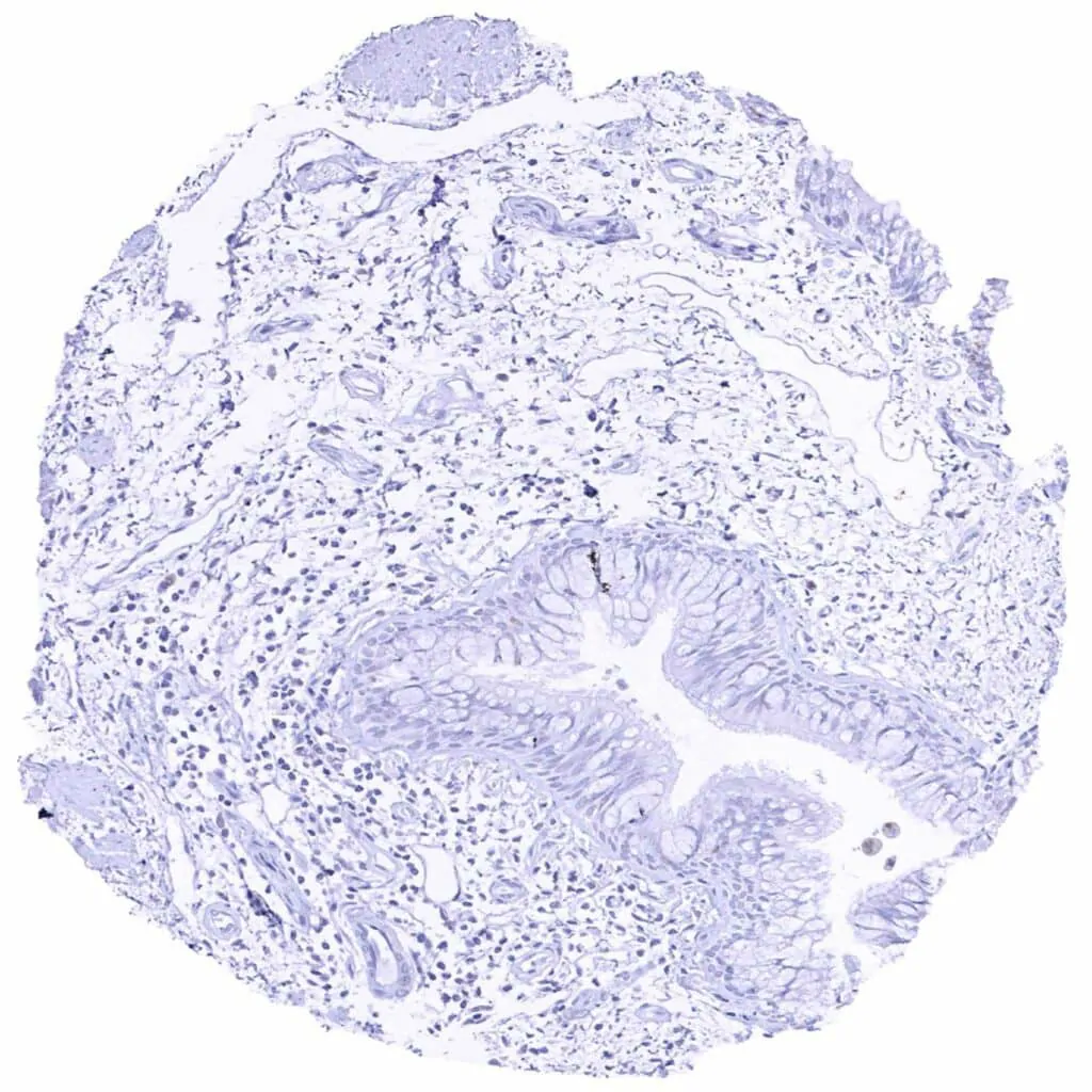
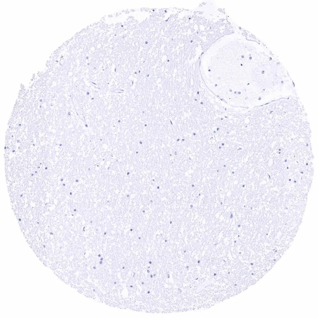
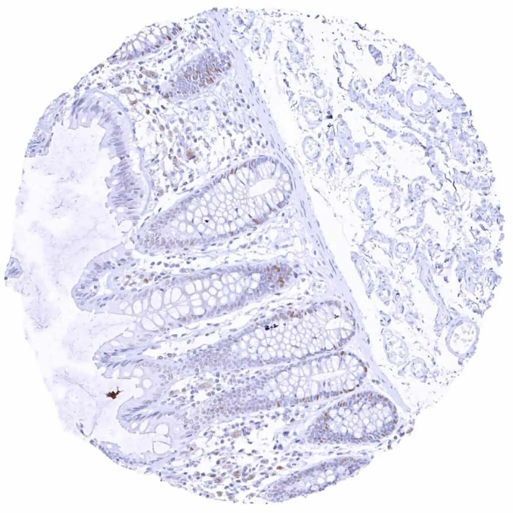
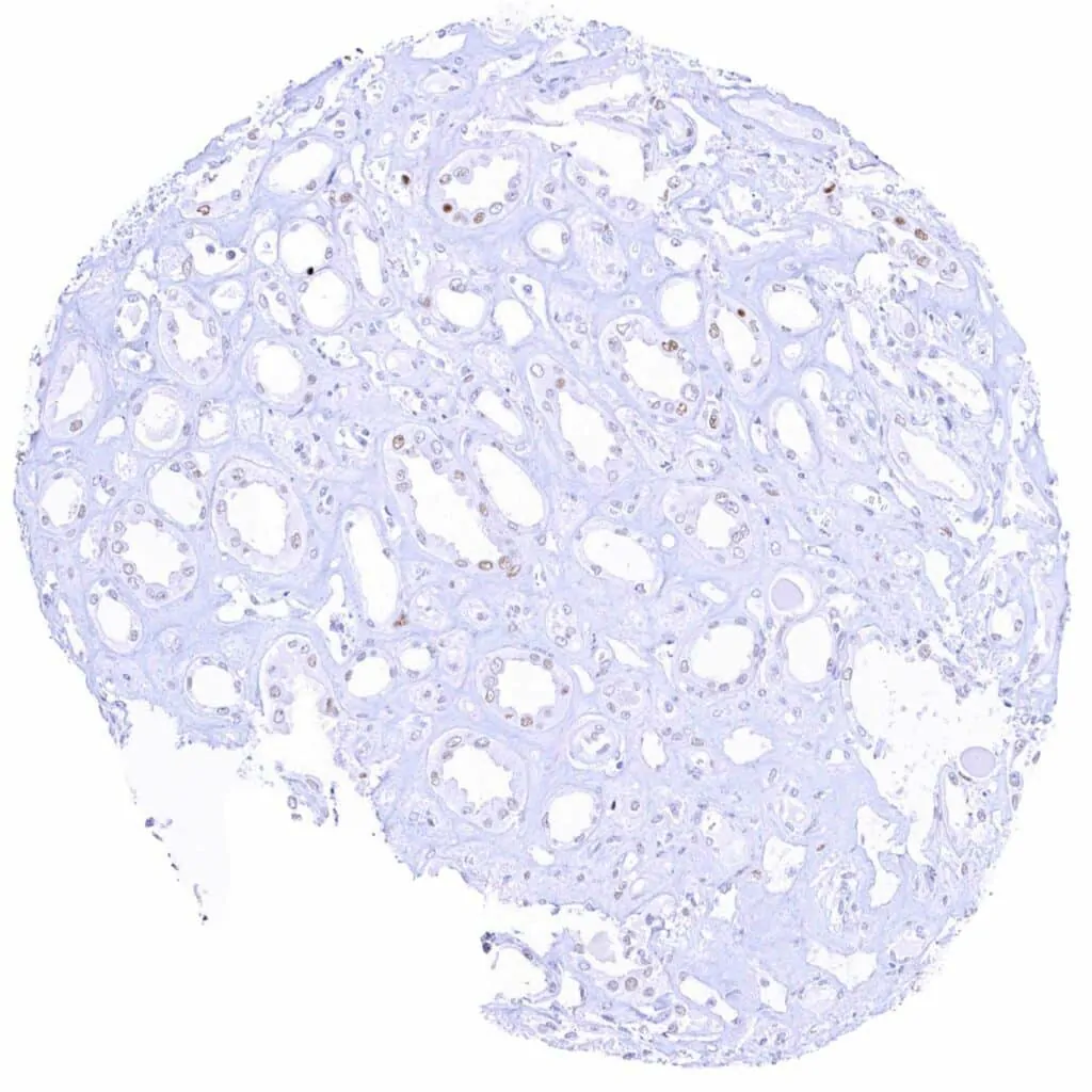
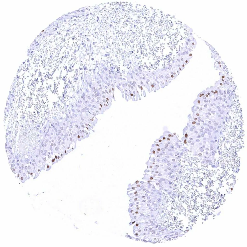
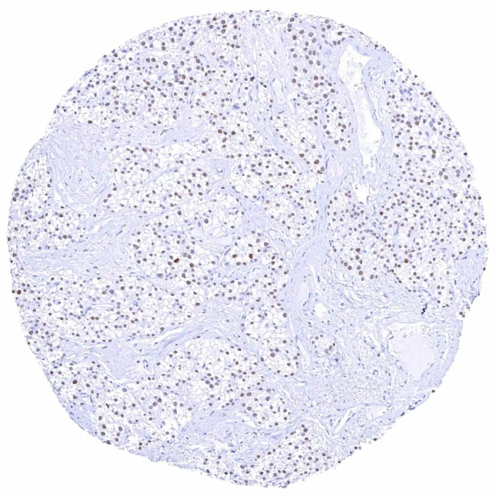
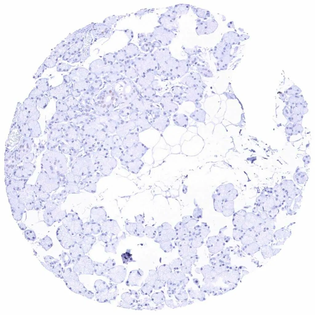
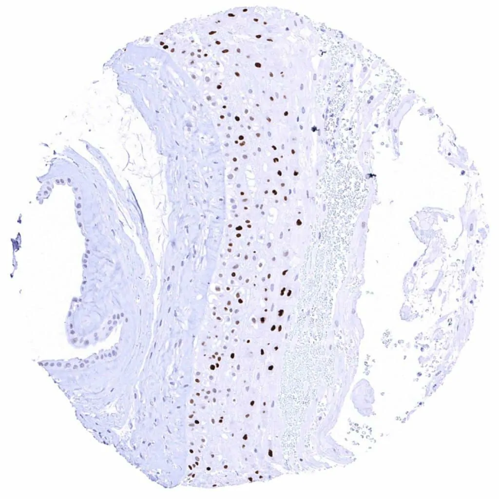
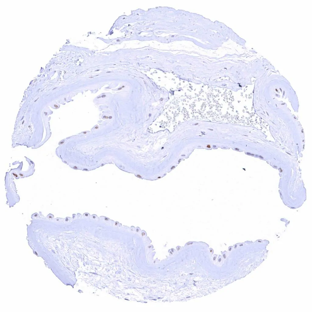
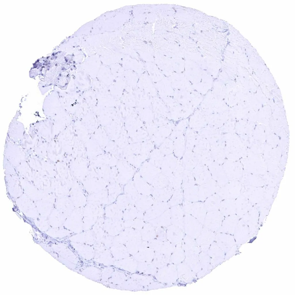
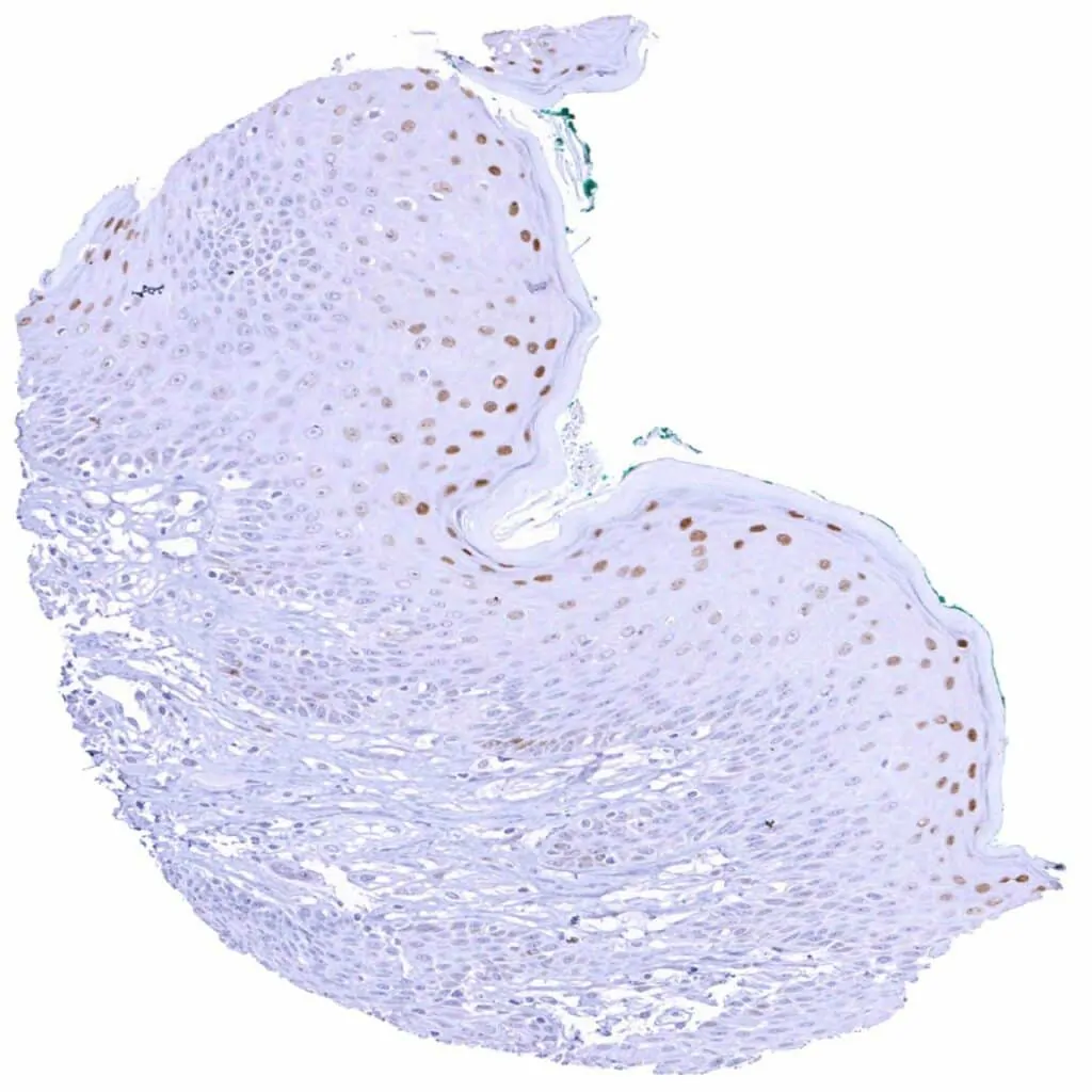
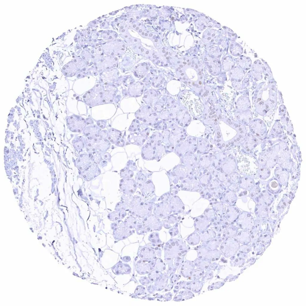
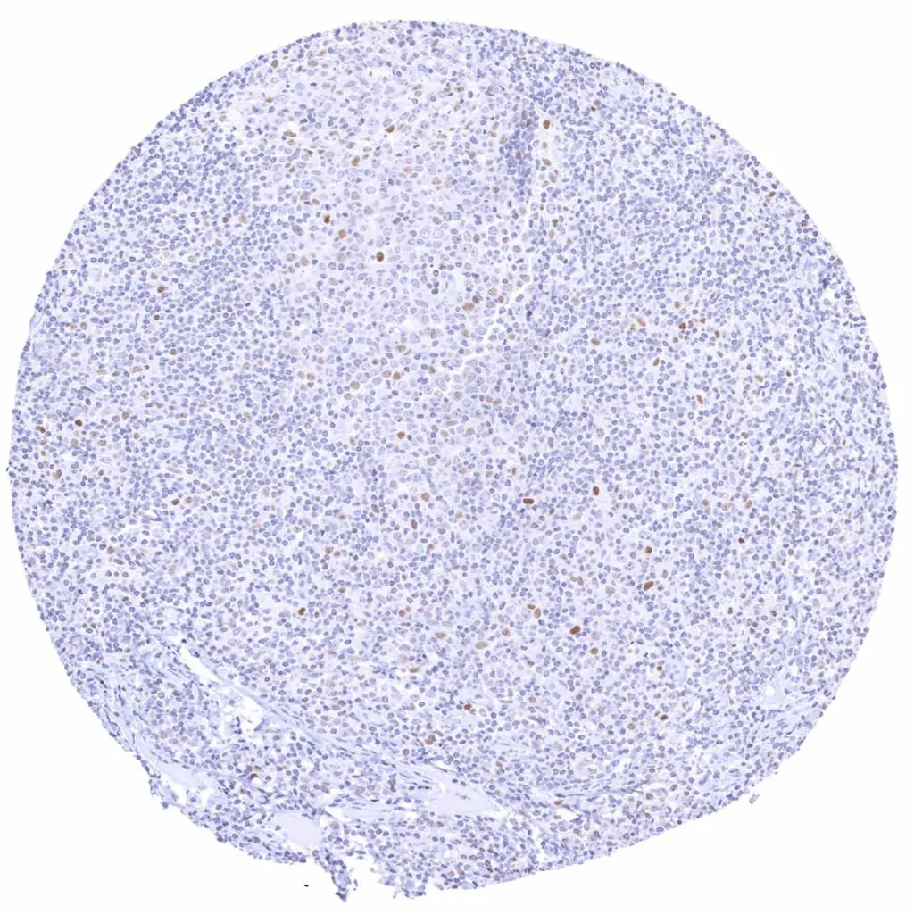
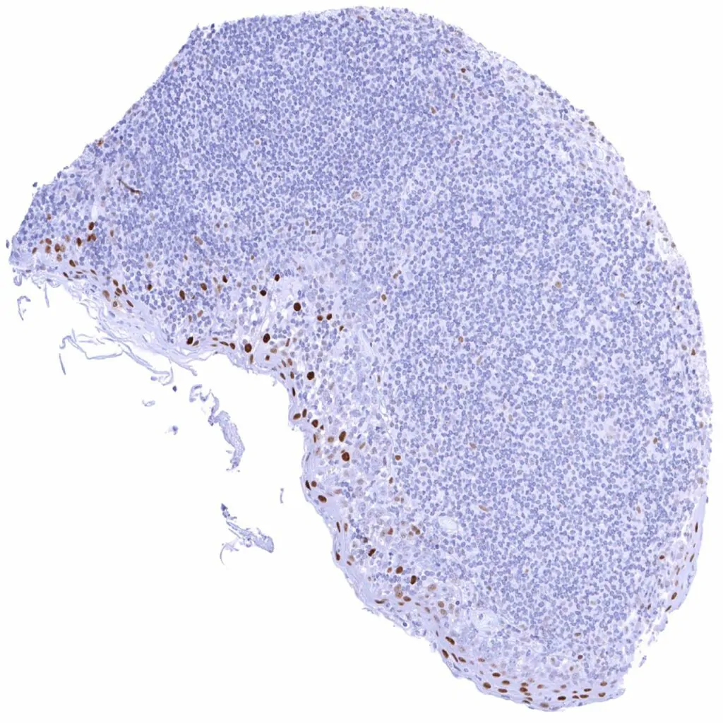
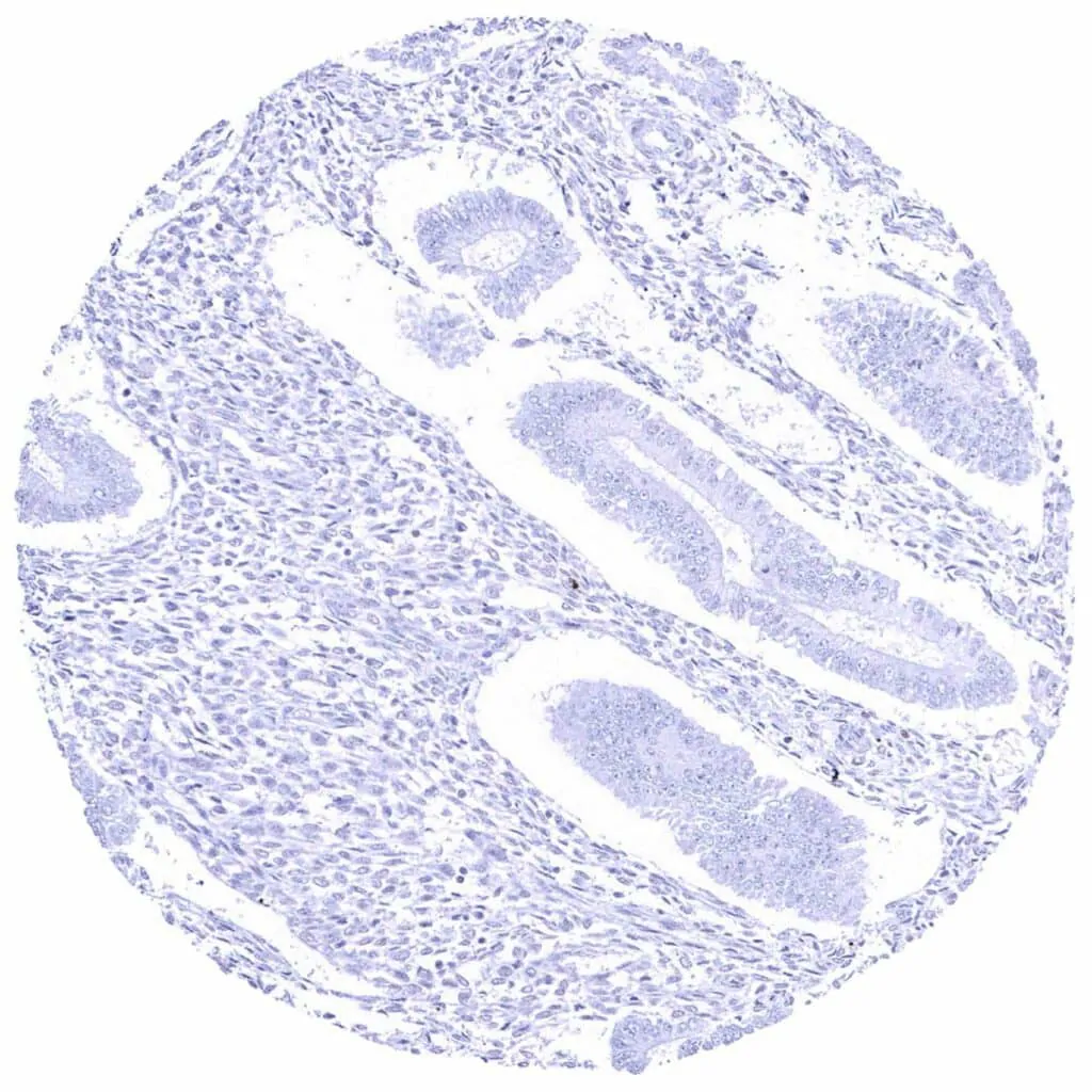
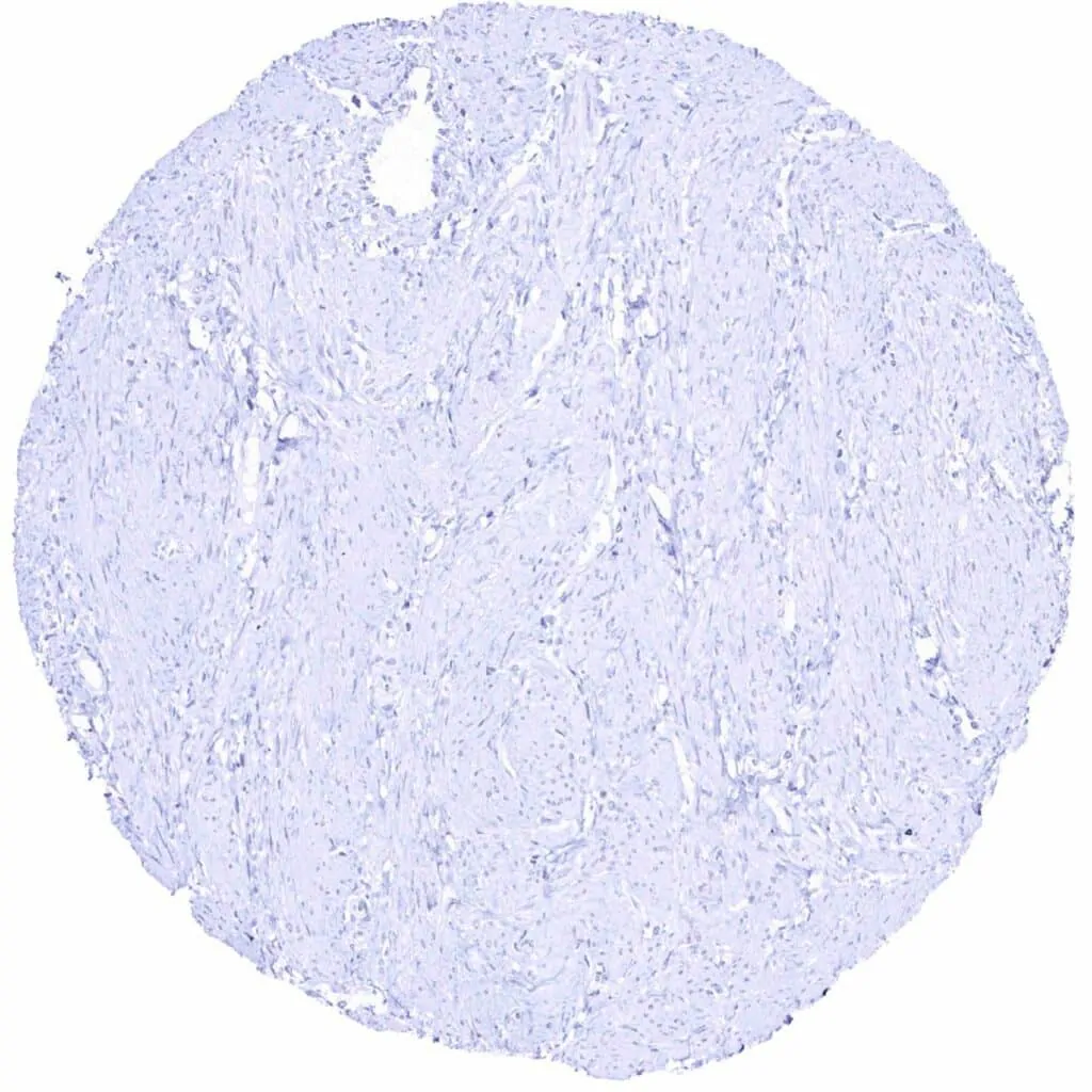
Cyclin E1 antibody [MSVA-898R] HistoMAX™


Go to Cancer Tissue Gallery
  
 Go back to product page
  

Go to HistoMAX page


Adrenal gland – Weak to moderate Cyclin E1 staining of adrenocortical cells

Aorta, media

Appendix, mucosa – Weak to moderate Cyclin E1 staining of a subset of crypt cells

Appendix, muscular wall – Weak to moderate Cyclin E1 staining of a subset of muscle cells. Moderate to strong Cyclin E1 positivity of ganglion cells


Bone marrow – Weak to strong Cyclin E1 staining of a subset of cells

Breast – Faint Cyclin E1 staining of few epithelial cells

Bronchus, mucosa

Cerebellum (white matter)


Cerebellum, cortex (molecular layer, Purkinje cell layer, granule cell layer)

Cerebrum, grey matter

Cerebrum, grey matter

Colon descendens, mucosa – Weak to moderate Cyclin E1 staining of a large subset of crypt cells


Colon descendens, muscular wall

Duodenum, Brunner gland

Duodenum, mucosa – Weak to moderate Cyclin E1 staining of a subset of crypt cells

Epididymis (Caput)


Epididymis (Cauda)

Esophagus, squamous epithelium Weak to strong nuclear Cyclin E1 staining of squamous epithelial cells in the upper half of the surface epithelium. The staining intensity gradually increases towards the surface

Fallopian tube, mucosa – Weak Cyclin E1 staining of few epithelial cells

Fat


Gallbladder, epithelium – Lack of Cyclin E1 staining in this sample

Gallbladder, epithelium – Weak Cyclin E1 staining of only few epithelial cells

Heart muscle

Ileum, mucosa – Weak to moderate Cyclin E1 staining of a subset of crypt cells


Kidney, cortex – Moderate to strong Cyclin E1 staining of a subset of inflammatory cells in a case of mild interstitial nephritis

Kidney, cortex

Kidney, medulla – Absence of Cyclin E1 staining in this sample

Kidney, medulla – Weak nuclear Cyclin E1 staining of a subset of collecting duct cells in this sample


Kidney, pelvis, urothelium – Weak to strong Cyclin E1 staining of urothelial cells is limited to the top cell layers of the urothelium. The staining intensity increases towards the surface

Liver

Lung

Lymph node – Weak Cyclin E1 staining of only a small fraction of cells


Ovary, corpus luteum – Weak Cyclin E1 staining of corpus luteum cells

Ovary, stroma

Pancreas

Parathyroid gland – Weak to moderate Cyclin E1 staining of epithelial cells


Parotid gland

Pituitary gland, anterior lobe

Pituitary gland, posterior lobe

Placenta (amnion and chorion) – Strong Cyclin E1 staining of most chorion cells. Amnion cells are largely negative in this sample


Placenta (amnion) – Weak to strong Cyclin E1 staining of a fraction of amnion cells

Placenta, first trimenon – Strong Cyclin E1 staining of most cytotrophoblast cells while stroma cells stain less intensively

Placenta, mature – Strong Cyclin E1 staining of many-most cytotrophoblast cells

Prostate


Rectum, mucosa – Weak to moderate Cyclin E1 staining of a large subset of crypt cells

Seminal vesicle

Sinus paranasales – Weak to moderate Cyclin E1 staining of a subset of inflammatory cells

Skeletal muscle – Very faint Cyclin E1 staining in few cells


Skin – Weak to strong Cyclin E1 staining of squamous epithelial cells of the upper half of the epidermis. The staining intensity gradually increases towards the surface

Spleen – Weak Cyclin E1 staining of only a small fraction of cells

Stomach, antrum – Weak to moderate Cyclin E1 staining of a subset of cells (predominantly neck cells)

Stomach, corpus – Faint Cyclin E1 staining of a subset of cells (predominantly neck cells)


Submandibular gland – Faint Cyclin E1 staining of few epithelial, mainly ductal cells

Testis – Weak to moderate Cyclin E1 staining of spermatocytes

Thymus – Strong Cyclin E1 staining of some regularly distributed cells (thymic epithelial cells_). Weak Cyclin E1 staining of some lymphocytic cells

Thyroid gland


Tonsil – Weak to moderate Cyclin E1 staining of a fraction of lymphocytic cells, especially in germinal centres

Tonsil, surface epithelium – Strong Cyclin E1 staining of most squamous epithelial cells in the upper half of the surface epithelium

Urinary bladder, muscular wall

Uterus, ectocervix – Faint Cyclin E1 staining of squamous epithelial cells of the upper 2-3 of the epithelial cell layer


Uterus, endocervix

Uterus, endometrium (pregnancy) – Strong Cyclin E1 staining of decidua cells

Uterus, endometrium (proliferation) – Strong Cyclin E1 staining of a large fraction of epithelial cells while staining is weak in a fraction of stroma cells

Uterus, endometrium (secretion)


Uterus, myometrium


| TOP |