GeneTex
  • Country / Location Selection

United States (US)

Normal Tissue Gallery- GTX04396

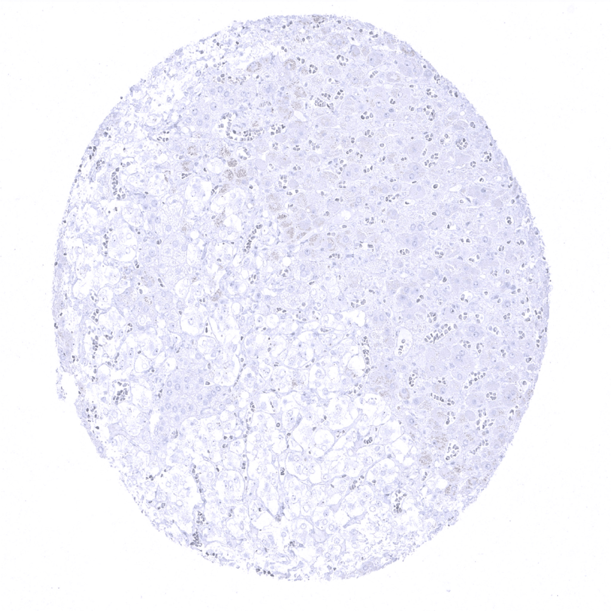
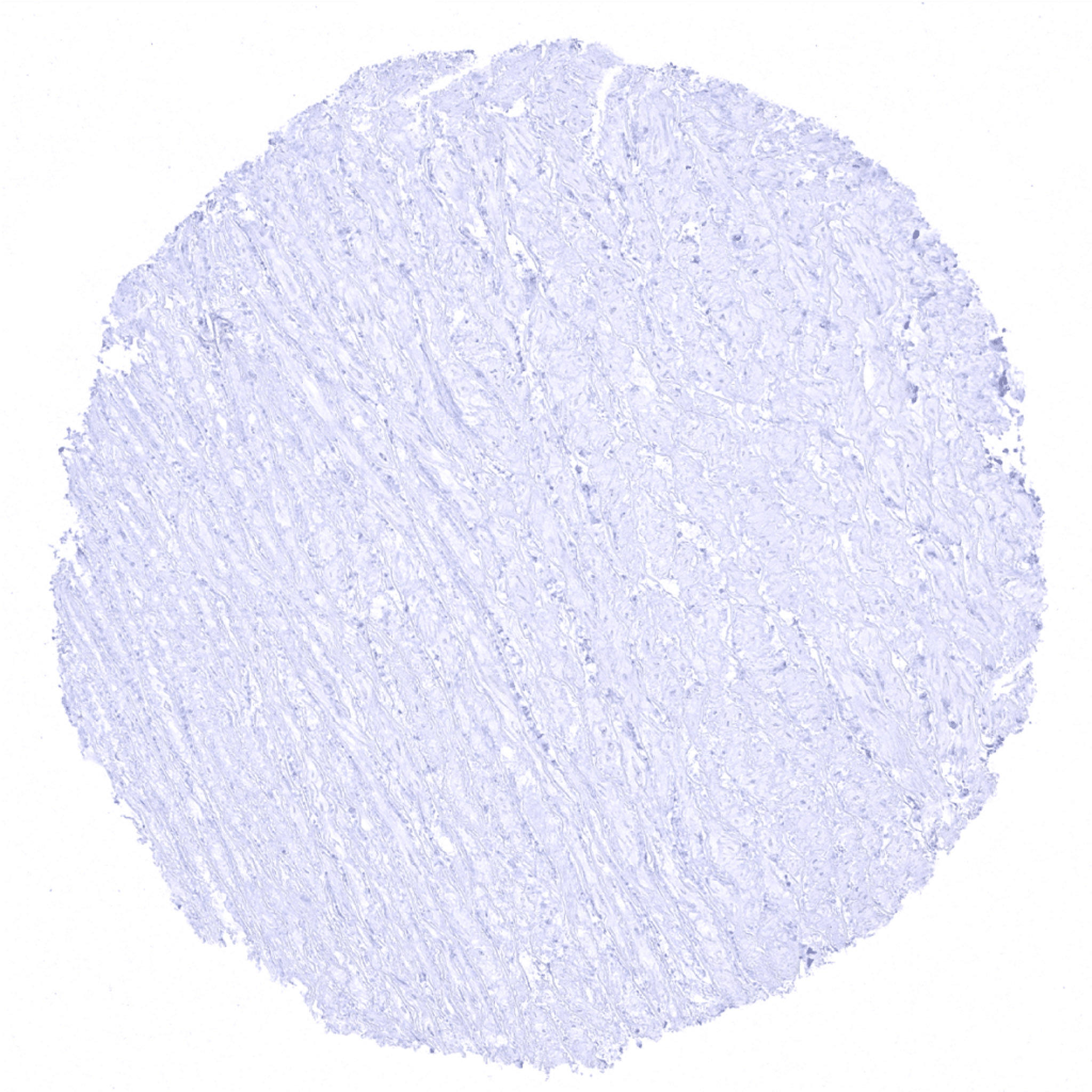
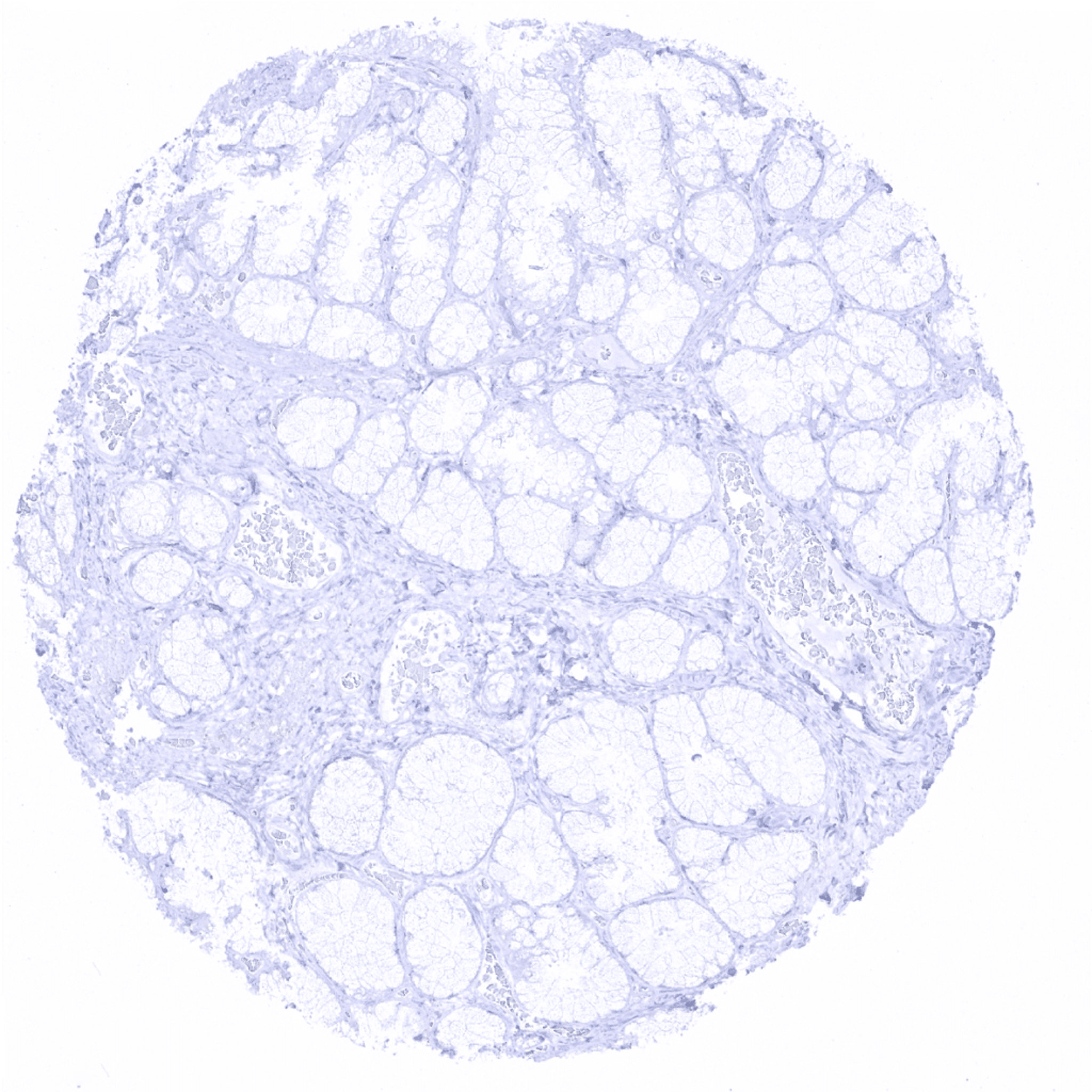
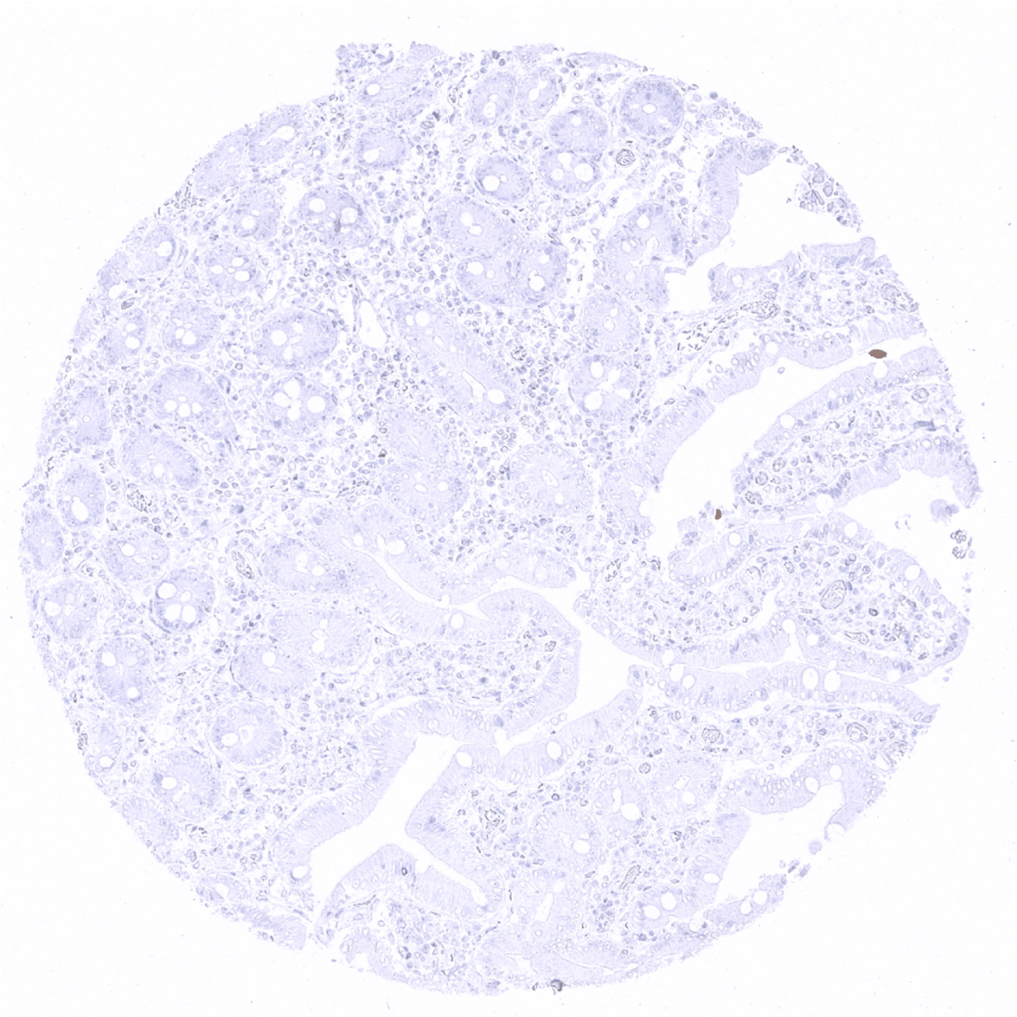
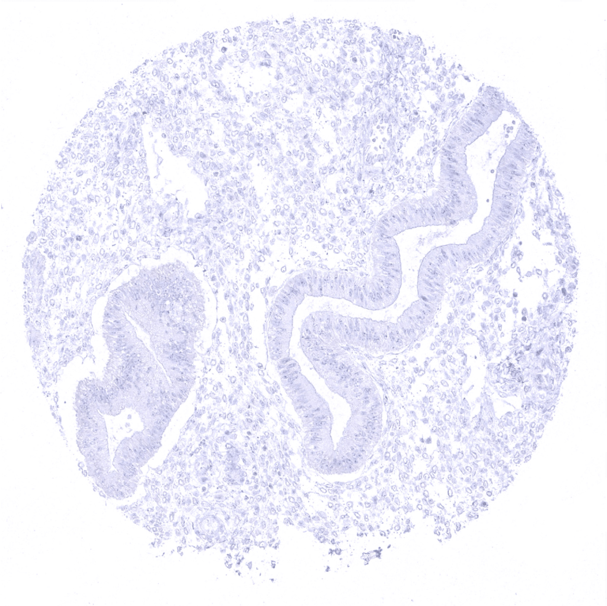
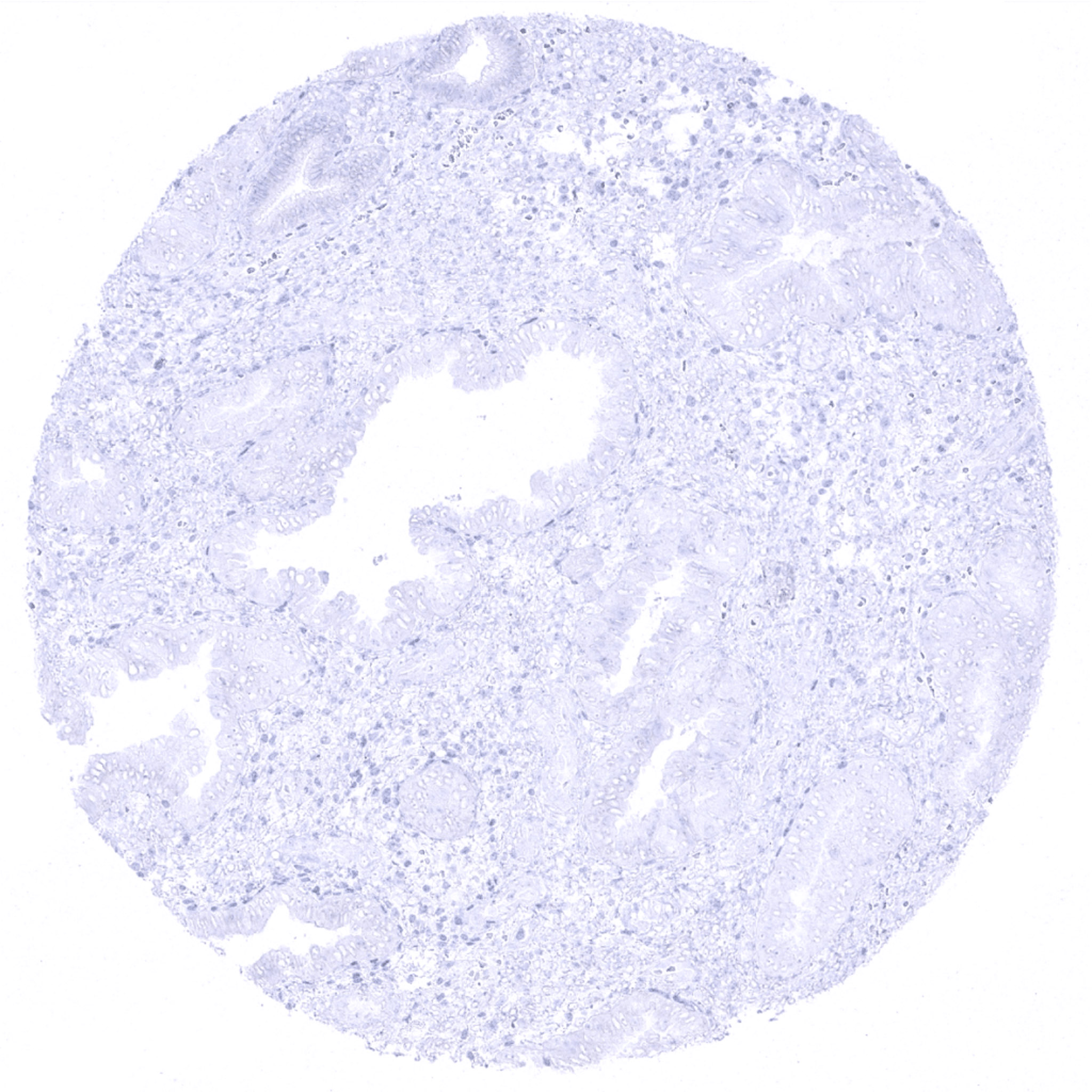
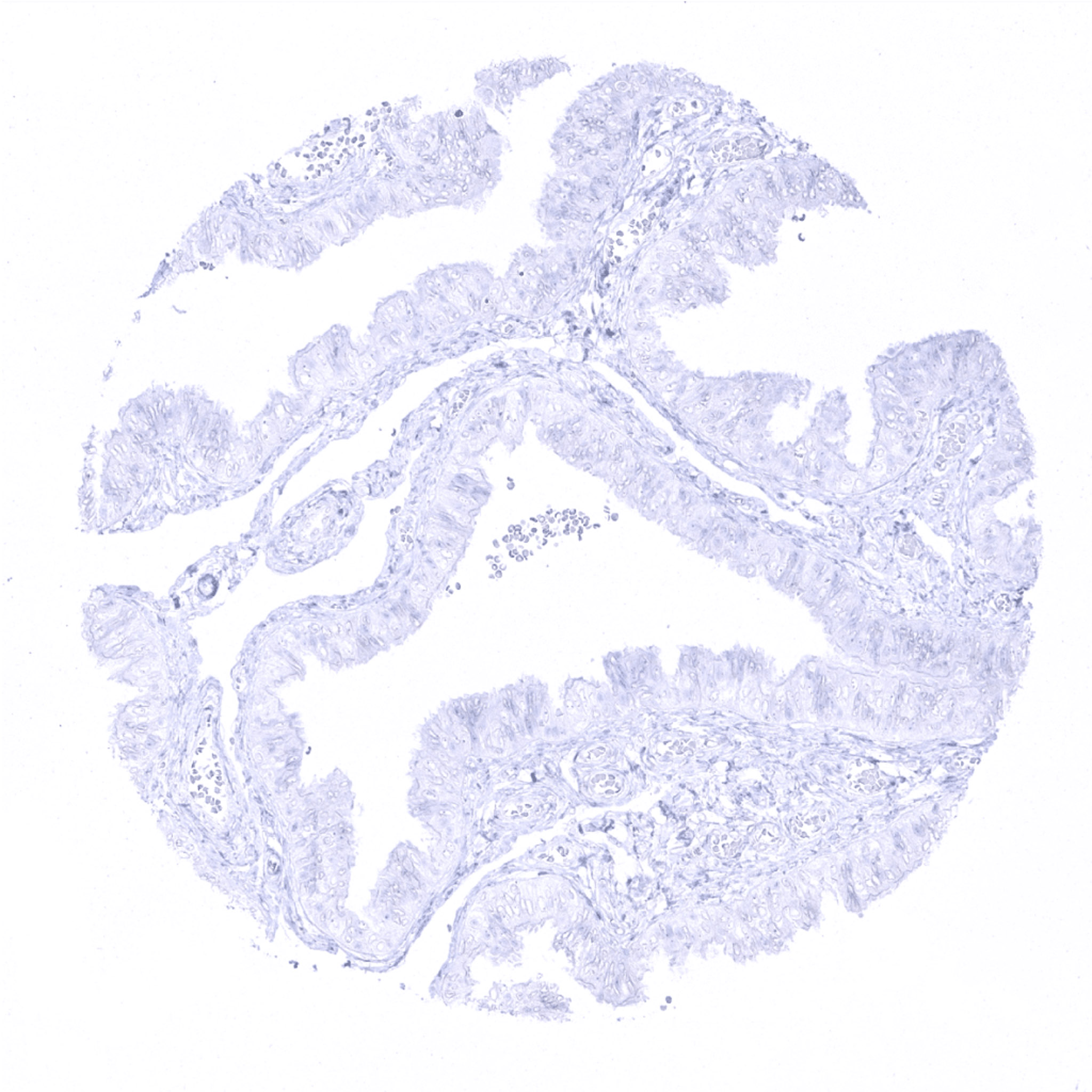
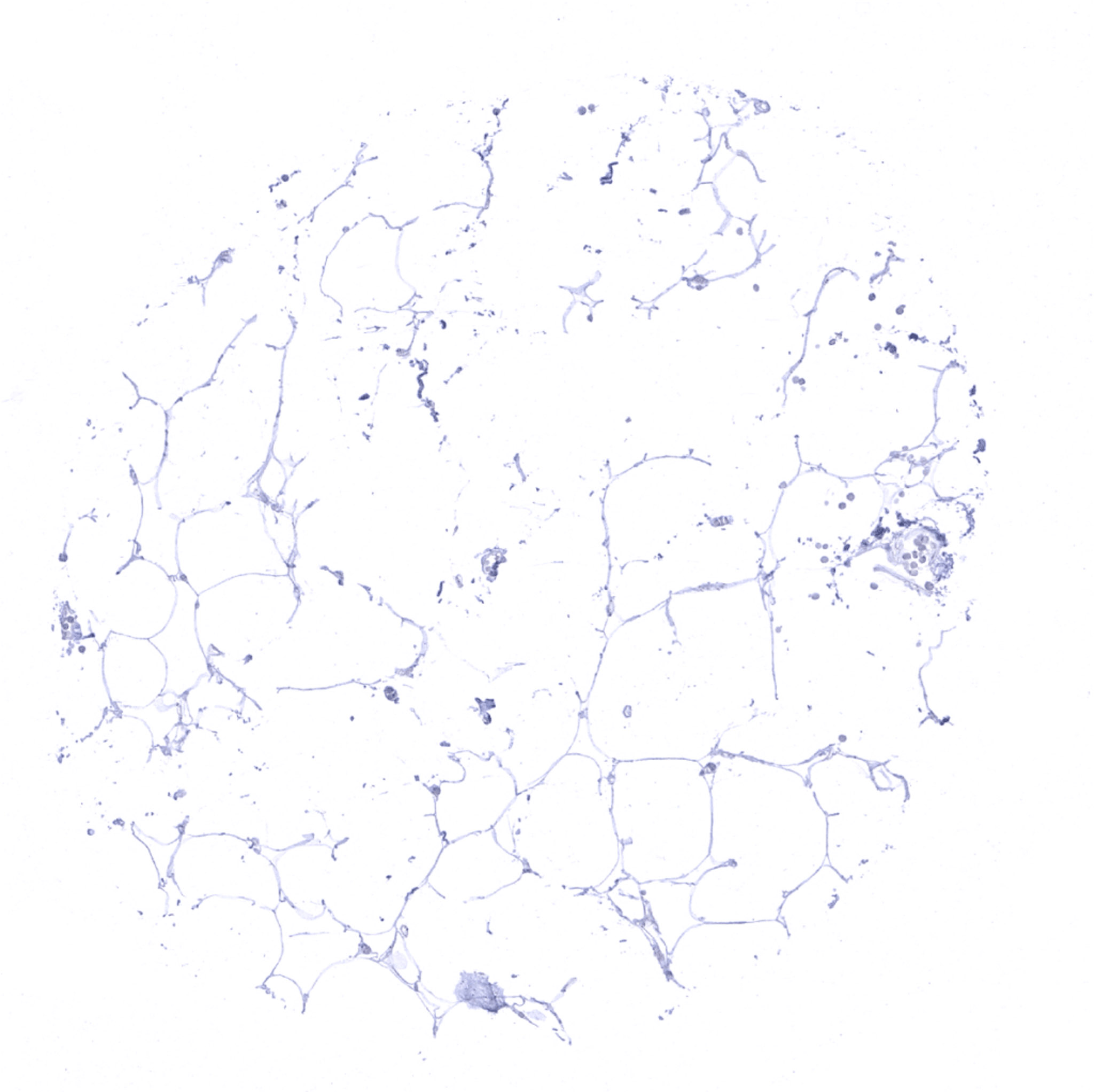
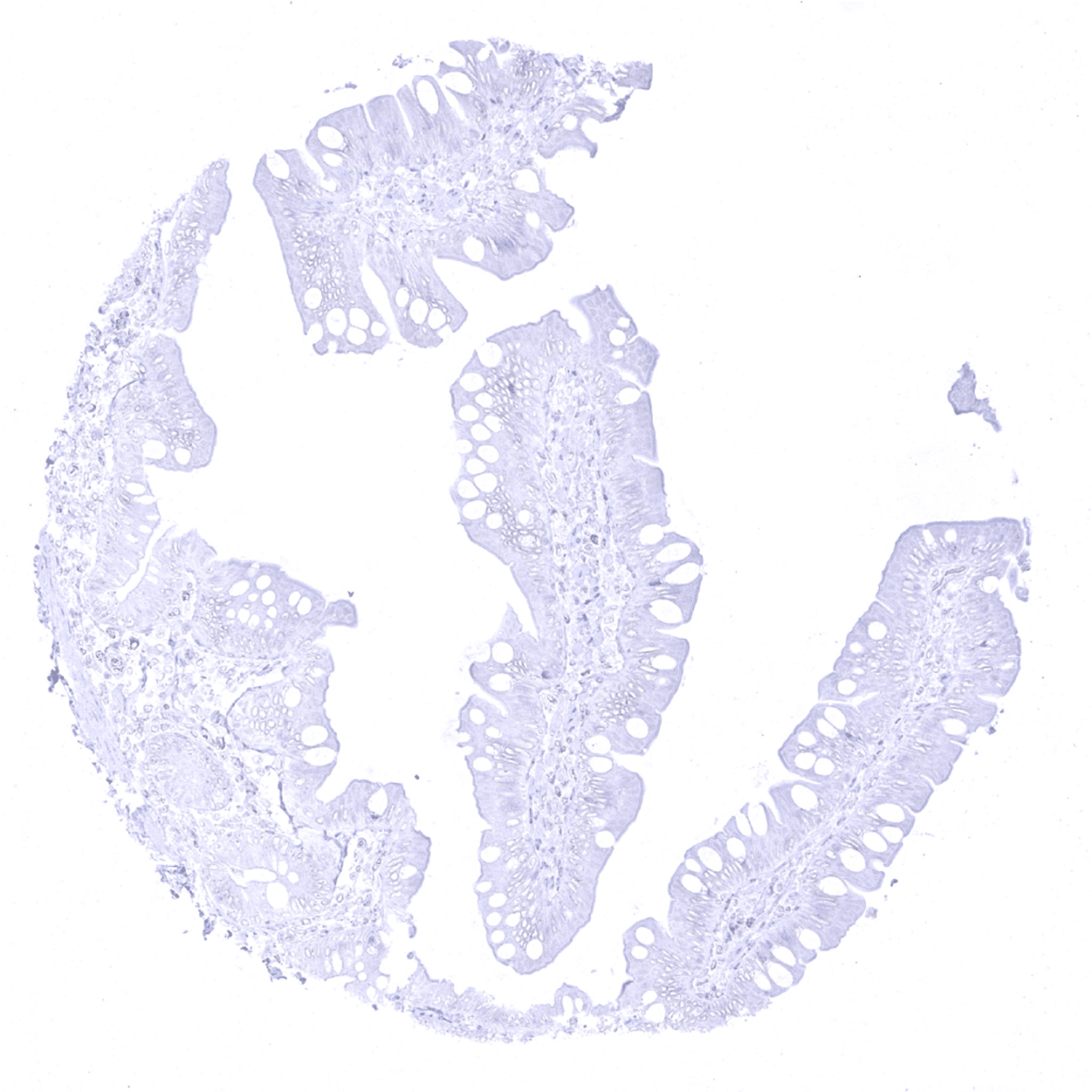
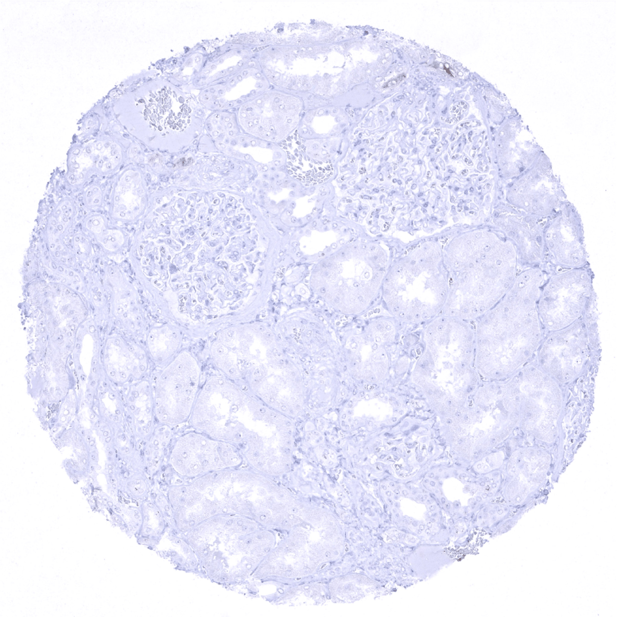
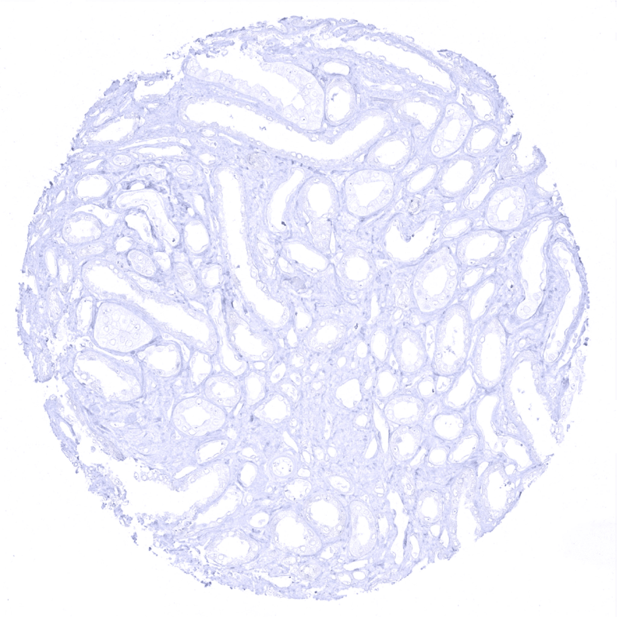
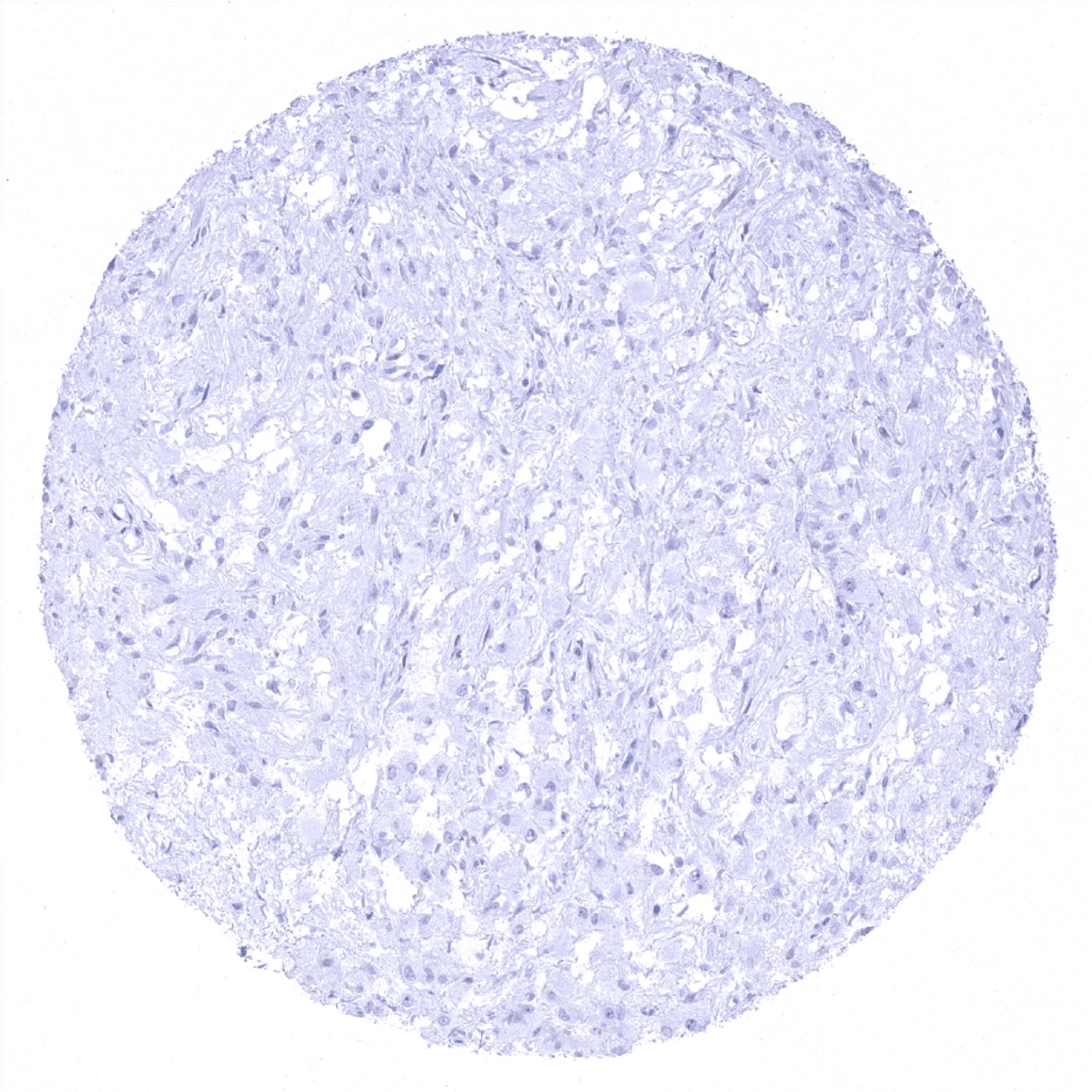
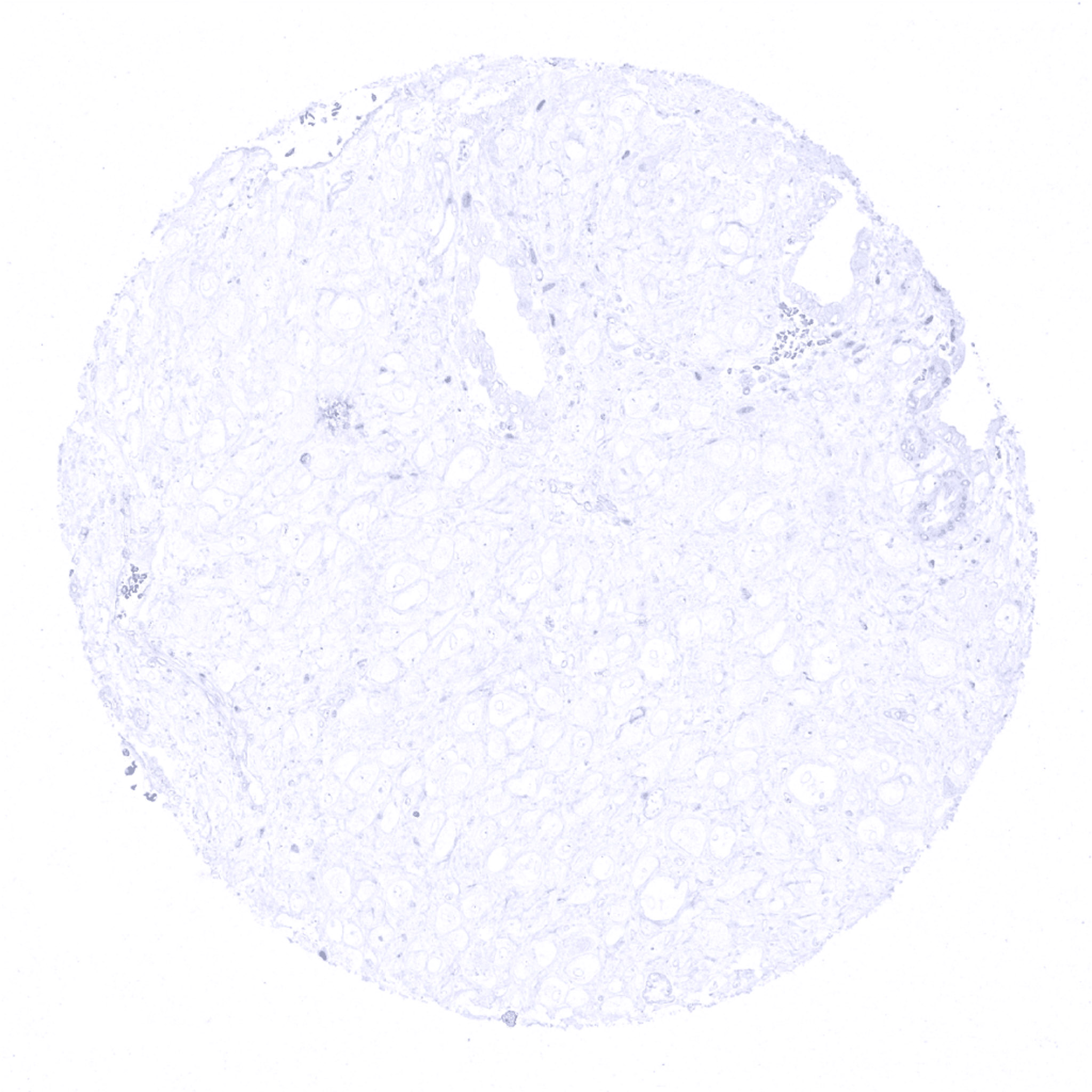
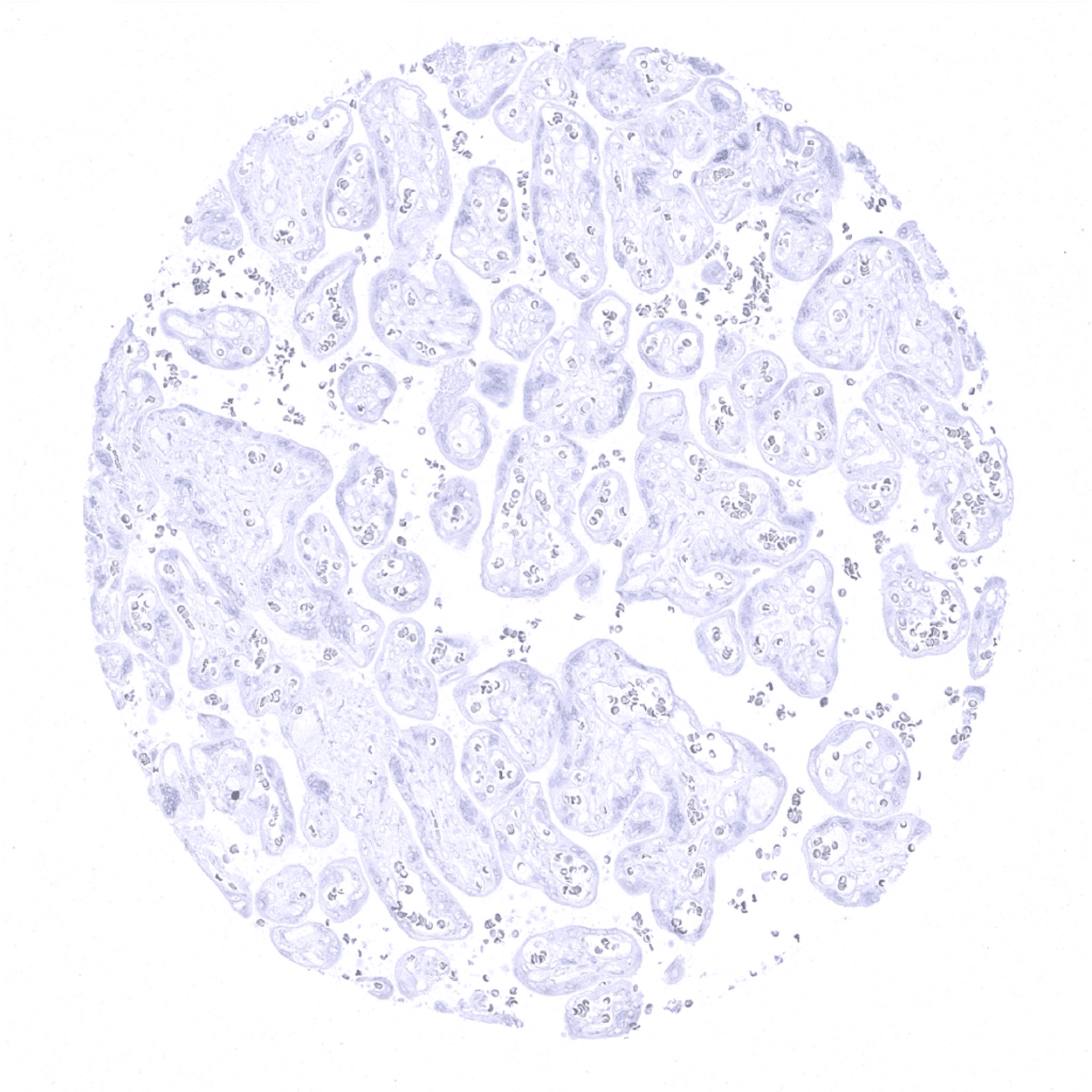
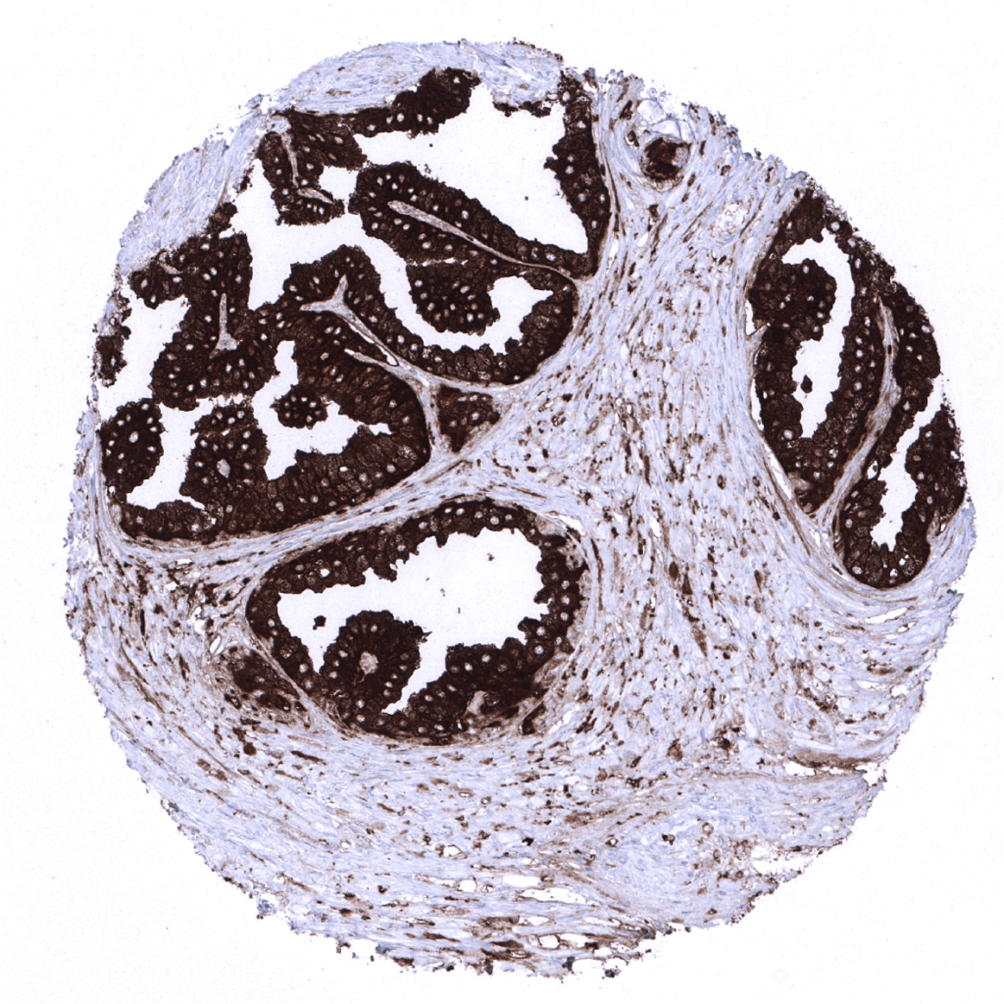
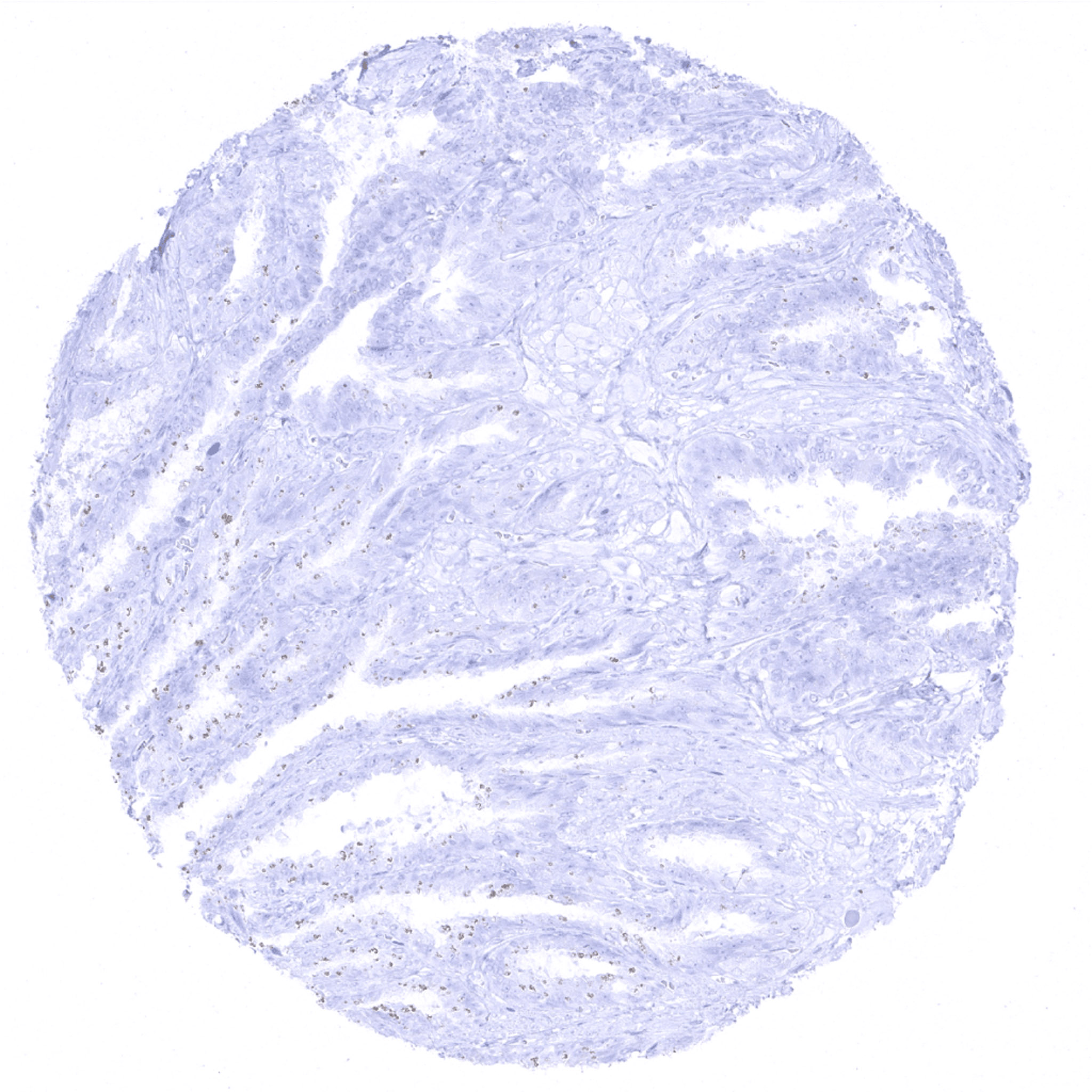
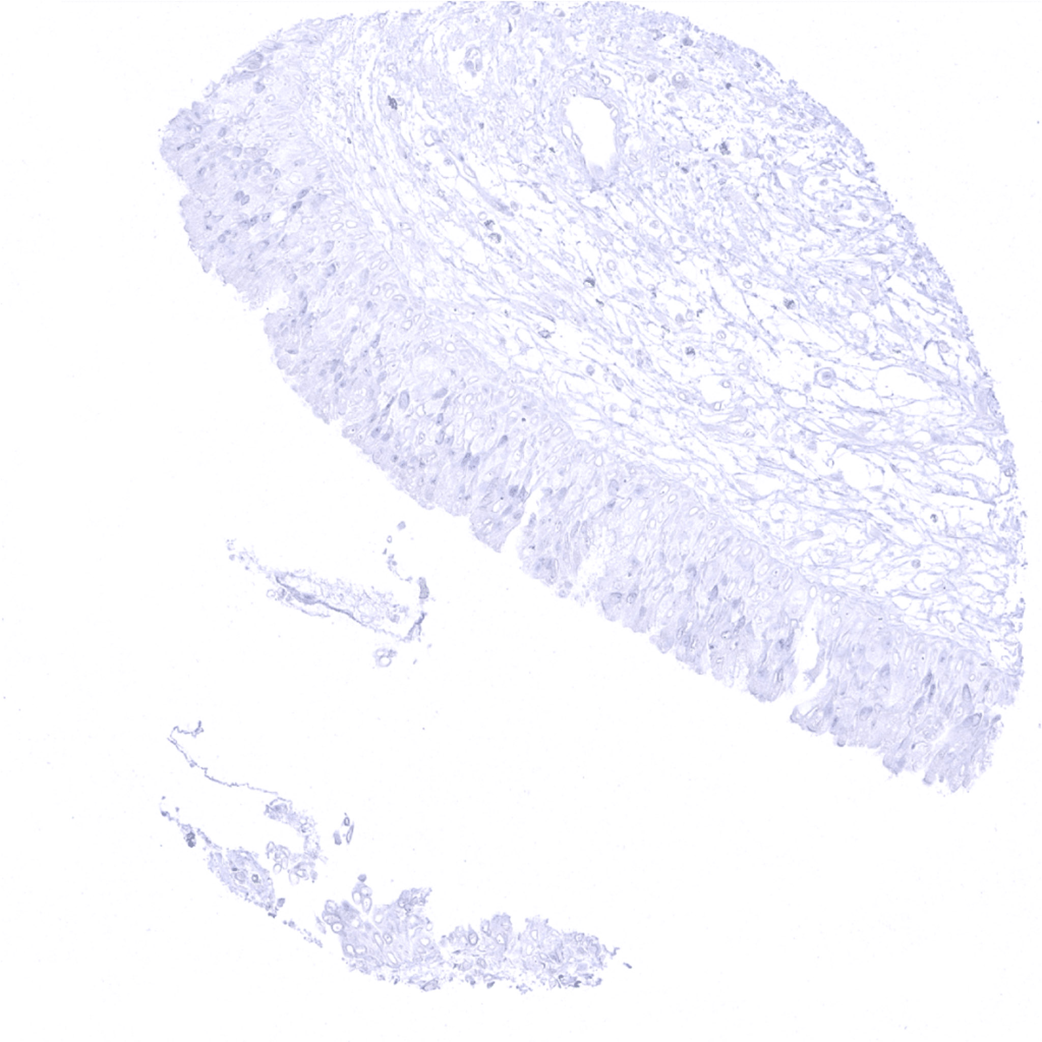
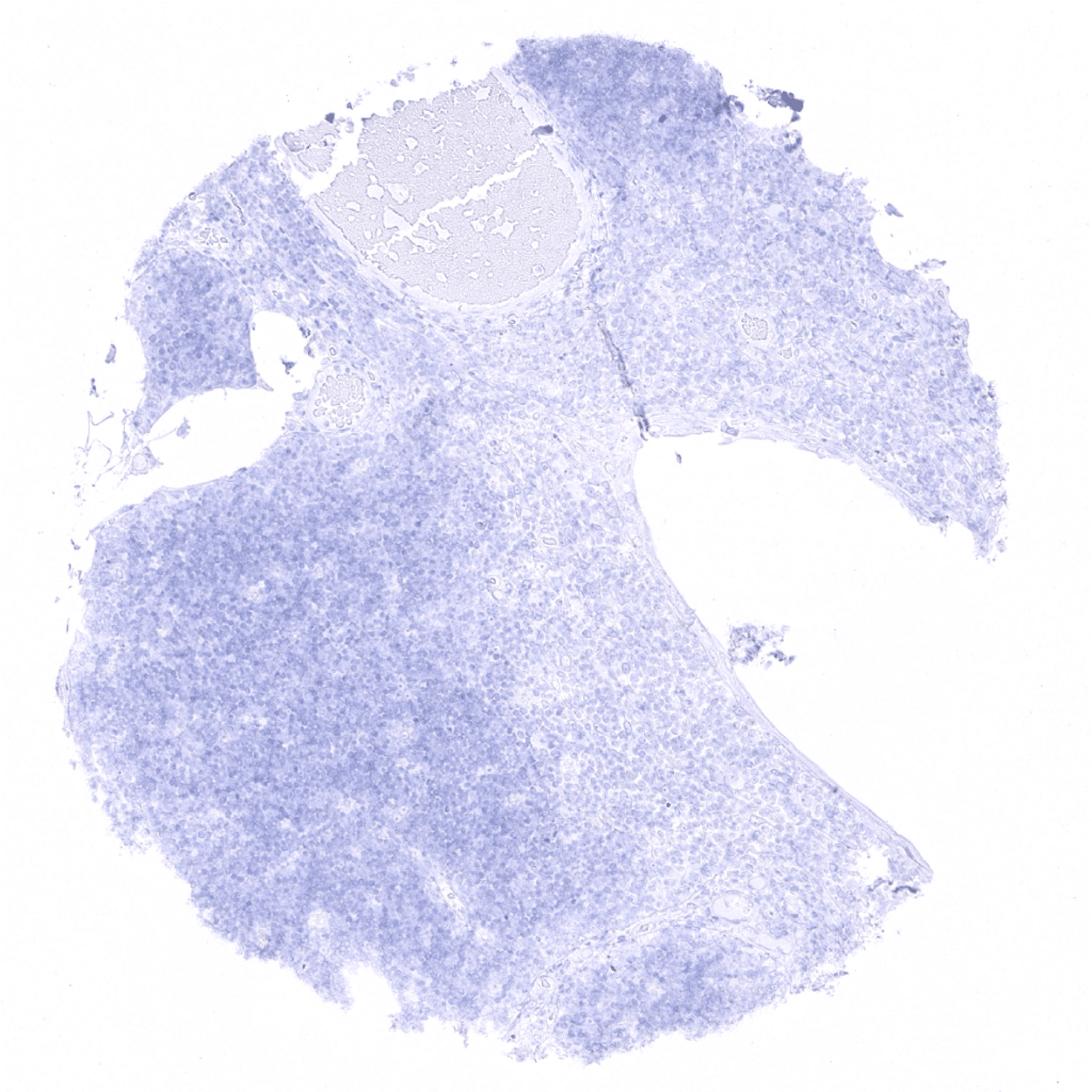
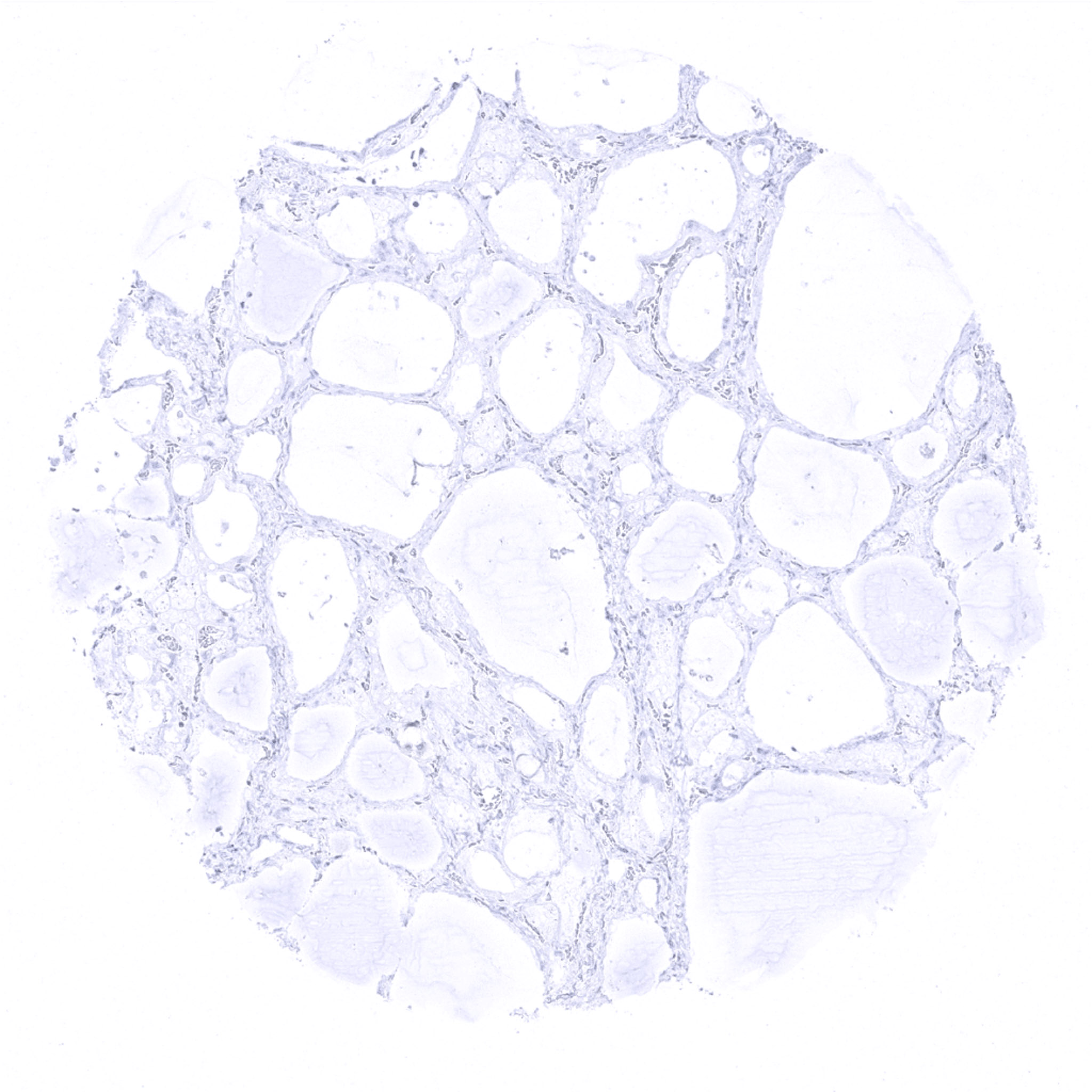
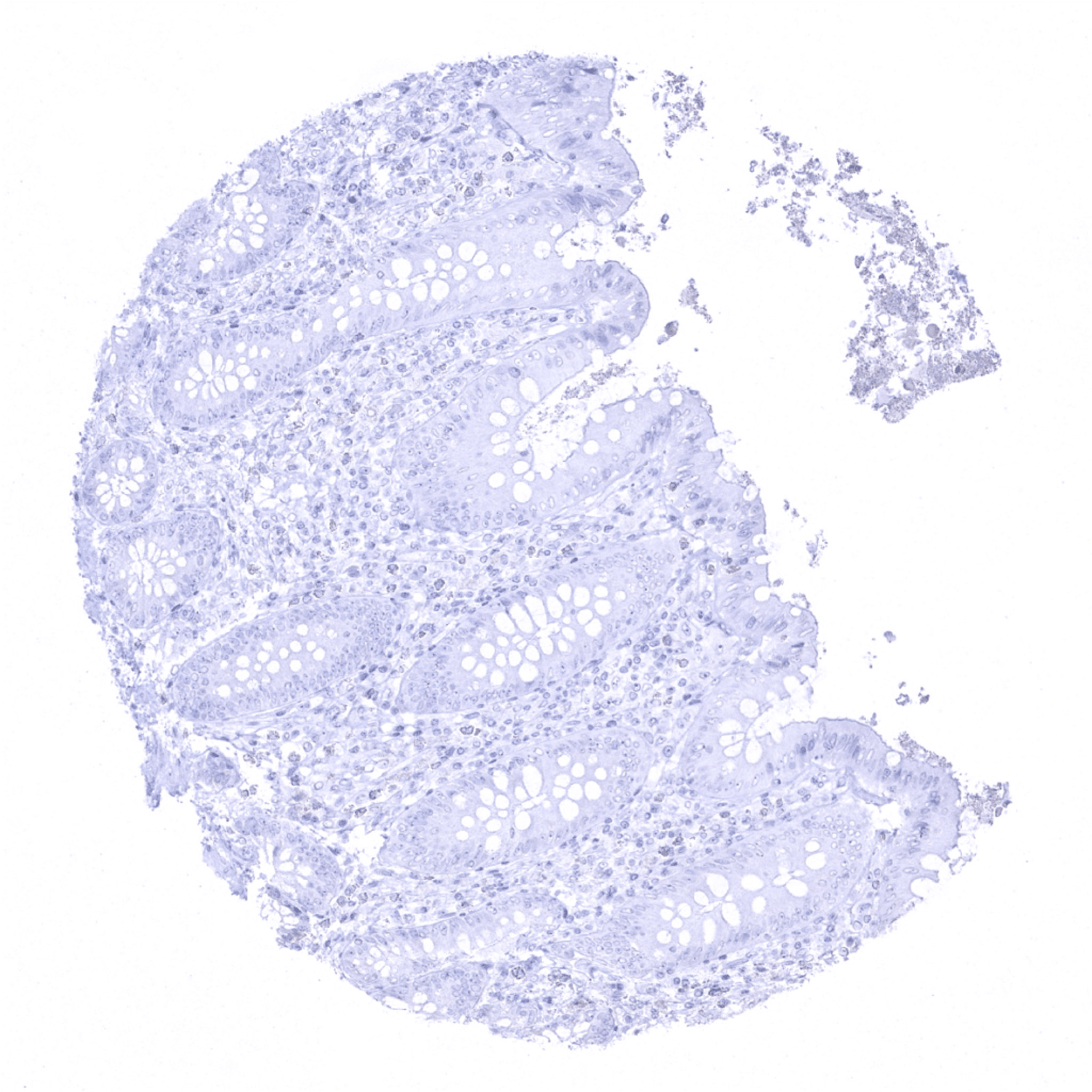
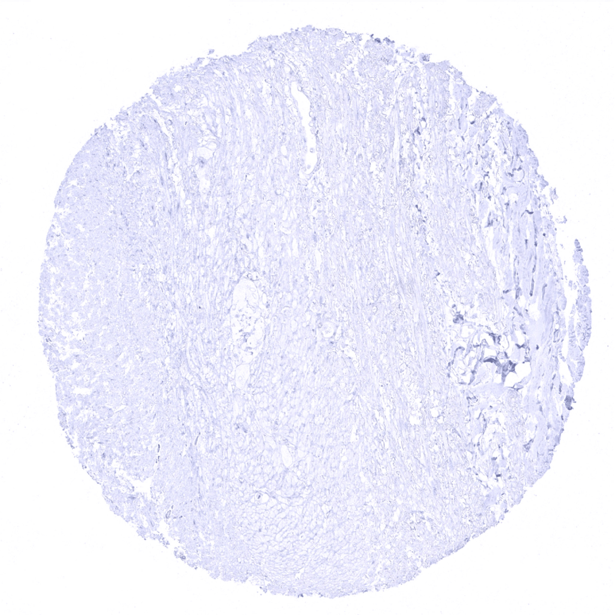
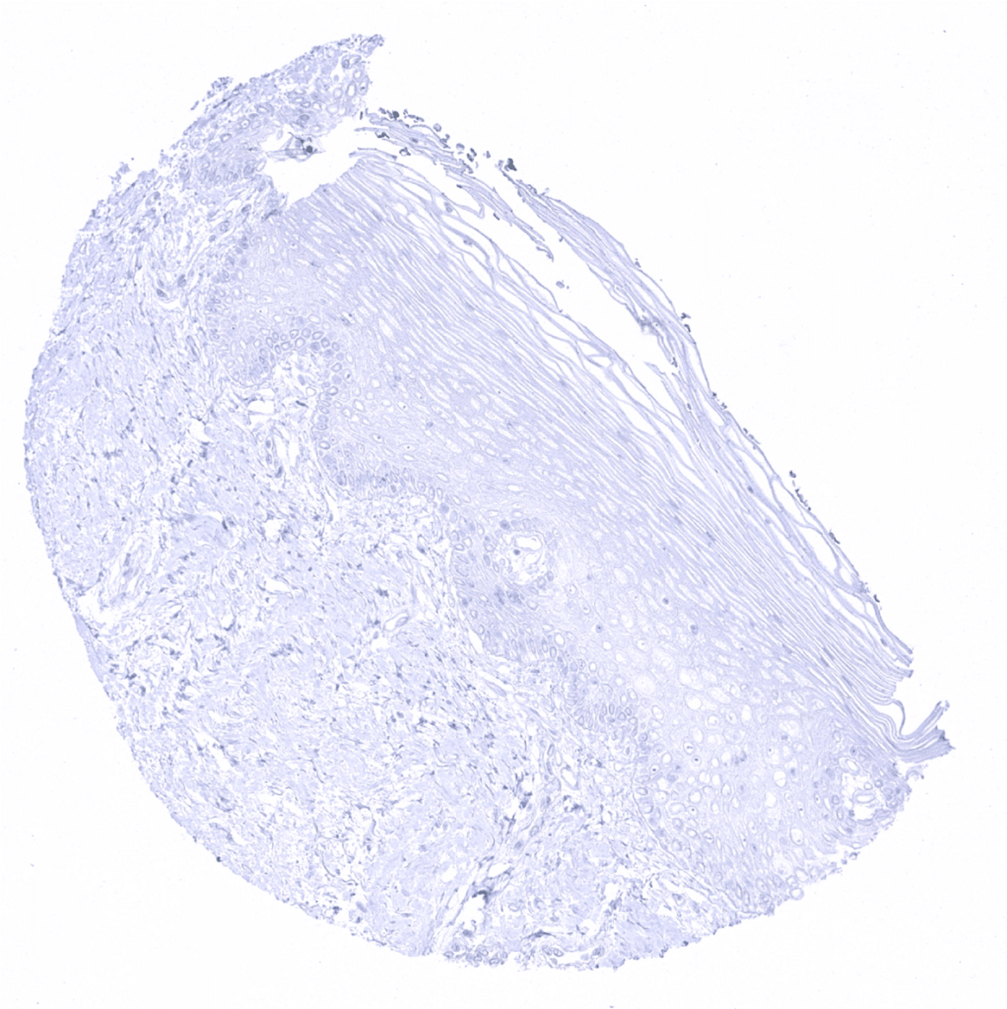
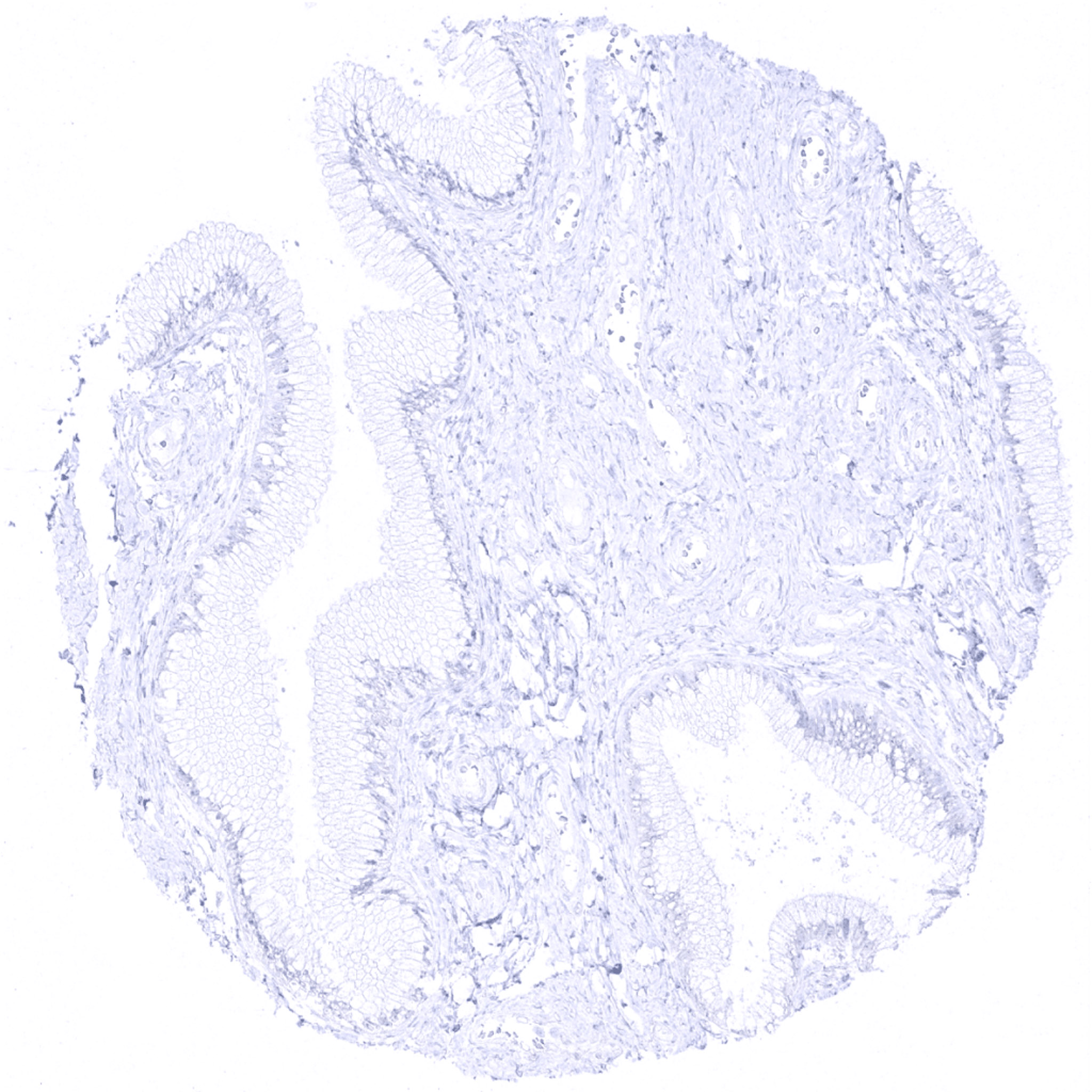
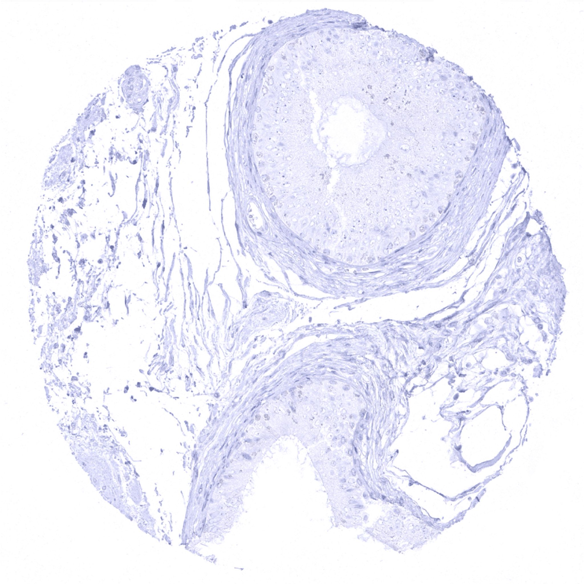
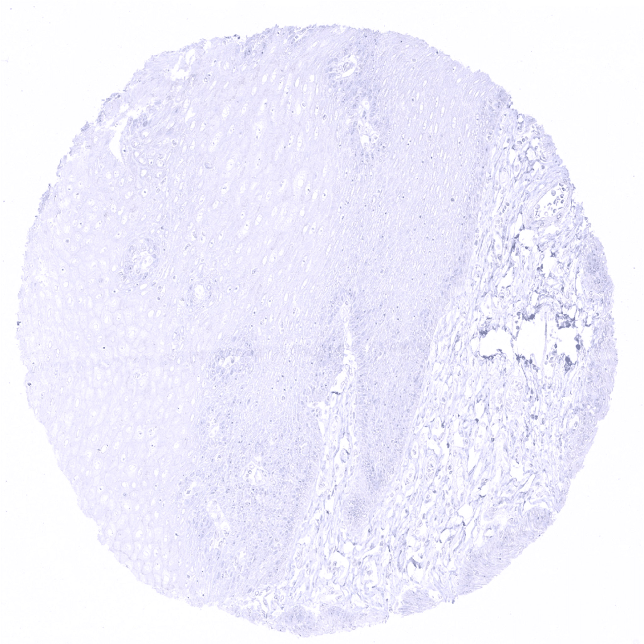
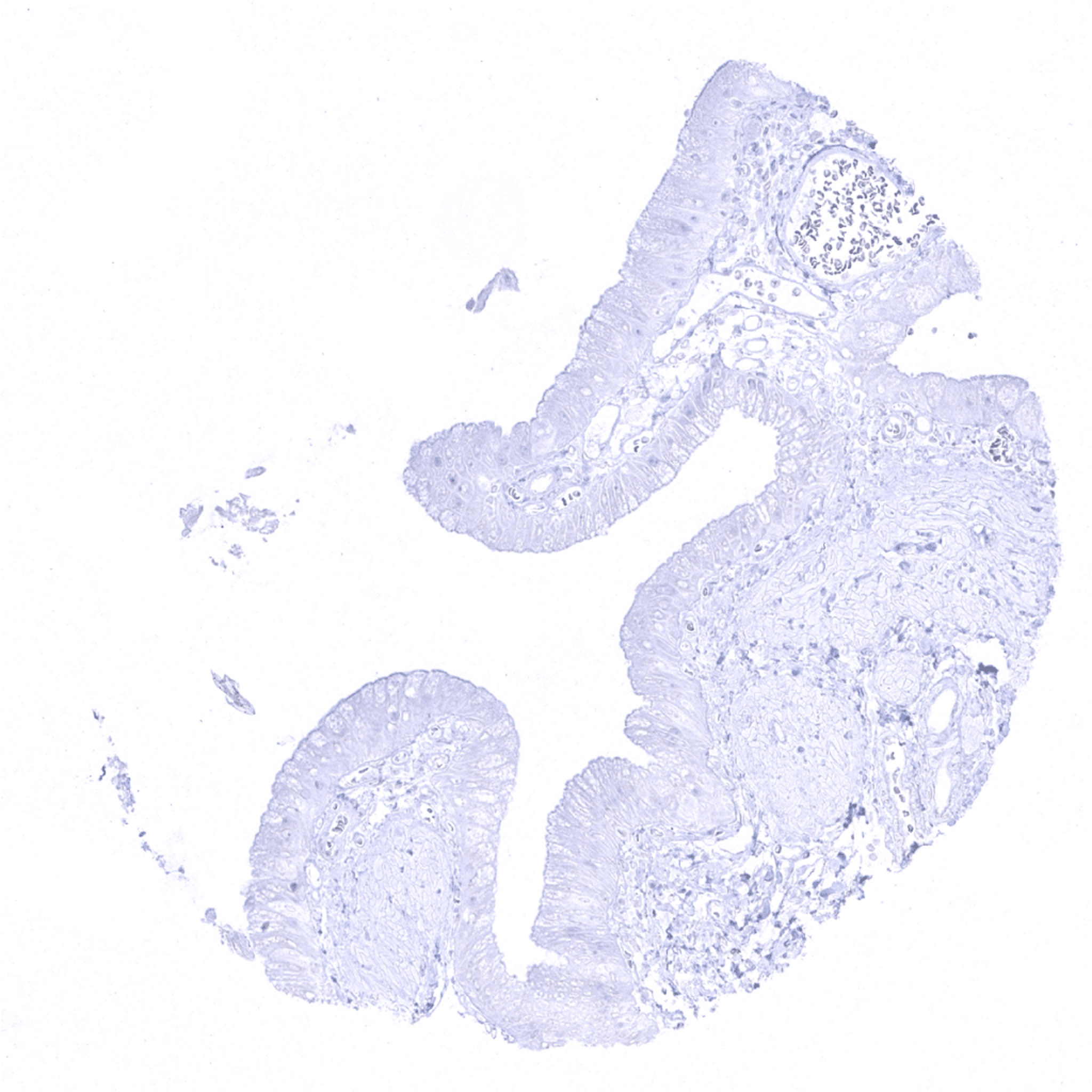
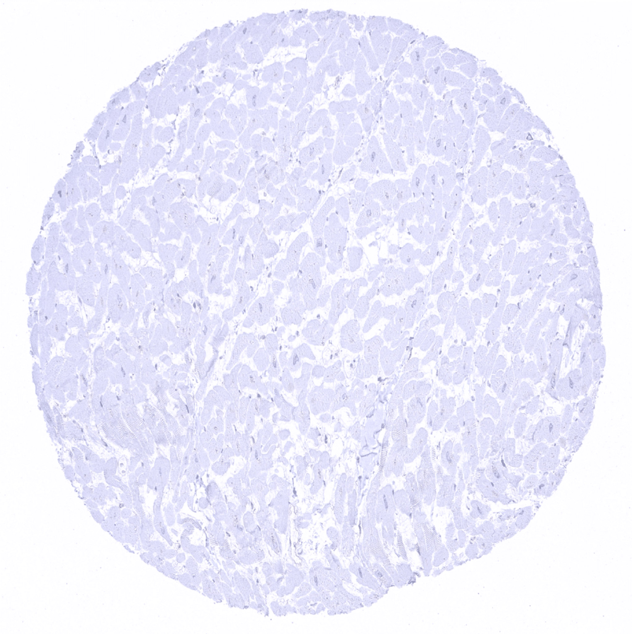
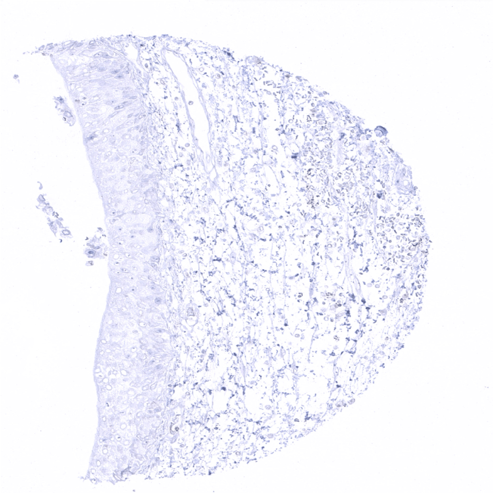
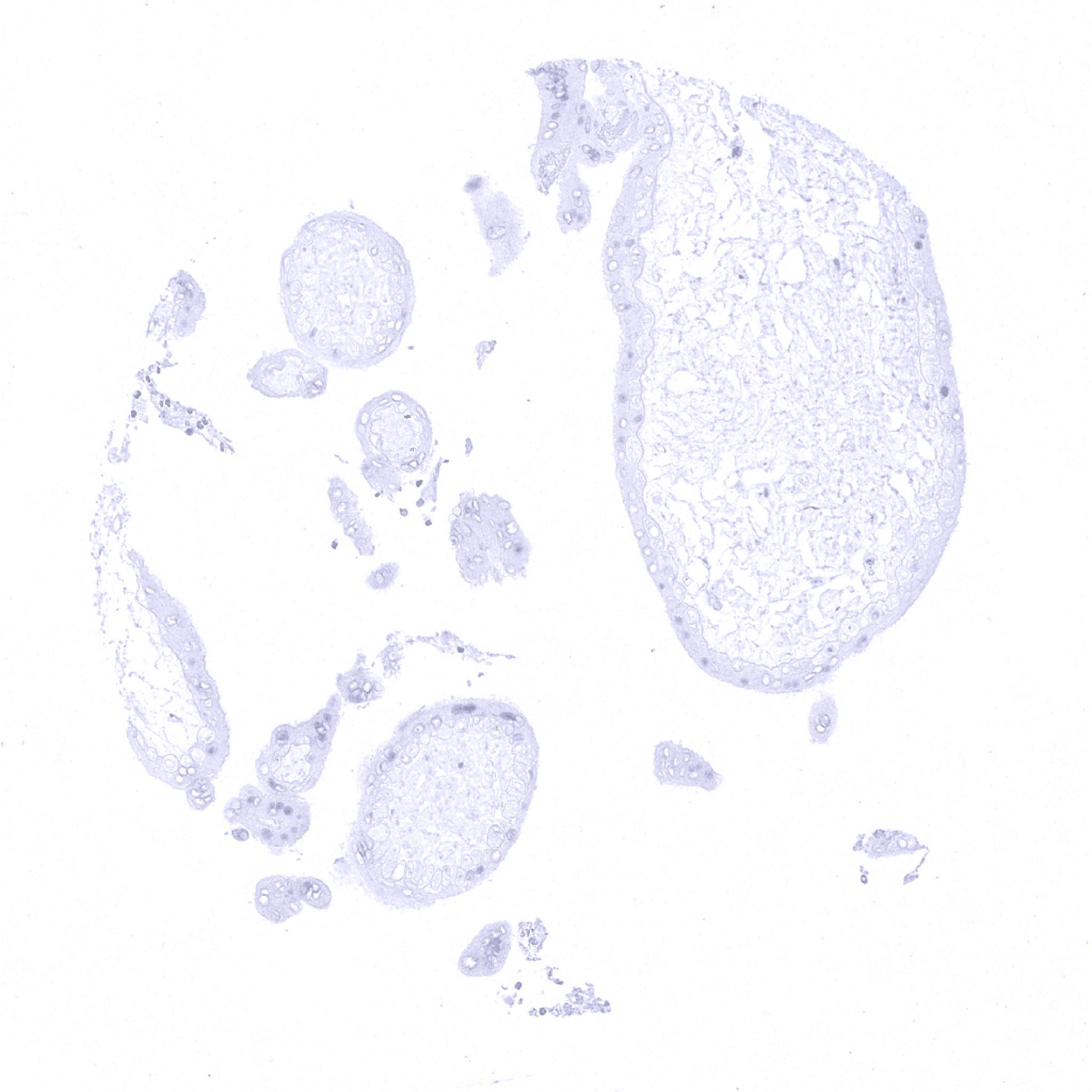
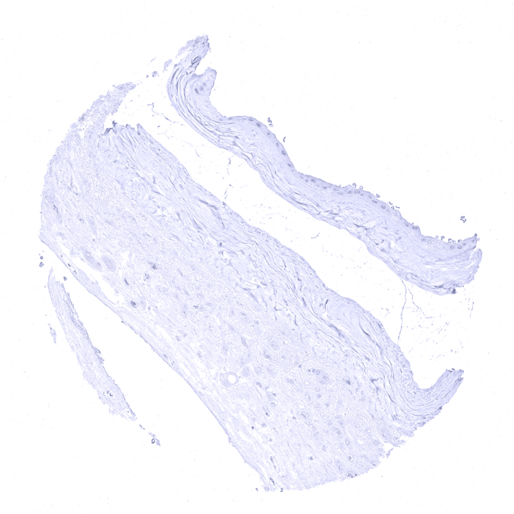
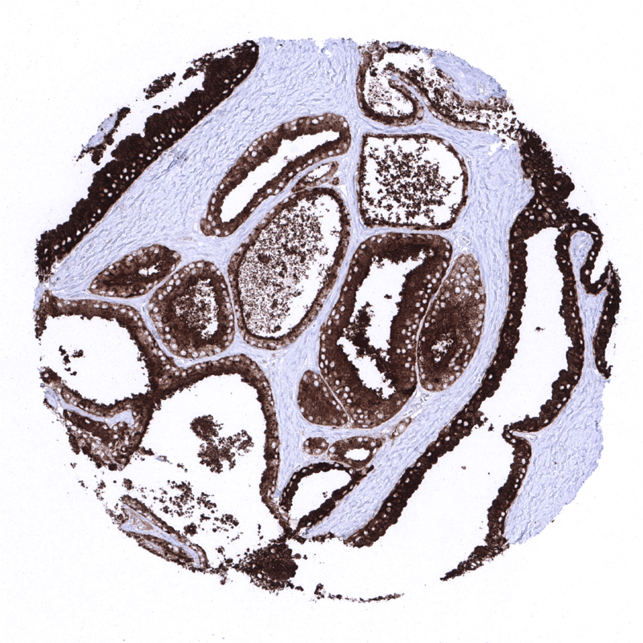
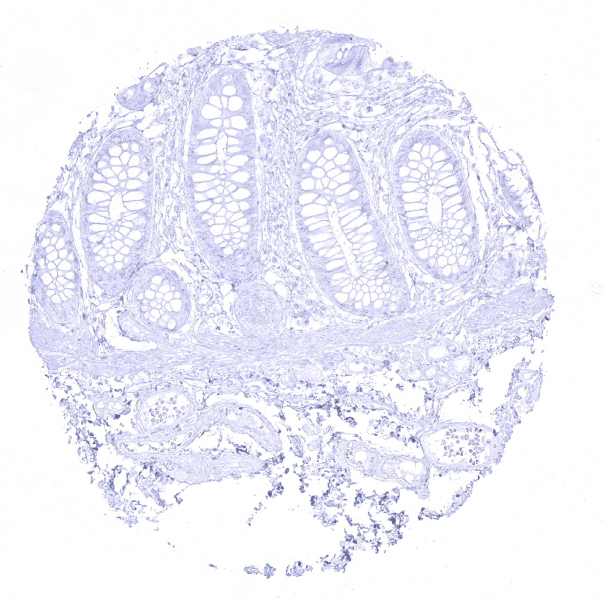
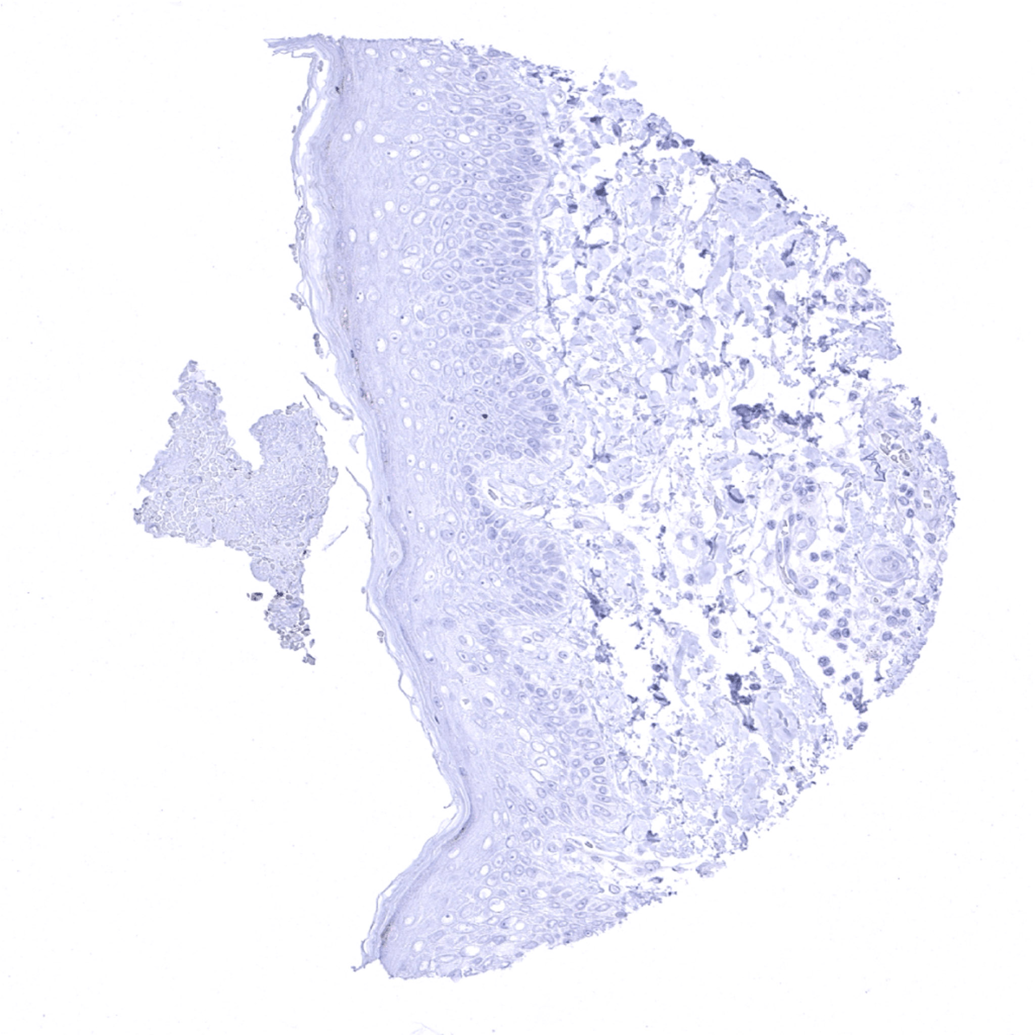
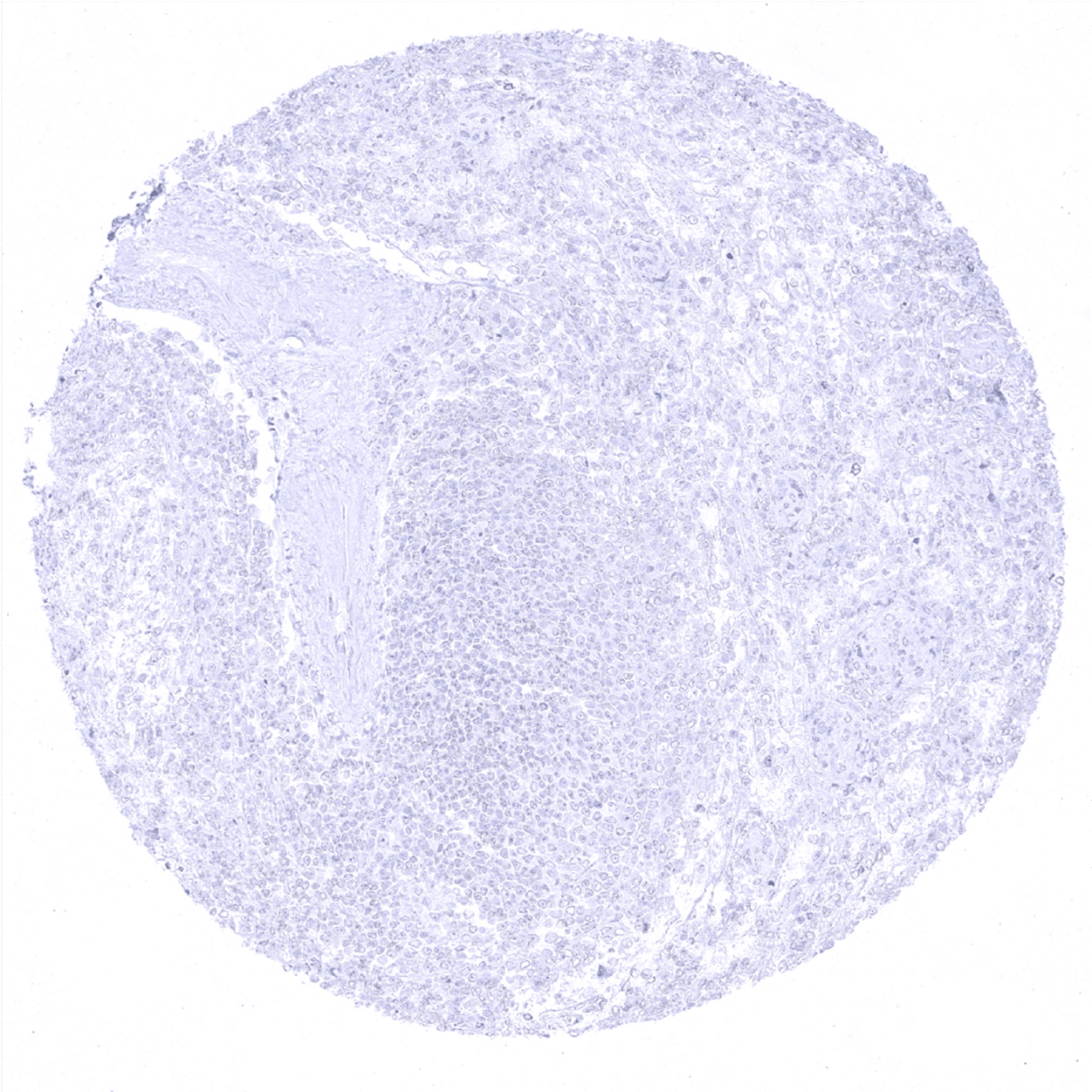
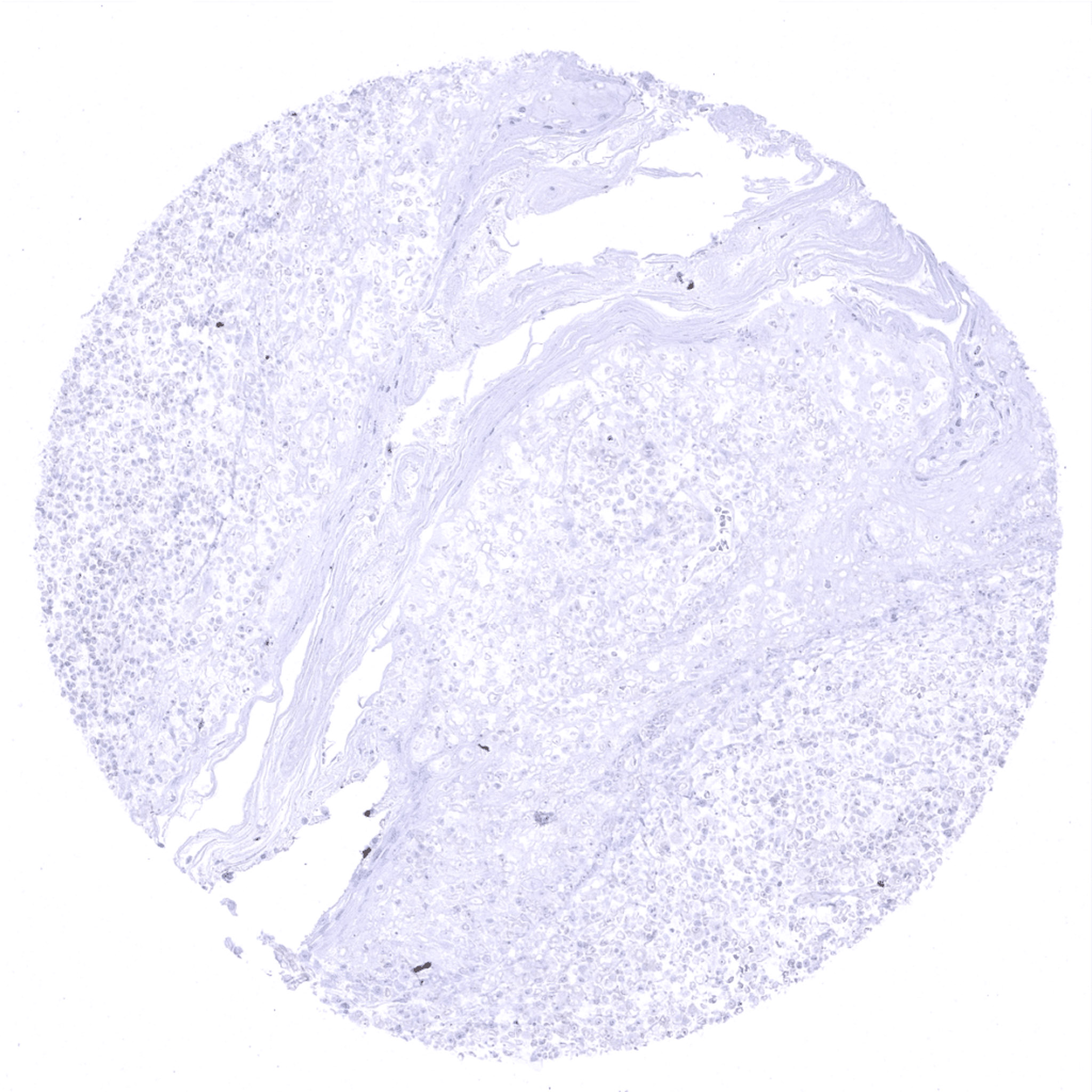
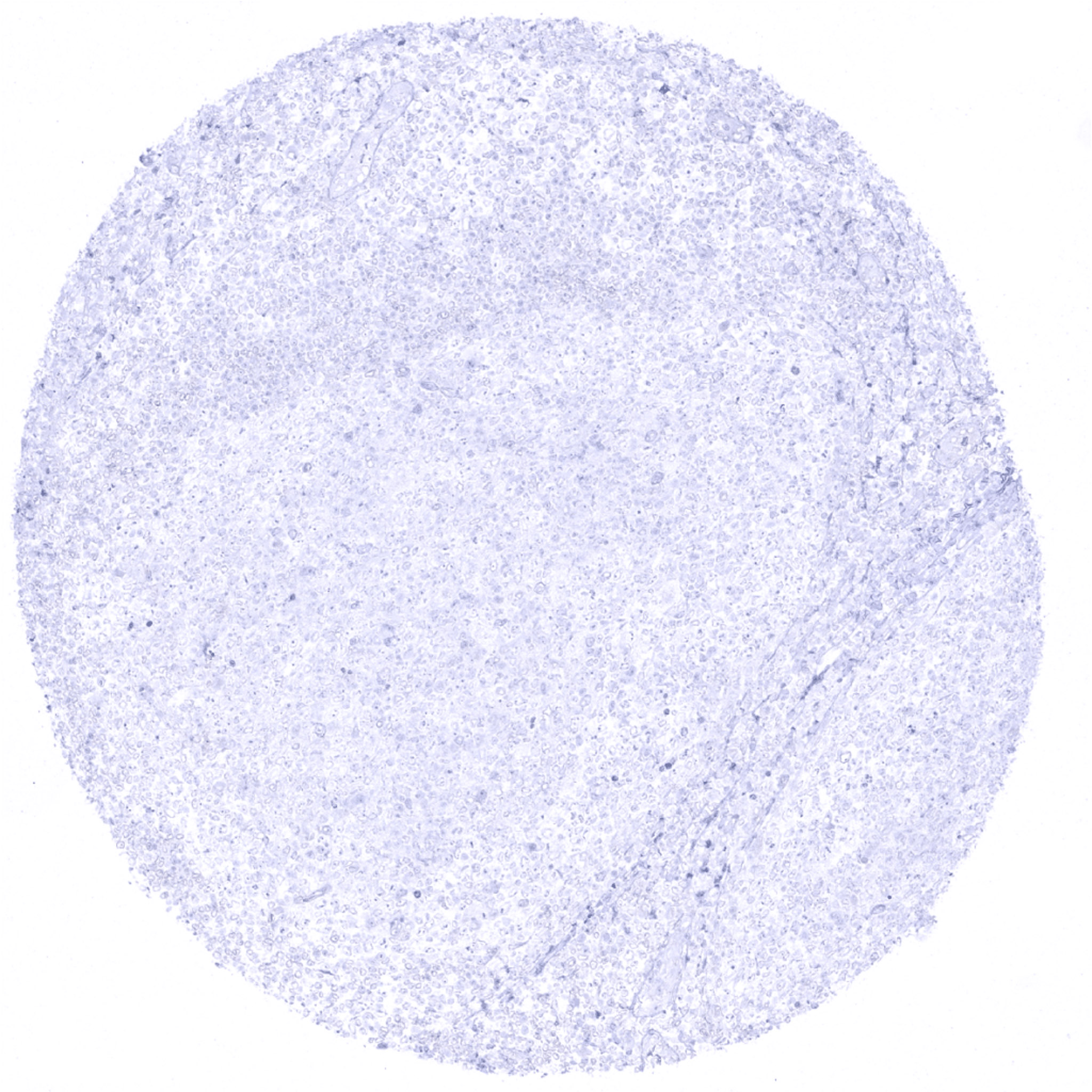
Prostatic Acid Phosphatase antibody [MSVA-452M] HistoMAXTM


Go to Cancer Tissue Gallery Go back to product page Go to HistoMAX page


Adrenal gland
Aorta, media
Appendix, mucosa – Complete absence of Prostatic Acid Phosphatase immunostaining in all epithelial and non-epithelial cell types of the appendix.
Appendix, muscular wall
Bone marrow
Breast
Bronchus, musosa
Cerebrum, white matter
Cerebrum, grey matter
Cerebellum (white matter)
Cerebellum (molecular layer, Purkinje cell layer, granule cell layer)
Colon descendens, mucosa
Colon descendens, muscular wall
Duodenum, Brunner gland
Duodenum, mucosa
Ectocervix
Endocervix
Endometrium, proliferation
Endometrium, secretion
Epididymis
Esophagus, squamous epithelium
Fallopian tube, mucosa
Fat
Gallbladder, epithelium
Heart
Ileum, mucosa
Kidney, cortex
Kidney, medulla
Kidney, pelvis, urothelium
Liver
Lung
Lymph node
Ovary, stroma
Pancreas
Parathyroid
Parotid gland
Pituitary gland, anterior lobe
Pituitary gland, posterior lobe
Pregnant uterus (decidua)
Placenta, early
Placenta (amnion and chorion)
Placenta, mature
Prostate – Strong Prostatic Acid Phosphatase immunostaining in all acinar epithelial cells of the prostate. Some contamination occurs in the stroma due to the very high Prostatic Acid Phosphatase expression levels in epithelial cells.
Prostate – Strong Prostatic Acid Phosphatase immunostaining in all acinar epithelial cells of the prostate.
Rectum, mucosa
Seminal vesicle
Sinus paranasales
Skin
Spleen
Stomach, antrum
Stomach, corpus
Striated muscle
Testis
Thymus
Thyroid gland
Tonsil, surface epithelium
Tonsil
Urinary bladder, muscular wall
Urinary bladder, urothelium
Uterus, myometrium

| TOP |