Normal Tissue Gallery- GTX646253

Bcl-XL antibody [HMV4724] HistoMAX™


Go to Cancer Tissue Gallery
  
 Go back to product page
  

Go to HistoMAX page


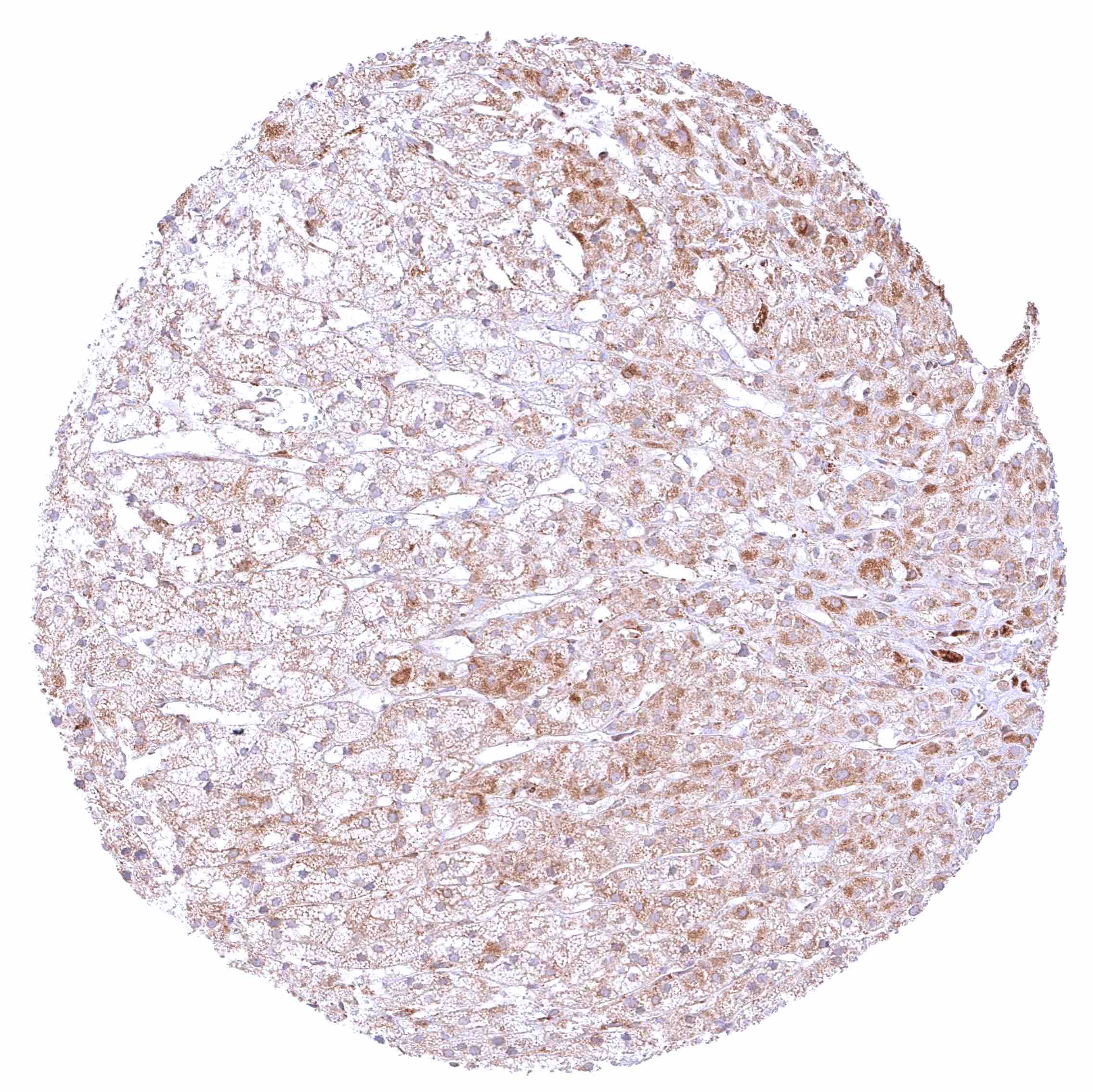
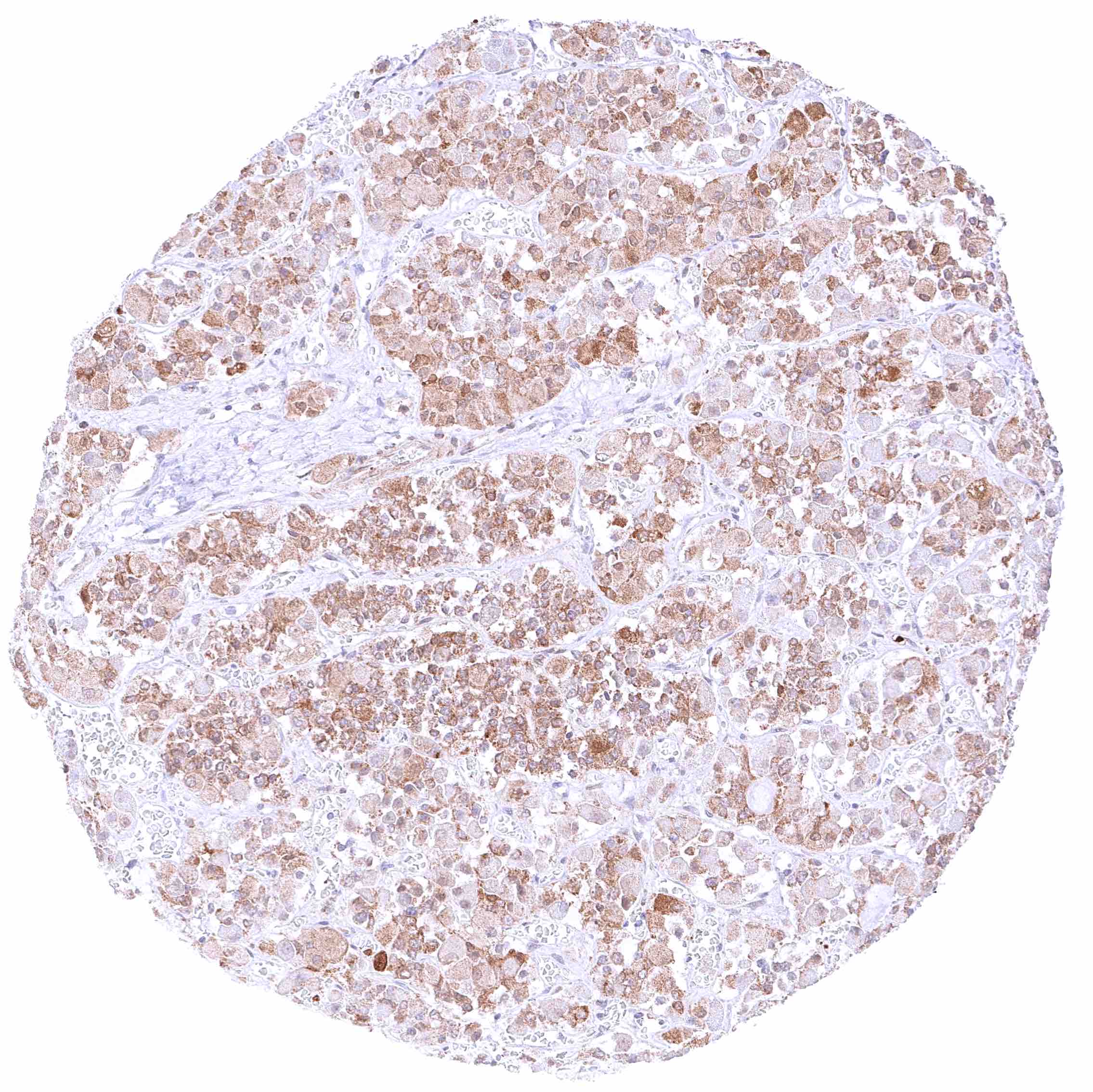
Adrenal gland – Moderate to strong cytoplasmic BCL-XL staining of epithelial cells.

Aorta, media

Appendix, mucosa – Distinct cytoplasmic BCL-XL staining of all epithelial cells and a subset of inflammatory cells (plasma cells).

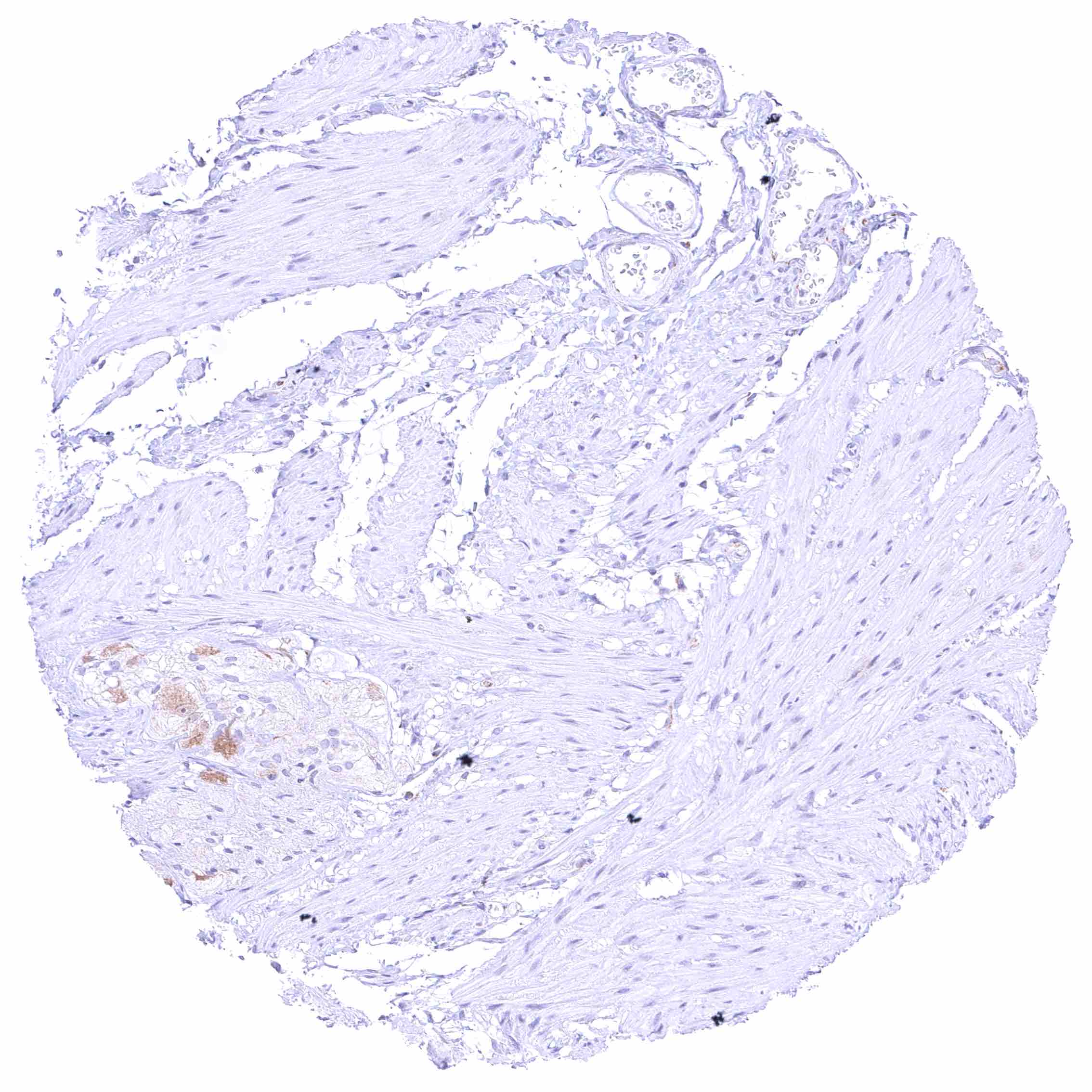
Appendix, muscular wall – Strong cytoplasmic BCL-XL staining of intramural ganglion cells.


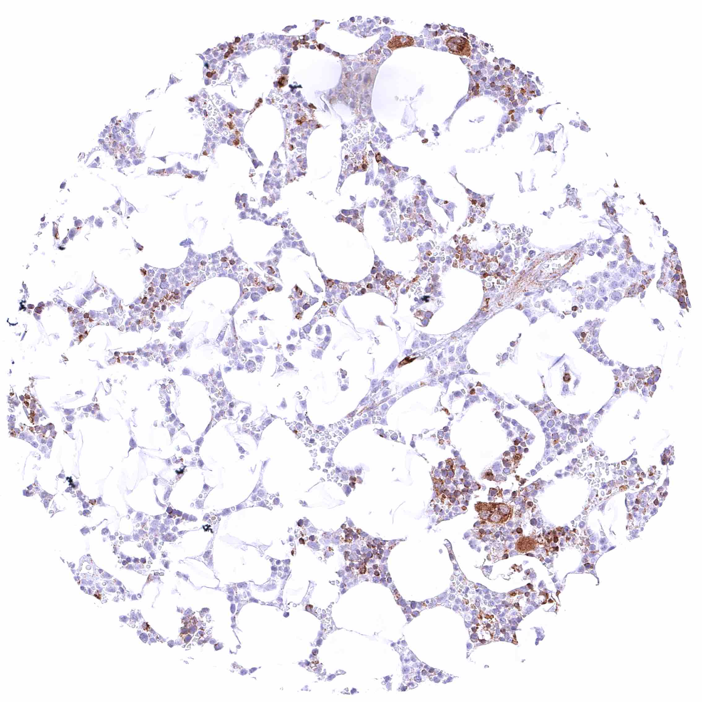
Bone marrow – Strong cytoplasmic BCL-XL staining of a subpopulation of bone marrow cells that includes megakaryocytes (and perhaps other cells).

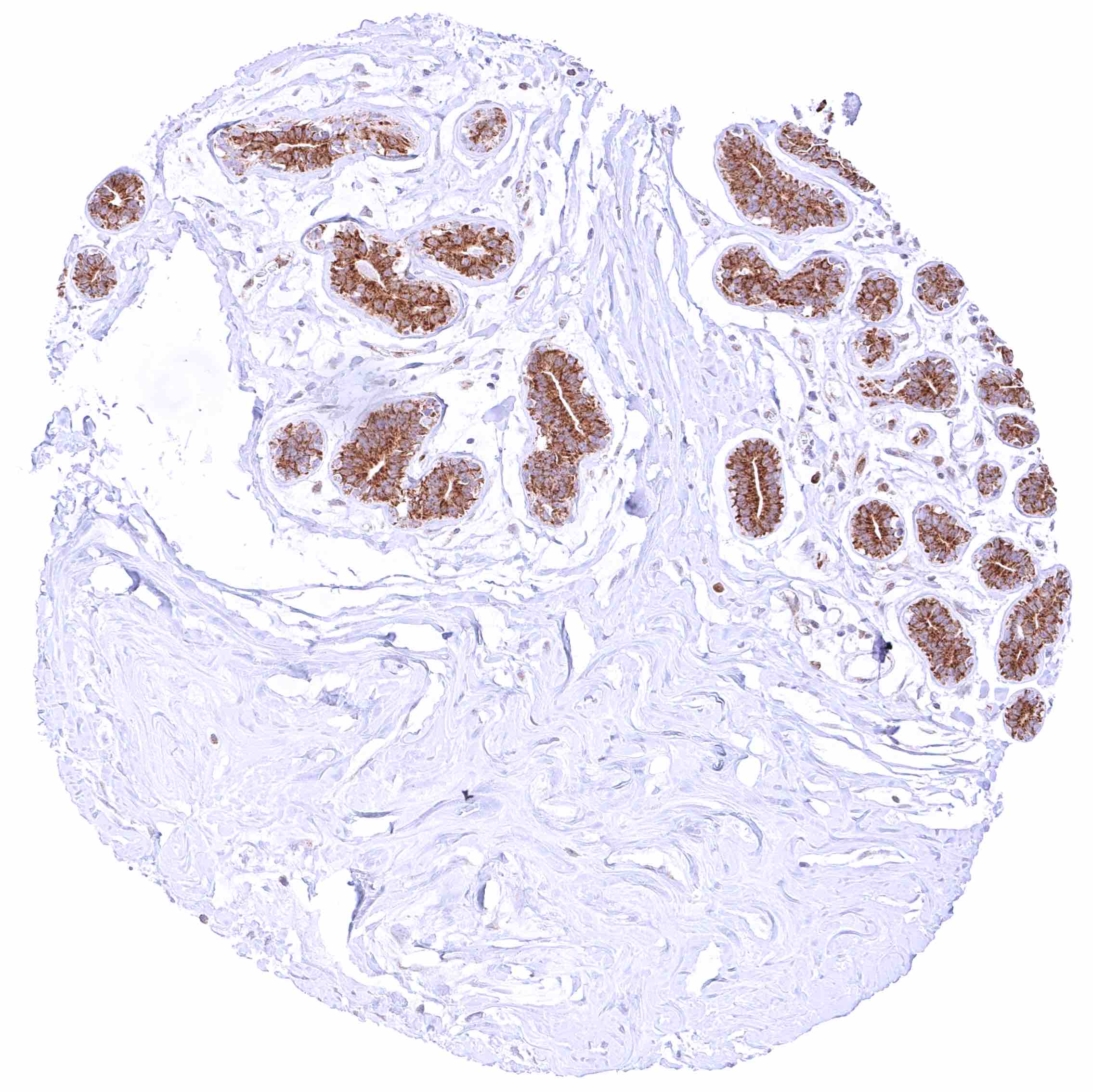
Breast – Moderate to strong cytoplasmic BCL-XL staining of all epithelial cells.

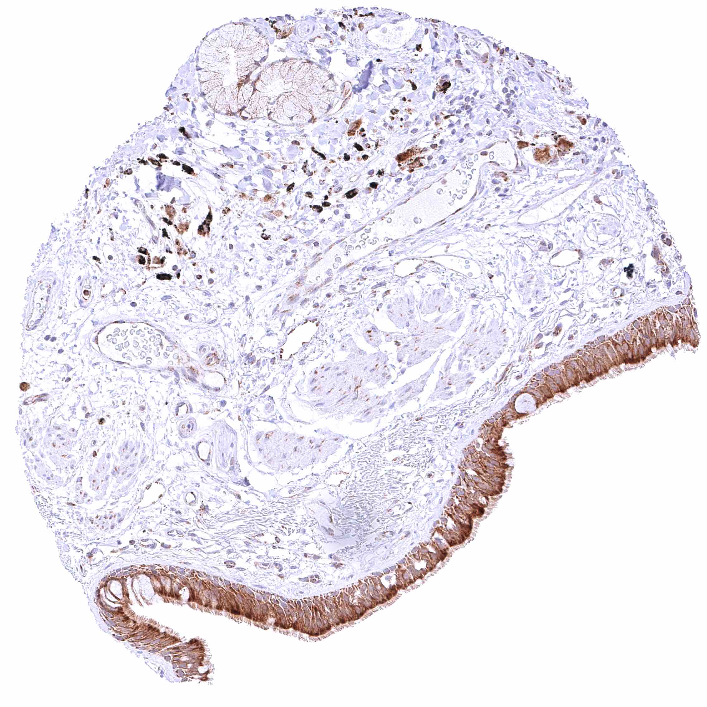
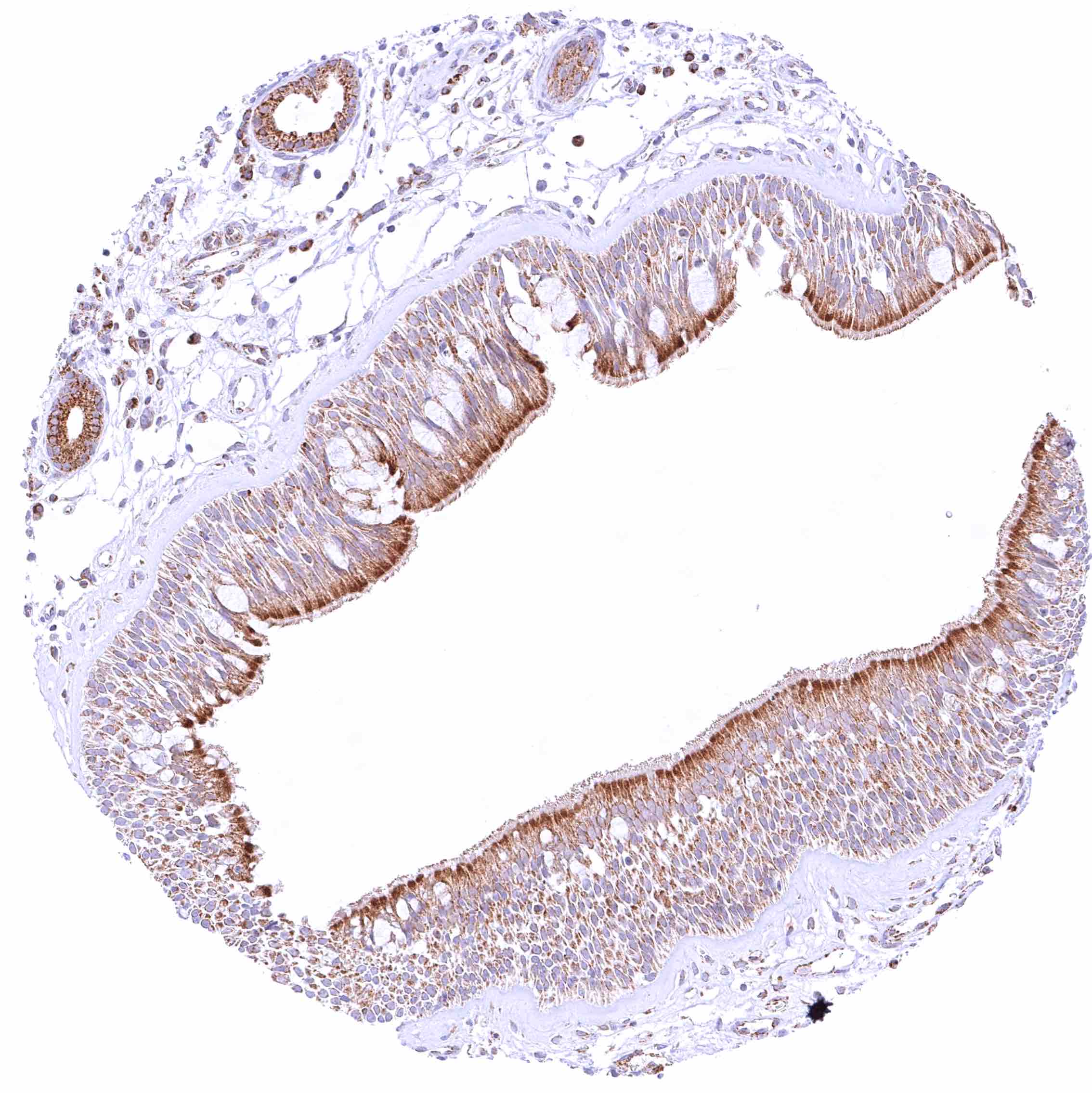
Bronchus, mucosa – Moderate to strong cytoplasmic LGALS3BP staining of respiratory epithelial cells, especially in the apical-luminal zone of the cells.

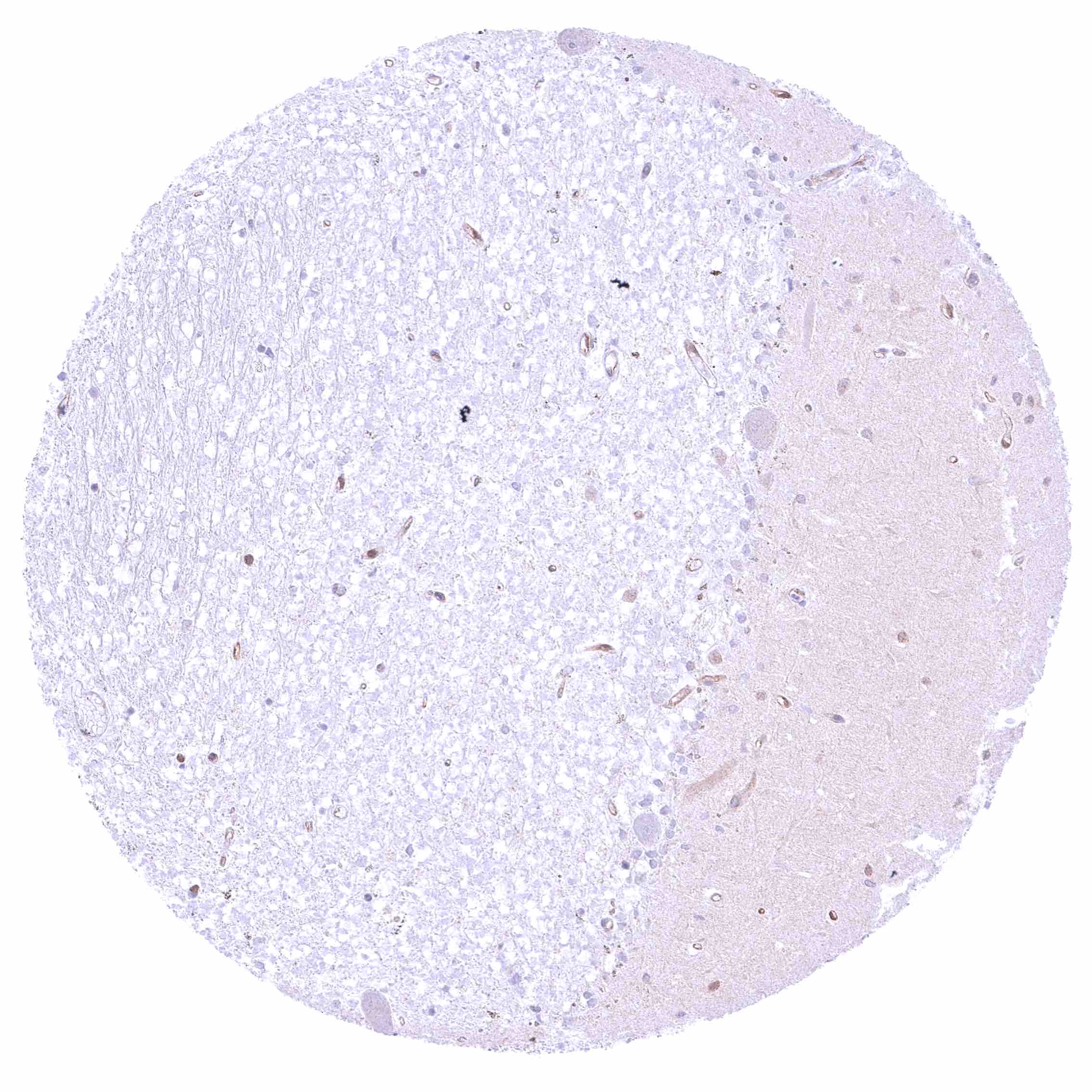
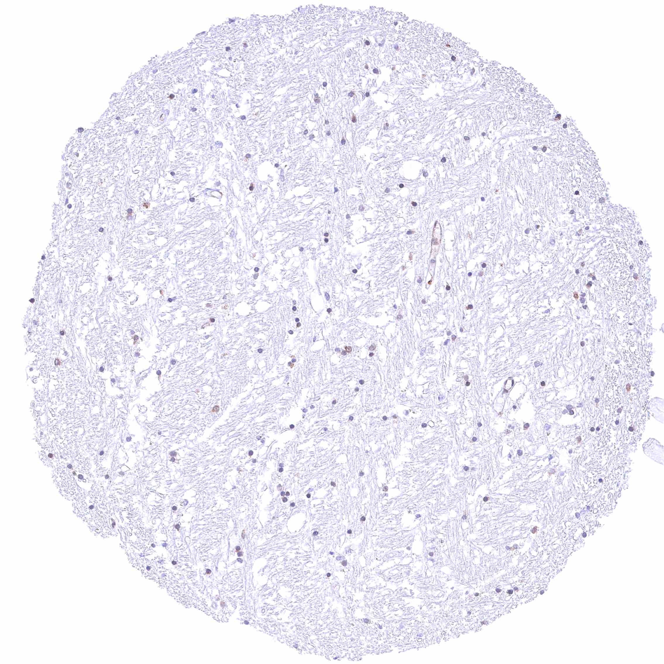
Cerebellum (white matter) – Moderate cytoplasmic LGALS3BP staining of glia cells.


Cerebellum, cortex (grey) – Weak cytoplasmic LGALS3BP staining of glia cells and of endothelial cells.

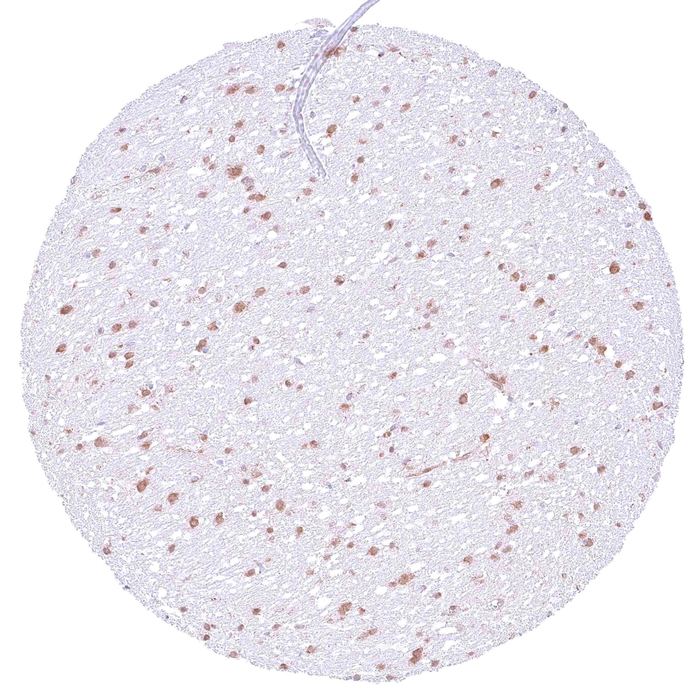
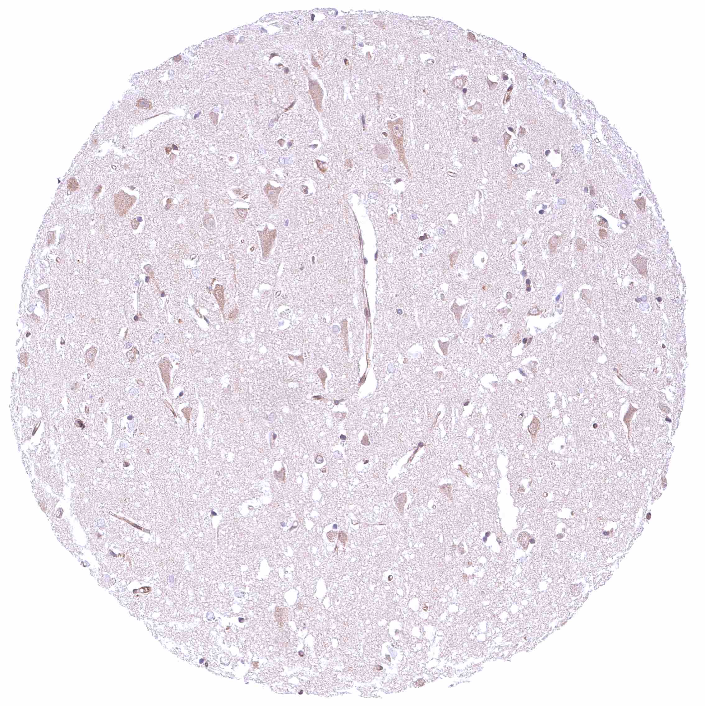
Cerebrum (grey matter) – Faint-weak BCL-XL staining of neurons.

Cerebrum (grey matter) – Faint-weak BCL-XL staining of neurons.

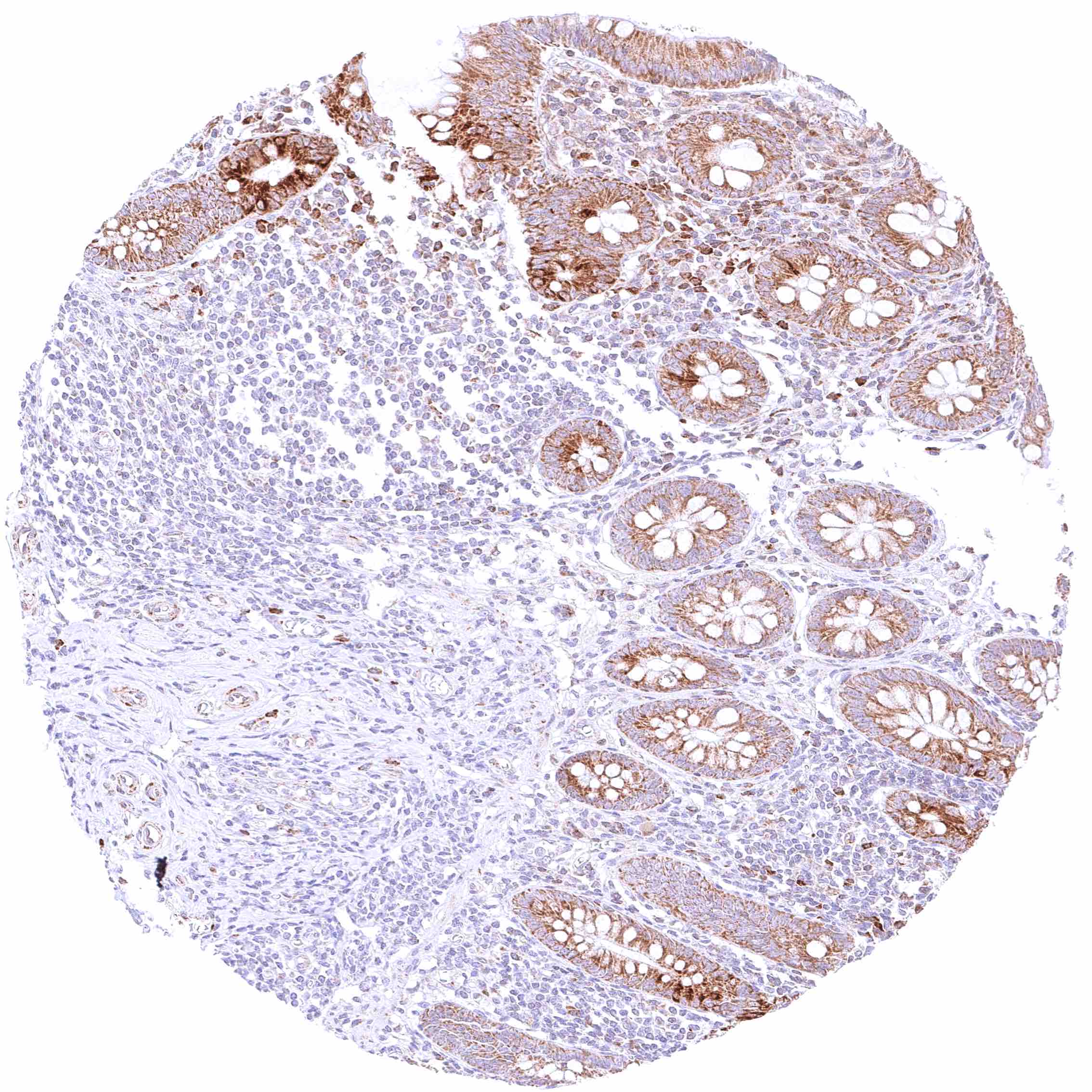
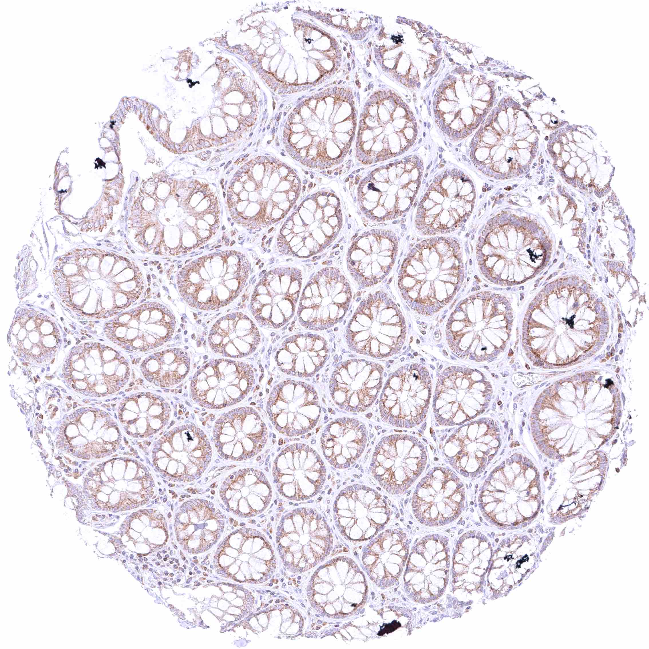
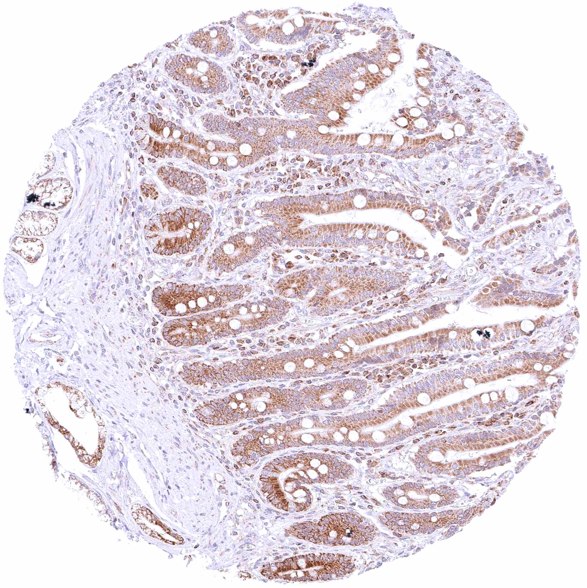
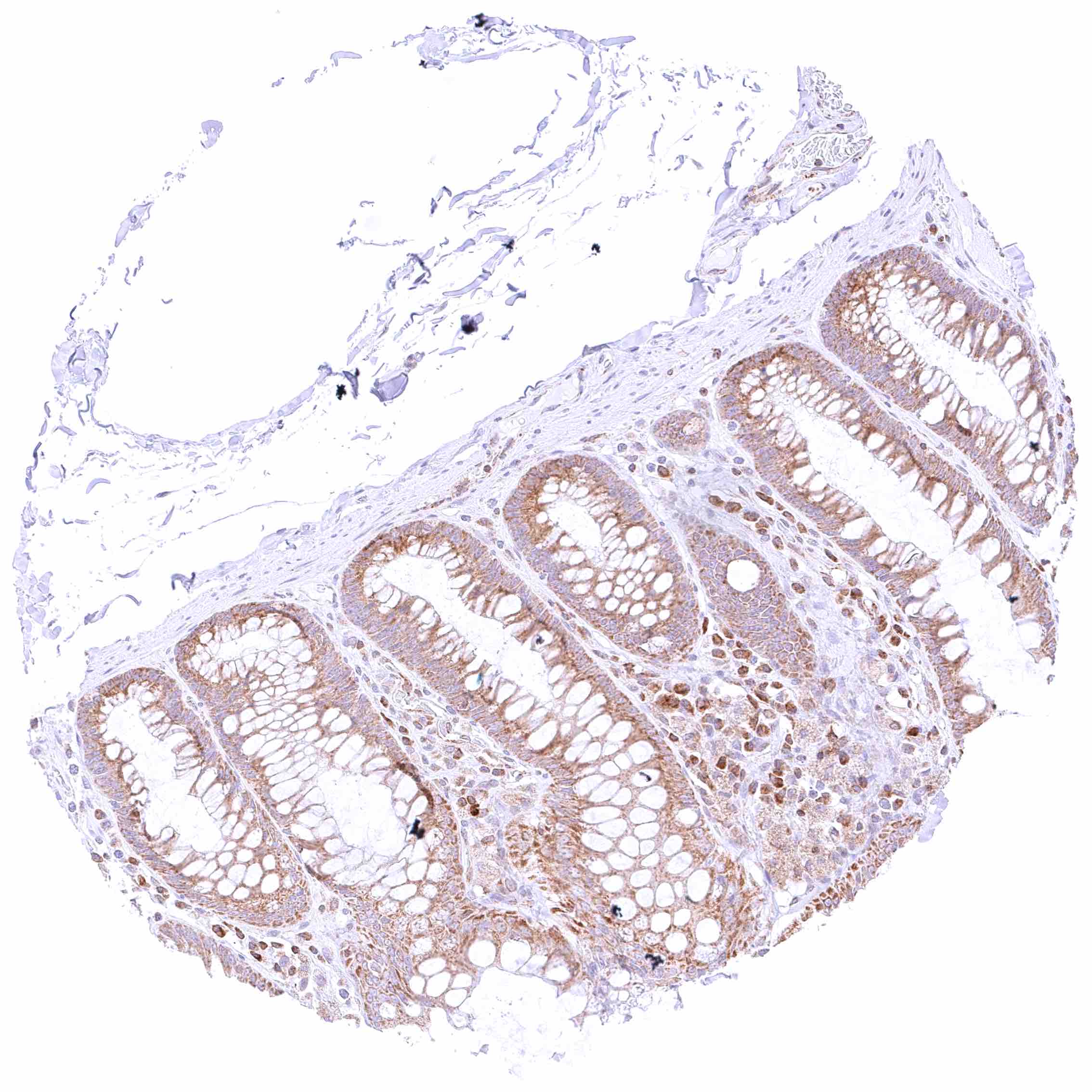
Colon descendens, mucosa – Distinct cytoplasmic BCL-XL staining of all epithelial cells.


Colon descendens, muscular wall – Moderate cytoplasmic BCL-XL staining of intramural ganglion cells.

Duodenum, mucosa – Distinct cytoplasmic BCL-XL staining of all epithelial cells and a subset of inflammatory cells (plasma cells)

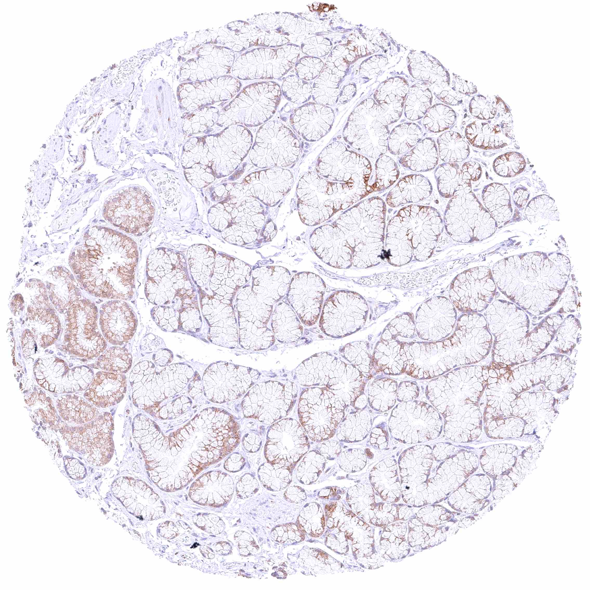
Duodenum, Brunner gland – Weak cytoplasmic BCL-XL staining of Brunner gland cells.

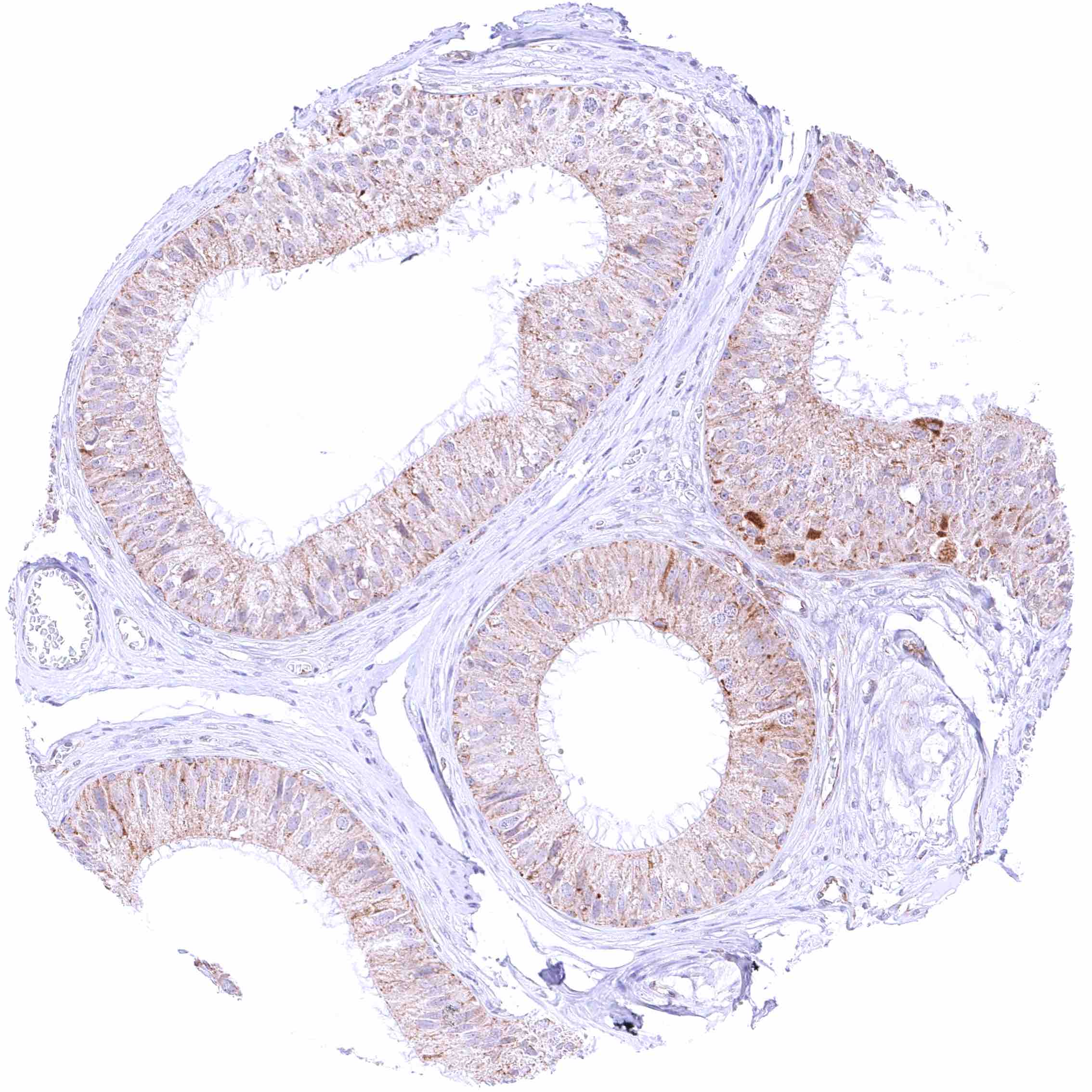
Epididymis (Caput) – Weak to moderate cytoplasmic BCL-XL staining of epithelial cells.


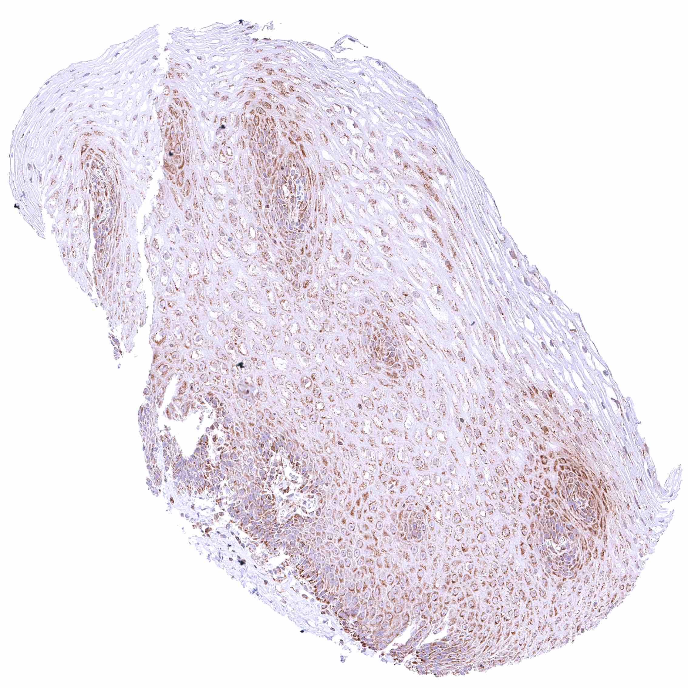
Esophagus, squamous epithelium – Distinct cytoplasmic BCL-XL staining of squamous epithelial cells (decrease of intensity towards the surface).

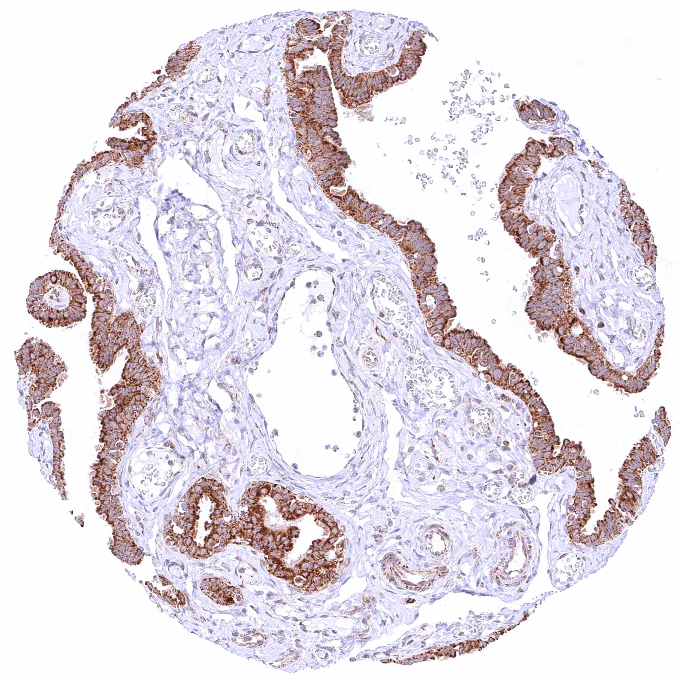
Fallopian tube, mucosa – Strong cytoplasmic BCL-XL staining of epithelial cells.

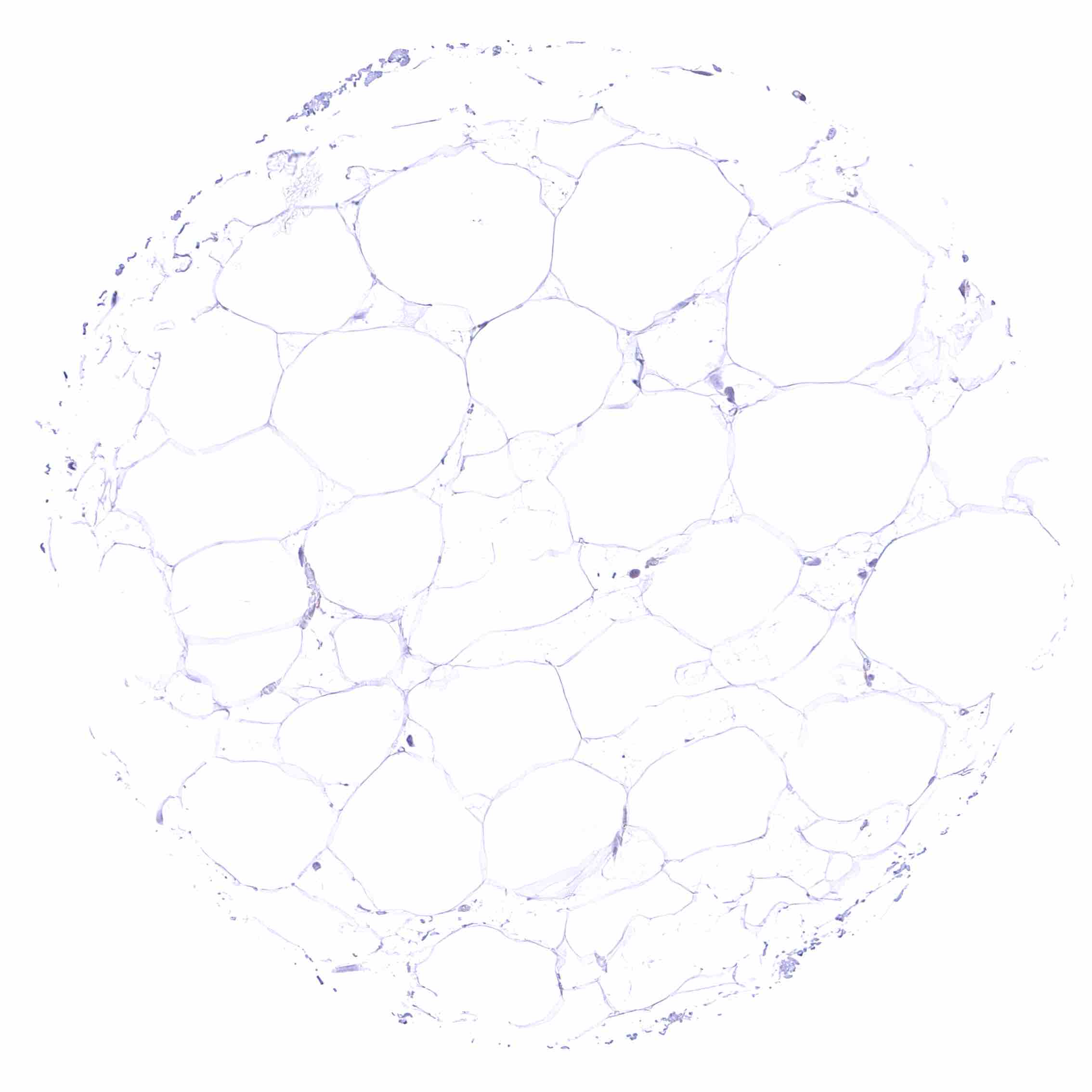
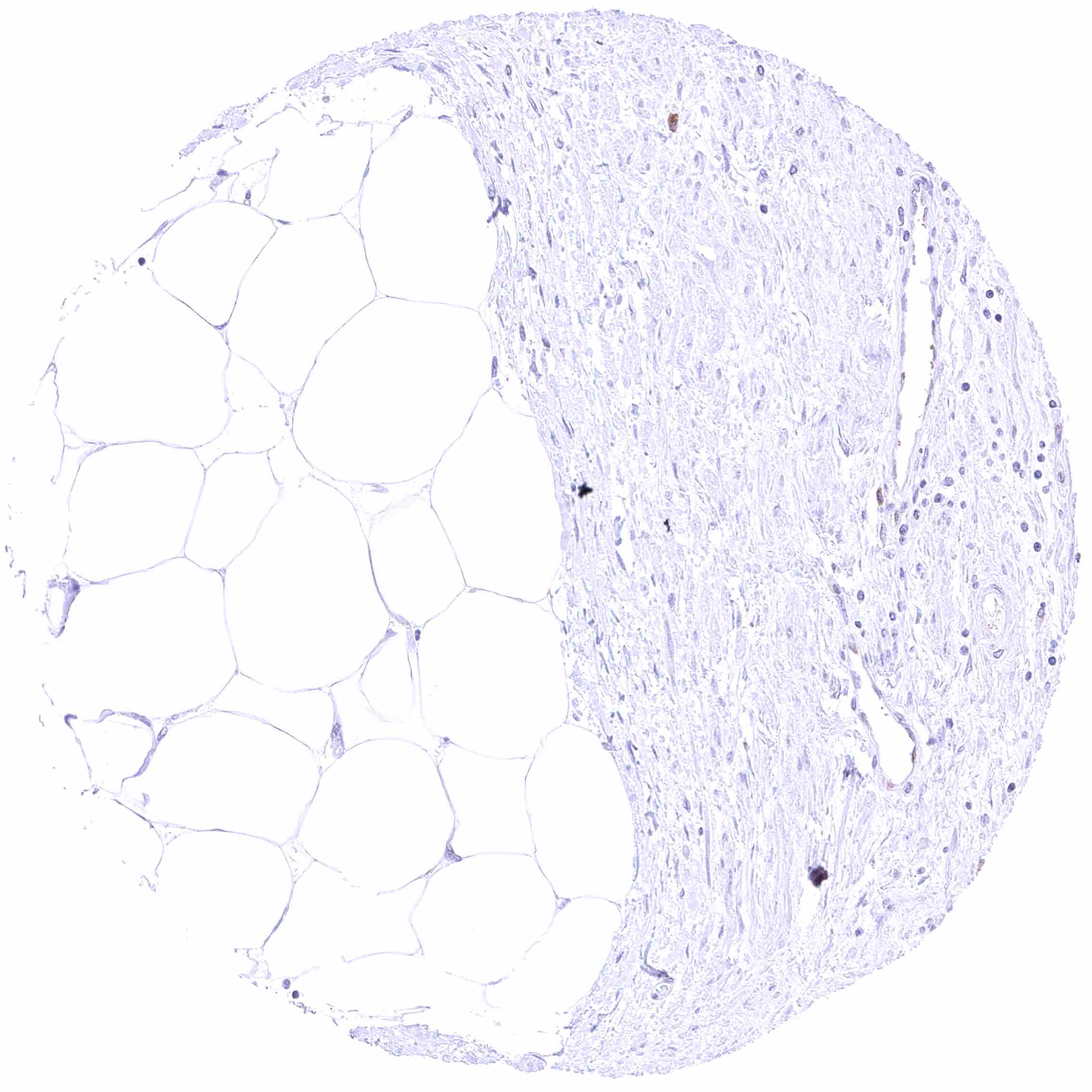
Fat – Faint cytoplasmic BCL-XL staining of fat cells.

Gallbladder, epithelium – Heterogeneous BCL-XL staining of epithelial cells (weak to intense cytoplasmic positivity).


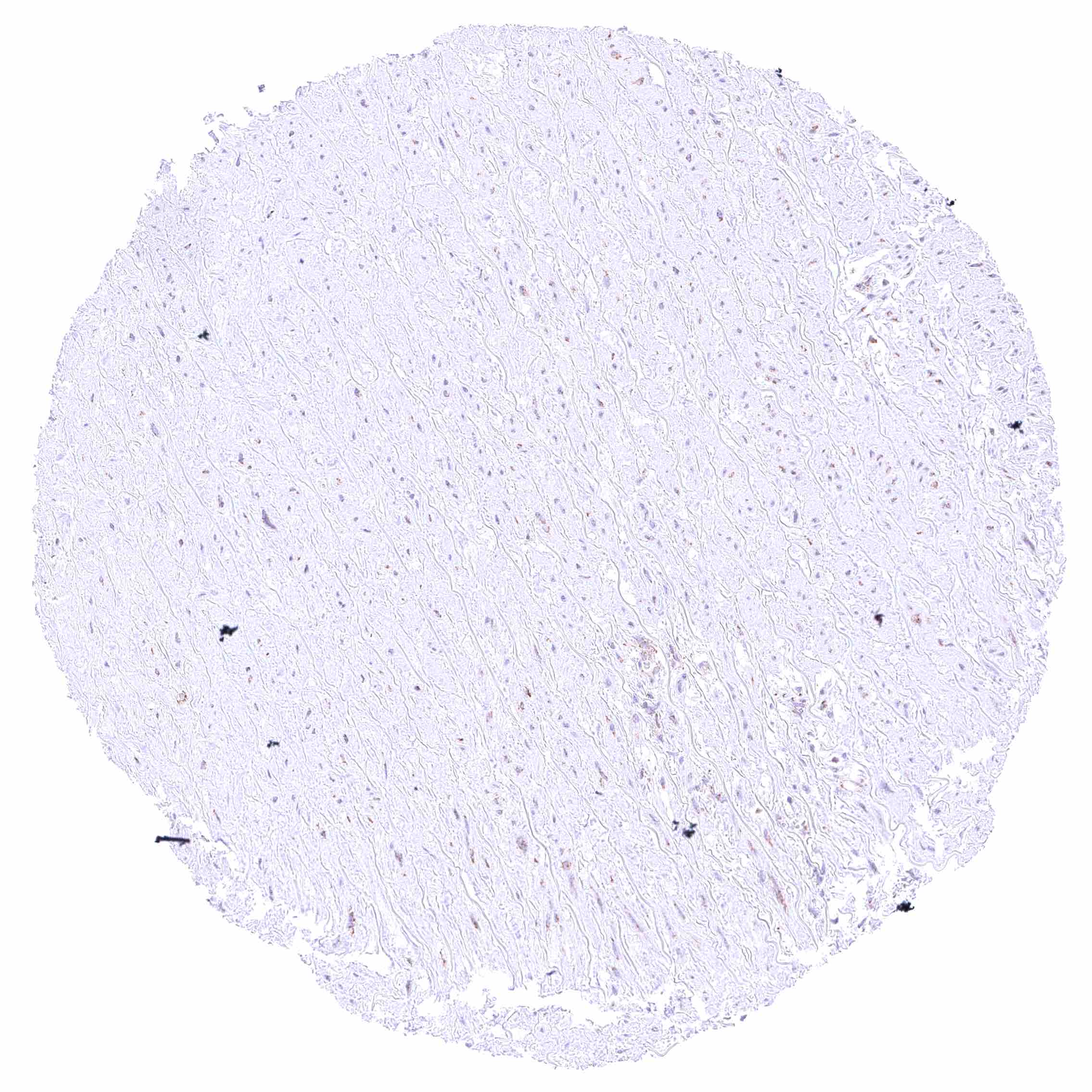
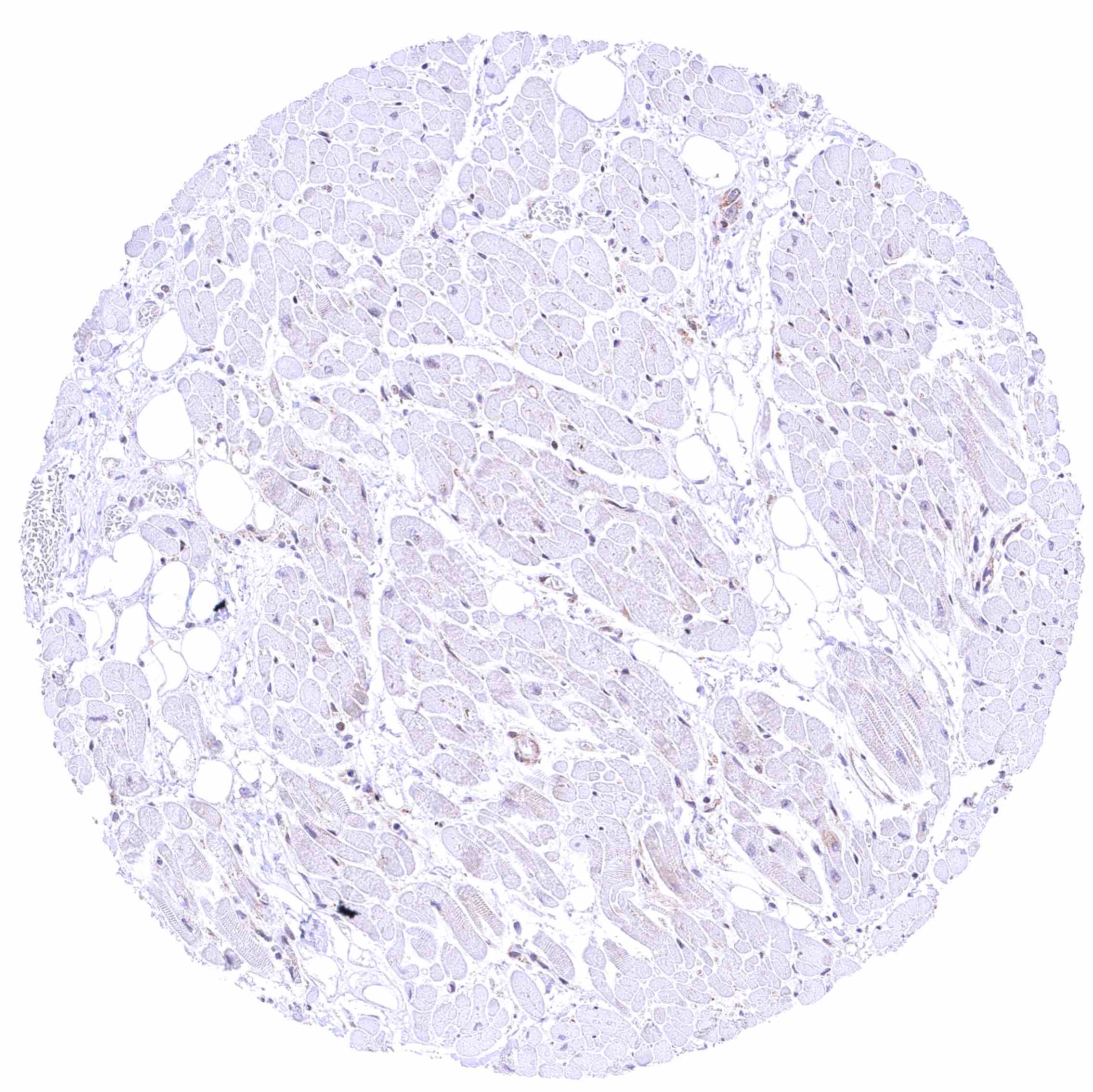
Heart muscle

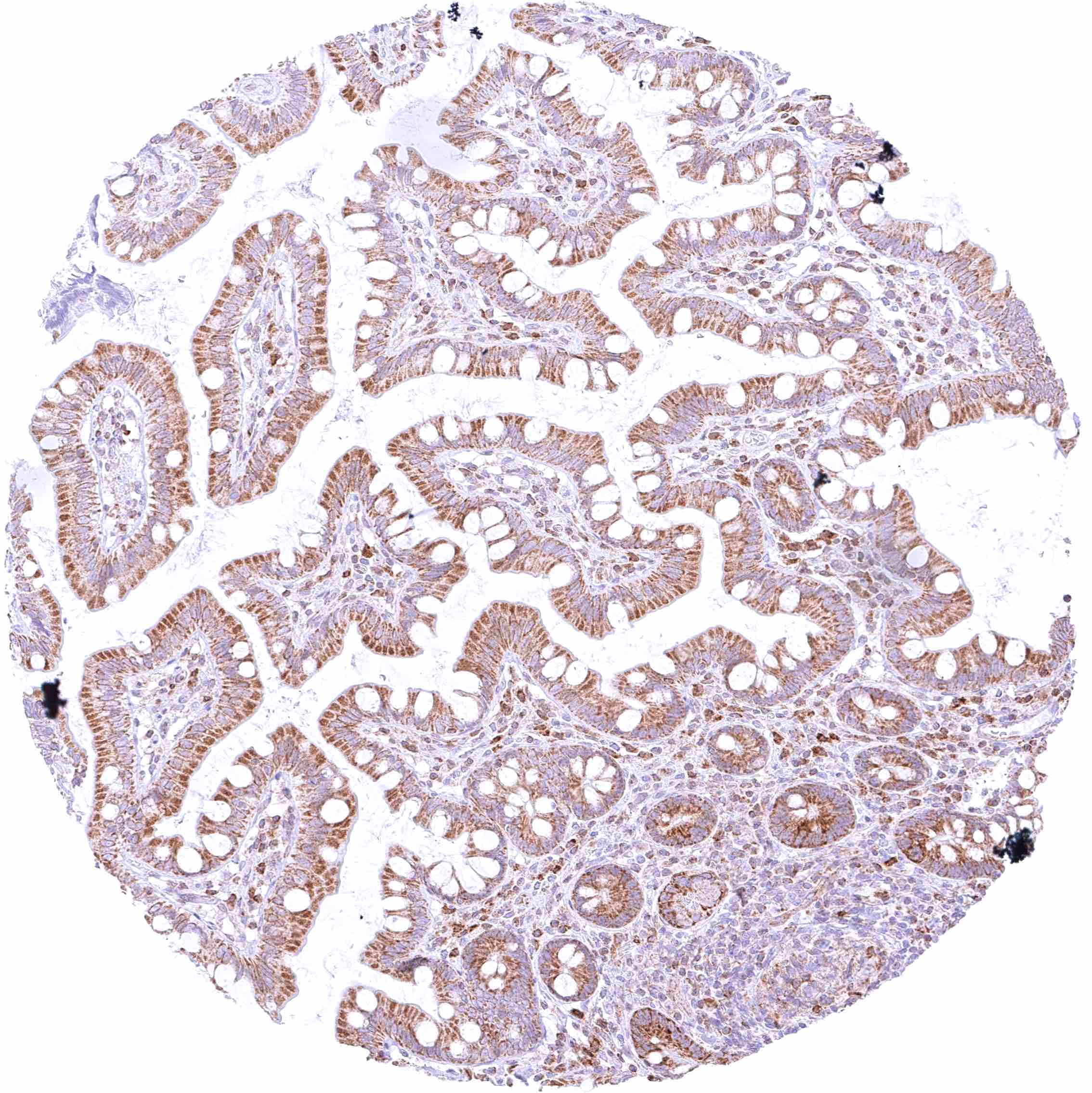
Ileum, mucosa – Distinct cytoplasmic BCL-XL staining of all epithelial cells and a subset of inflammatory cells (plasma cells_).

Ileum, muscular wall – Weak cytoplasmic BCL-XL staining of intramural ganglion cells.

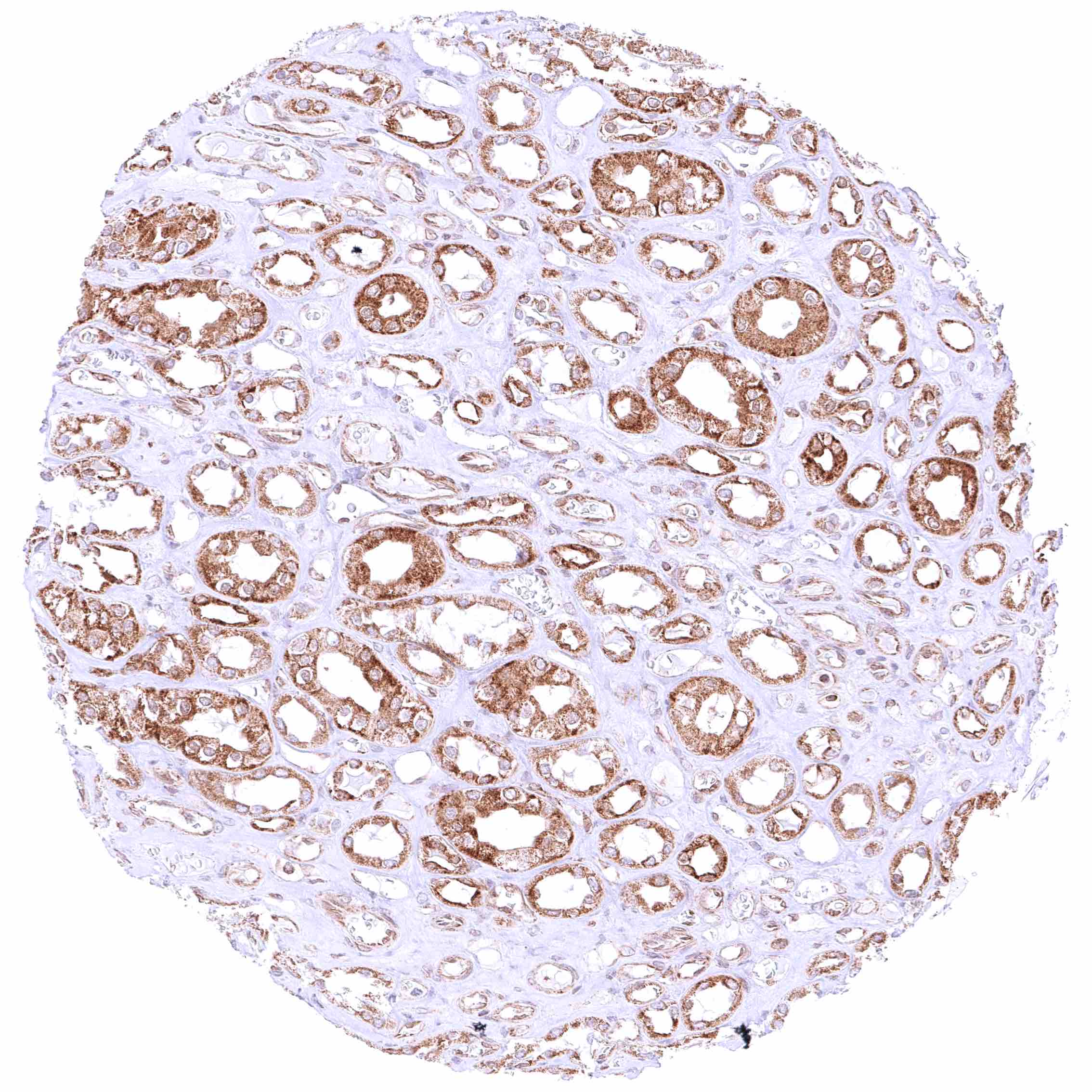
Kidney, medulla – Collecting duct cells show a variable BCL-XL staining ranging from negative to strong.


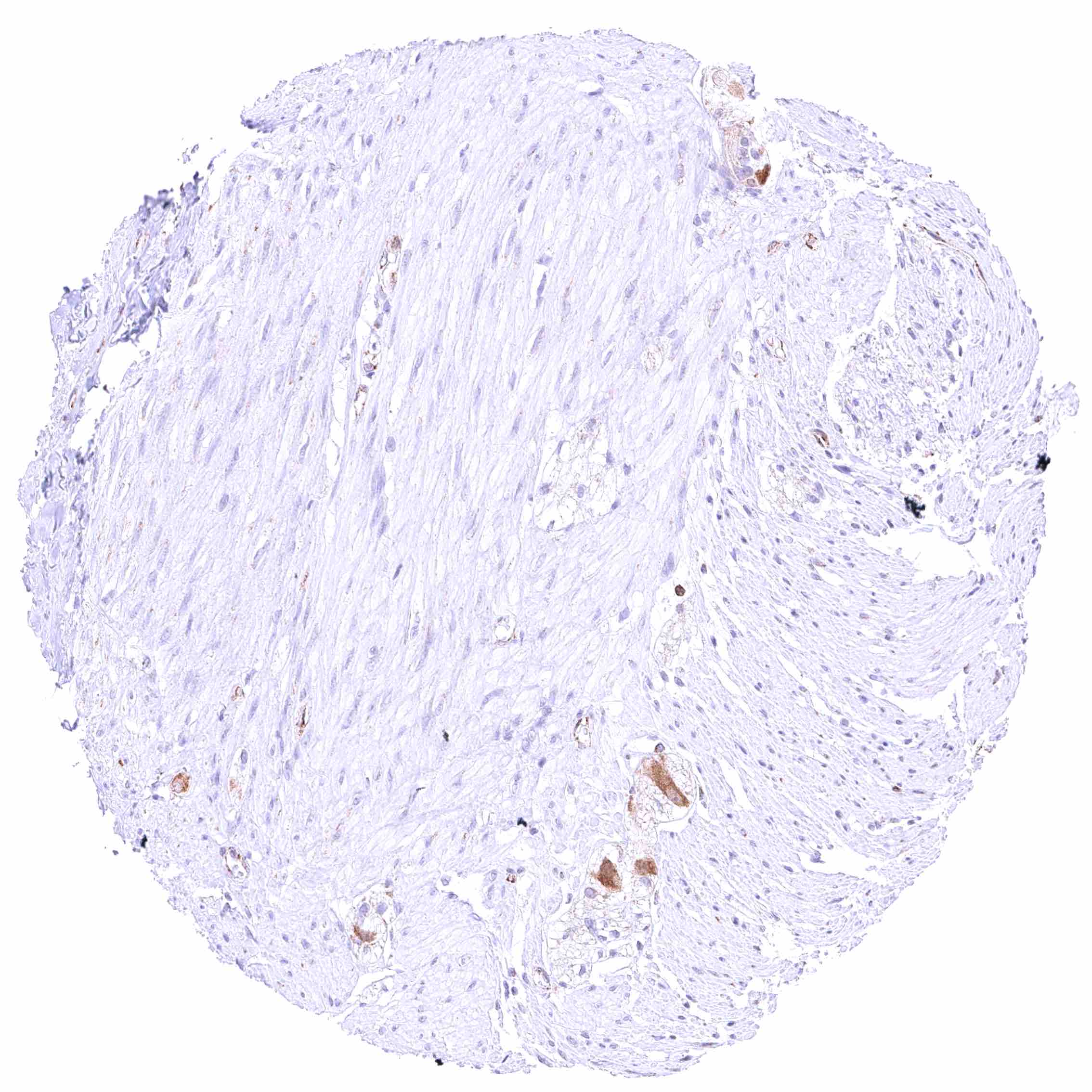
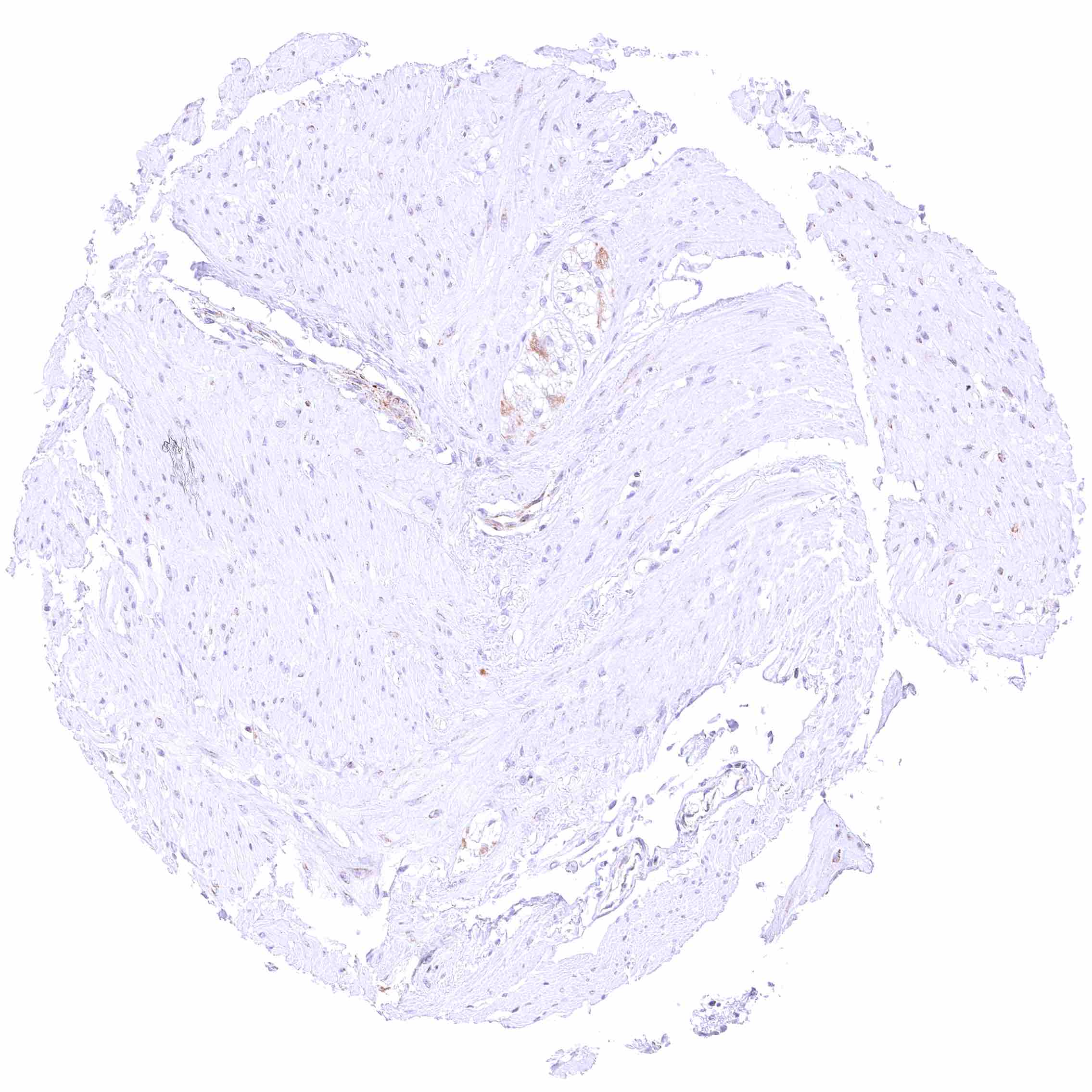
Kidney, pelvis, muscular wall

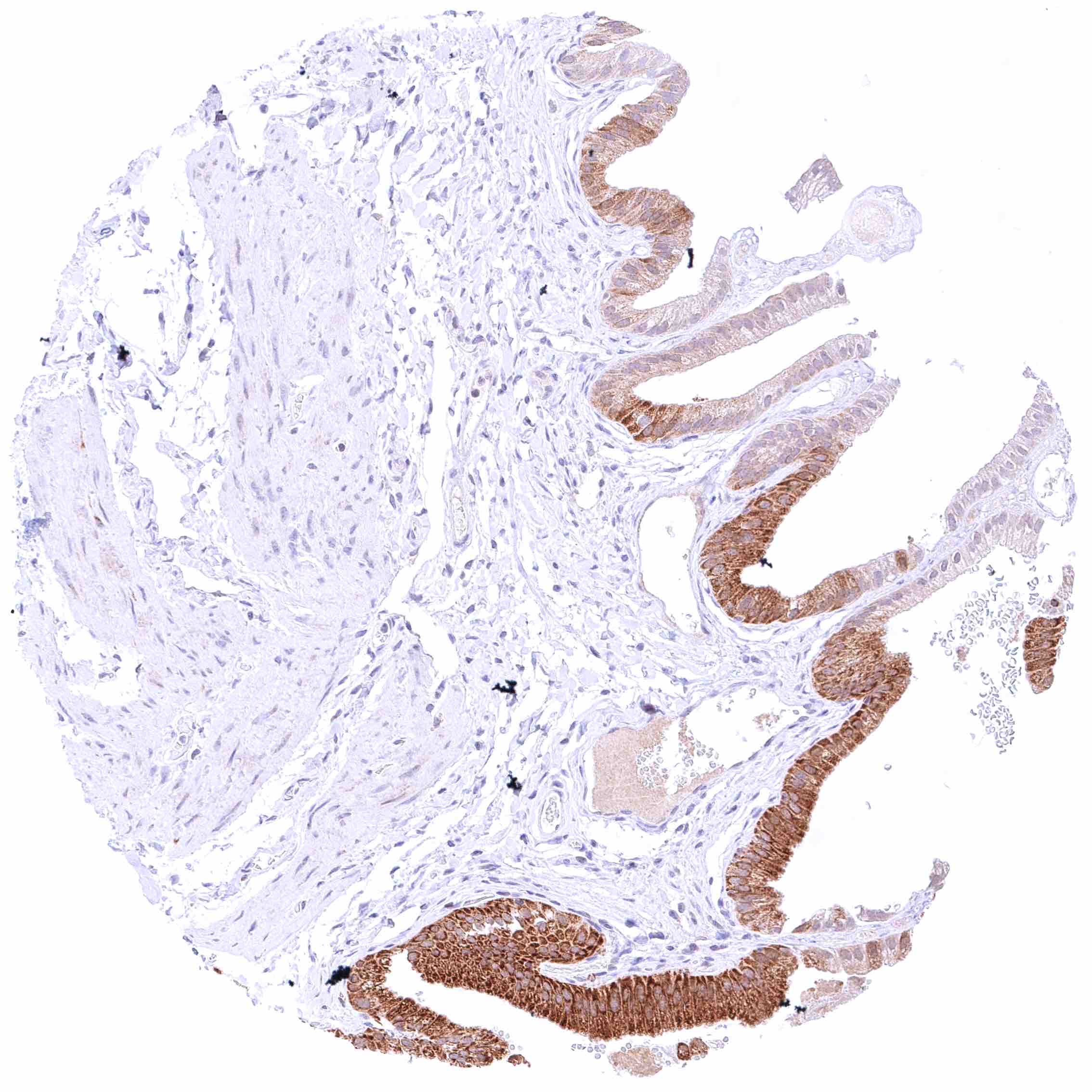
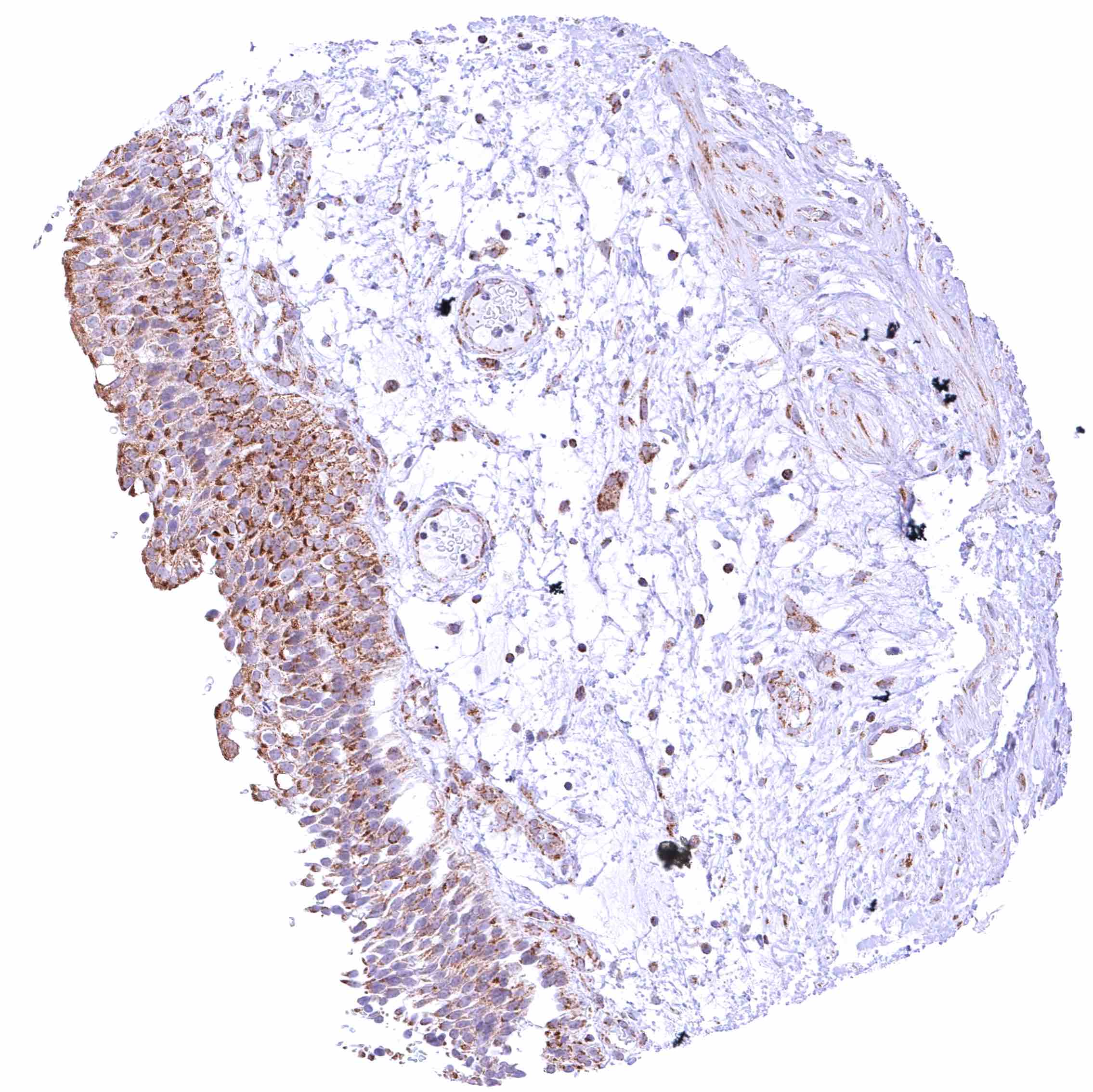
Kidney, pelvis, urothelium – Distinct cytoplasmic BCL-XL staining of urothelial cells.

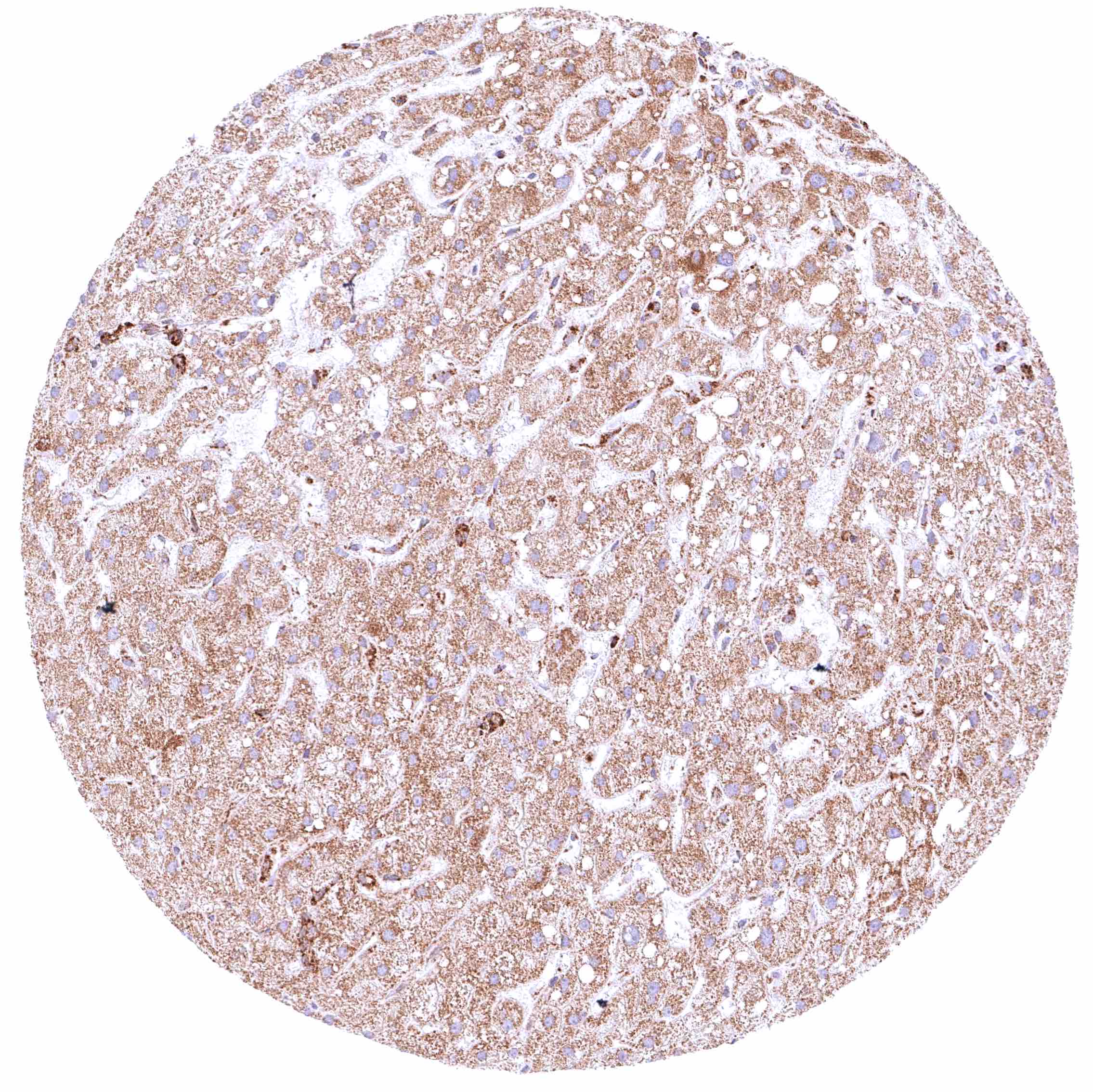
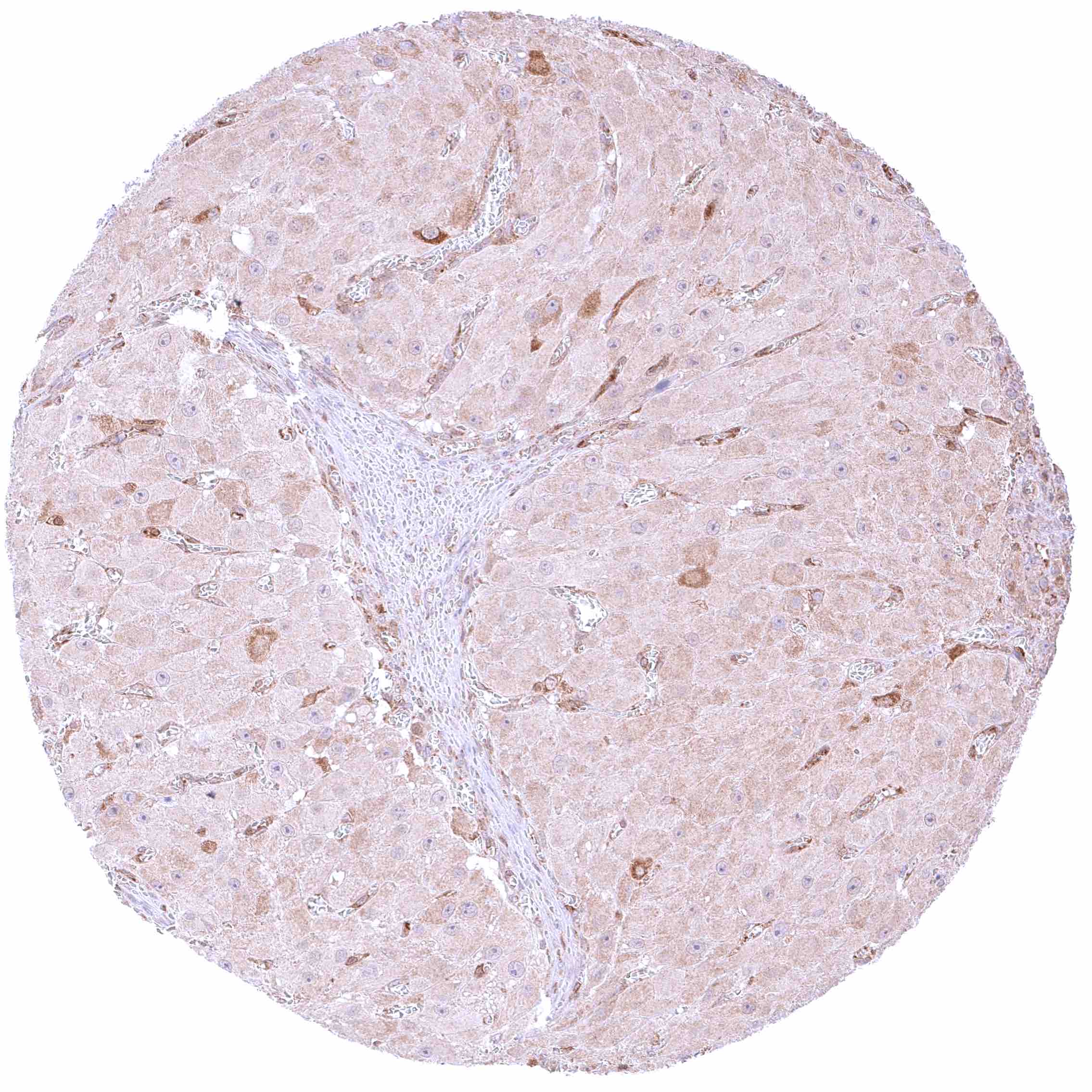
Liver – Moderate cytoplasmic BCL-XL staining of hepatocytes.

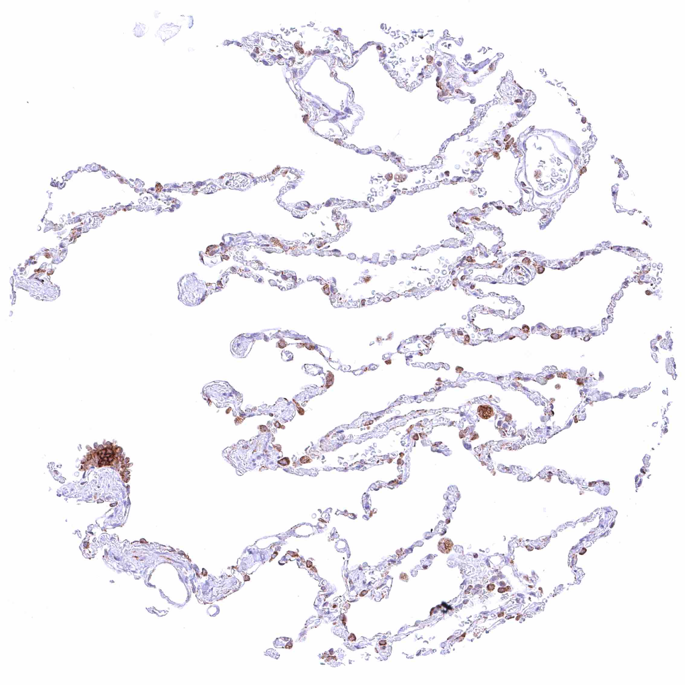
Lung – Moderate cytoplasmic BCL-XL staining of most pneumocytes.


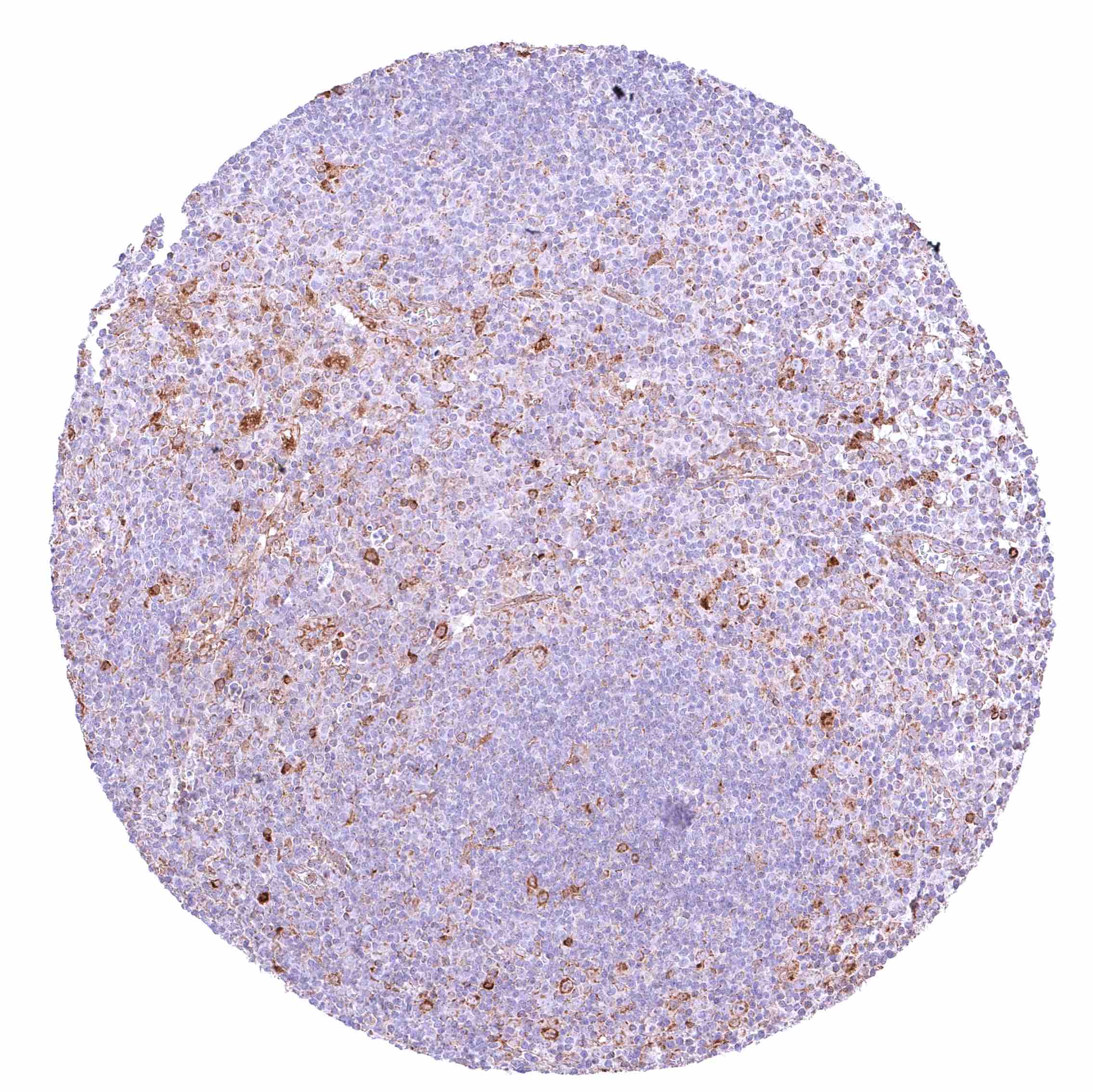
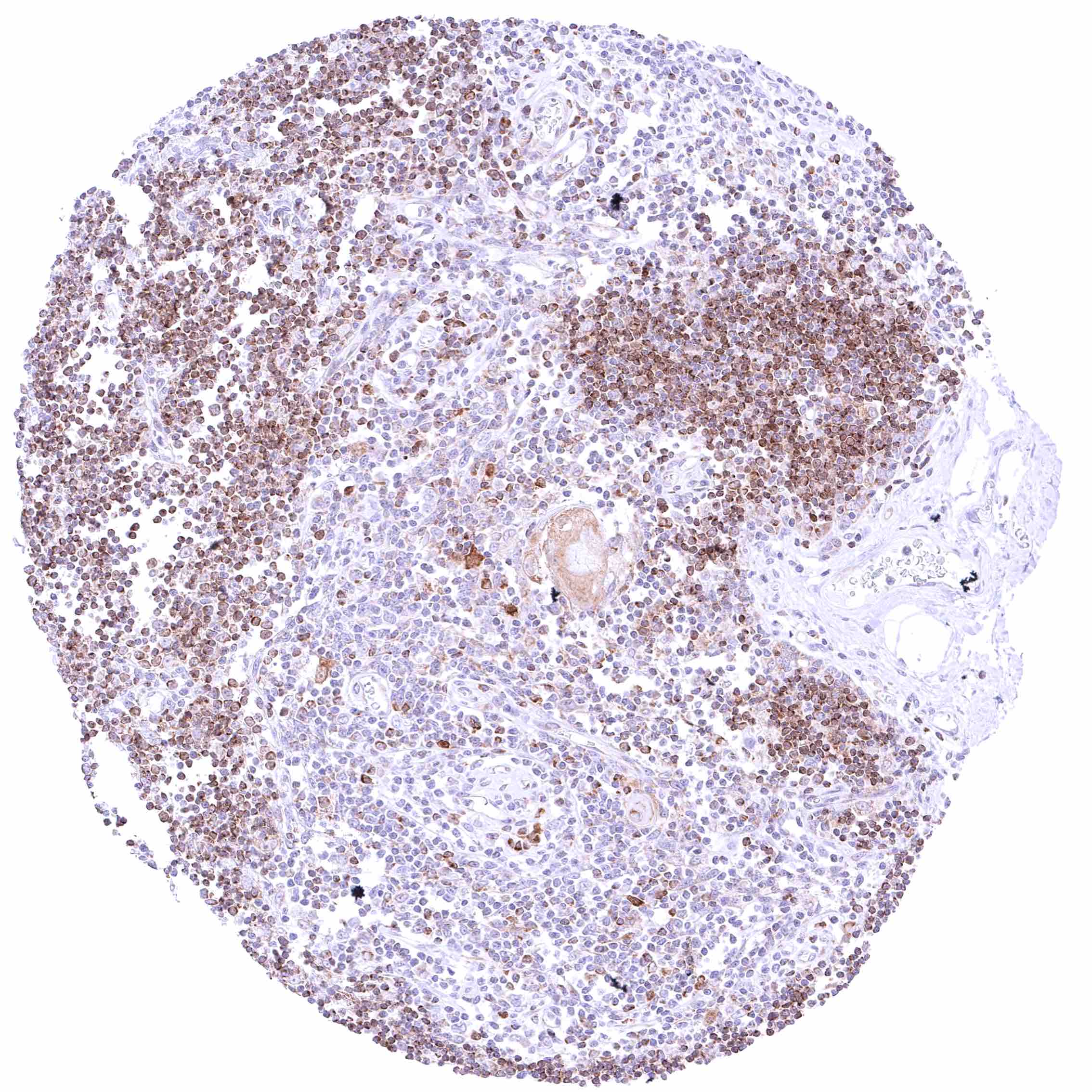
Lymph node – Most lymphocytes, especially in the interfollicular area, are BCL-XL negative or only show a faint staining. Some cell types show a distinct BCL-XL staining, often in germinal centres.

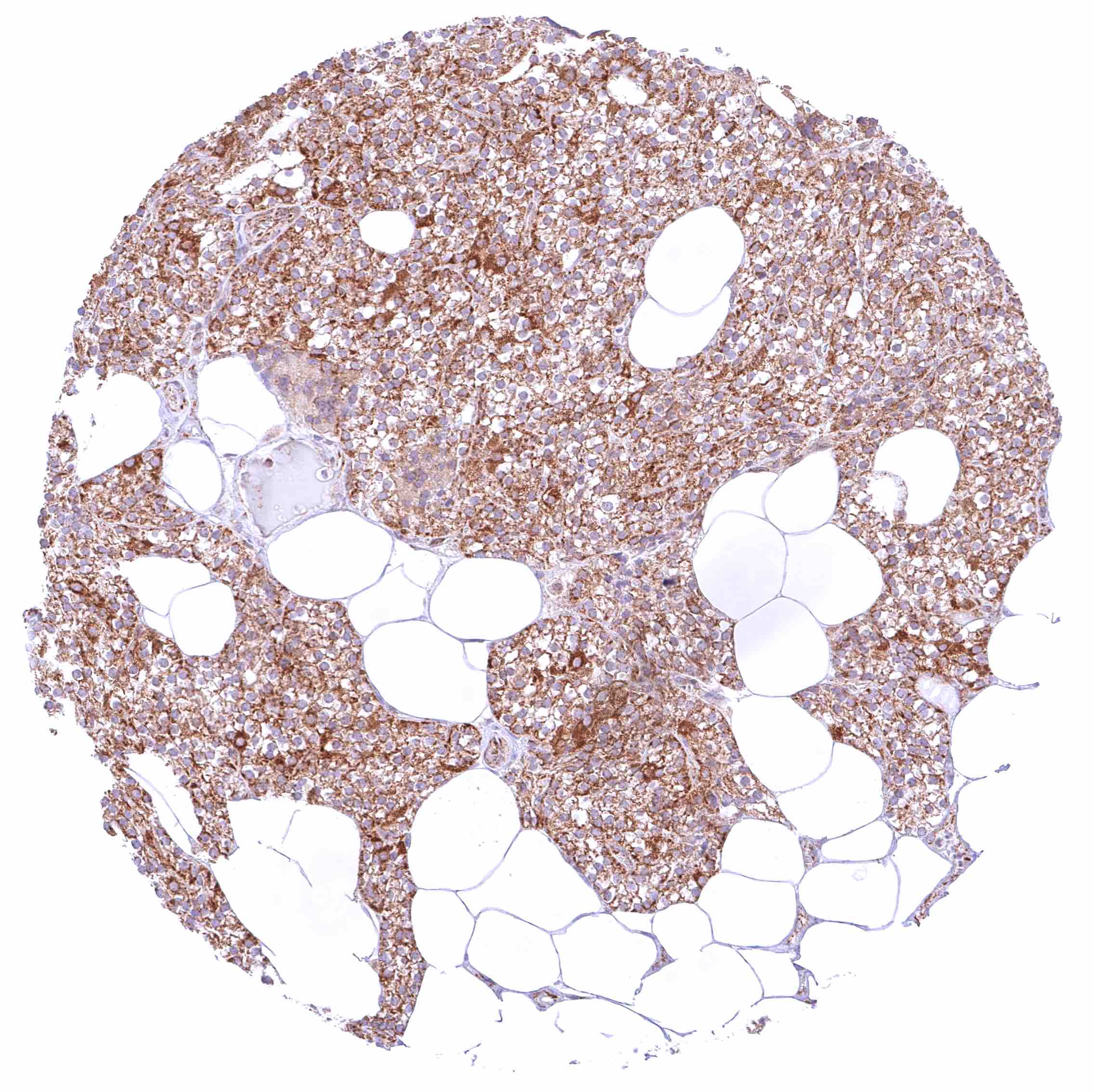
Ovary, corpus luteum – Weak to moderate cytoplasmic BCL-XL staining of corpus luteum cells.

Ovary, follicular cyst – Weak cytoplasmic BCL-XL staining of theca interna and externa cells.

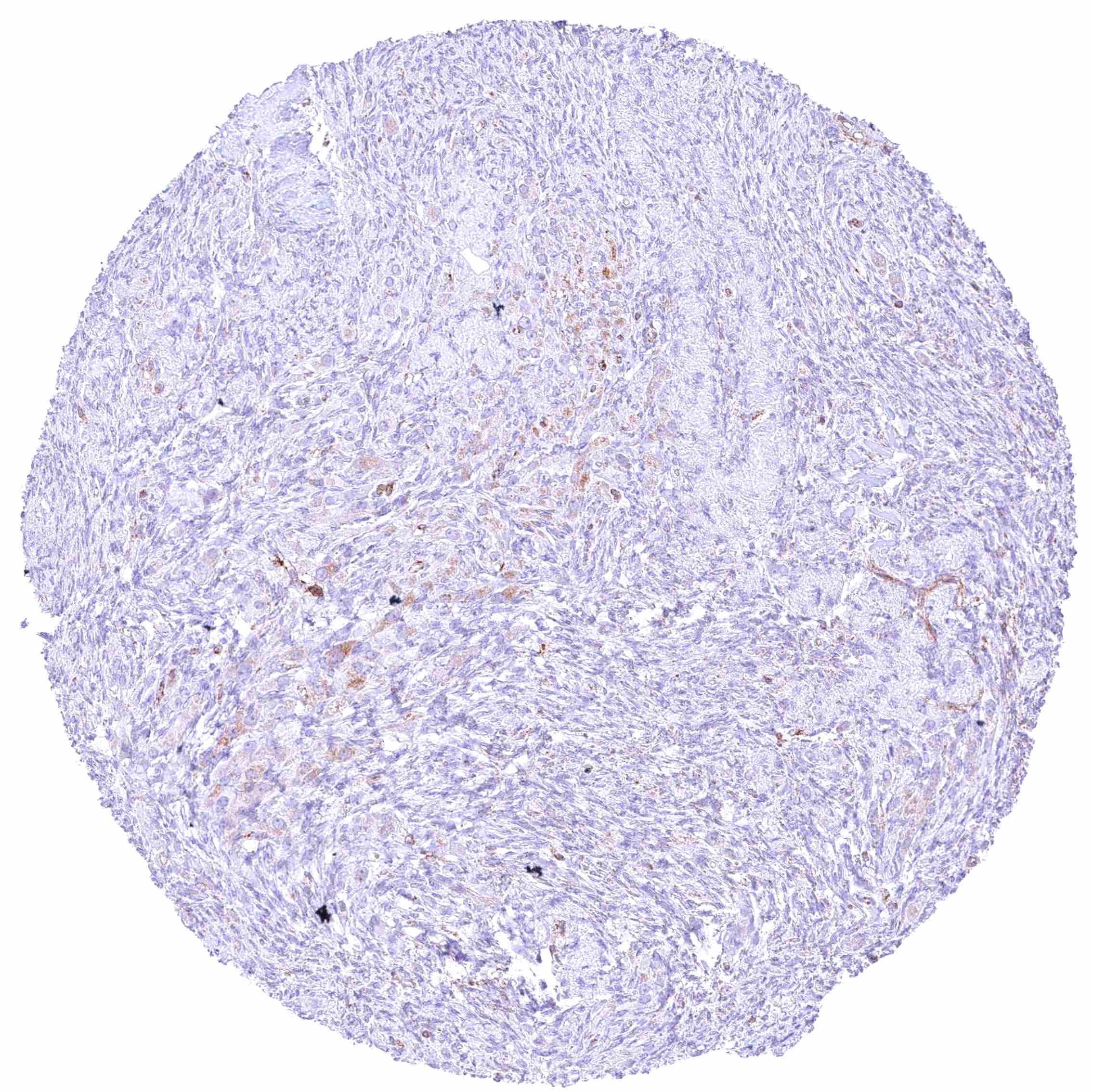
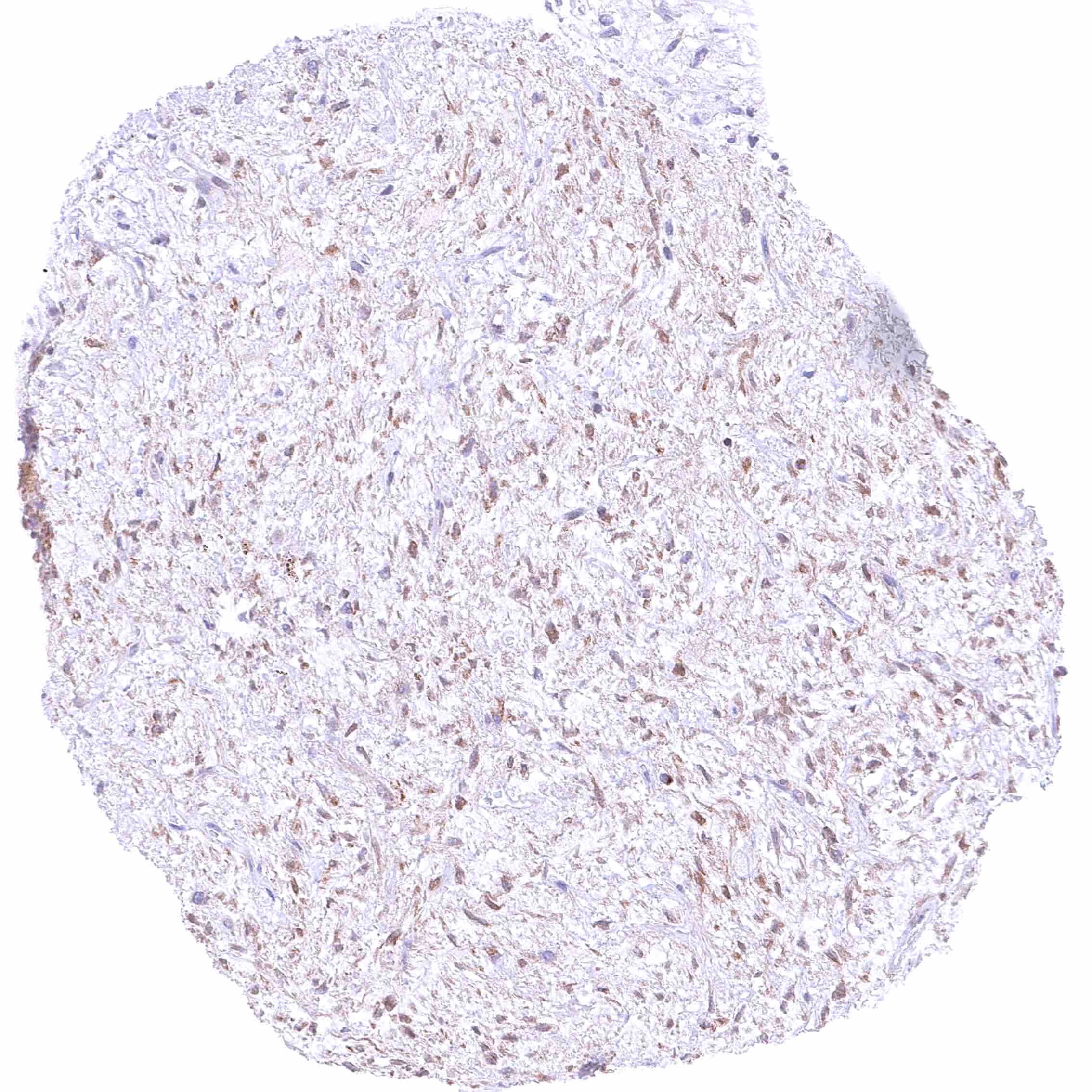
Ovary, stroma – Faint-weak cytoplasmic BCL-XL staining of stroma cells.


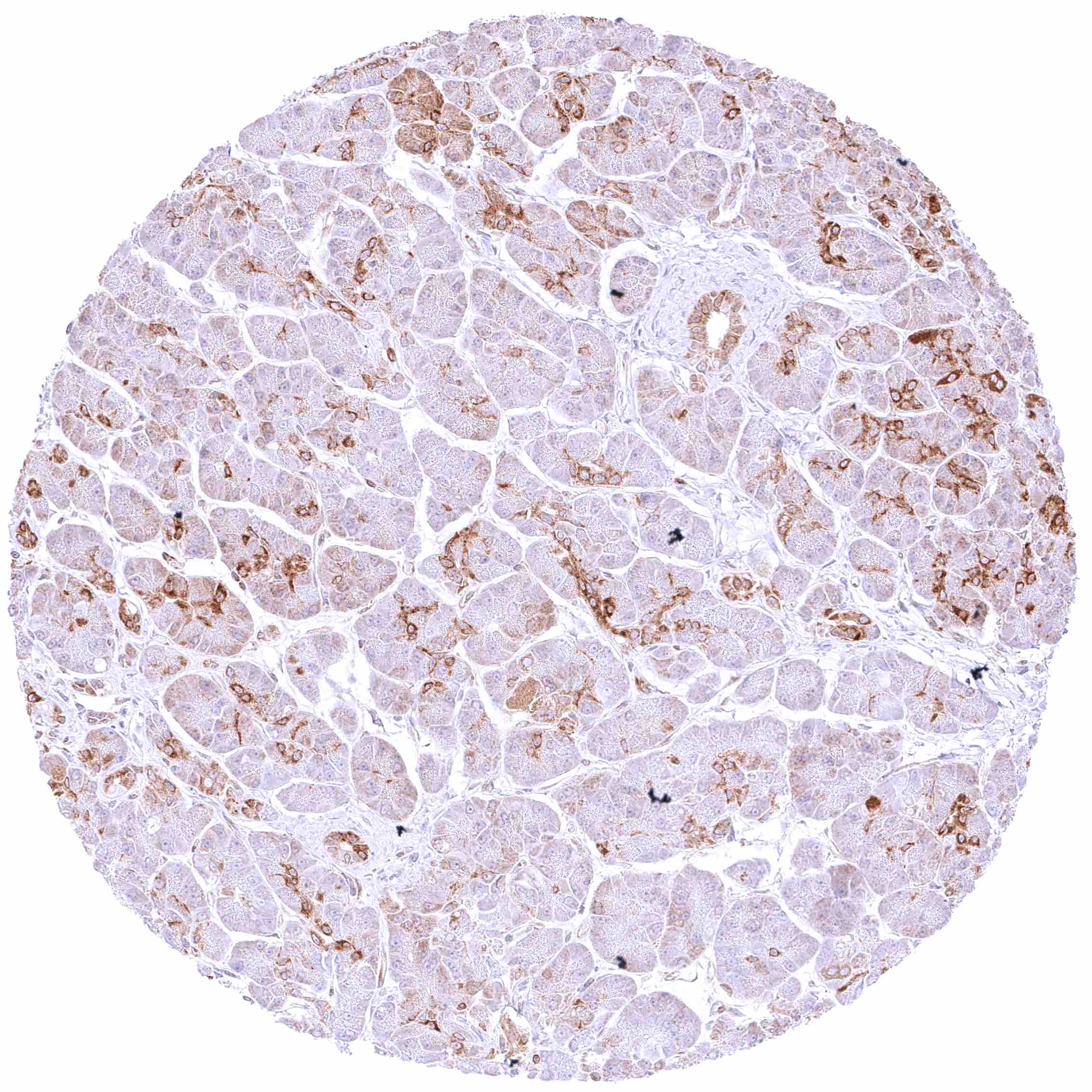
Pancreas – Moderate to strong cytoplasmic BCL-XL staining of intercalated ducts while staining is weak to moderate in excretory ducts and weak or absent in acinar cells.

Parathyroid gland – Strong cytoplasmic BCL-XL staining of epithelial cells.

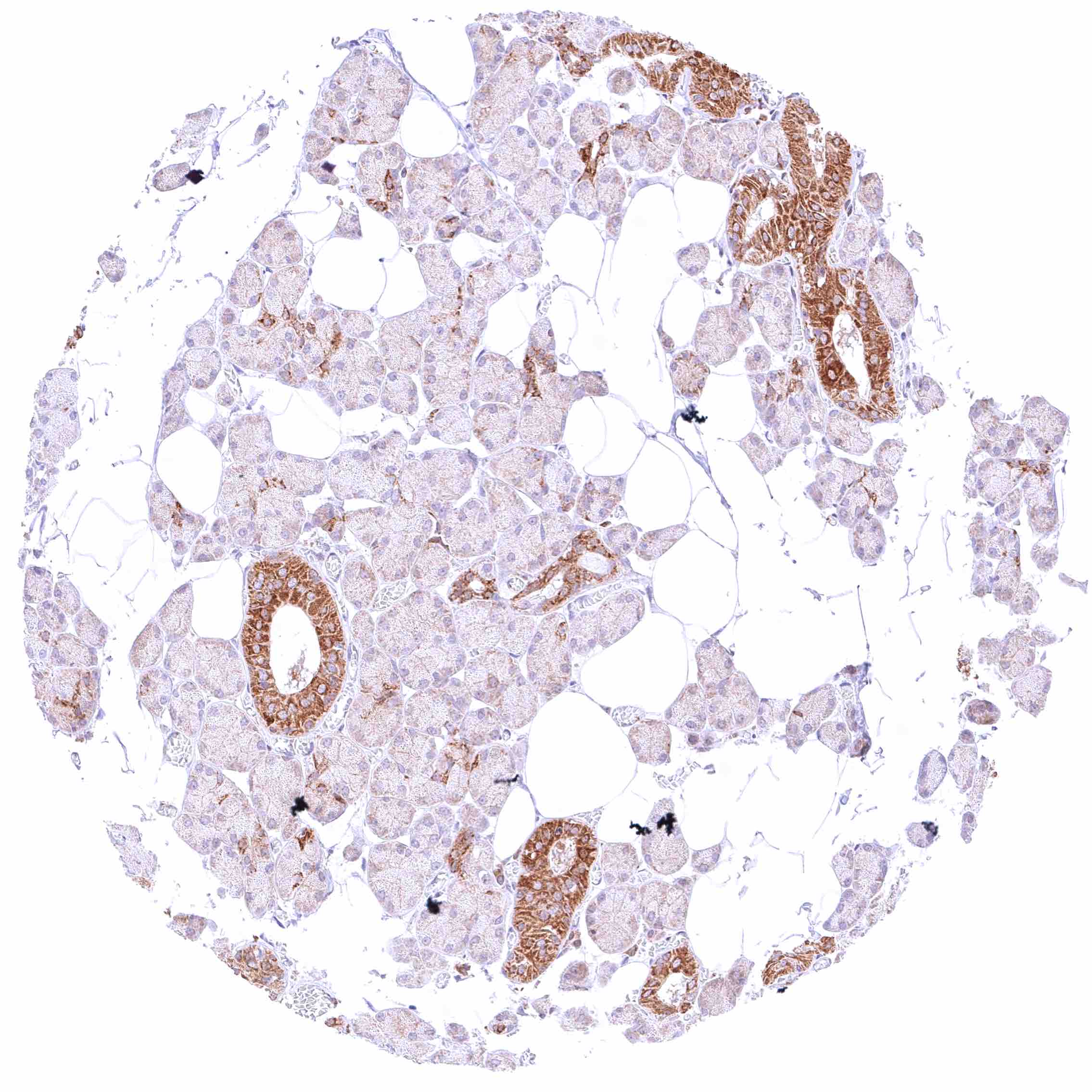
Parotid gland – Strong cytoplasmic BCL-XL positivity of excretory ducts while the staining is markedly less pronounced in glandular cells.

Pituitary gland, anterior lobe – Weak to moderate cytoplasmic BCL-XL staining of a large subset of epithelial cells.


Pituitary gland, posterior lobe – Weak to moderate fibrillar and BCL-XL staining and weak cytoplasmic staining of pituicytes.

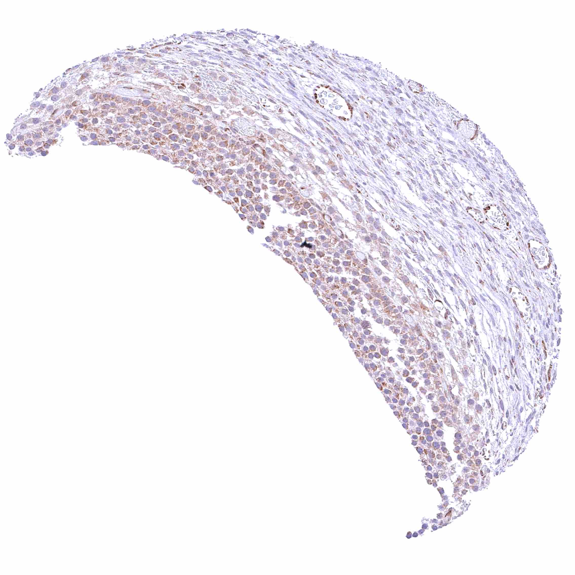
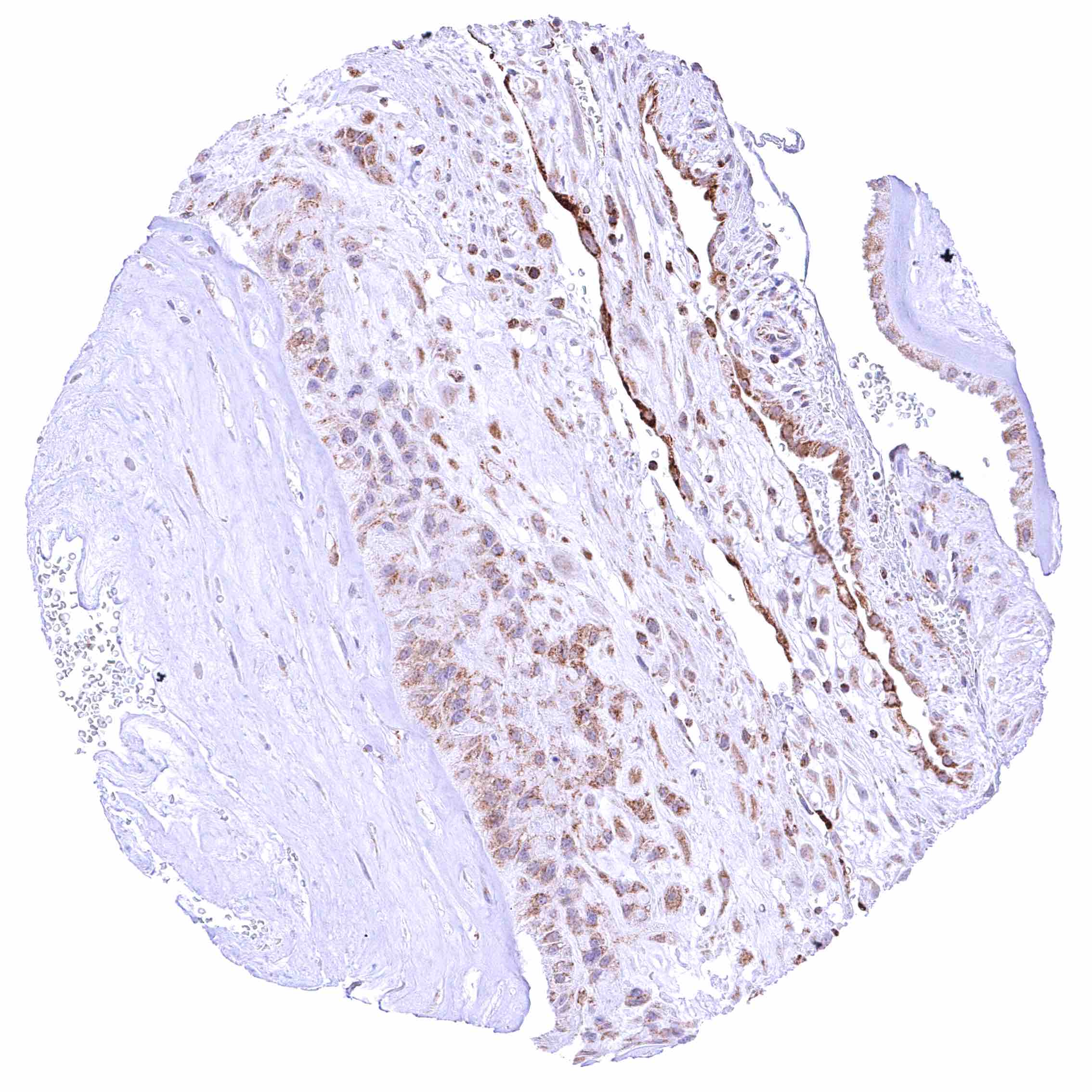
Placenta (amnion and chorion) – Moderate to strong cytoplasmic BCL-XL staining of chorion cells. Weak cytoplasmic BCL-XL staining of amnion cells. Intense staining of endothelial cells.

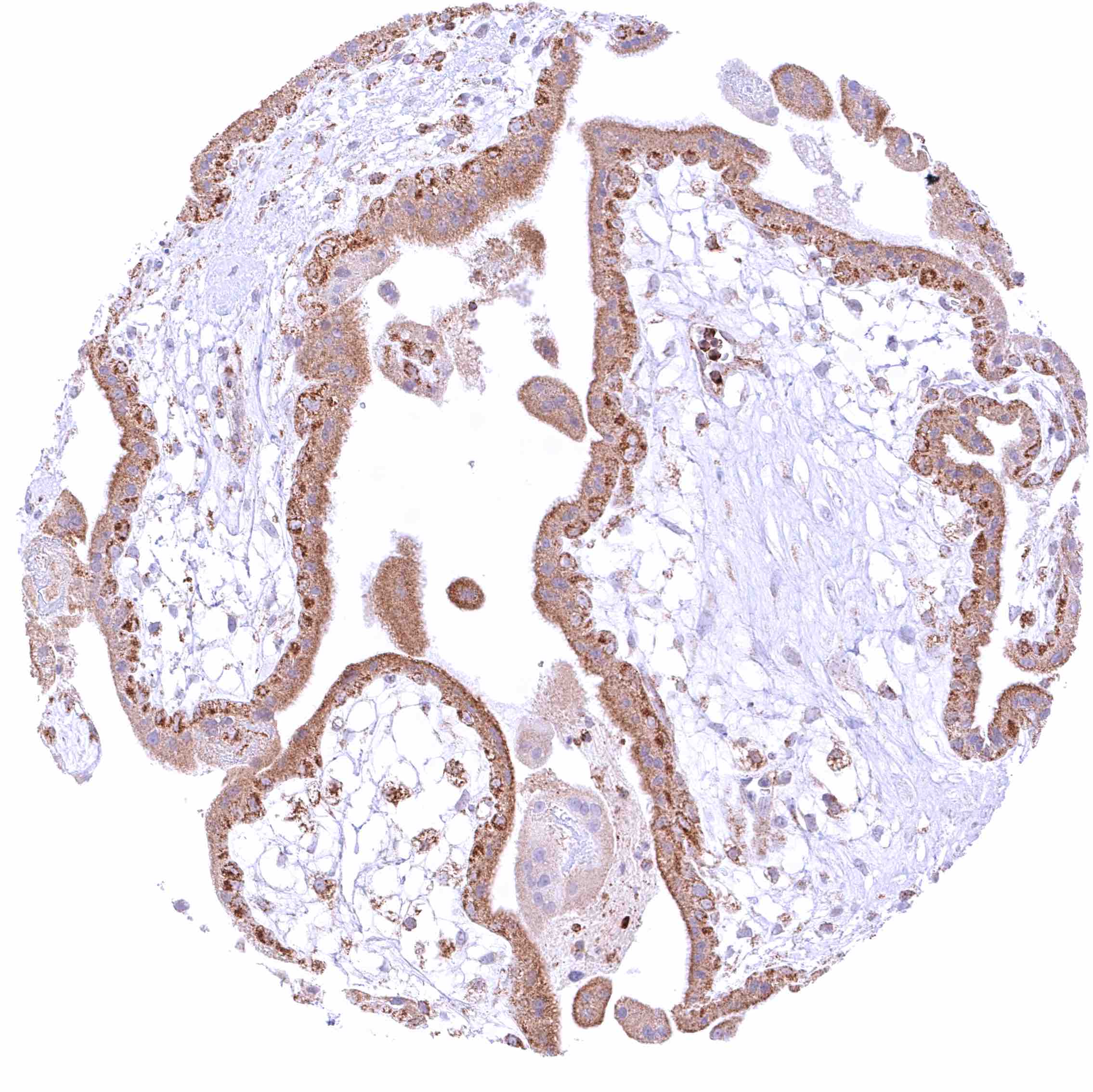
Placenta, early – Strong cytoplasmic BCL-XL staining of trophoblast cells (most intense in the cytotrophoblast).

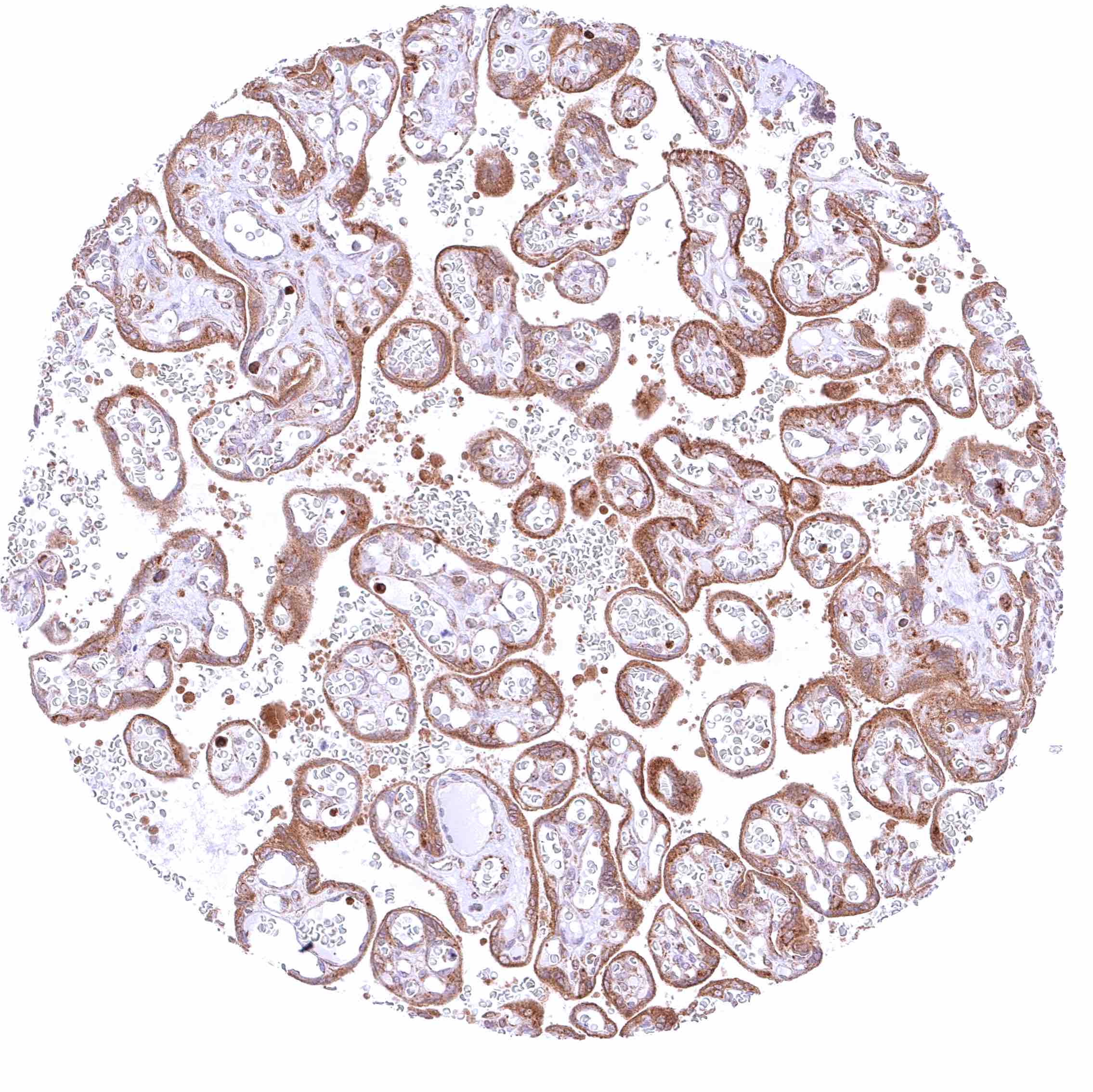
Placenta, mature – Strong cytoplasmic BCL-XL staining of trophoblast cells.


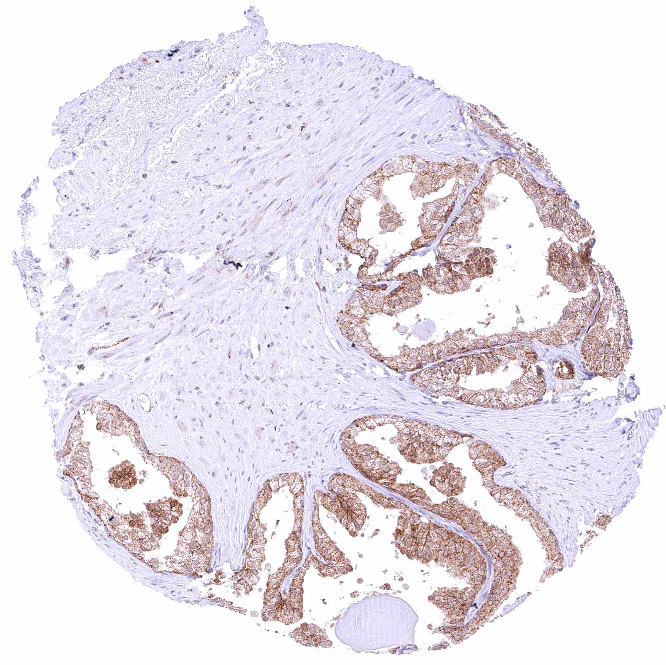
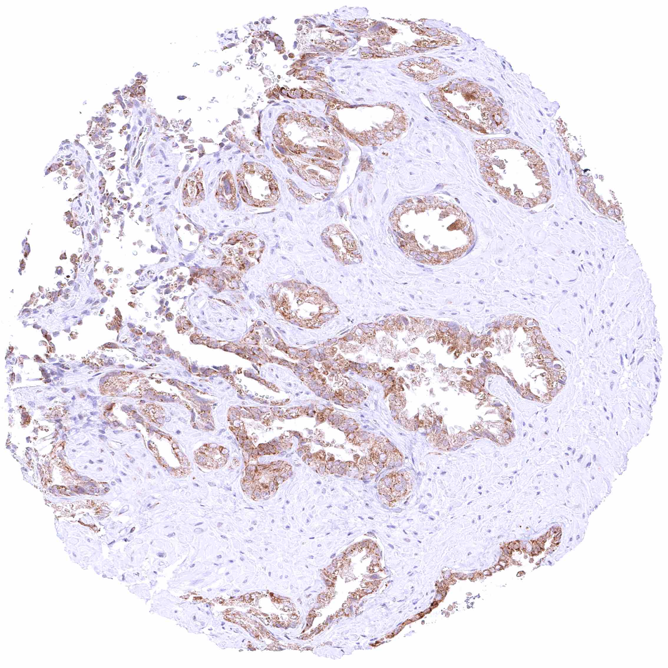
Prostate – Moderate to strong cytoplasmic BCL-XL staining of all epithelial cells.

Rectum, mucosa – Distinct cytoplasmic BCL-XL staining of all epithelial cells and a subset of inflammatory cells (plasma cells_).

Seminal vesicle – Moderate BCL-XL staining of all epithelial cells.

Sinus paranasales – Moderate to strong cytoplasmic LGALS3BP staining of respiratory epithelial cells, especially in the apical-luminal compartment of the cells.


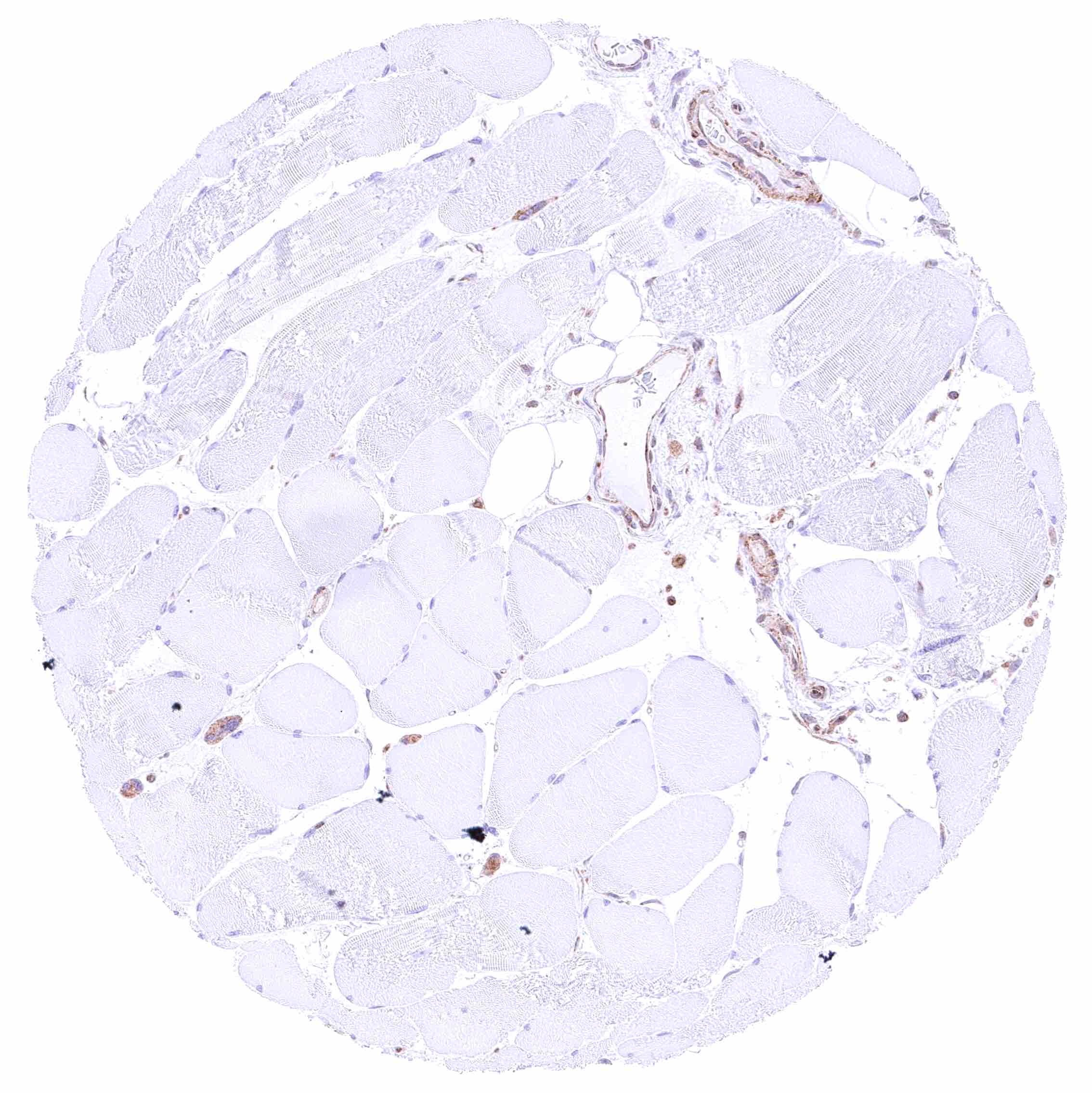
Skeletal muscle – BCL-XL staining is largely absent in skeletal muscle cells.

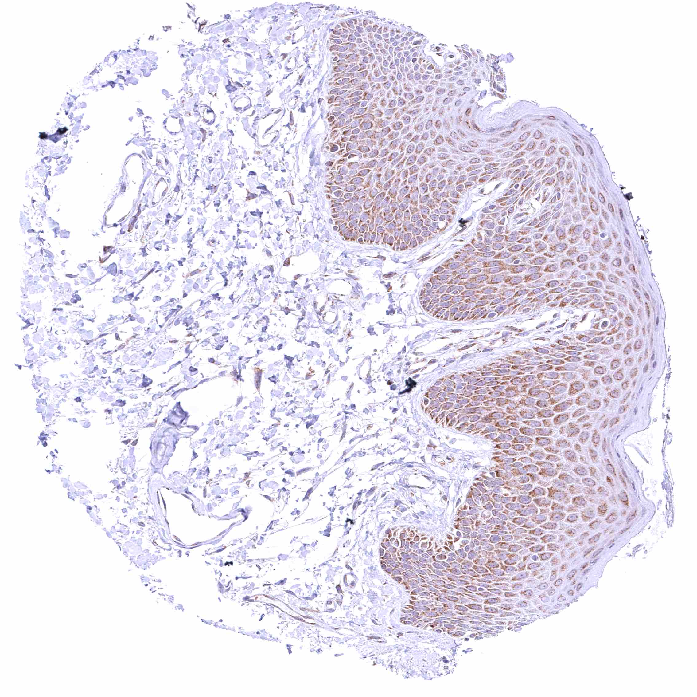
Skin – Weak to moderate cytoplasmic BCL-XL staining of squamous epithelial cells of all cell layers (slight decrease of intensity towards the surface).

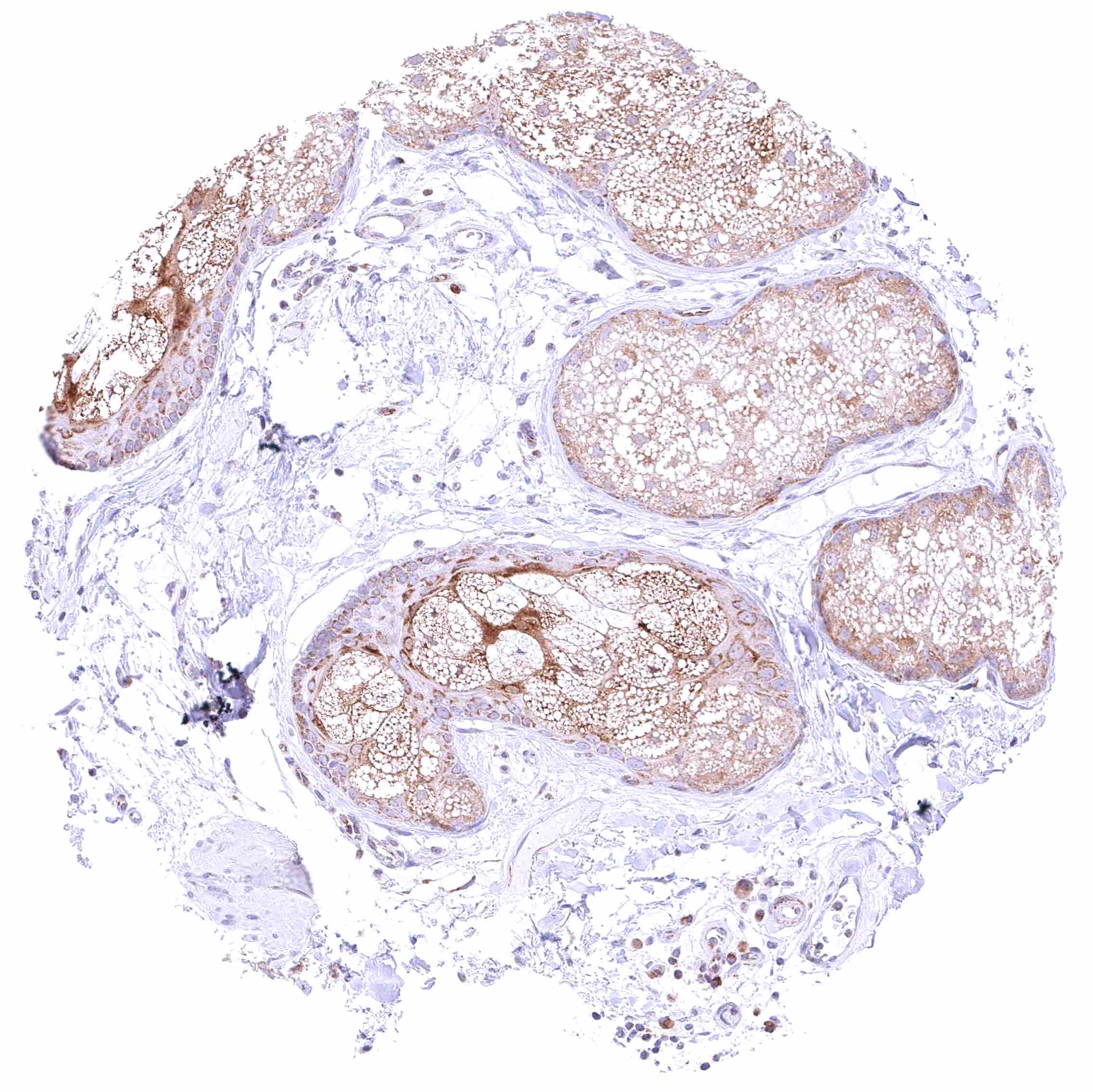
Skin, sebaceous glands – Moderate to strong cytoplasmic BCL-XL staining of sebaceous cells.

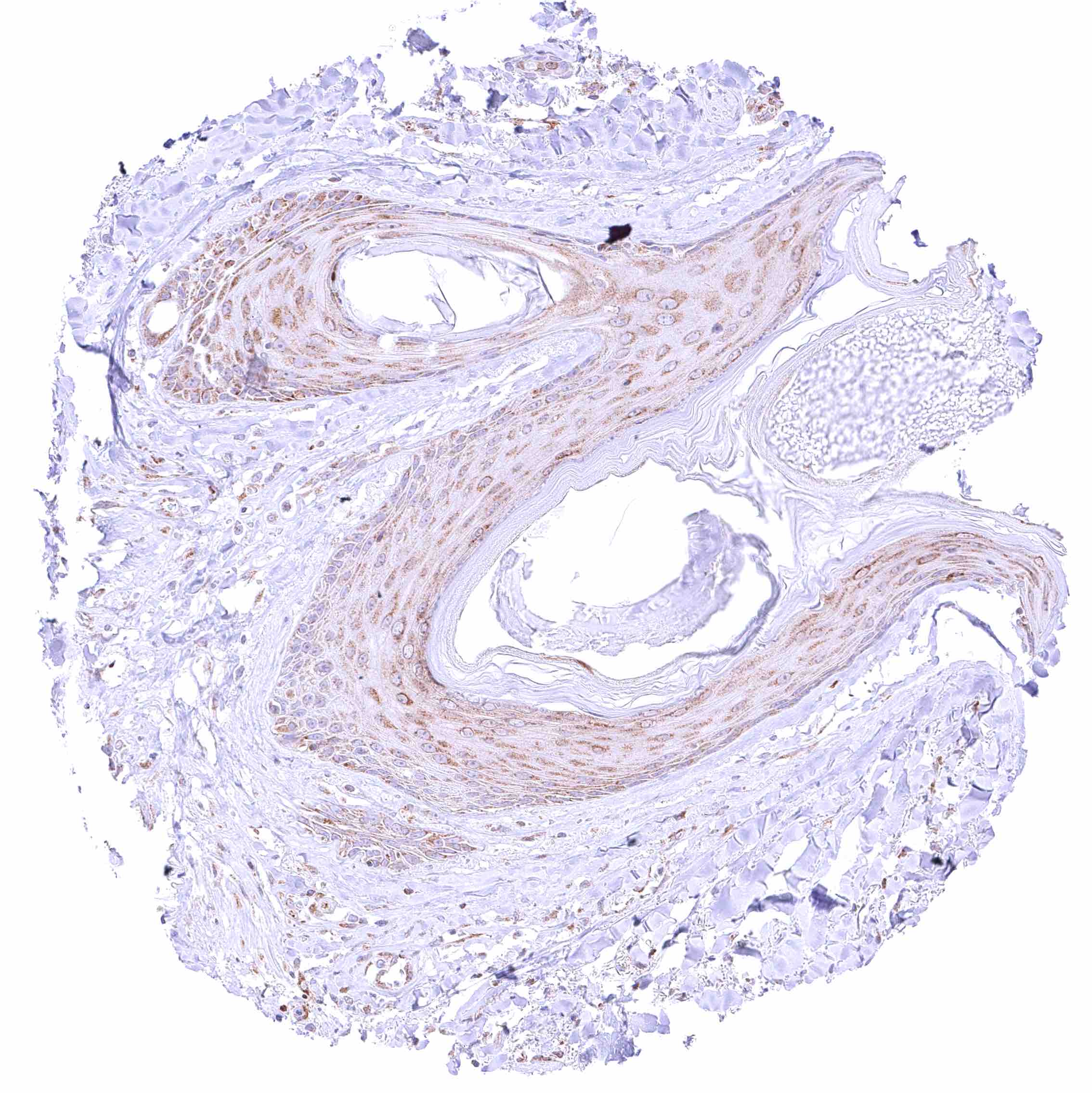
Skin, hairfollicle – Weak to moderate cytoplasmic BCL-XL staining of squamous epithelial cells of hair follicles.


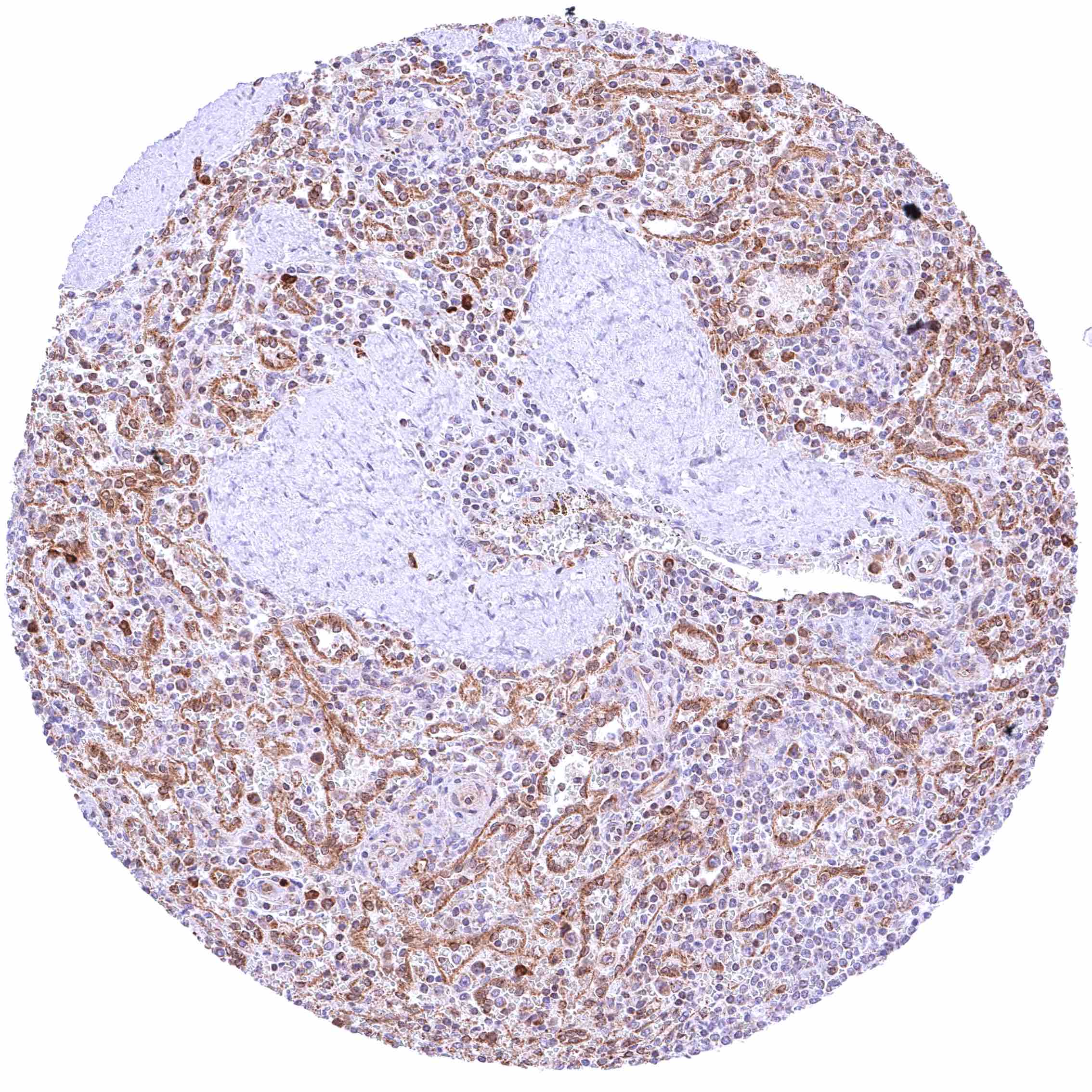
Spleen – Moderate to strong BCL-XL staining of endothelial cells.

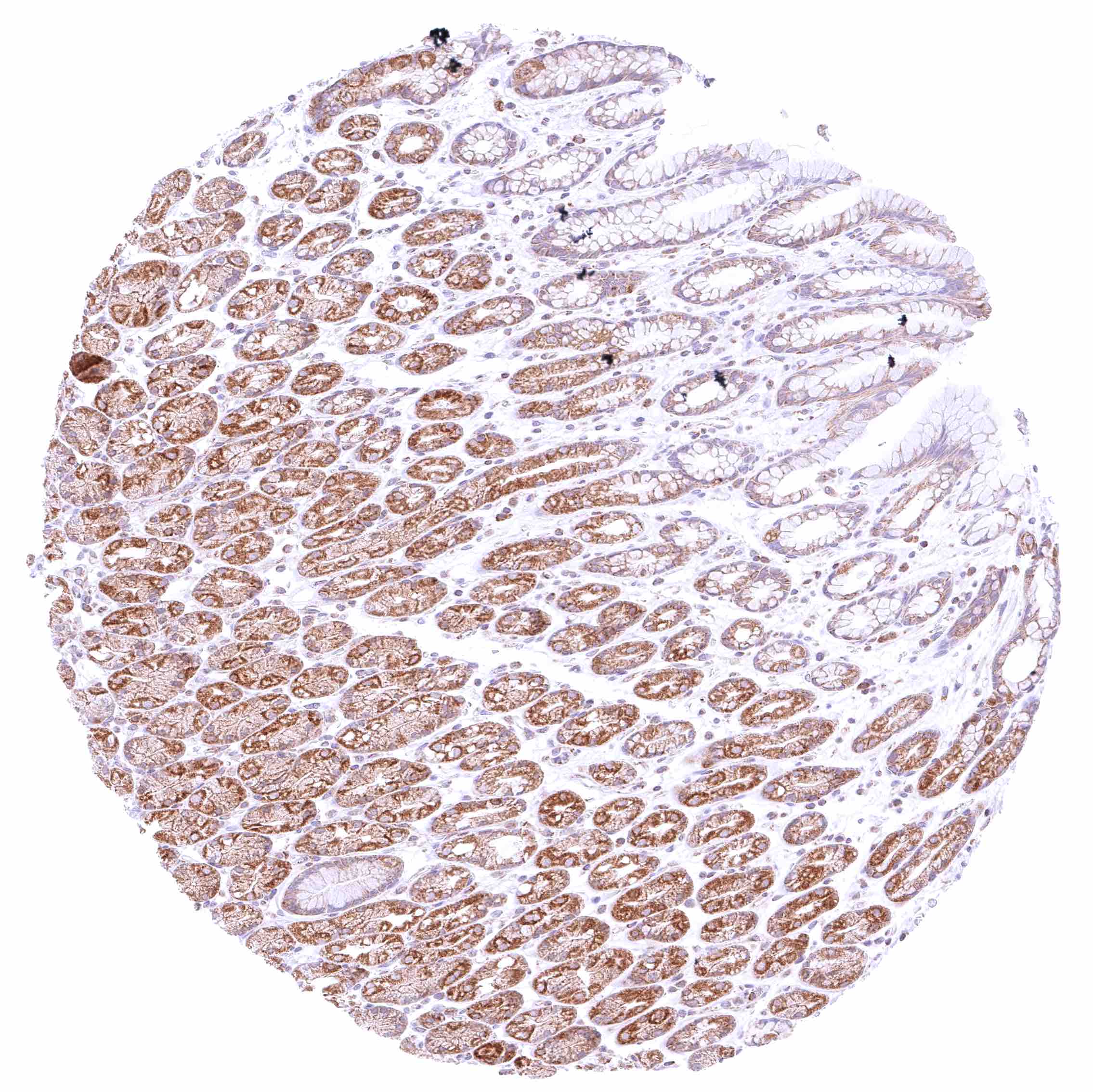
Stomach, corpus – Cytoplasmic BCL-XL staining is moderate in gastric glands but faint in superficial epithelial cells.

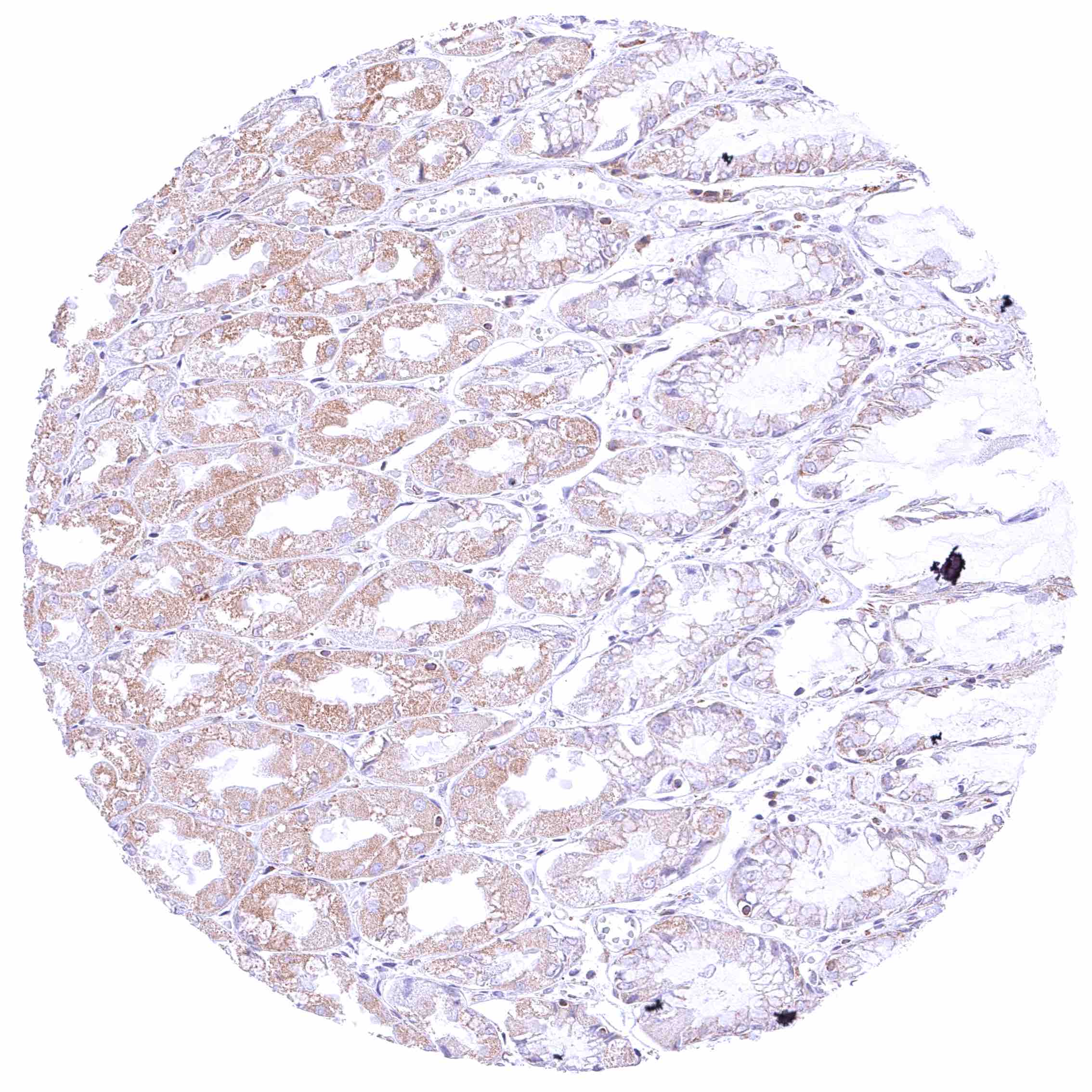
Stomach, antrum – Cytoplasmic BCL-XL staining is significant but variable according to the cell types in gastric glands while BCL-XL staining is only weak in superficial epithelial cells.

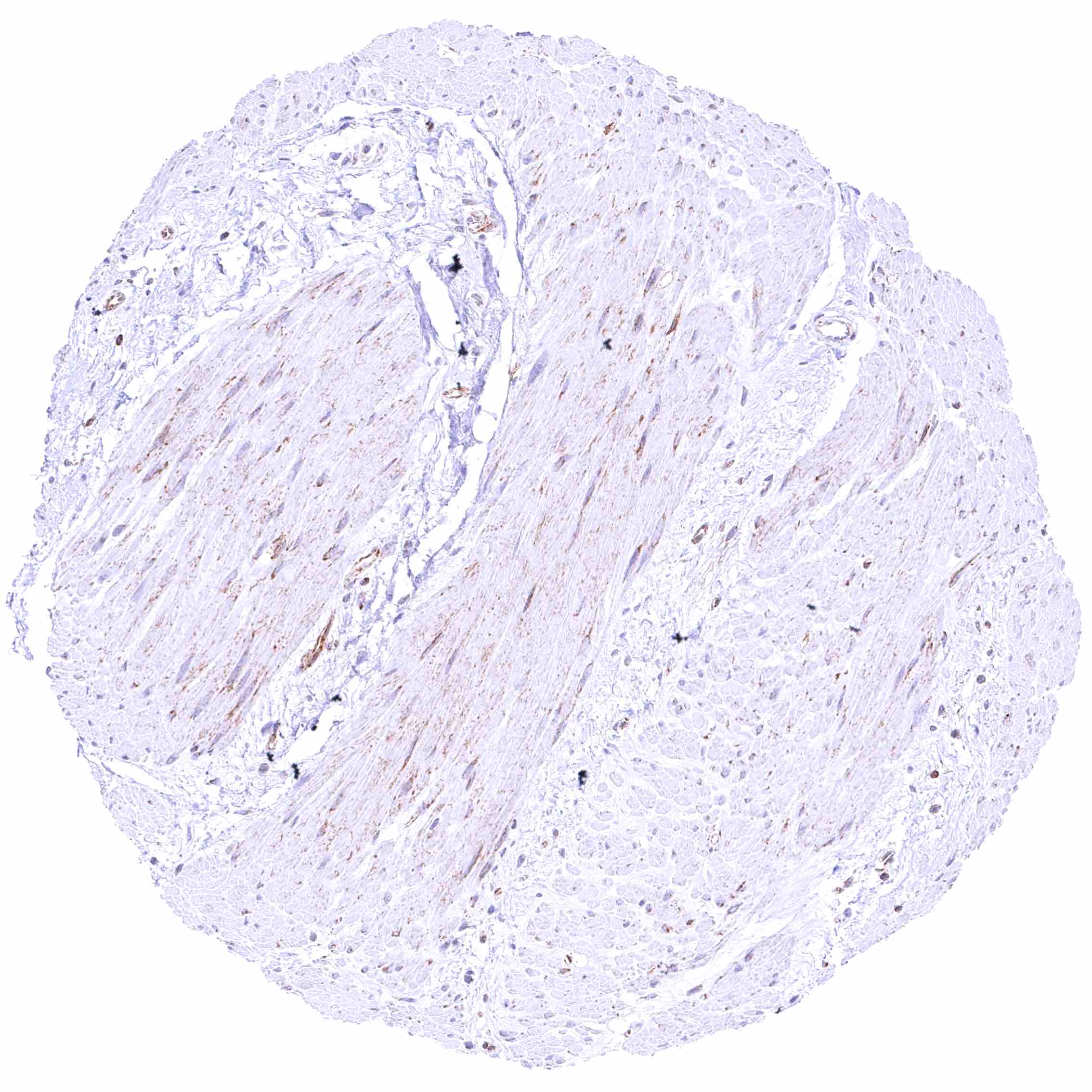
Stomach, muscular wall – Weak cytoplasmic BCL-XL staining of smooth muscle cells.


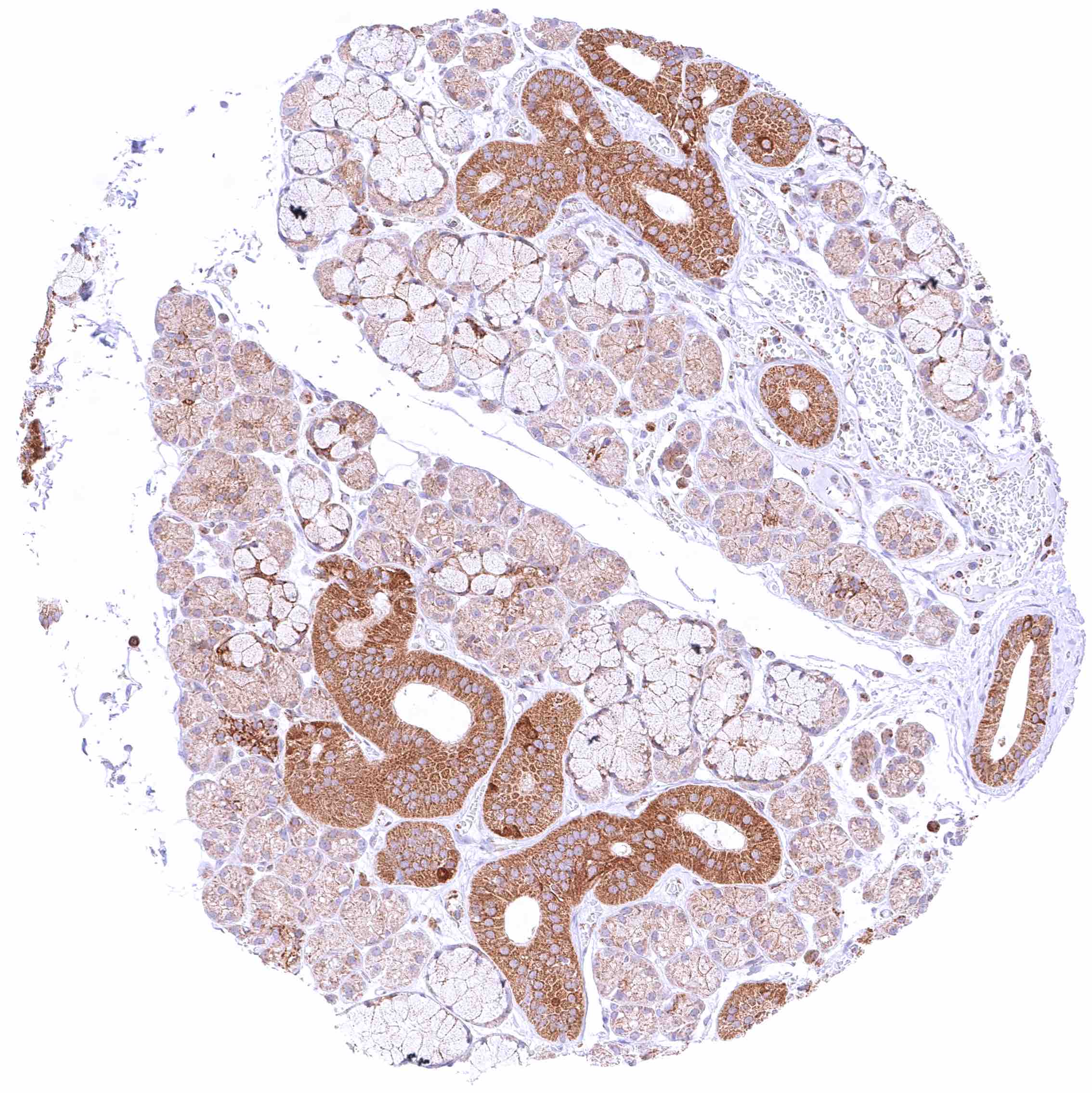
Submandibular gland – Strong cytoplasmic BCL-XL positivity of excretory ducts while the staining is markedly less pronounced in glandular cells.

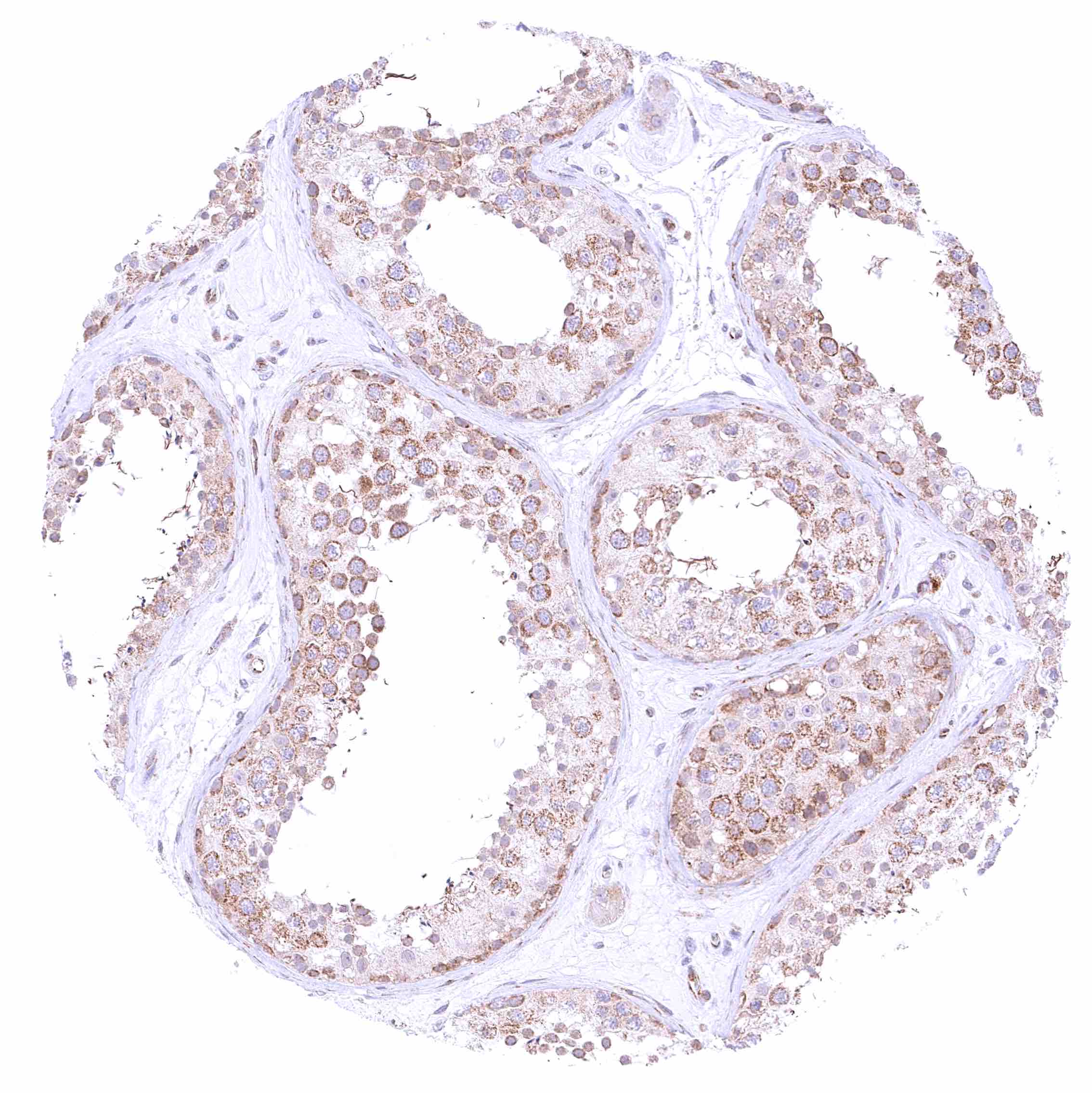
Testis – Weak to moderate cytoplasmic BCL-XL staining of germ cells while the staining is less intense in Sertoli cells. Faint cytoplasmic BCL-XL staining of Leydig cells.

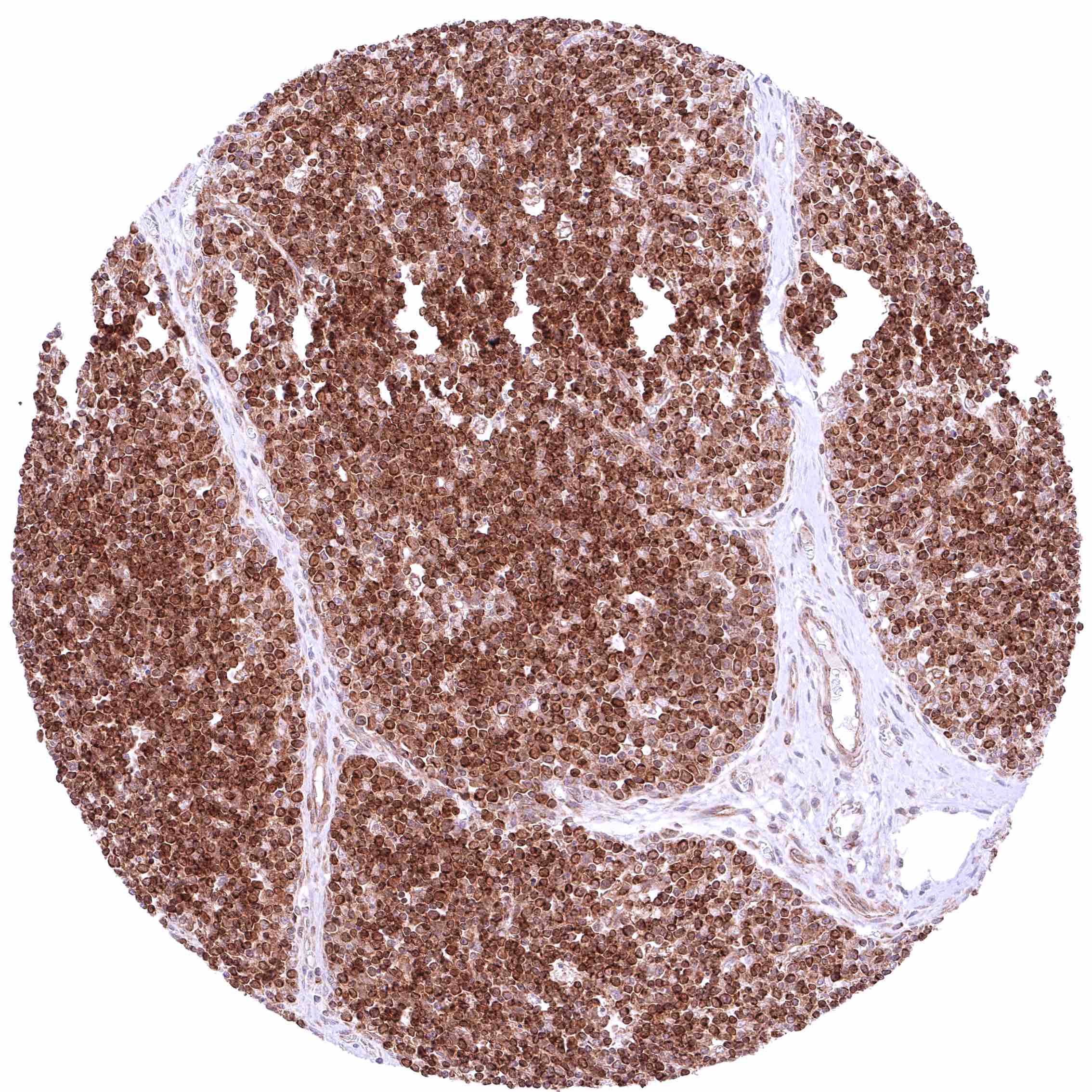
Thymus – Most lymphocytes of the cortex display a strong BCL-XL staining while medullary cells are mostly negative.

Thymus – Most lymphocytes of the cortex display a strong BCL-XL staining.


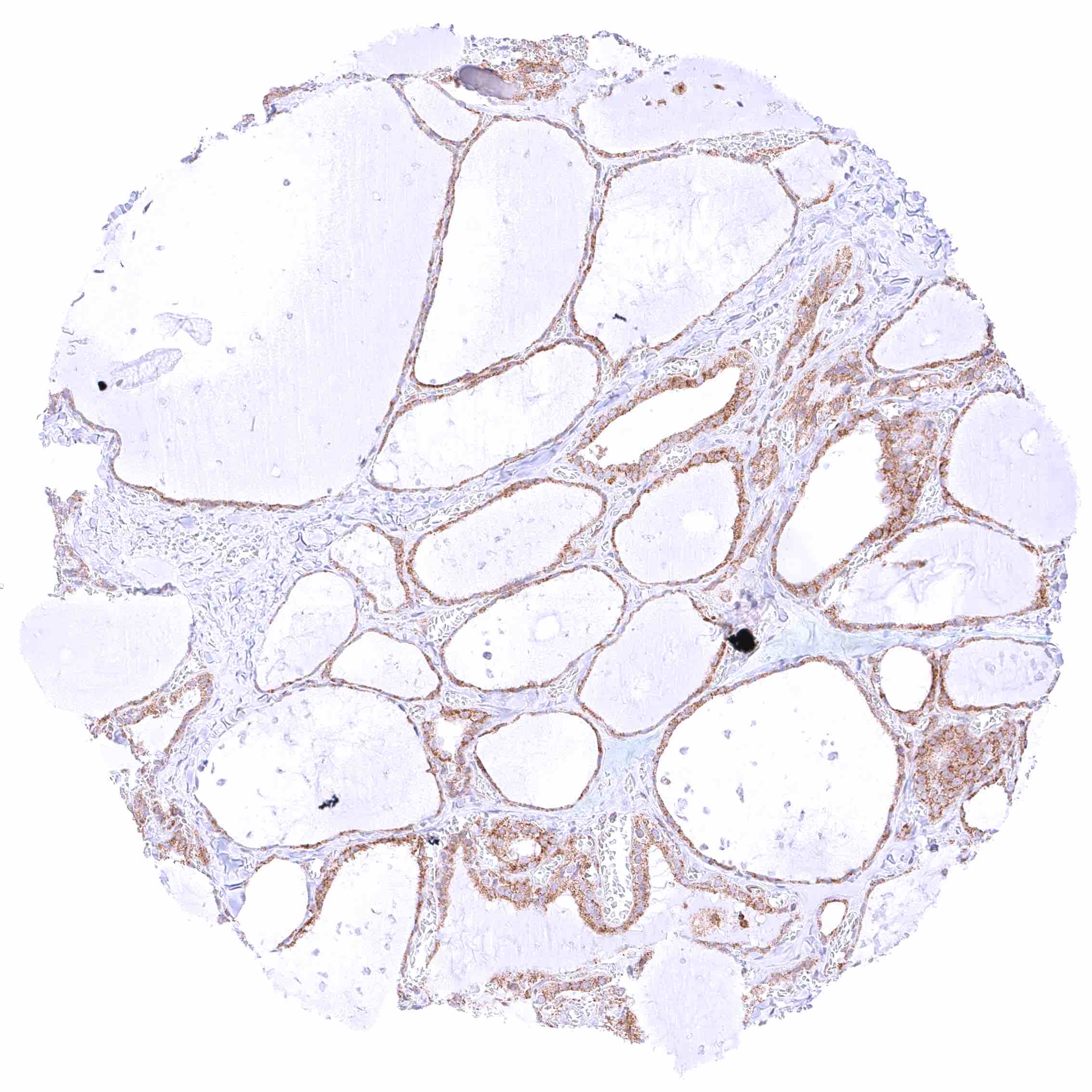
Thyroid gland – Moderate cytoplasmic BCL-XL staining of epithelial cells.

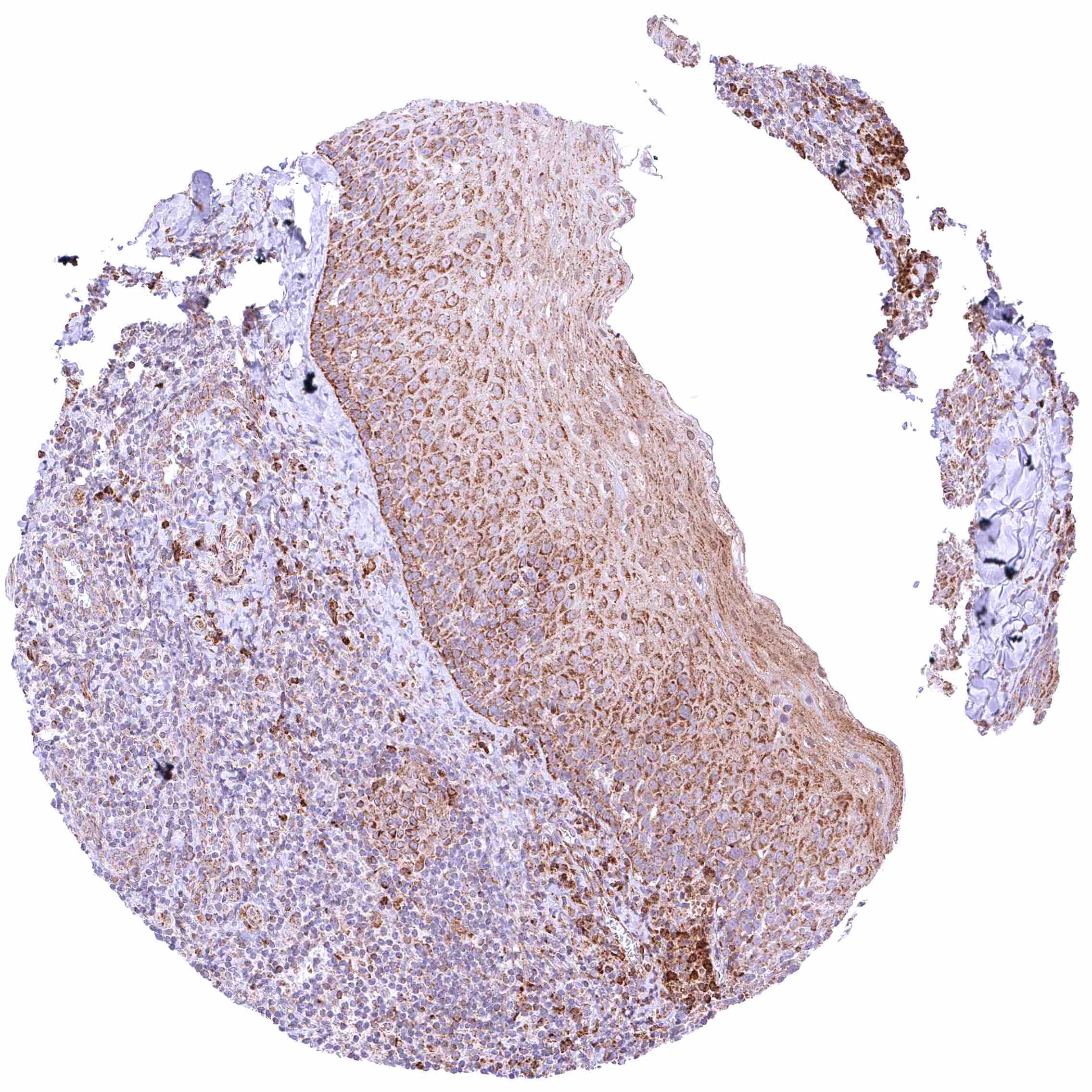
Tonsil, Surface epithelium – Moderate to strong BCL-XL staining of squamous epithelial cells.

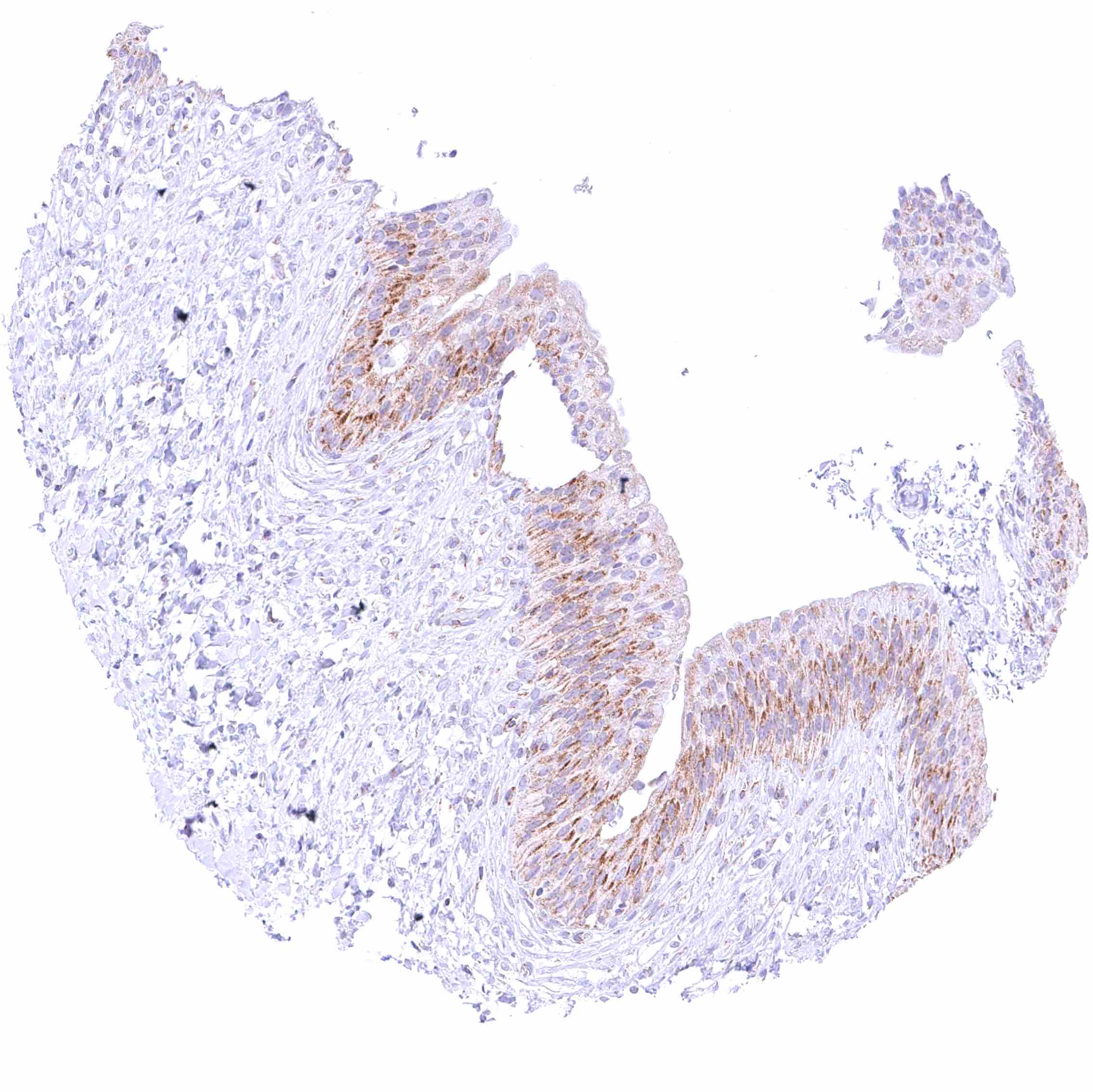
Urinary bladder, urothelium – Distinct cytoplasmic BCL-XL staining of urothelial cells.

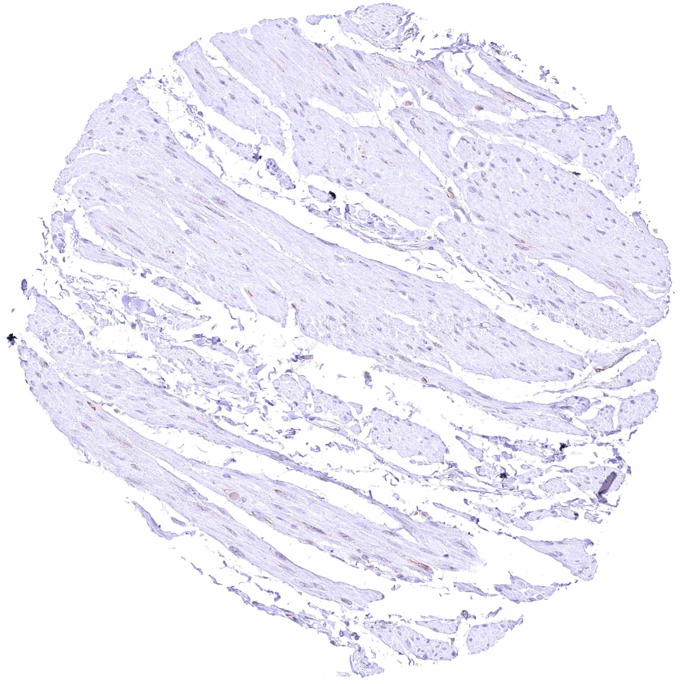
Urinary bladder, muscular wall – Faint cytoplasmic BCL-XL staining of smooth muscle cells.


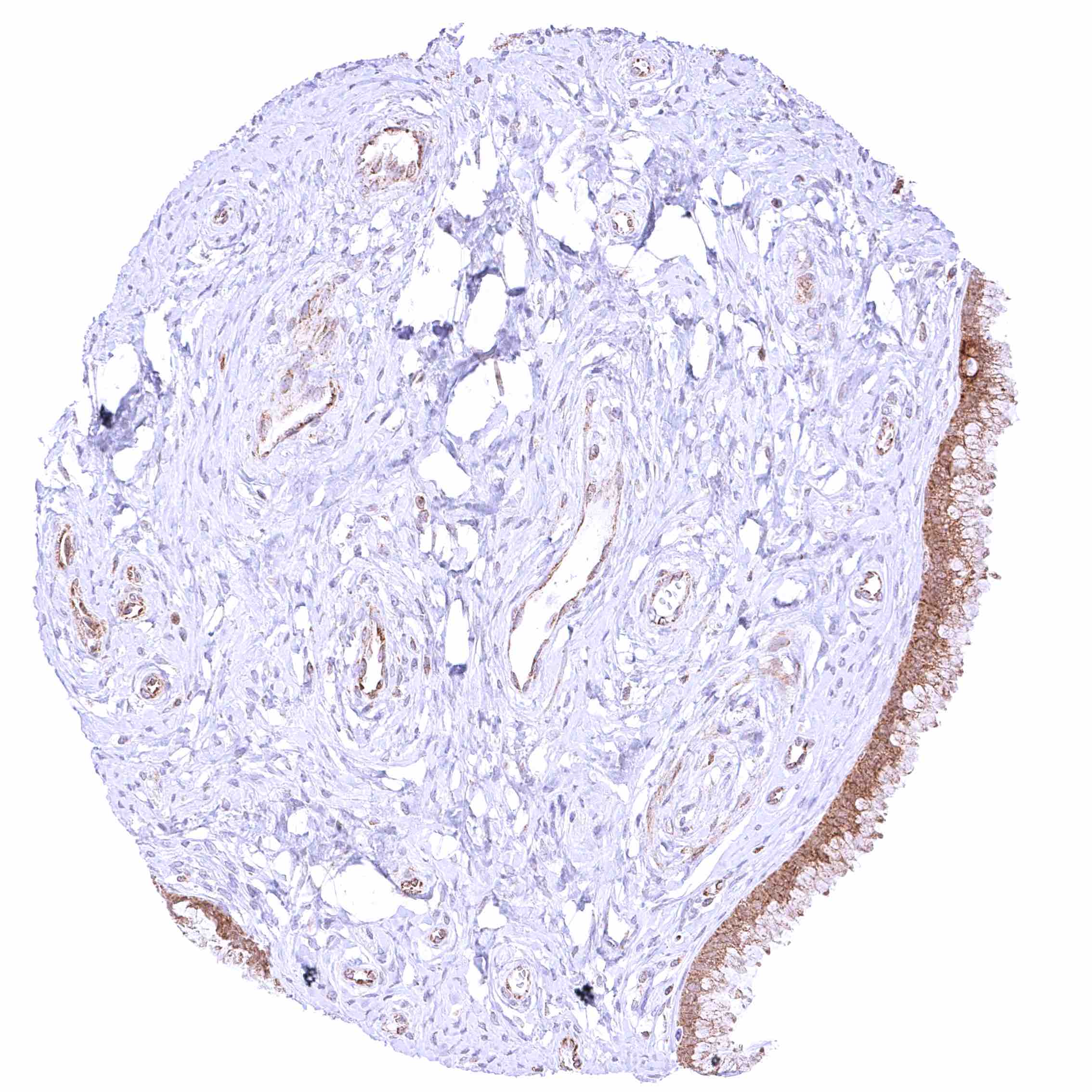
Uterus, endocervix – Strong cytoplasmic BCL-XL staining of epithelial cells.

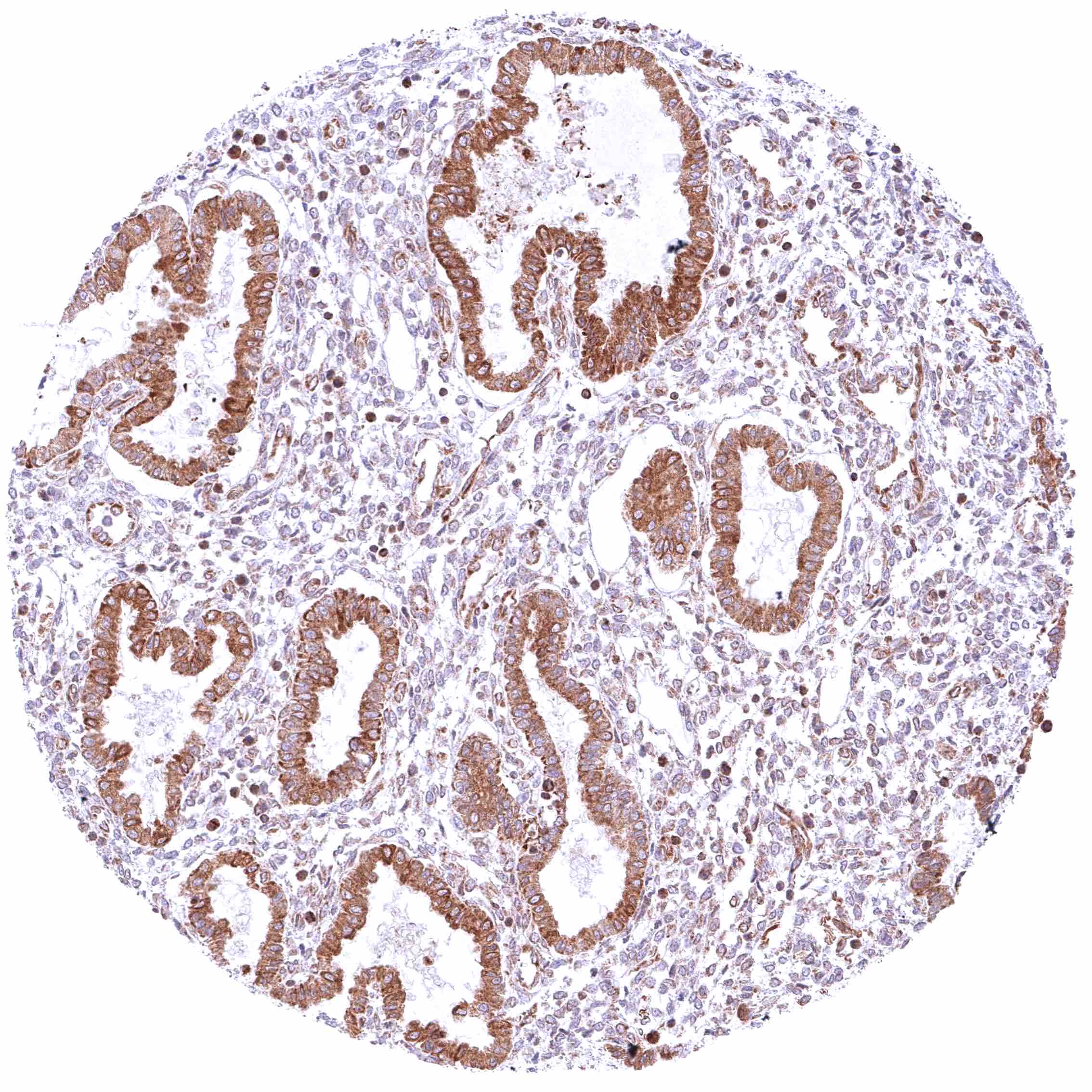
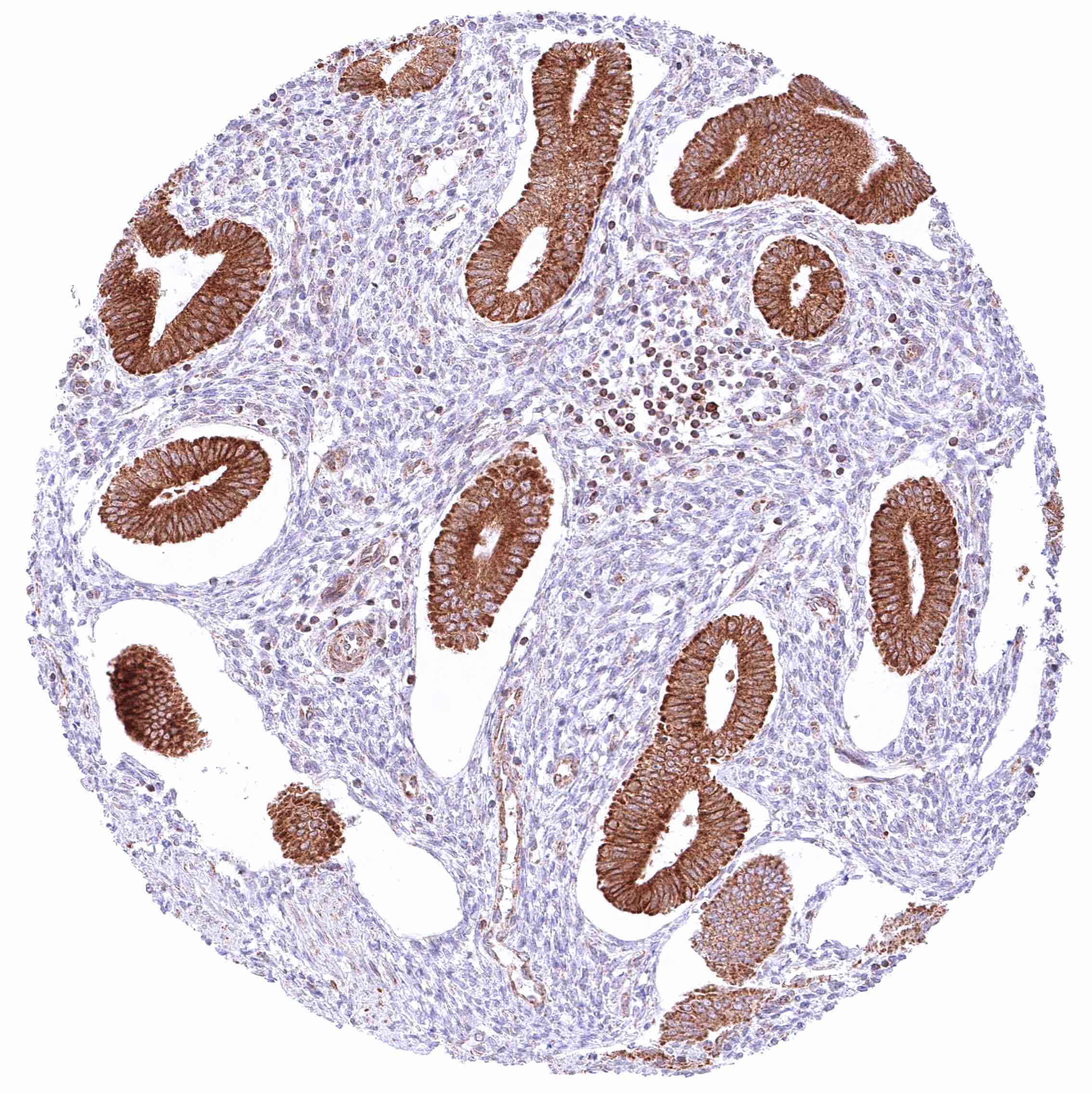
Uterus, endometrium (secretion) – Moderate to strong cytoplasmic BCL-XL staining of endometrial epithelial cells.

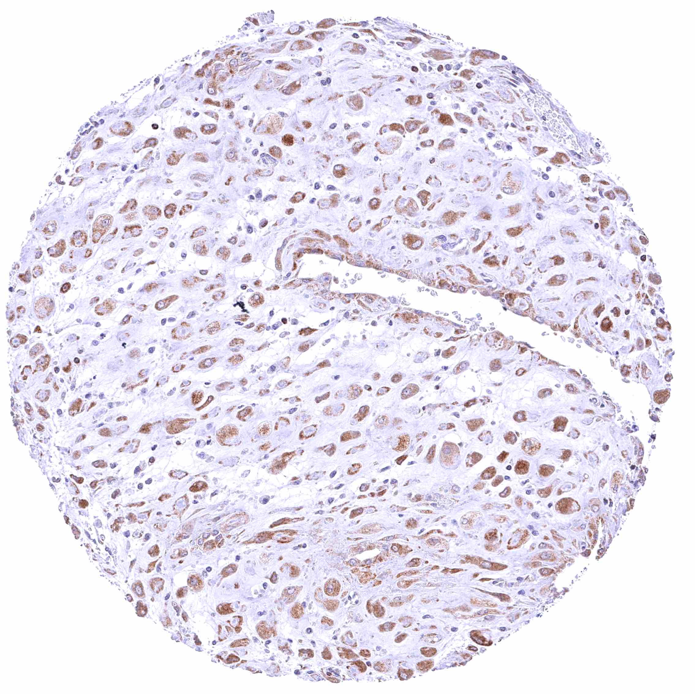
Uterus, endometrium (pregnancy) – Moderate to strong cytoplasmic BCL-XL staining of decidua cells.

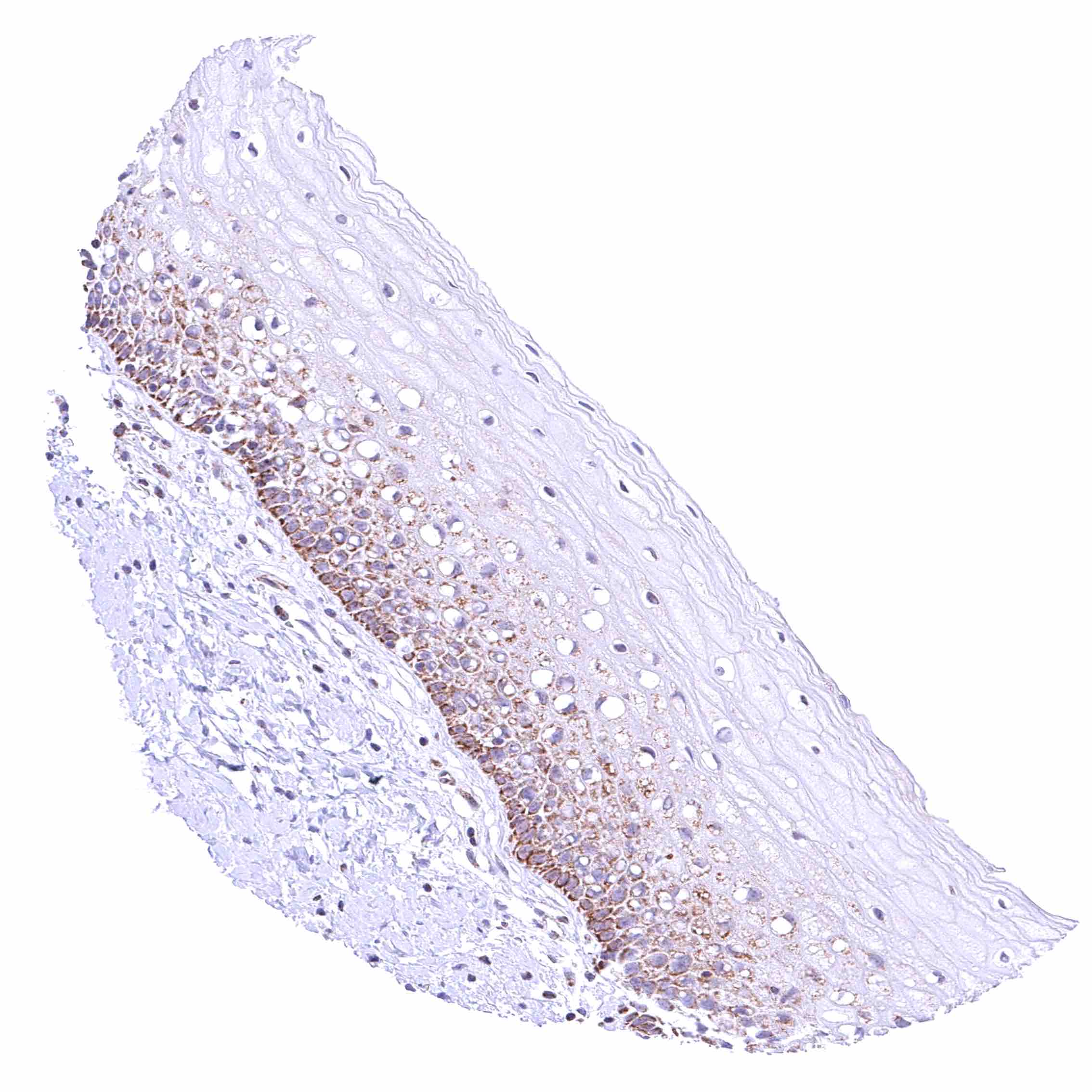
Uterus, ectocervix – Weak to moderate cytoplasmic BCL-XL staining of squamous epithelial cells of basal and suprabasal cell layers (marked decrease of intensity towards the surface).


Uterus, endometrium (proliferation) – Strong cytoplasmic BCL-XL staining of endometrial epithelial cells.

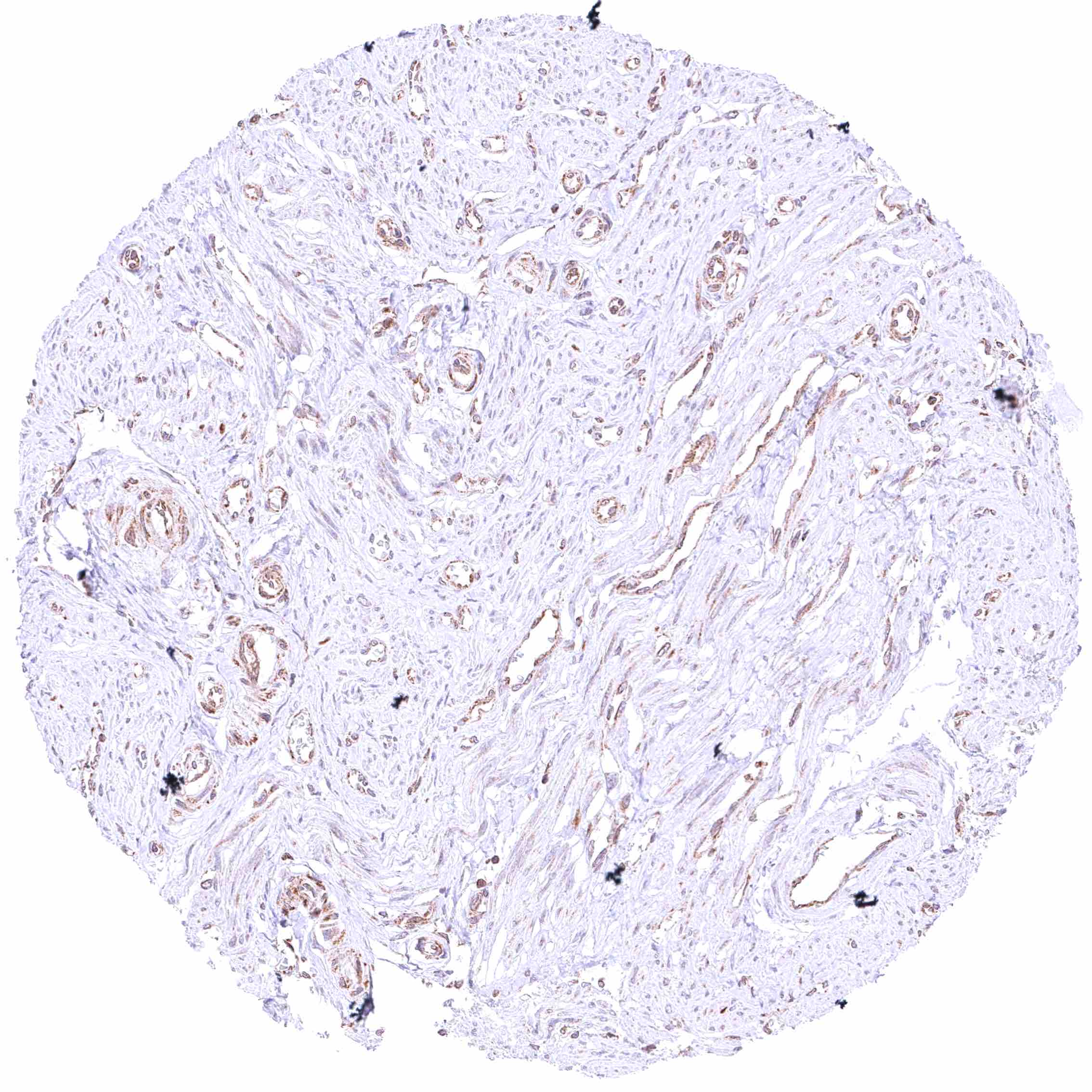
Uterus, myometrium – Faint cytoplasmic BCL-XL staining of myometrium cells.


| TOP |