Normal Tissue Gallery- GTX04967

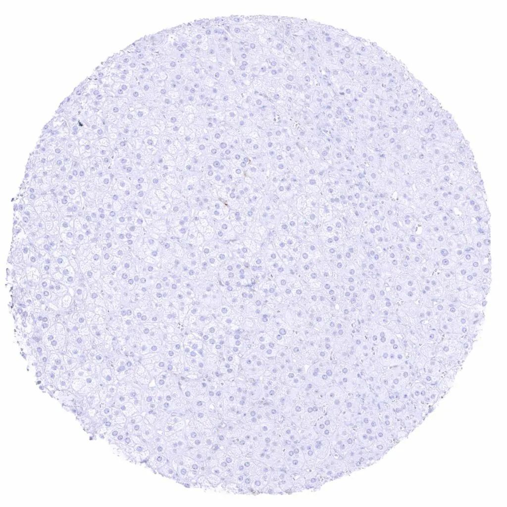
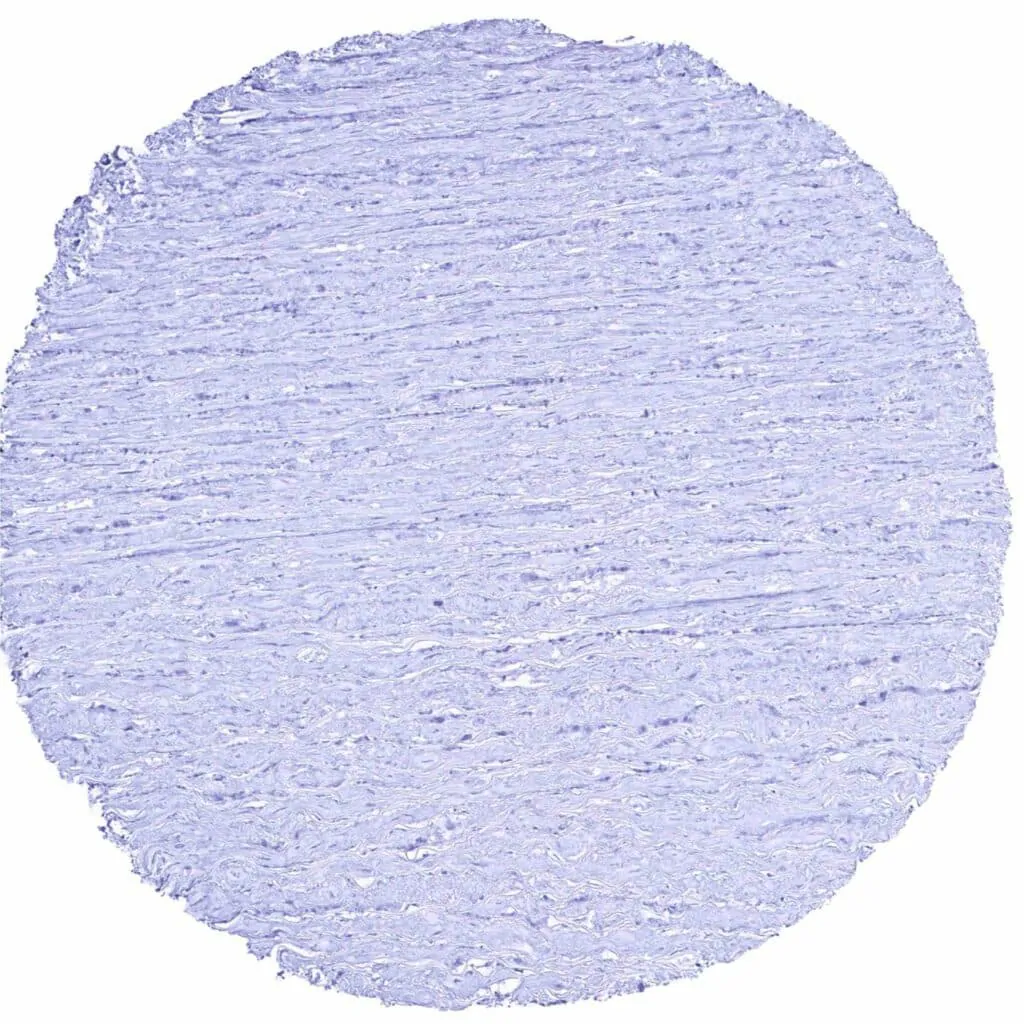
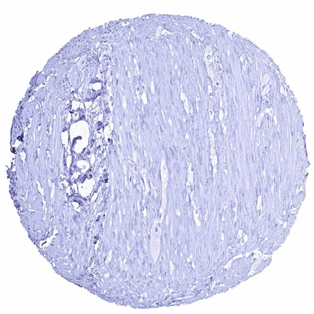
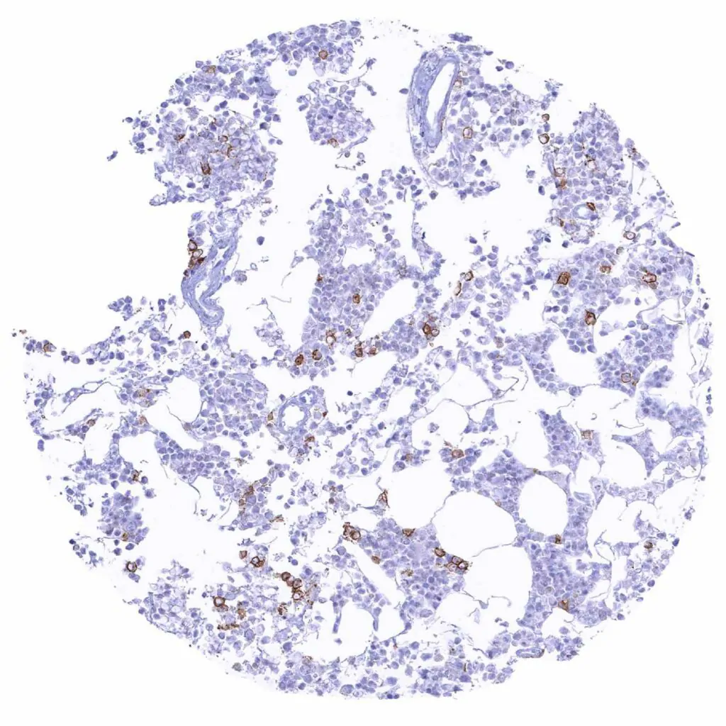
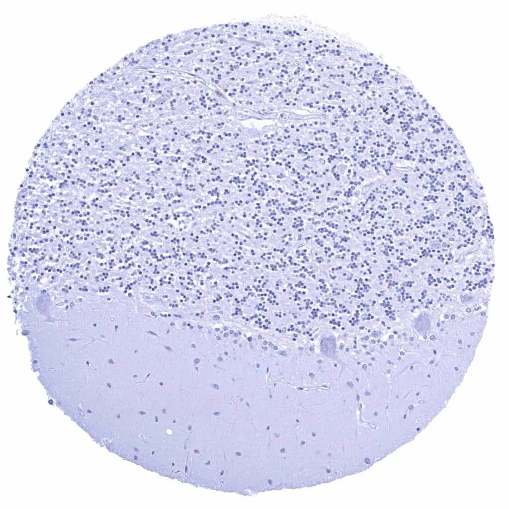
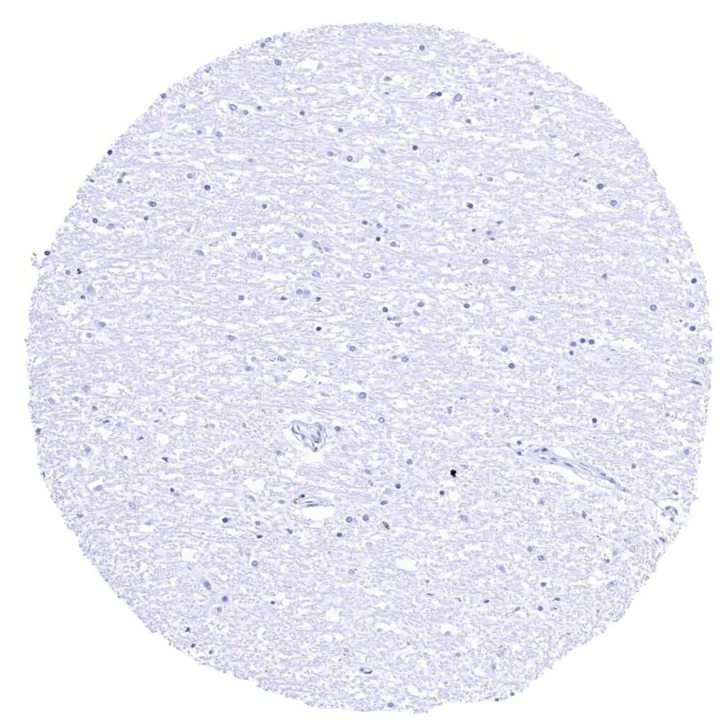
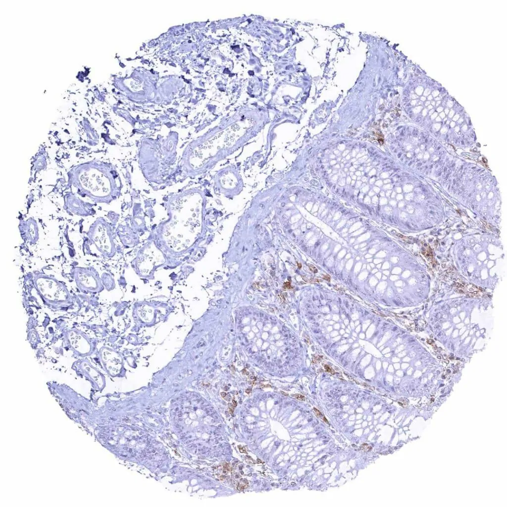
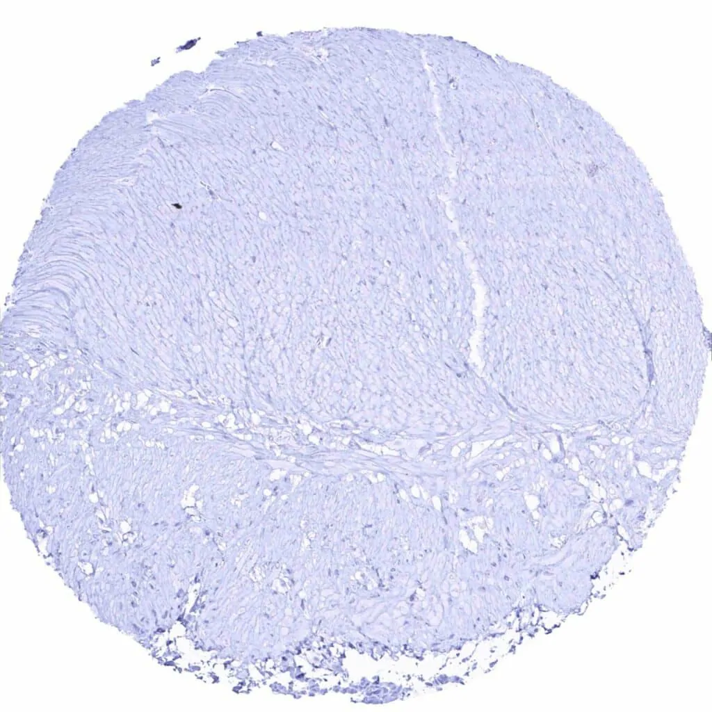
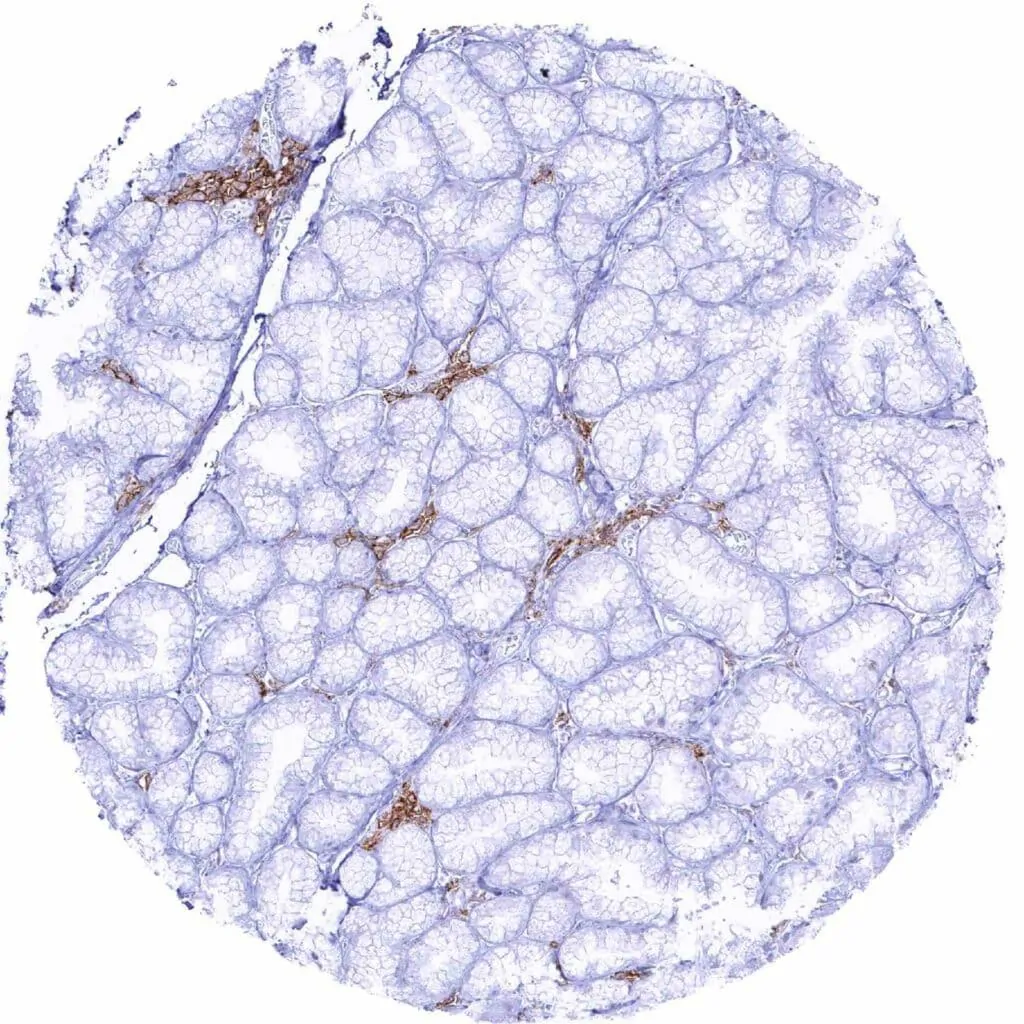
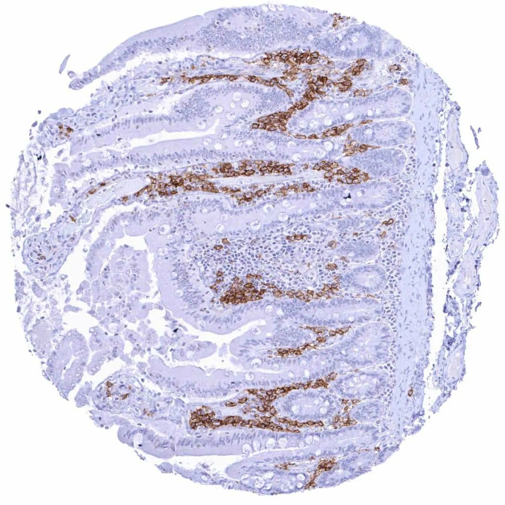
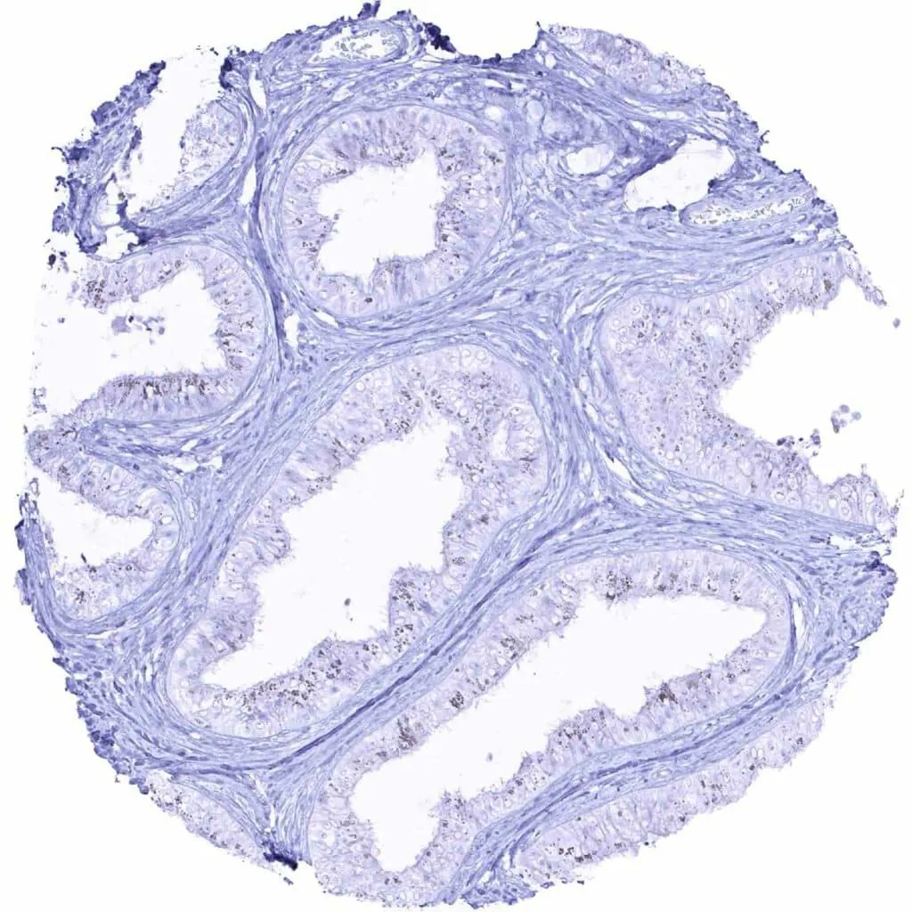
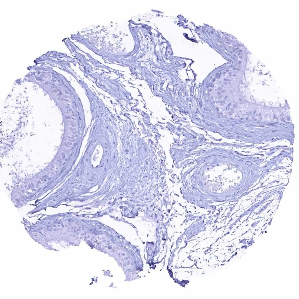
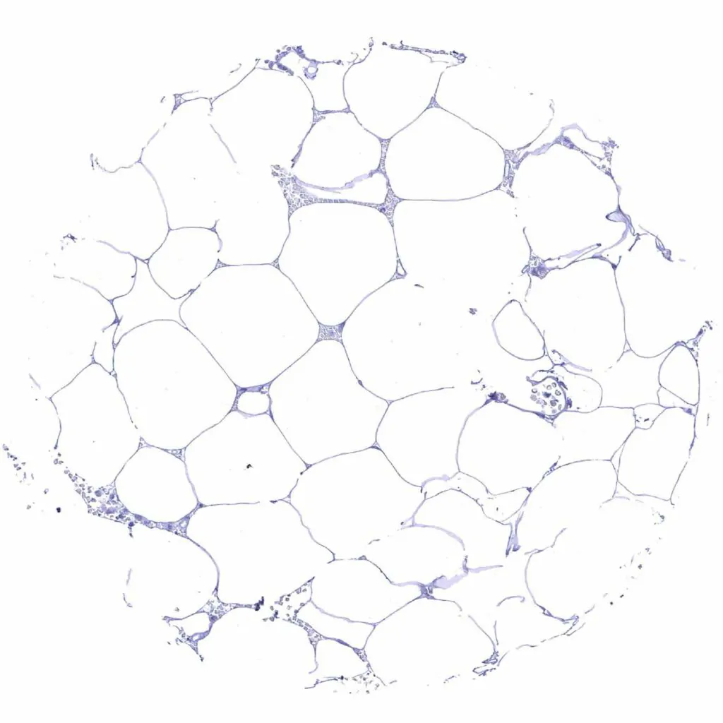
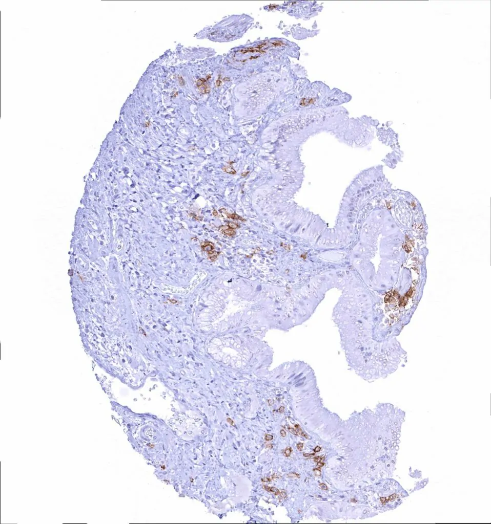
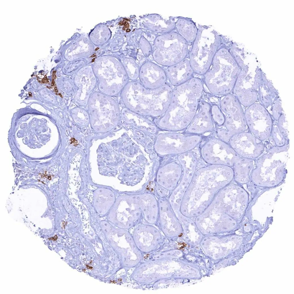
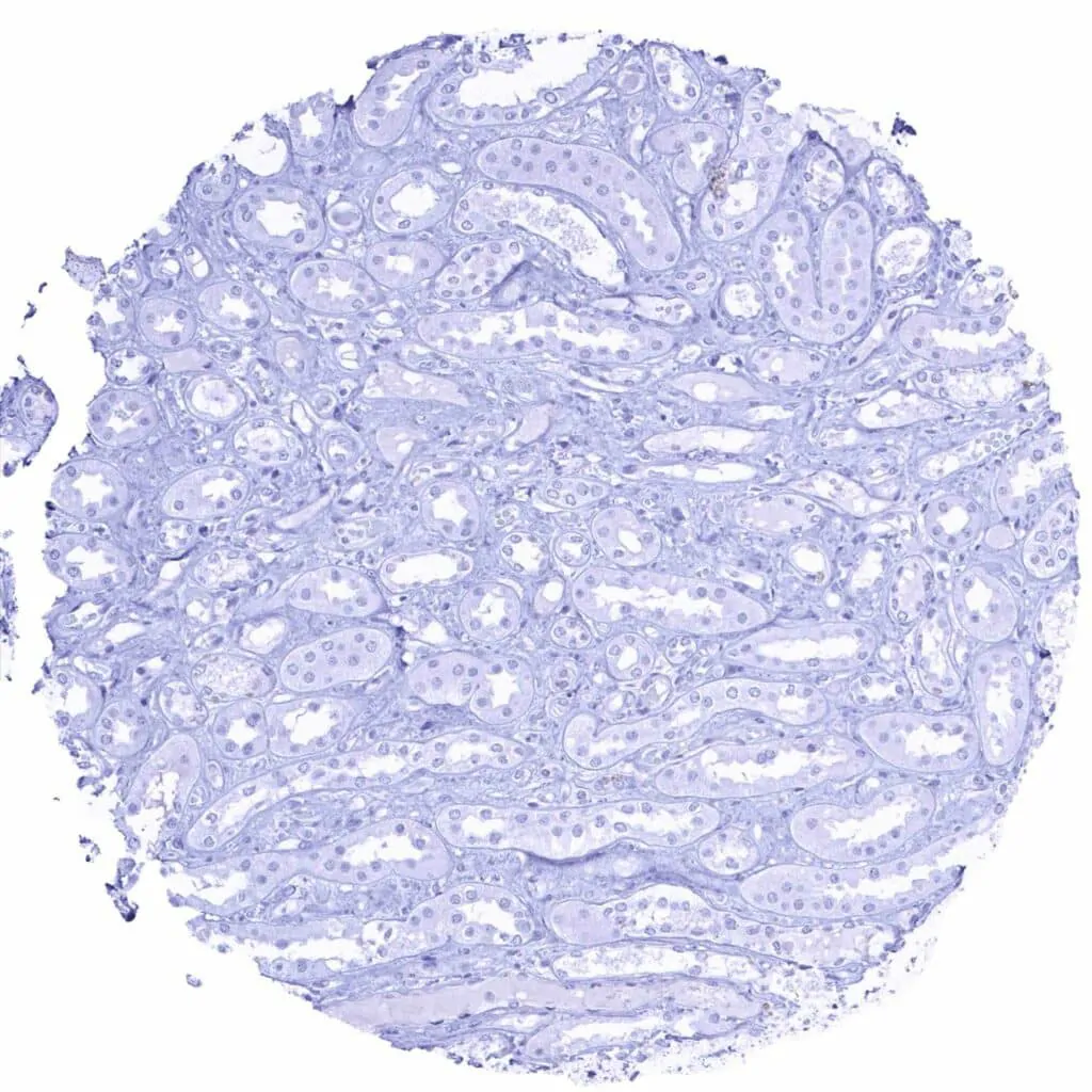
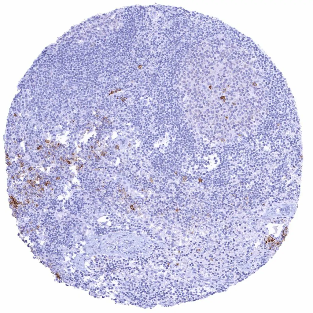
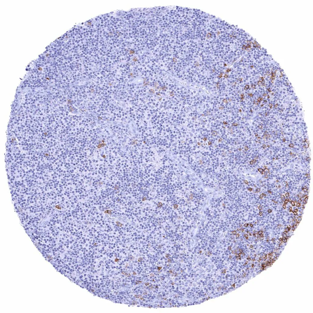
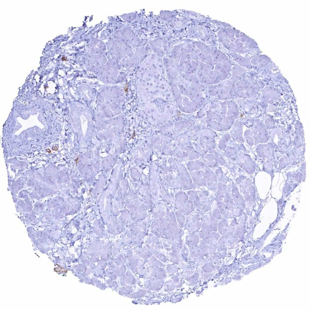
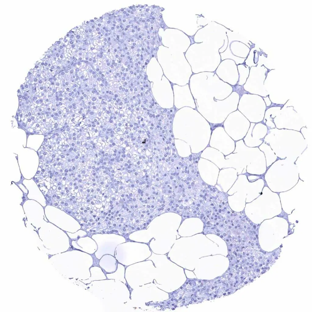
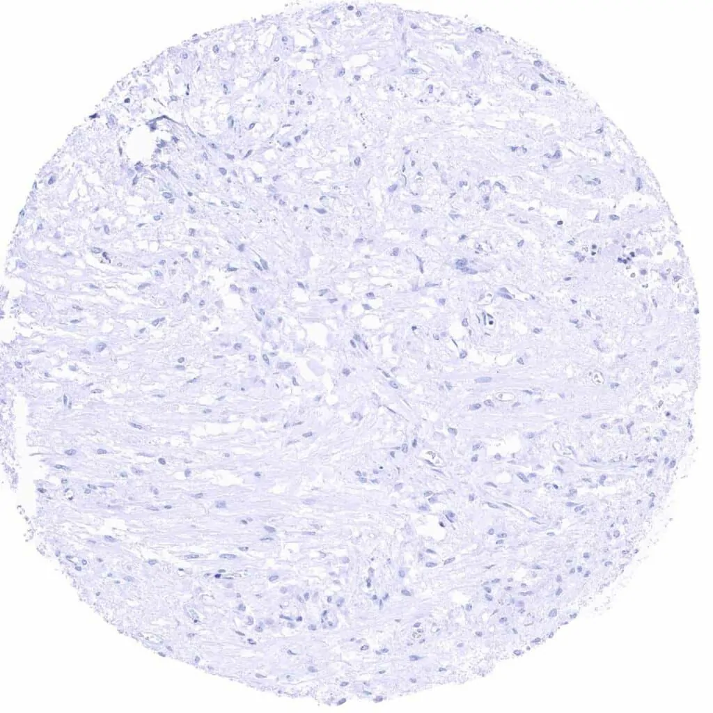
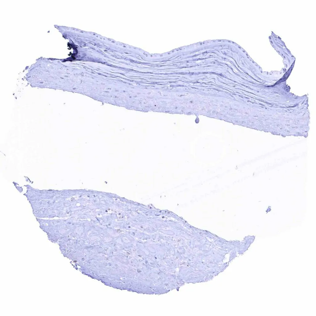
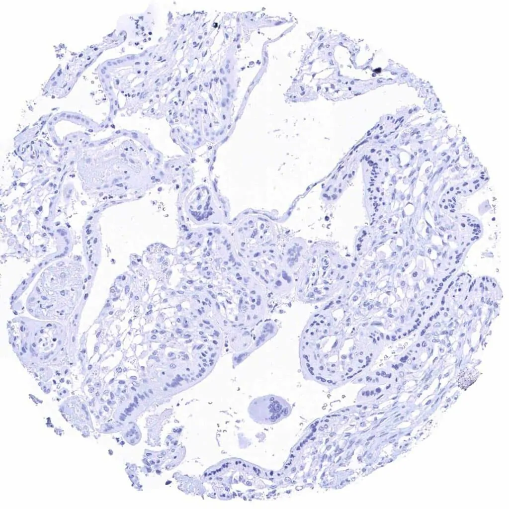
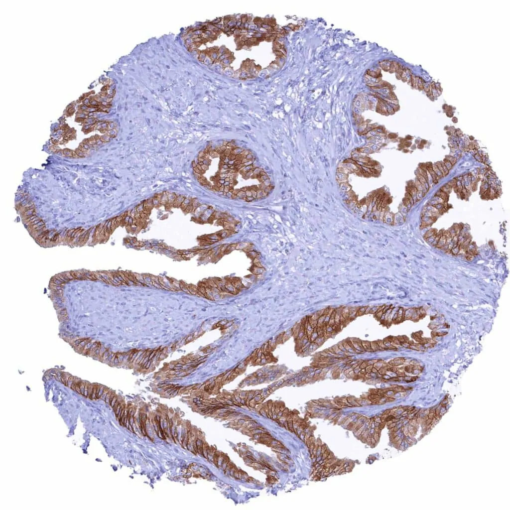
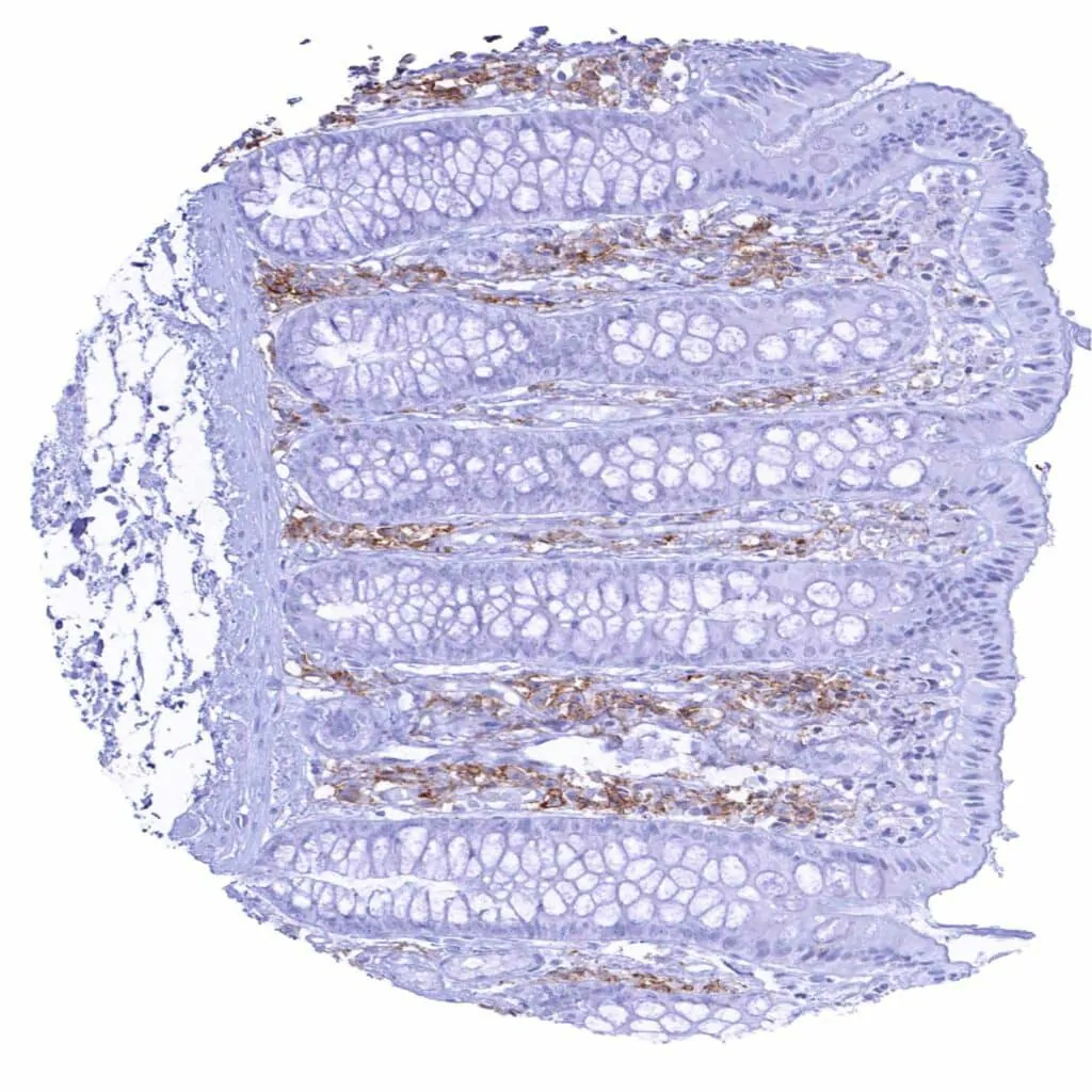
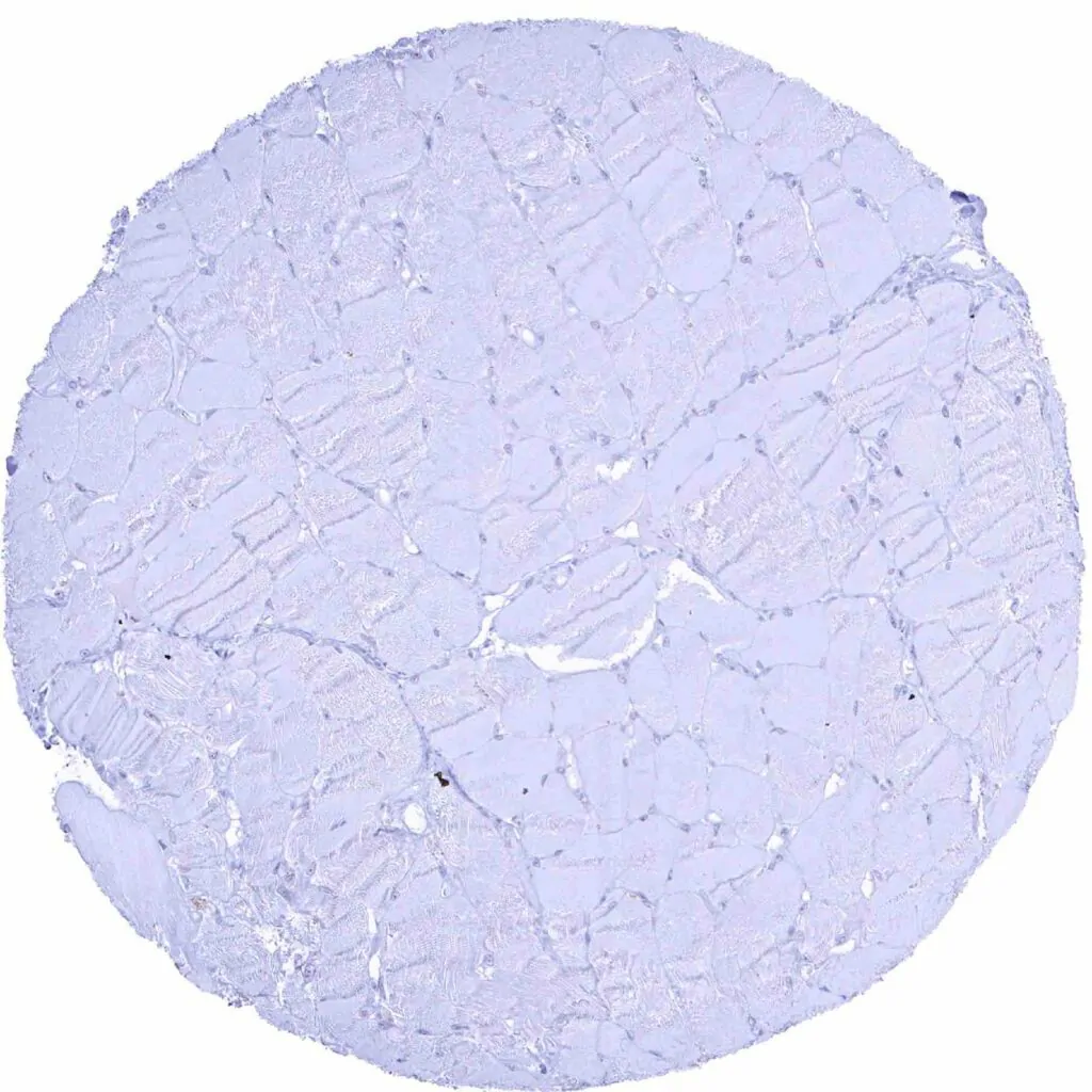
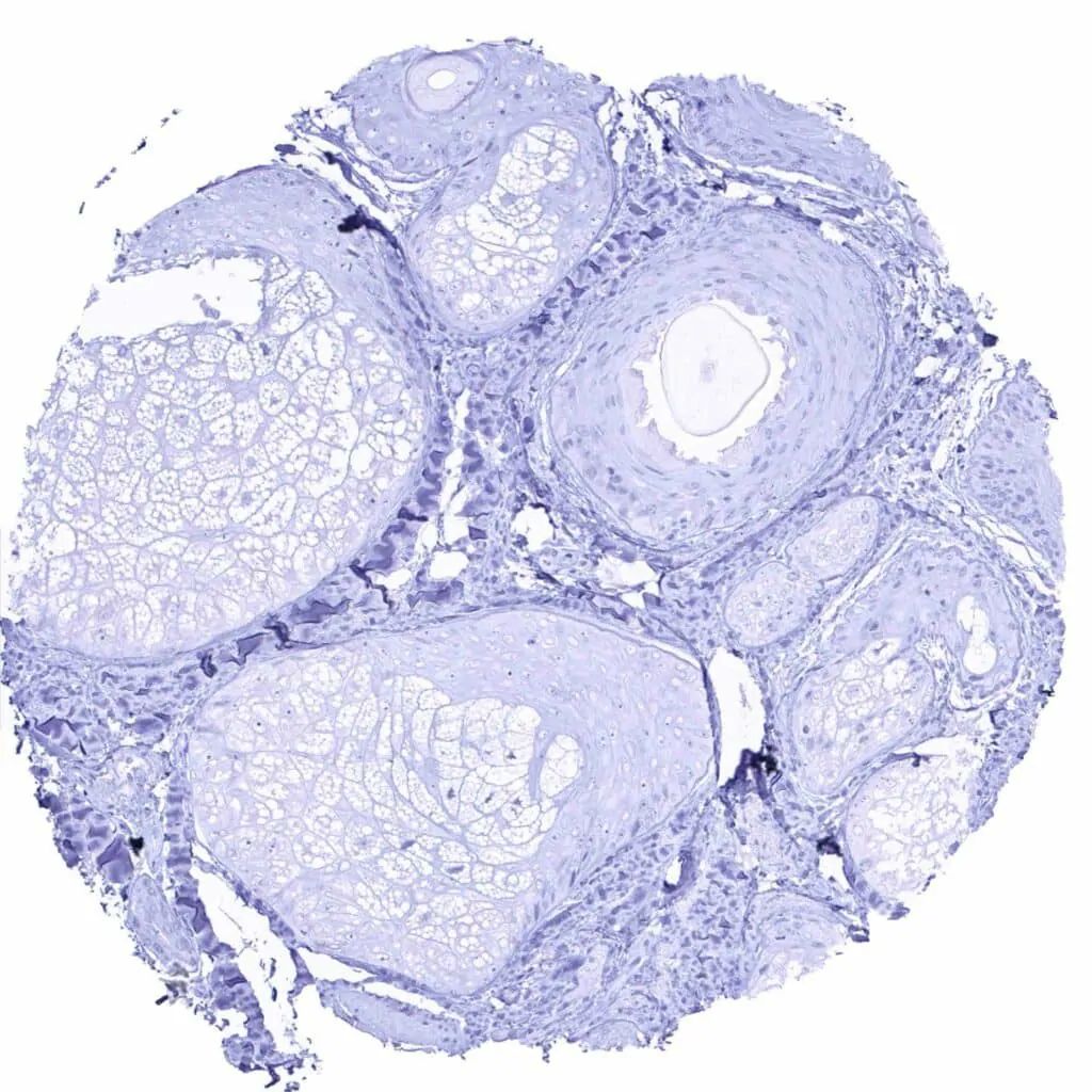
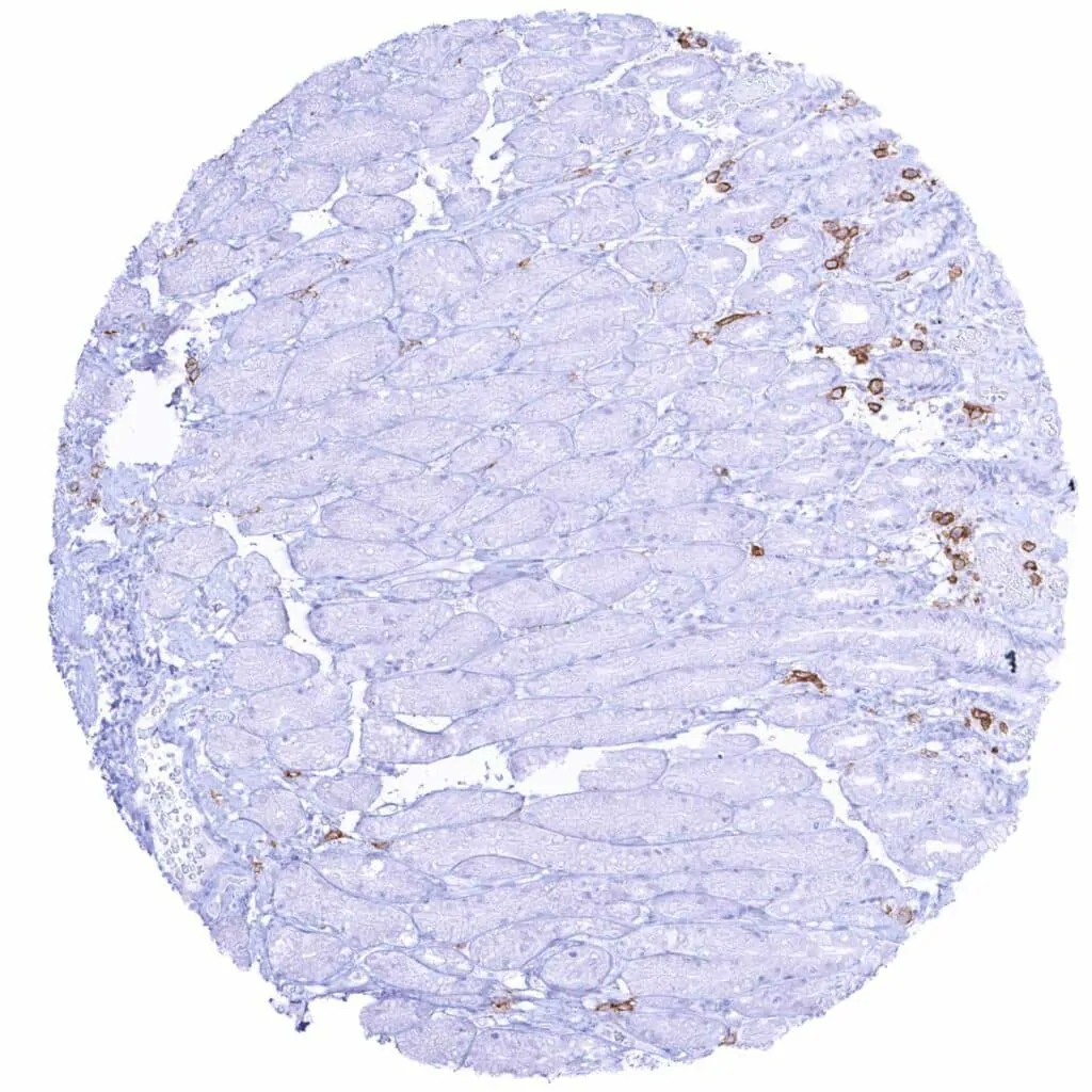
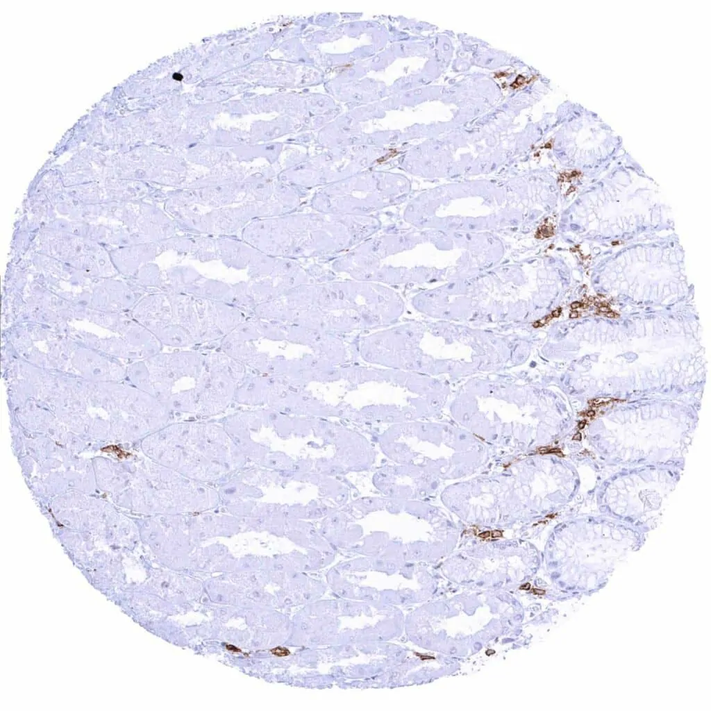
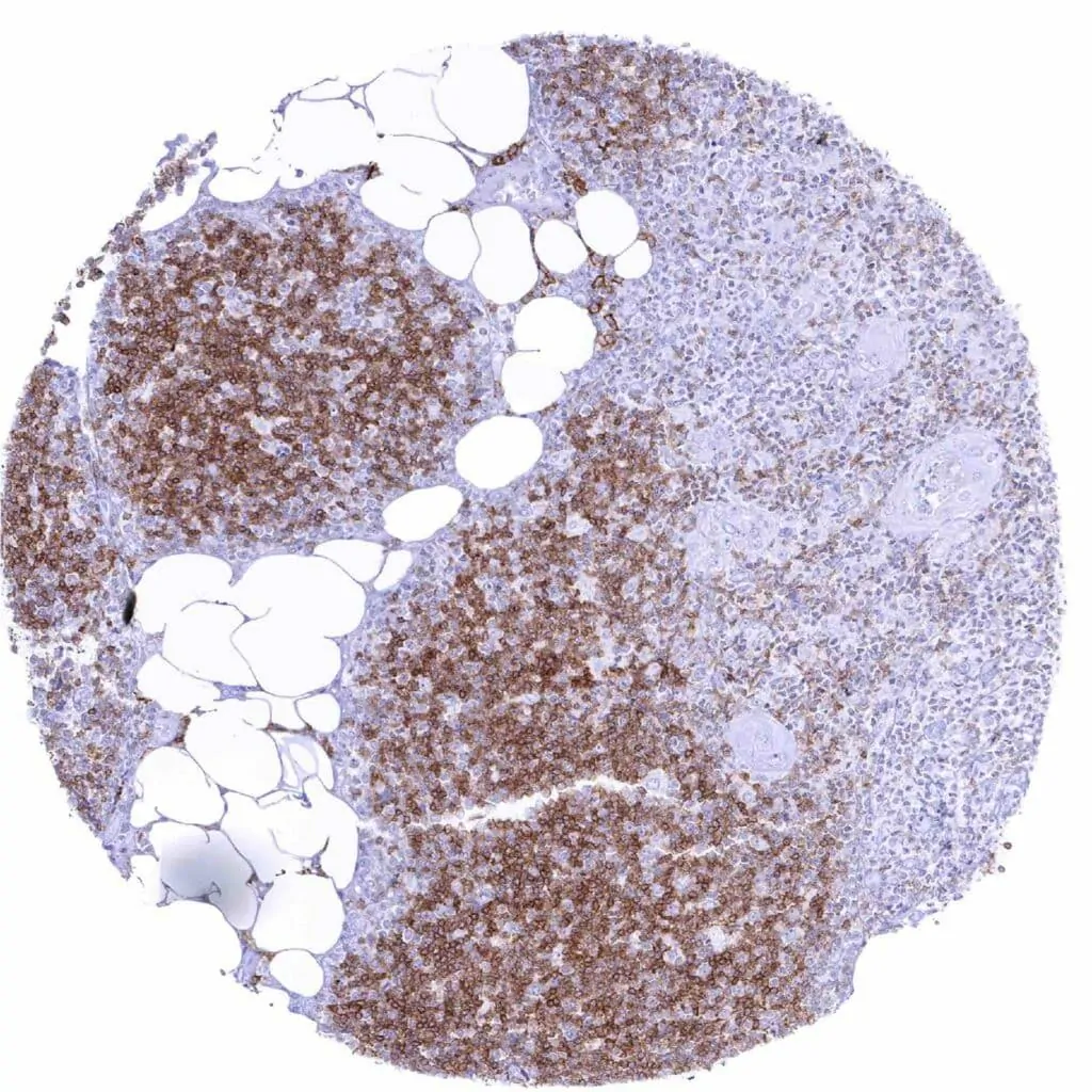
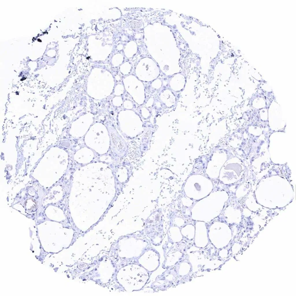
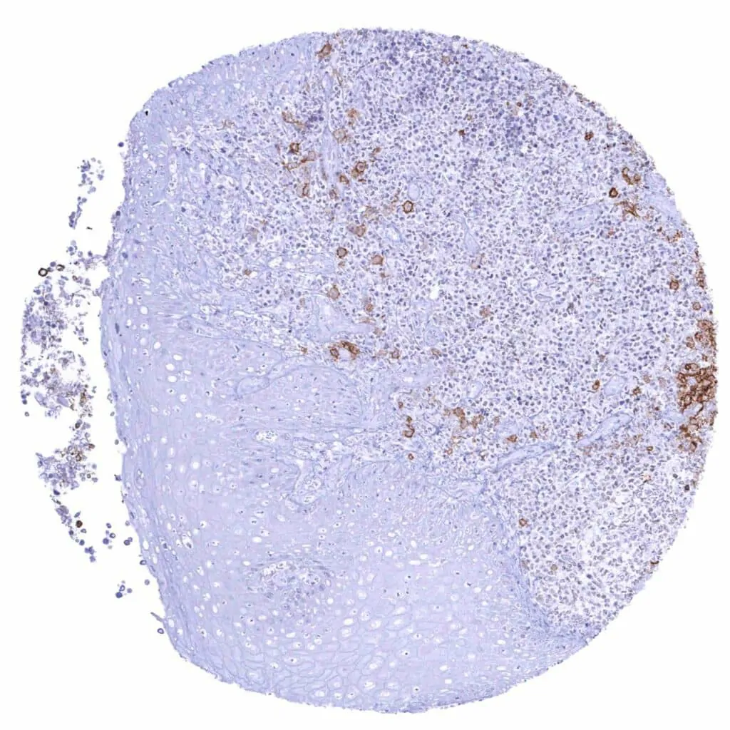
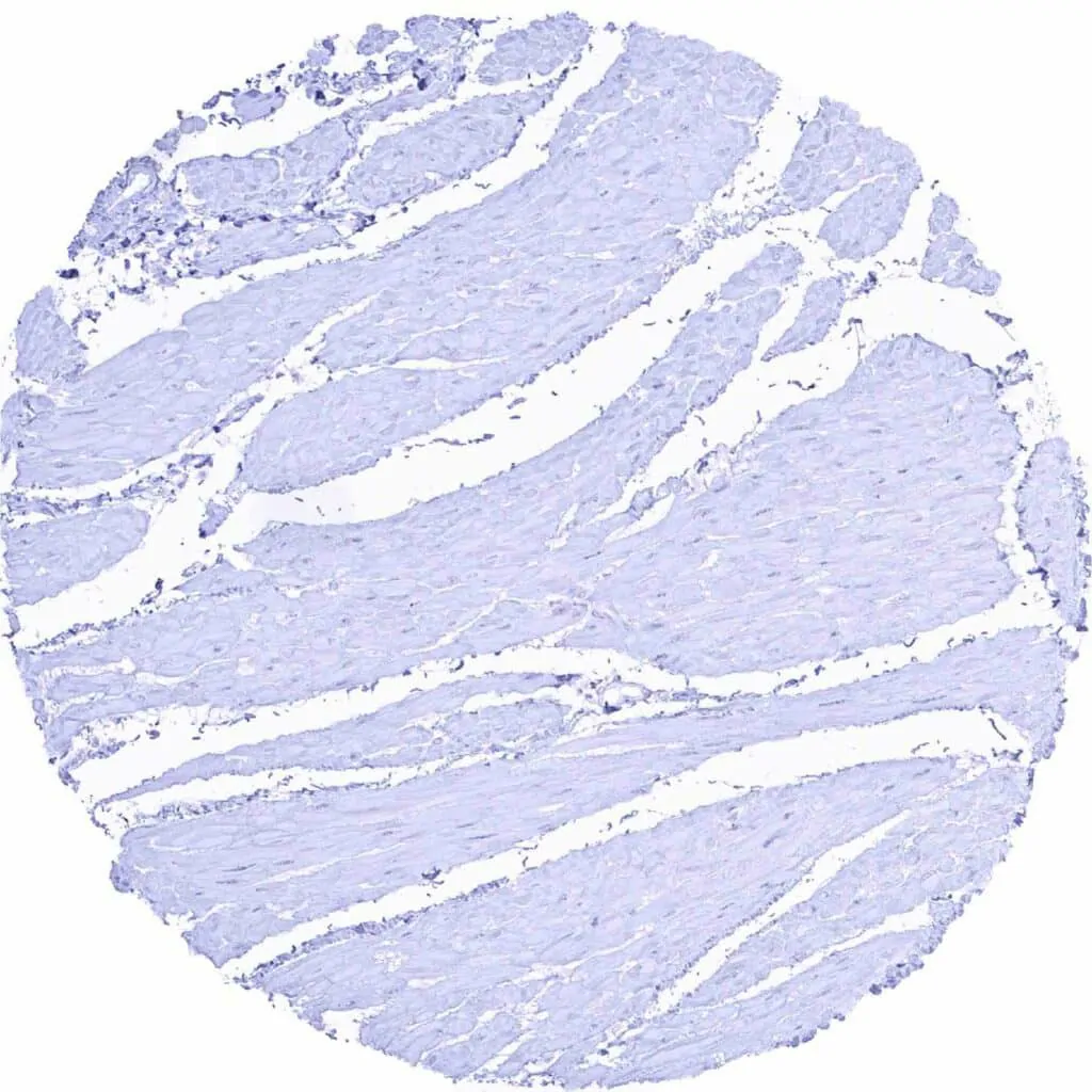
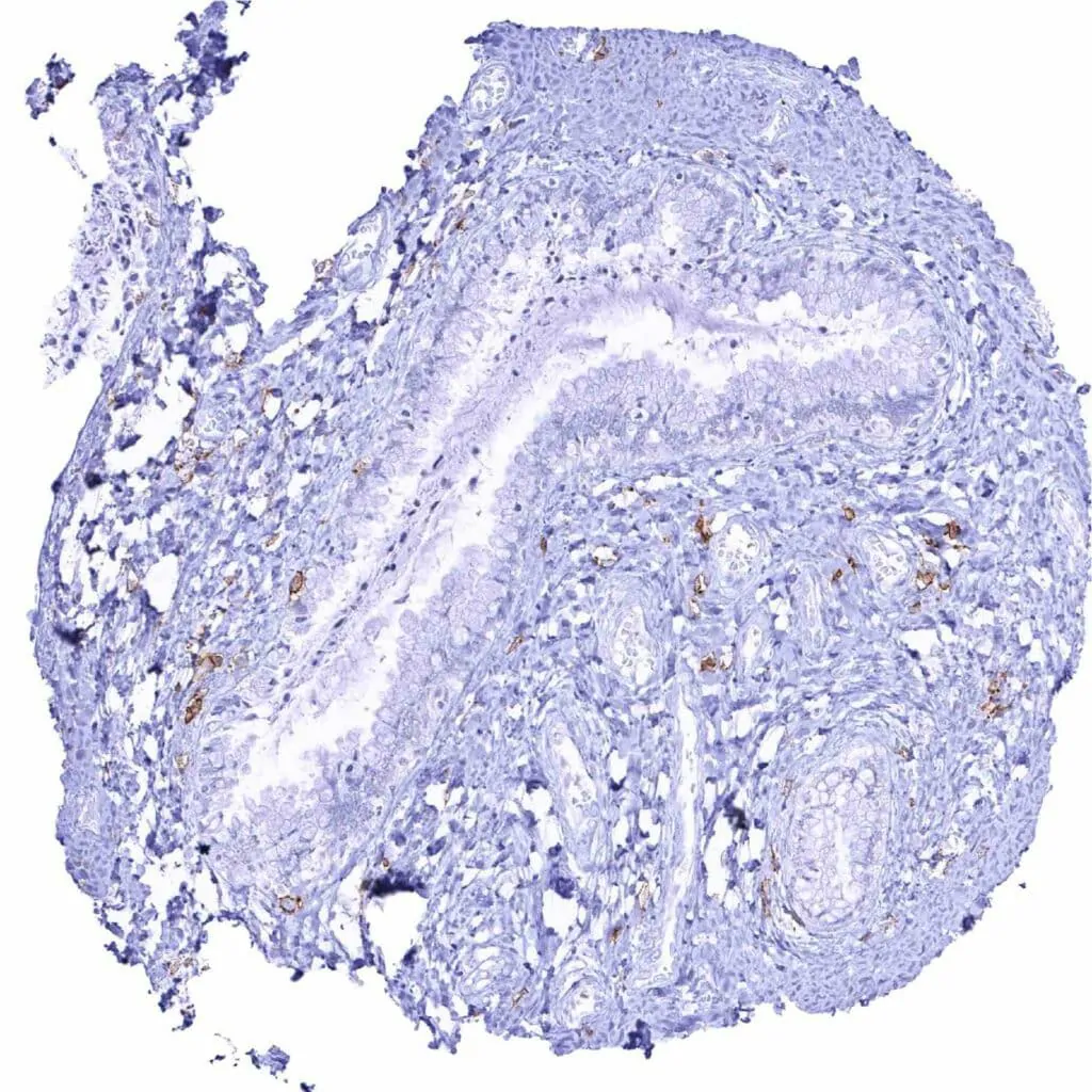
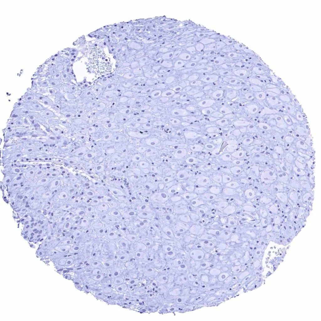
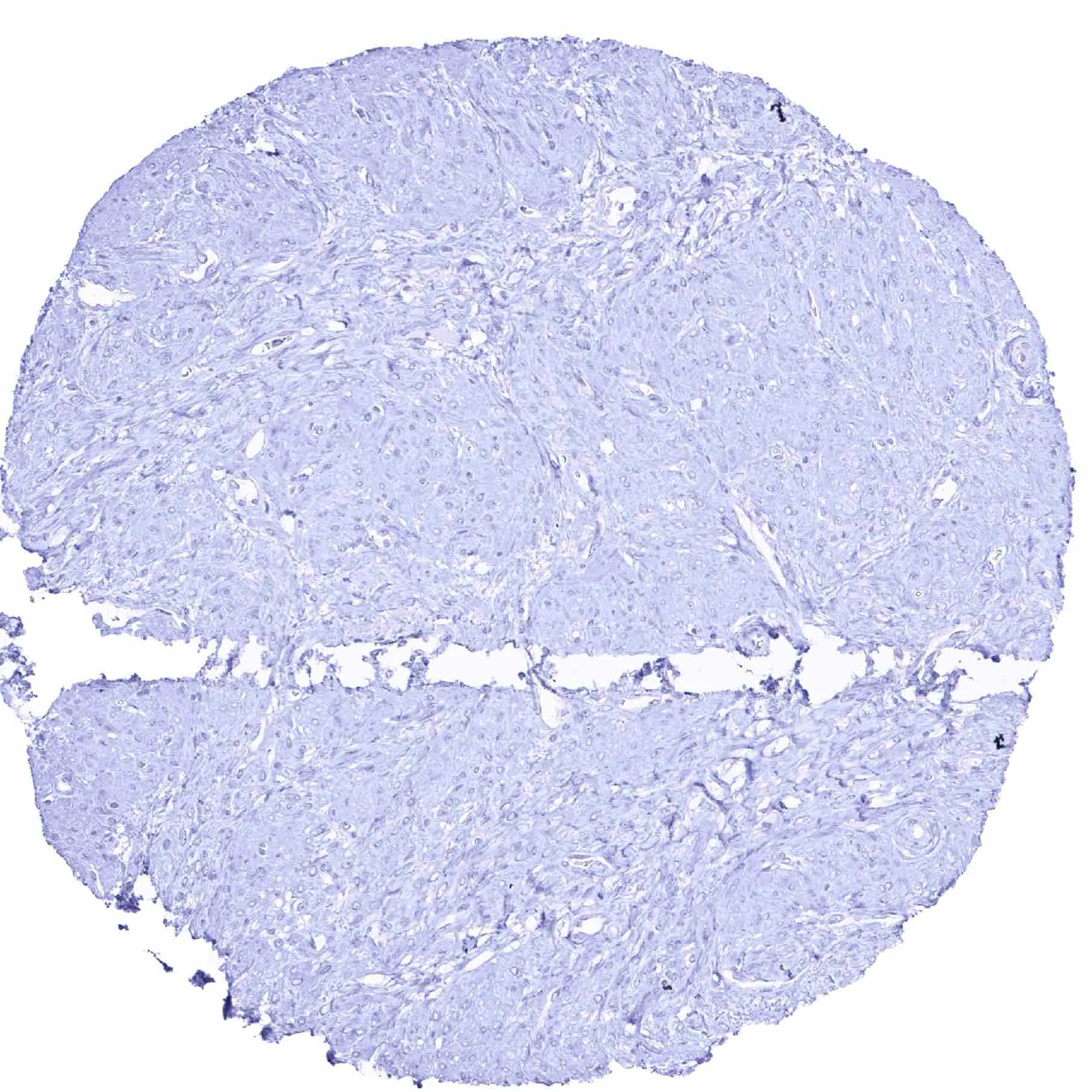
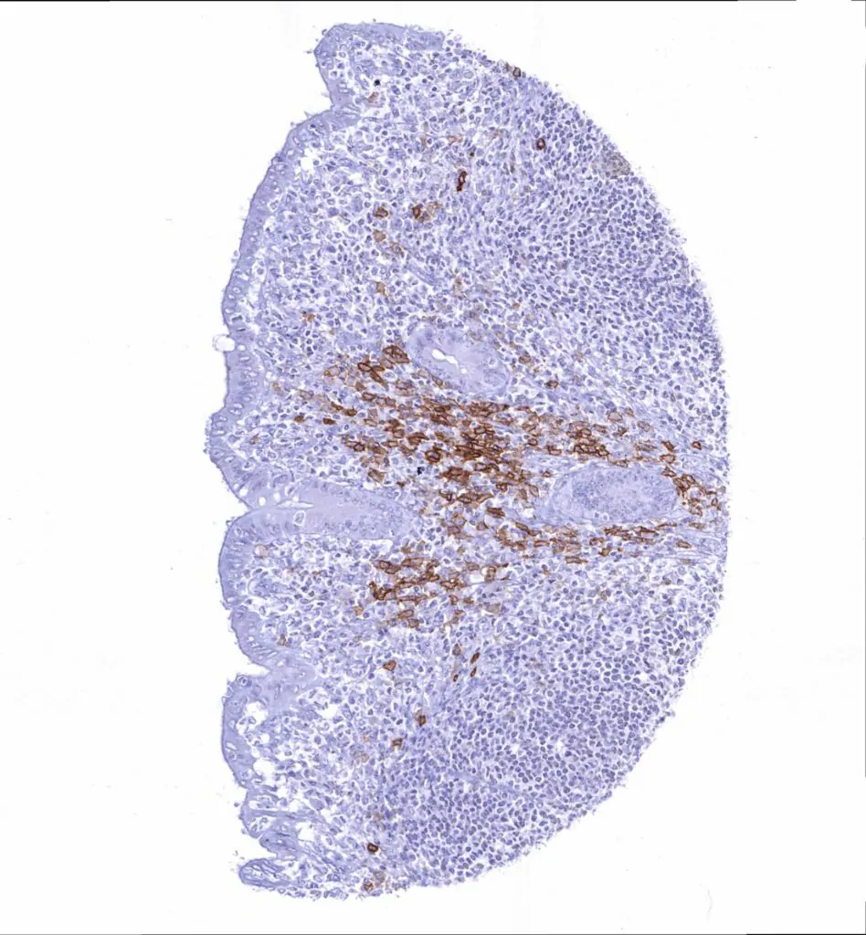
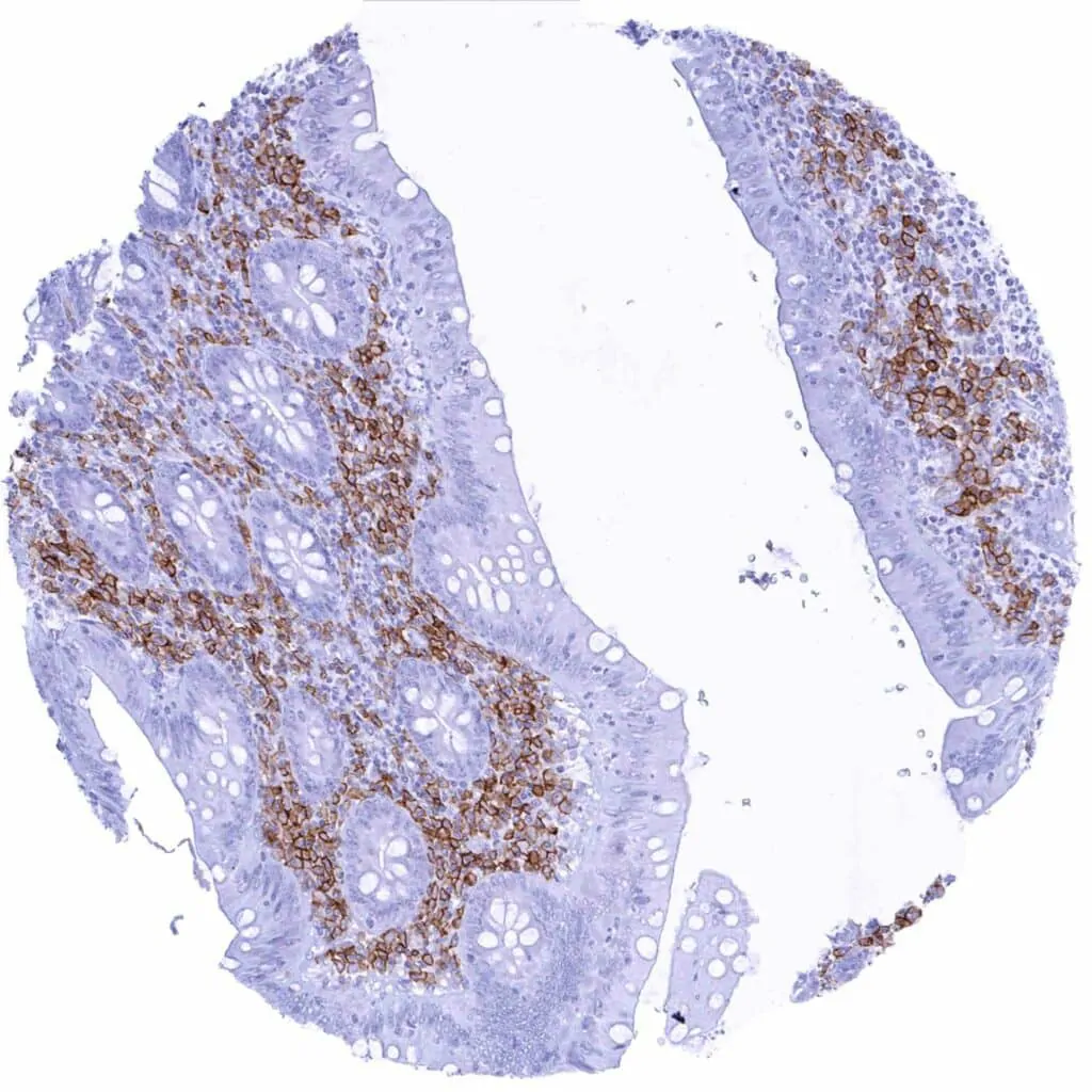
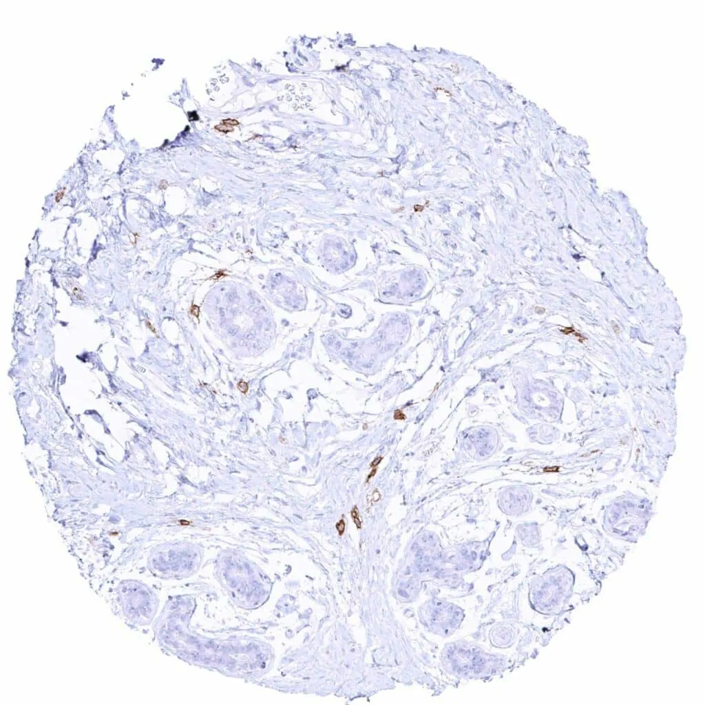
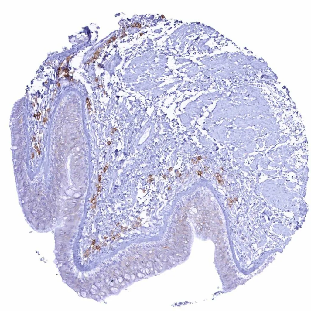
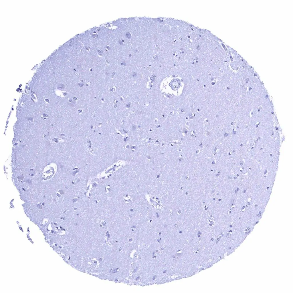
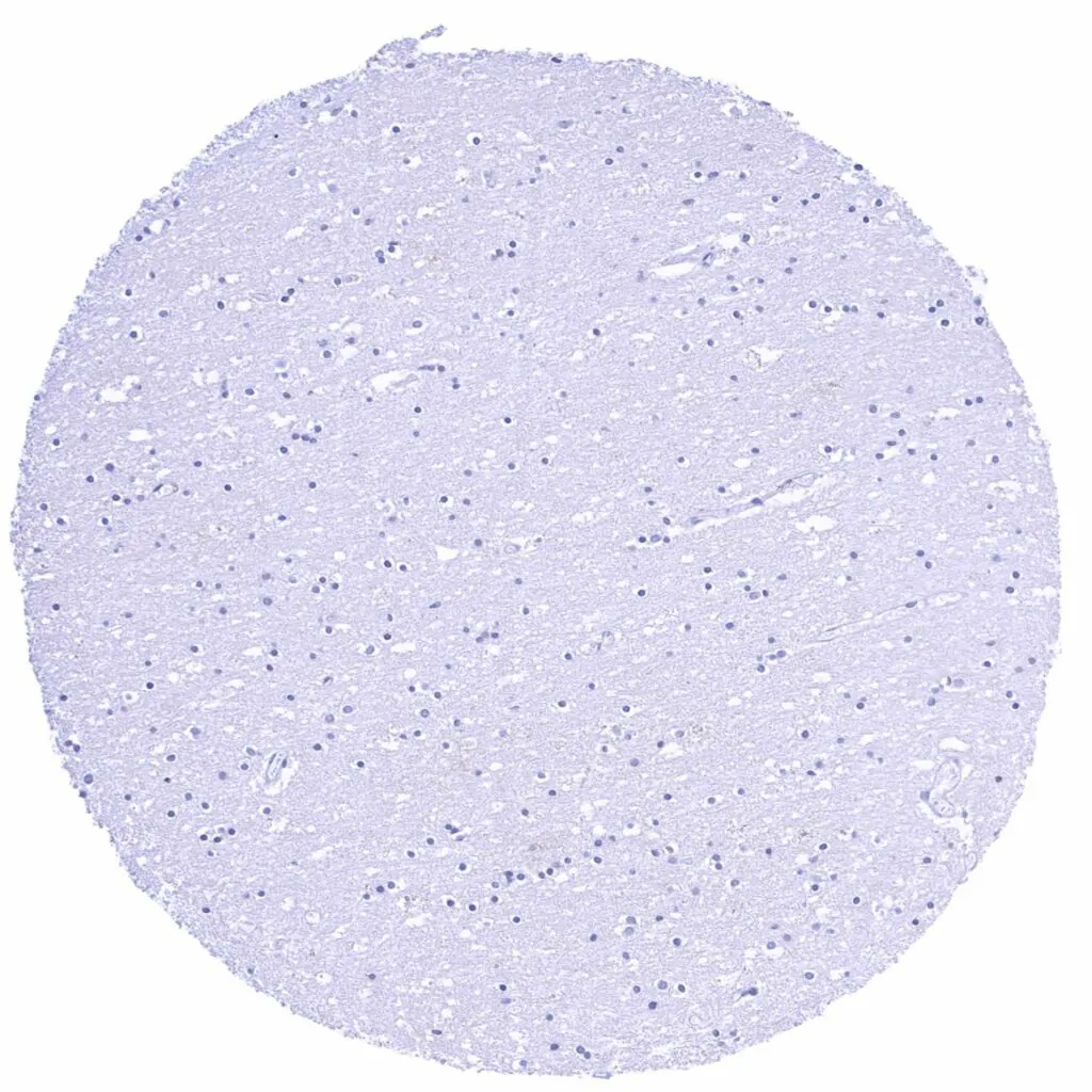
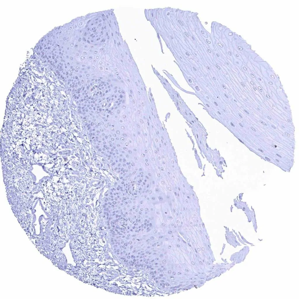
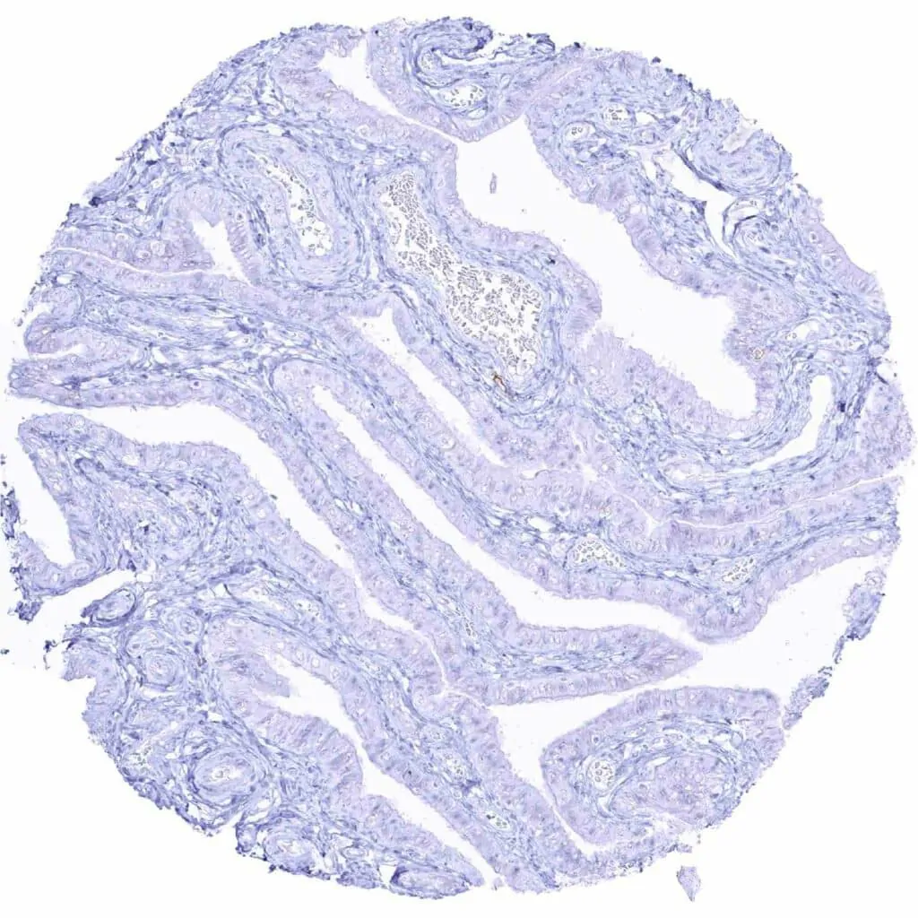
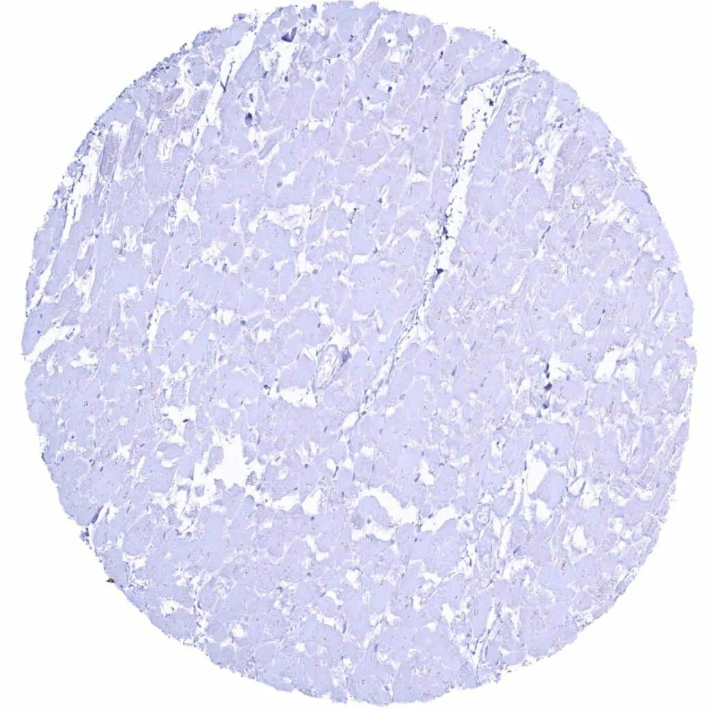
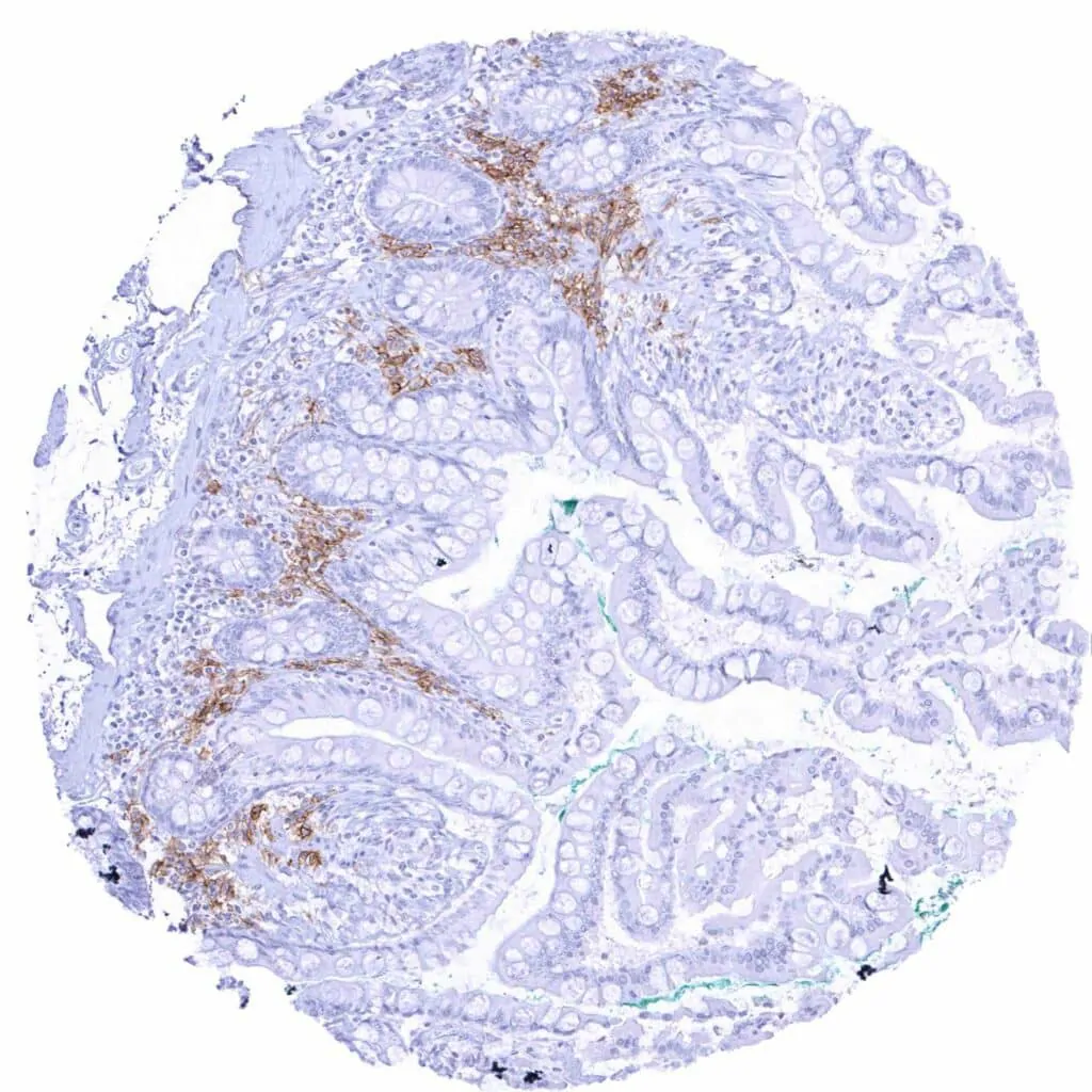
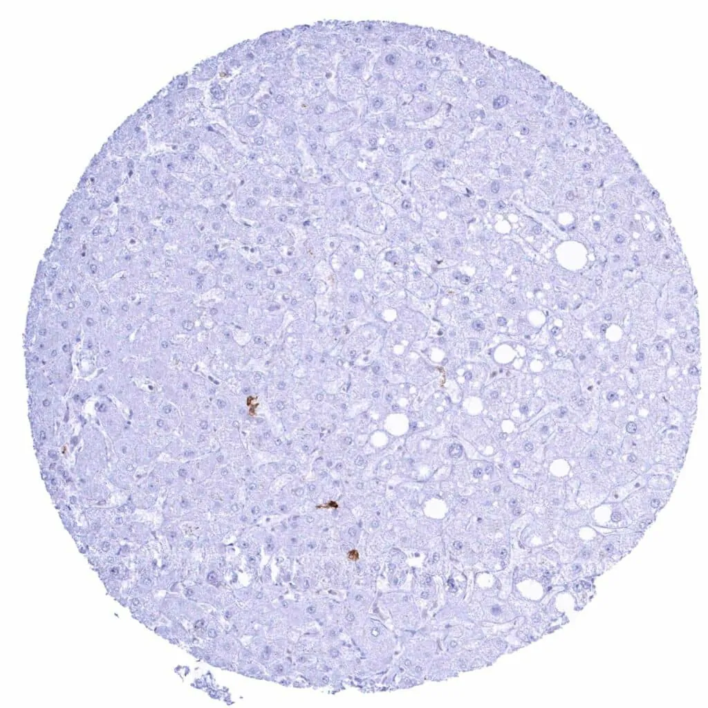
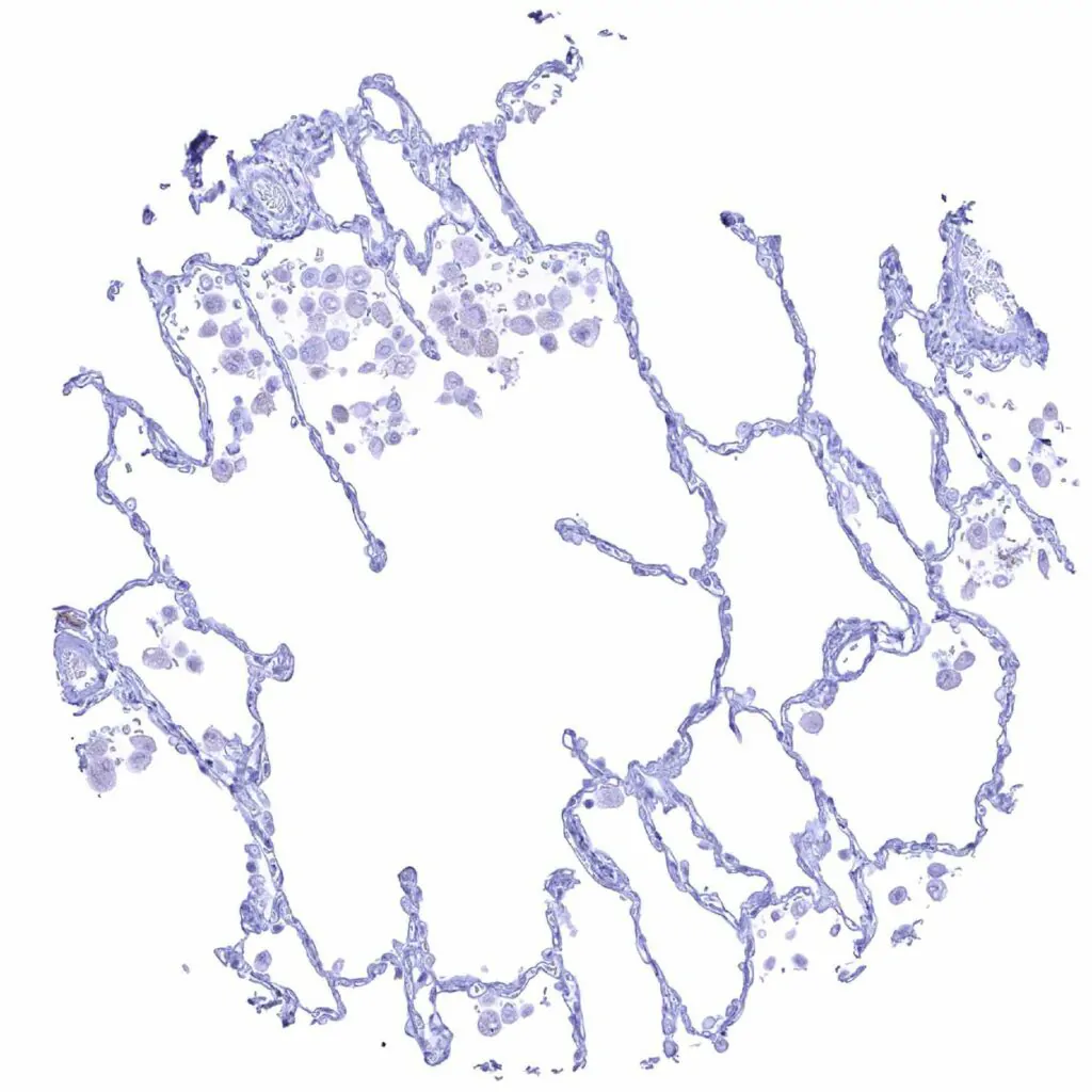
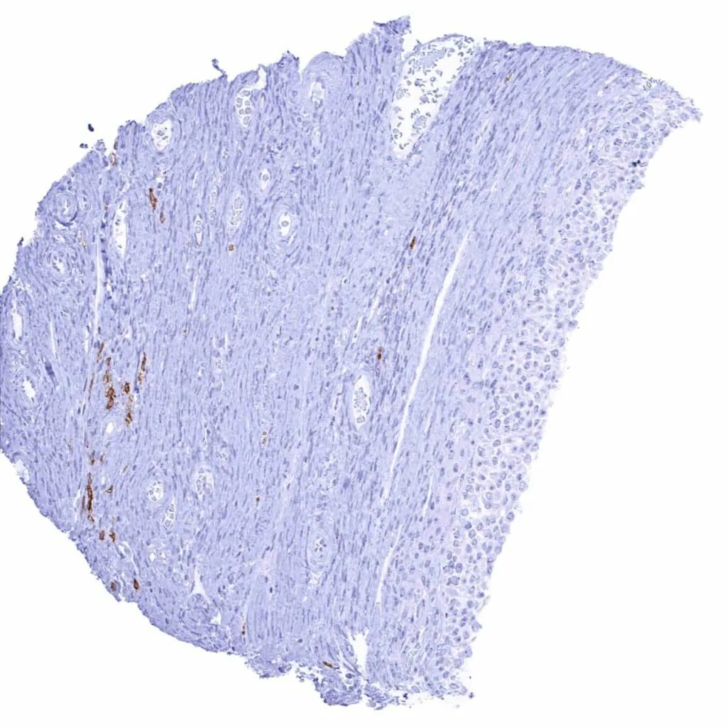
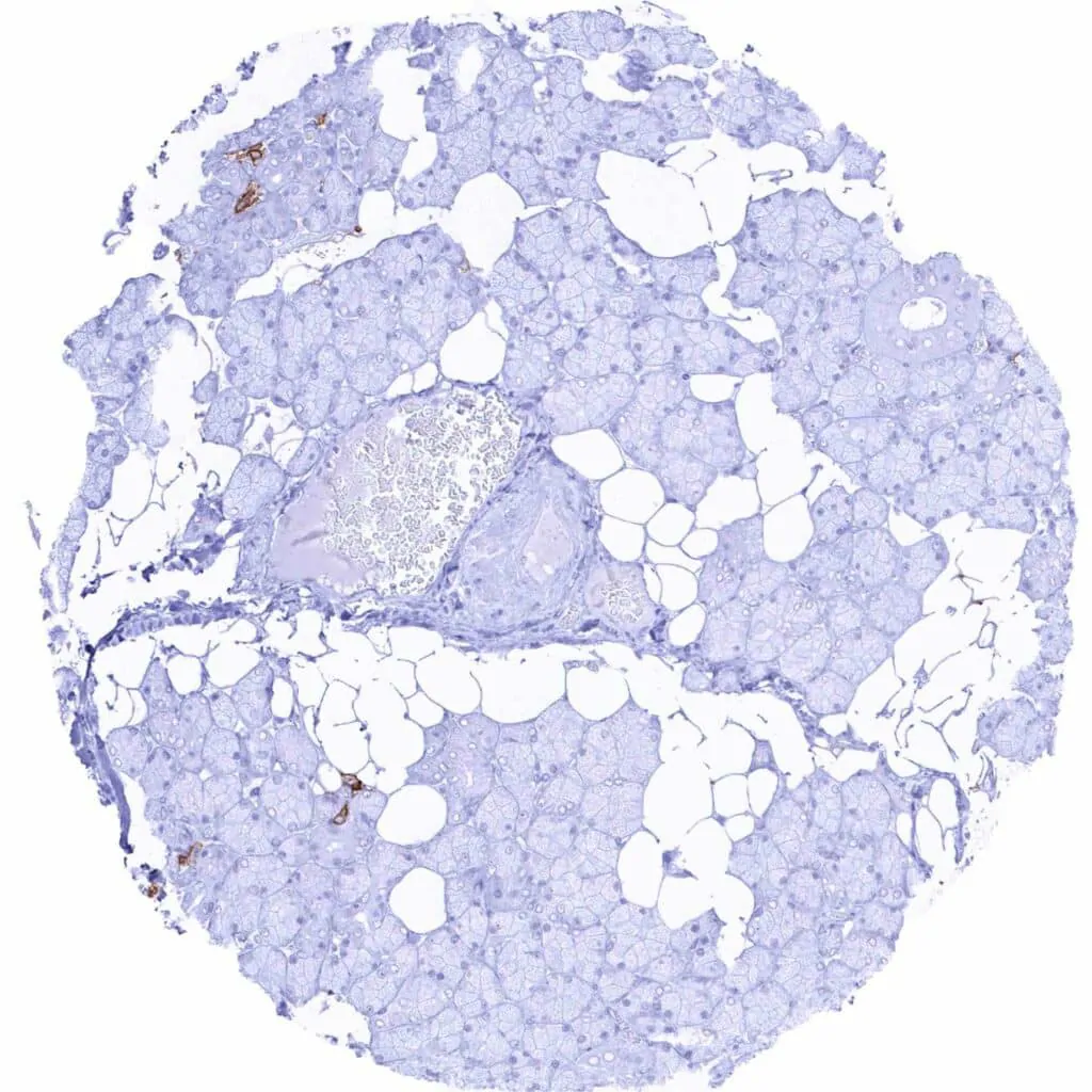
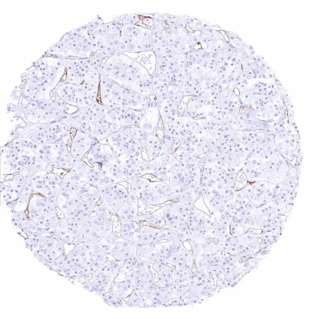
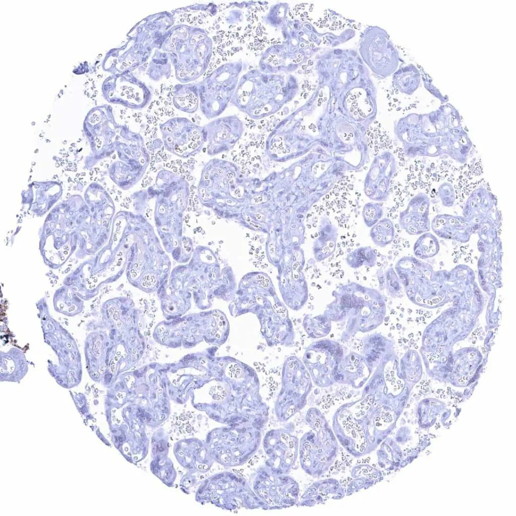
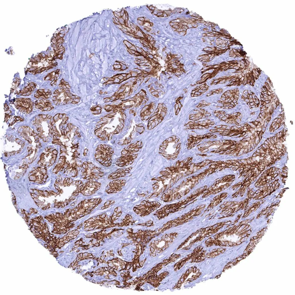
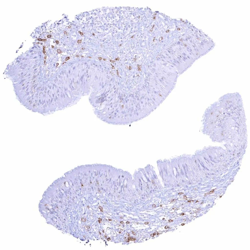
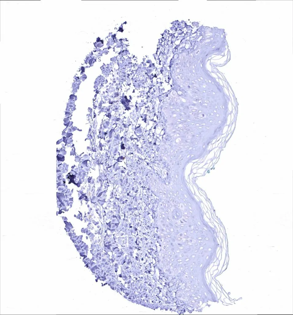
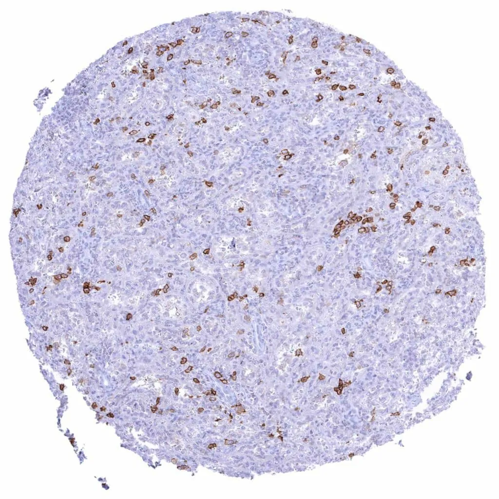
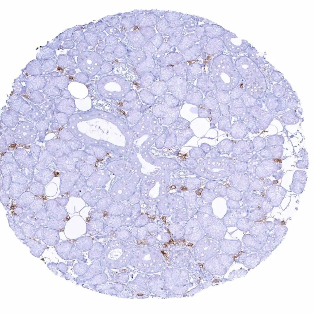
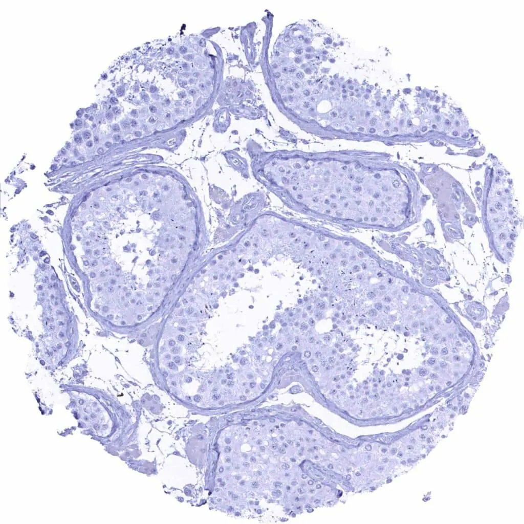
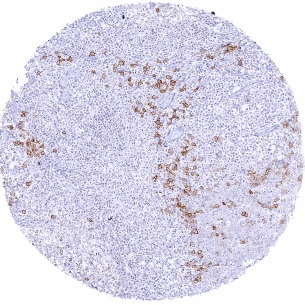
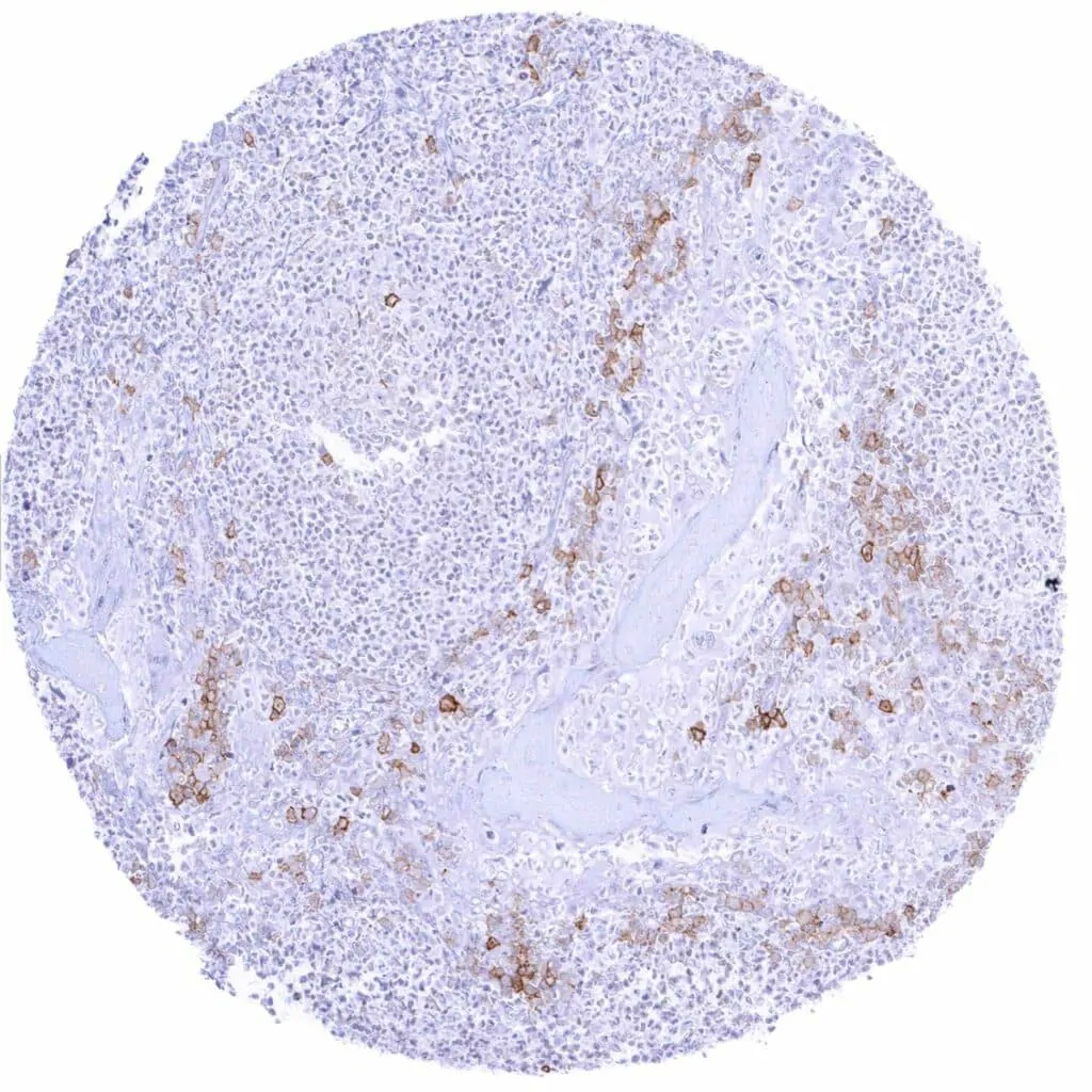
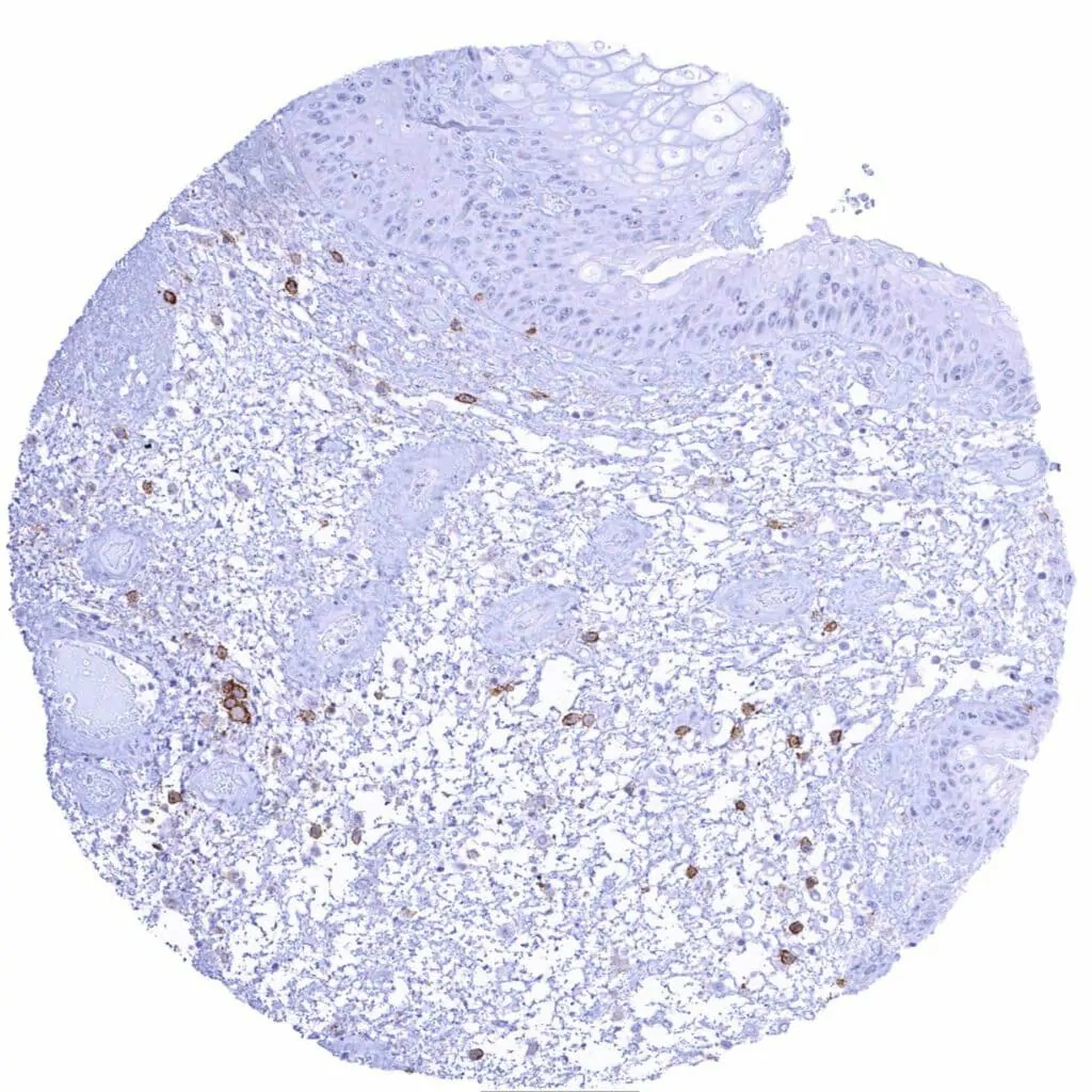
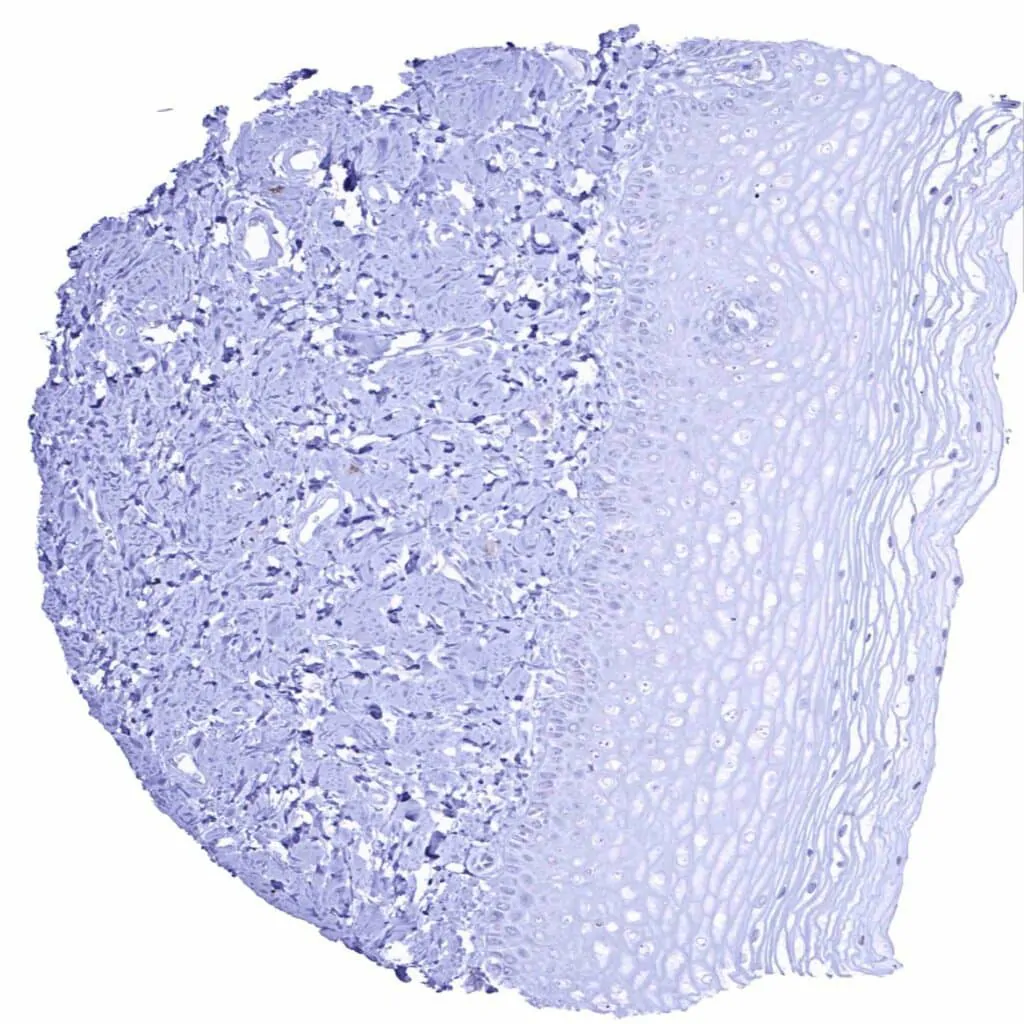
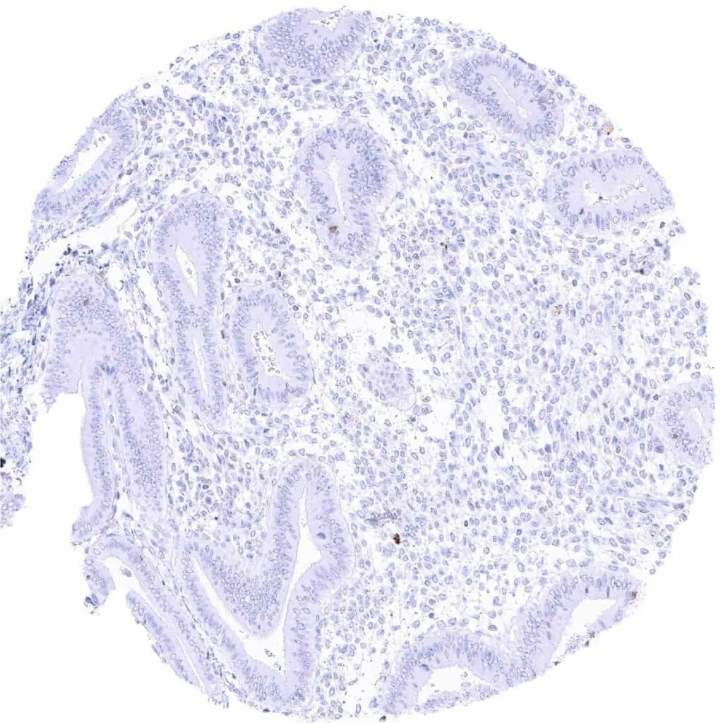
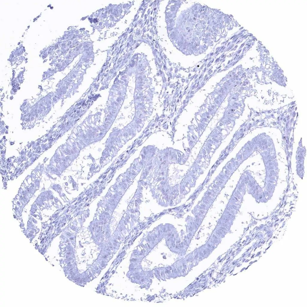
CD38 antibody [MSVA-038R] HistoMAX™


Go to Cancer Tissue Gallery
  
 Go back to product page
  

Go to HistoMAX page


Adrenal gland

Aorta, media

Appendix, mucosa – Strong CD38 staining of a subset of inflammatory cells

Appendix, mucosa


Appendix, muscular wall

Bone marrow – Distinct CD38 staining of a subset of cells

Breast – CD38 staining of some inflammatory cells

Bronchus, mucosa – A weak membranous CD38 staining occurs in a fraction of respiratory epithelial cells


Cerebellum (molecular layer, Purkinje cell layer, granule cell layer)

Cerebellum (white matter)

Cerebrum, grey matter

Cerebrum, white matter


Colon descendens, mucosa

Colon descendens, muscular wall

Duodenum, Brunner gland – CD38 staining of some inflammatory cells

Duodenum, mucosa – Distinct CD38 staining of a subset of inflammatory cells


Epididymis – CD38 staining is lacking in the cauda

Epididymis – CD38 staining is lacking in the corpus

Esophagus, squamous epithelium

Fallopian tube, mucosa


Fat

Gallbladder, epithelium – Distinct CD38 staining of a subset of inflammatory cells

Heart muscle

Ileum, mucosa – Distinct CD38 staining of a some inflammatory cells


Kidney, cortex – CD38 staining of some inflammatory cells

Kidney, medulla

Liver – Distinct CD38 staining of few cells

Lung


Lymph node – Distinct CD38 staining of a subset of lympho–monocytic cells.

Lymph node – Distinct CD38 staining of a subset of lympho–monocytic cells

Ovary, follicular cyst – CD38 staining of some inflammatory cells

Ovary, stroma


Pancreas

Parathyroid gland

Parotid gland

Pituitary gland, anterior lobe – Weak to moderate CD38 staining of endothelial cells in at least a subset small blood vessels


Pituitary gland, posterior lobe

Placenta (amnion and chorion)

Placenta, early

Placenta, mature


Prostate – Strong membranous CD38 staining of most epithelial cells. Staining is weaker or absent in basal cells

Rectum, mucosa

Seminal vesicle – Strong membranous CD38 staining of epithelial cells

Sinus paranasales – A weak membranous CD38 staining occurs in a fraction of respiratory epithelial cells


Skeletal muscle

Skin, hairfollicel and sebaceous glands

Skin

Spleen – Distinct CD38 staining of a subset of lympho–monocytic cells


Stomach, antrum – Distinct CD38 staining of a some inflammatory cells

Stomach, corpus – Distinct CD38 staining of a some inflammatory cells

Submandibular gland

Testis


Thymus – CD38 staining intensity is highest in cortical thymocytes

Thyroid gland

Tonsil – Distinct CD38 staining of a subset of lympho– histiocytic cells. CD38 staining is particularly prominent in cells (plasma cells_) located near the tonsil crypts

Tonsil – Distinct CD38 staining of asubset of lympho– histiocytic cells. CD38 staining is particularly prominent in cells (plasma cells_) located near the tonsil crypts


Tonsil, surface epithelium

Urinary bladder, muscular wall

Urinary bladder, urothelium – Urothelial cells are CD38 negative. Some inflammatory cells show distinct staining

Uterus, ectocervix


Uterus, endocervix – CD38 staining of some inflammatory cells

Uterus, endometrium (pregnancy)

Uterus, endometrium (proliferation)

Uterus, endometrium (secretion)


Uterus, myometrium


| TOP |