Normal Tissue Gallery- GTX04964

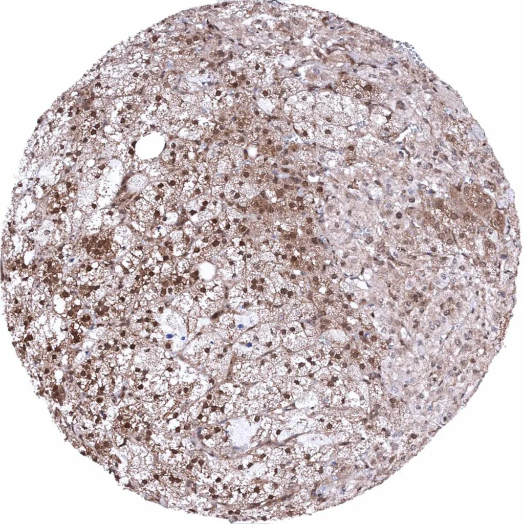
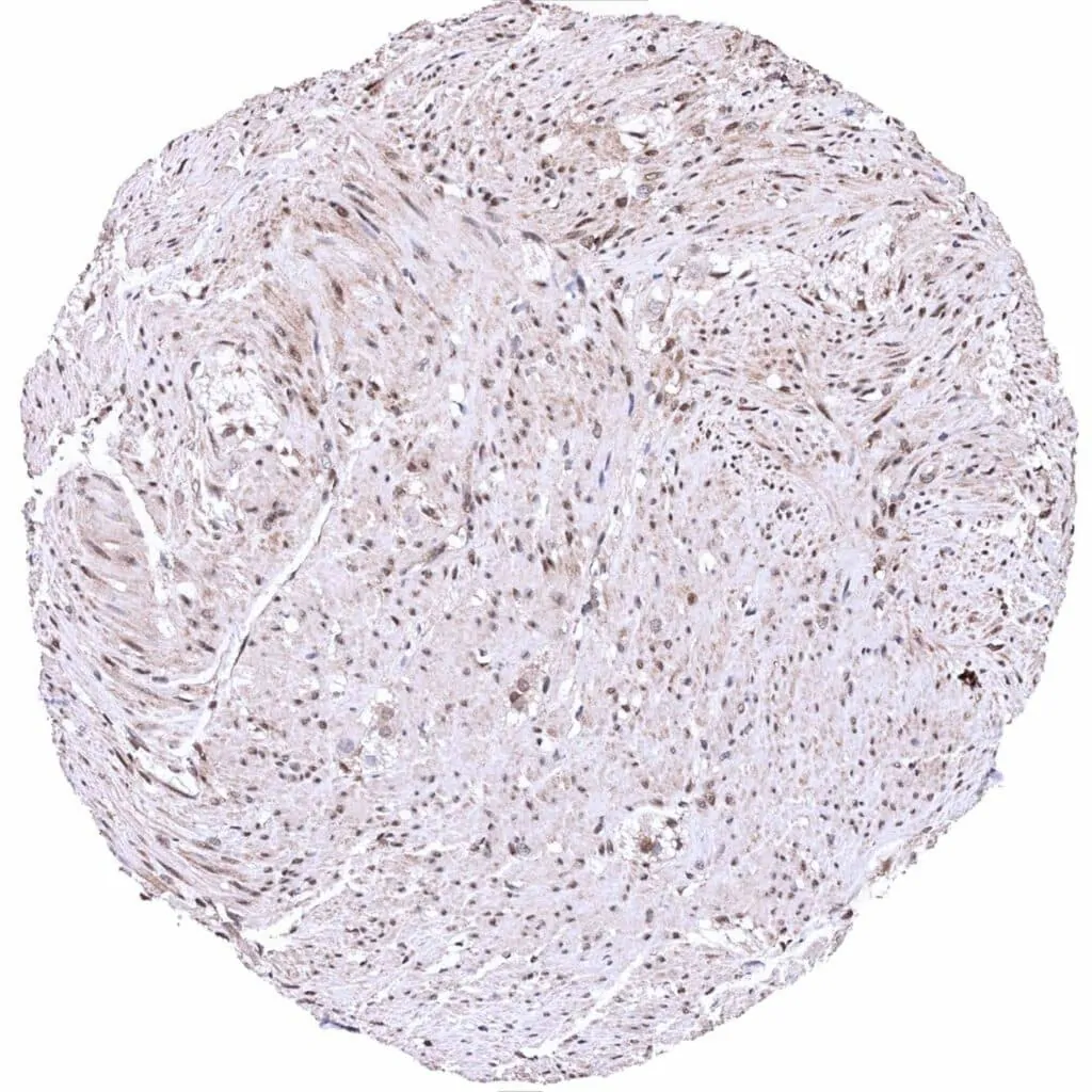
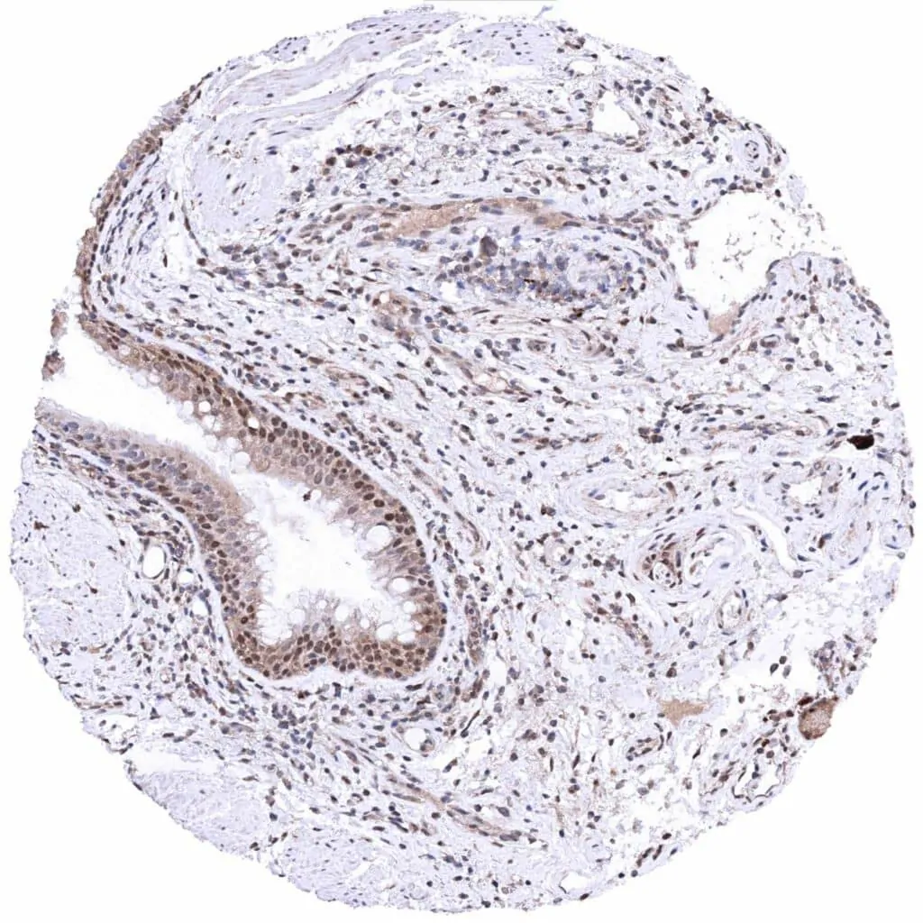
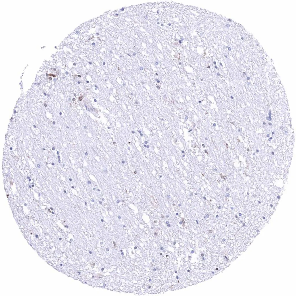
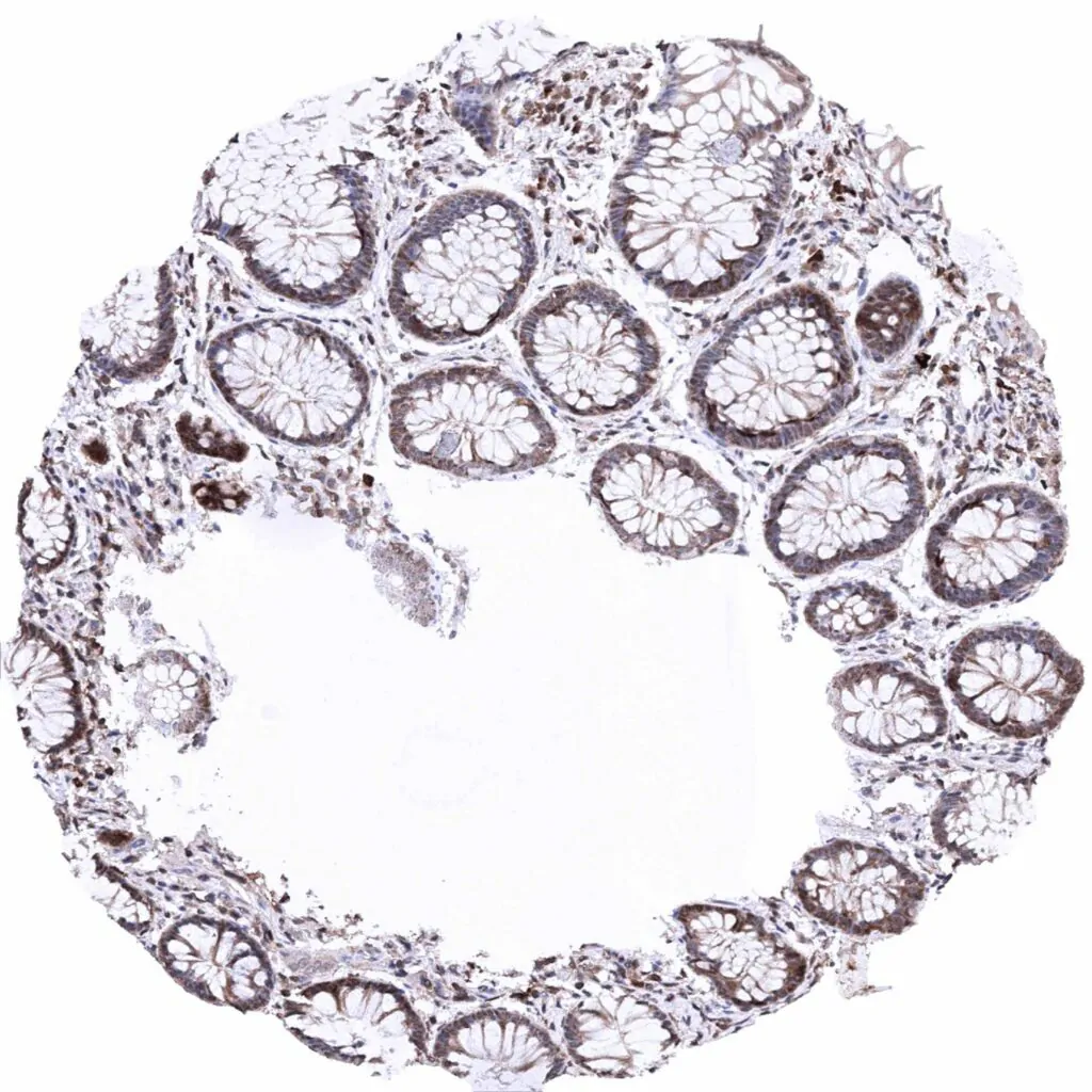
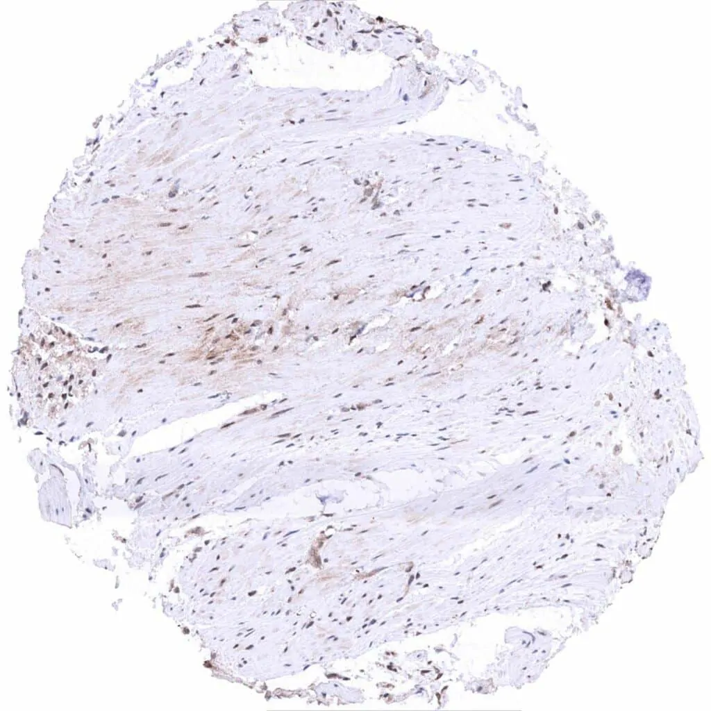
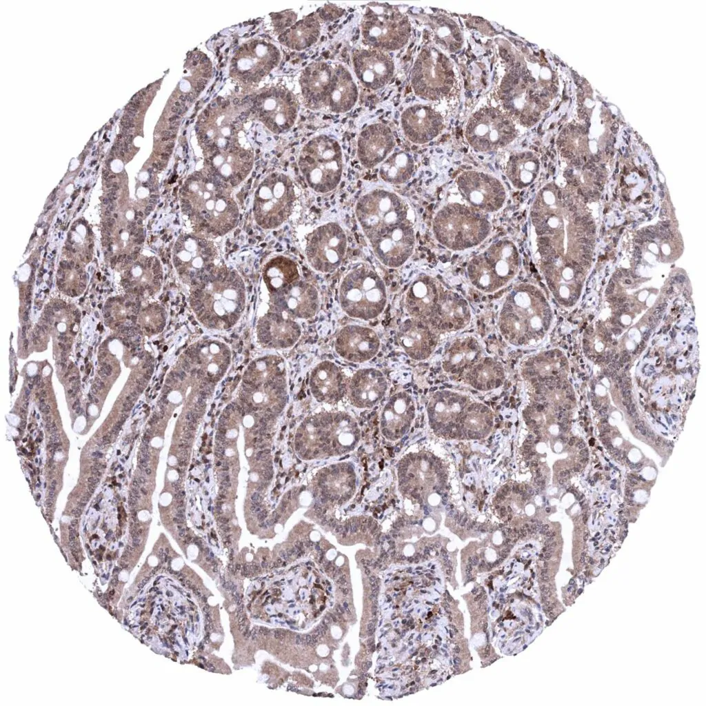
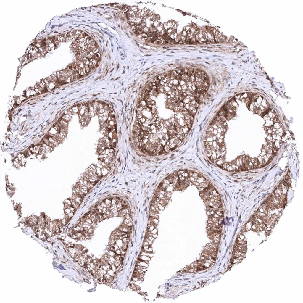
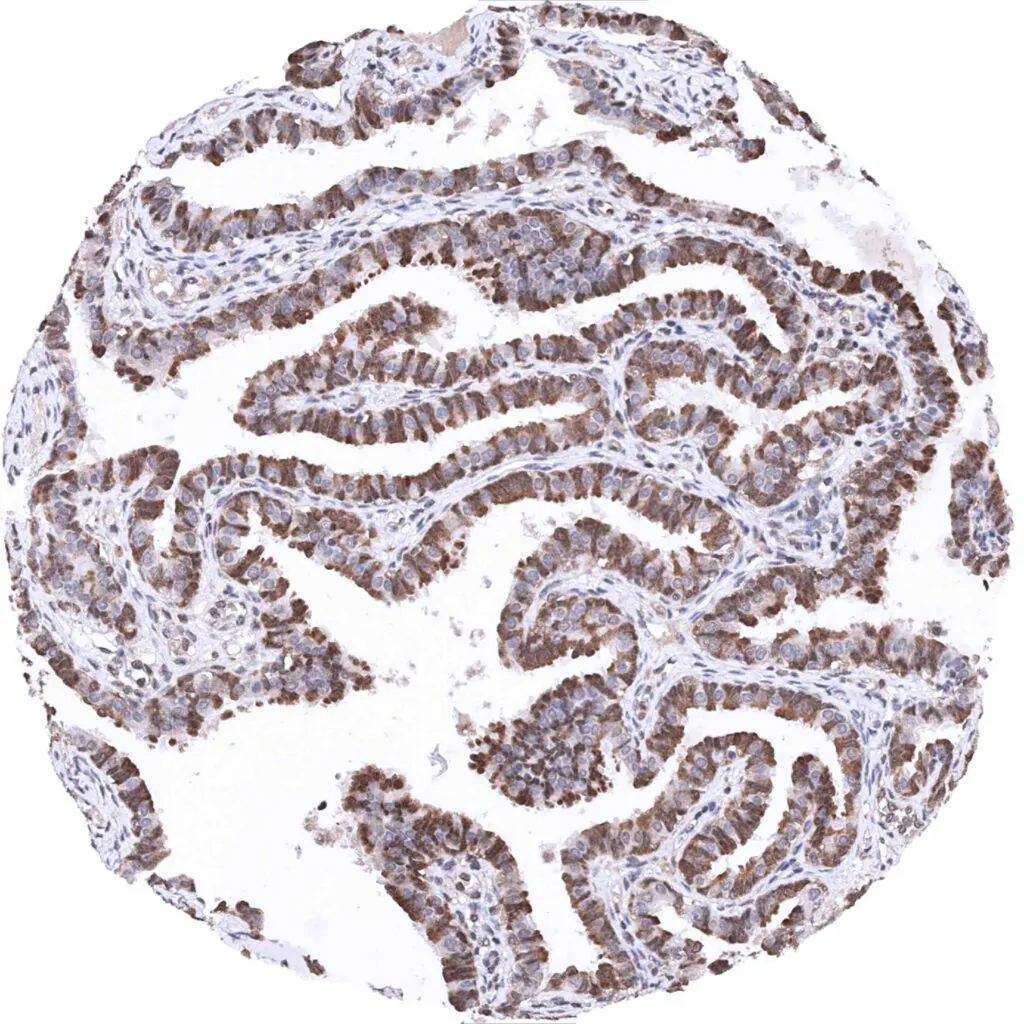
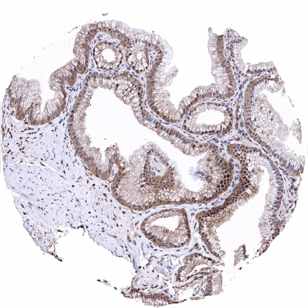
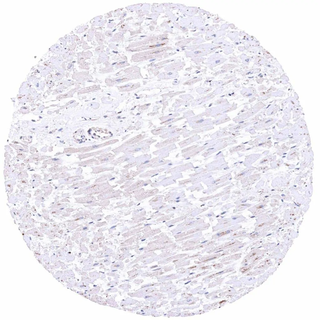
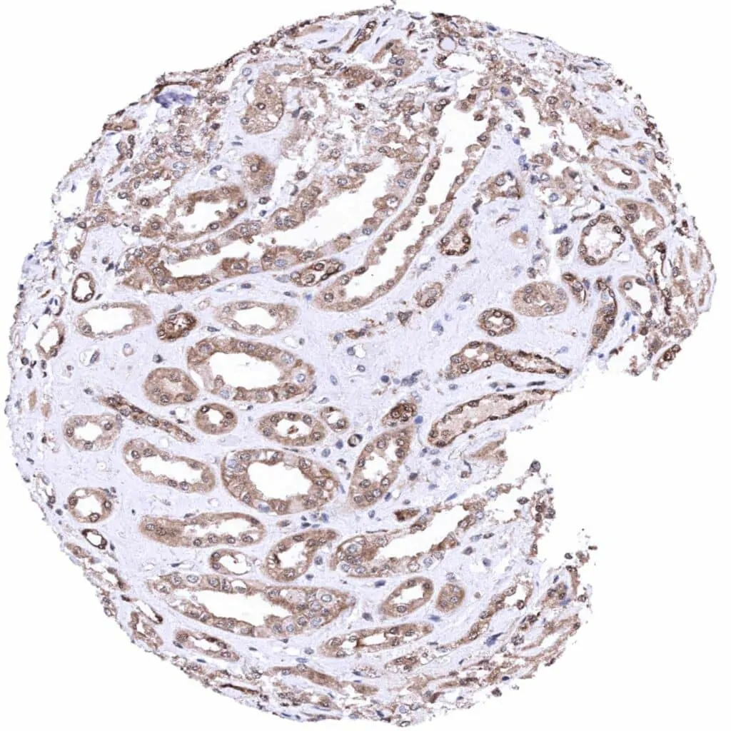
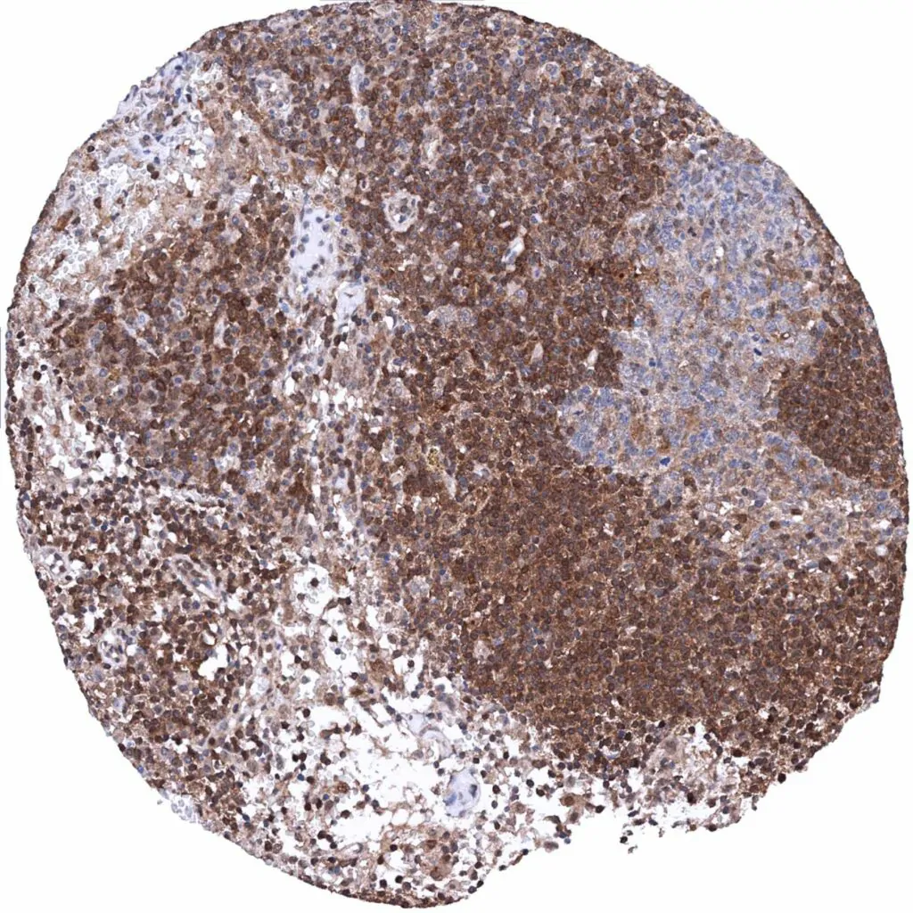
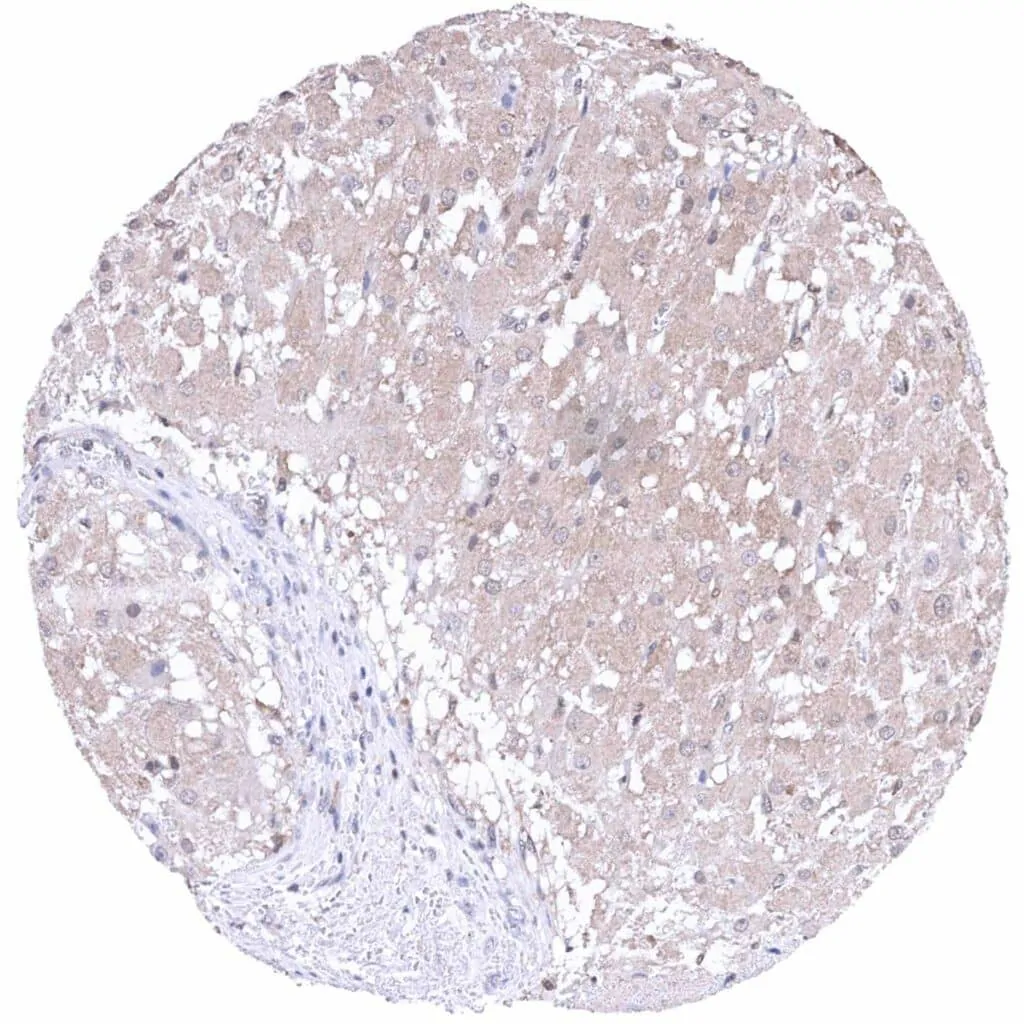
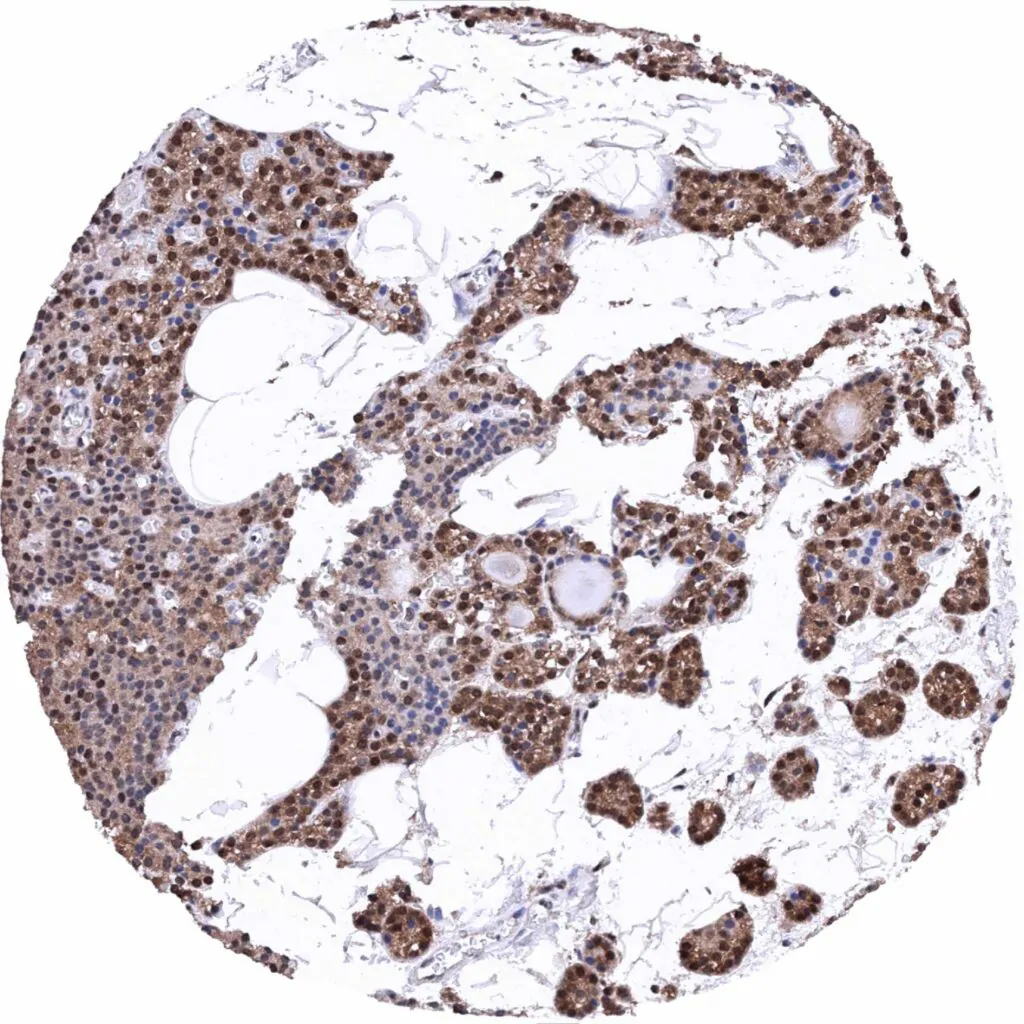
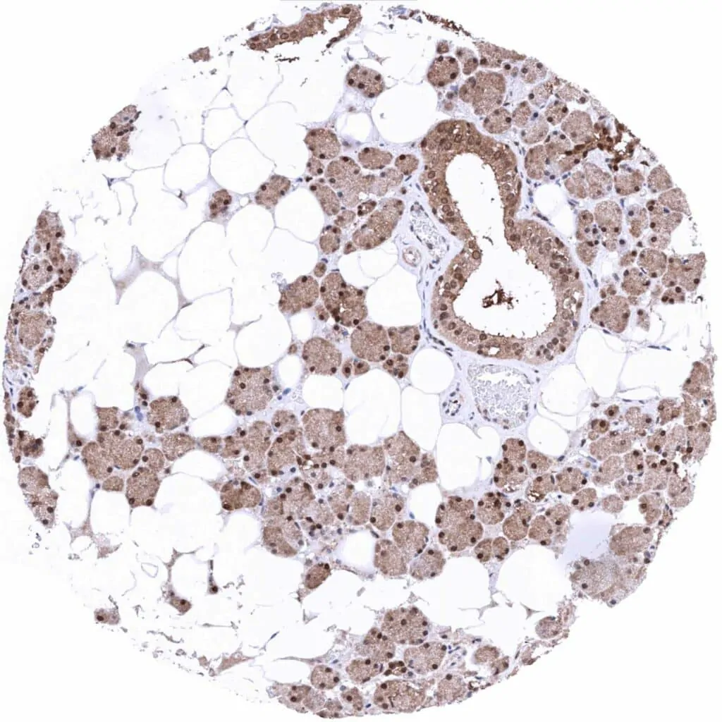
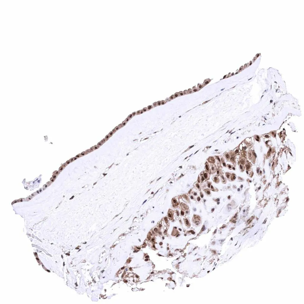
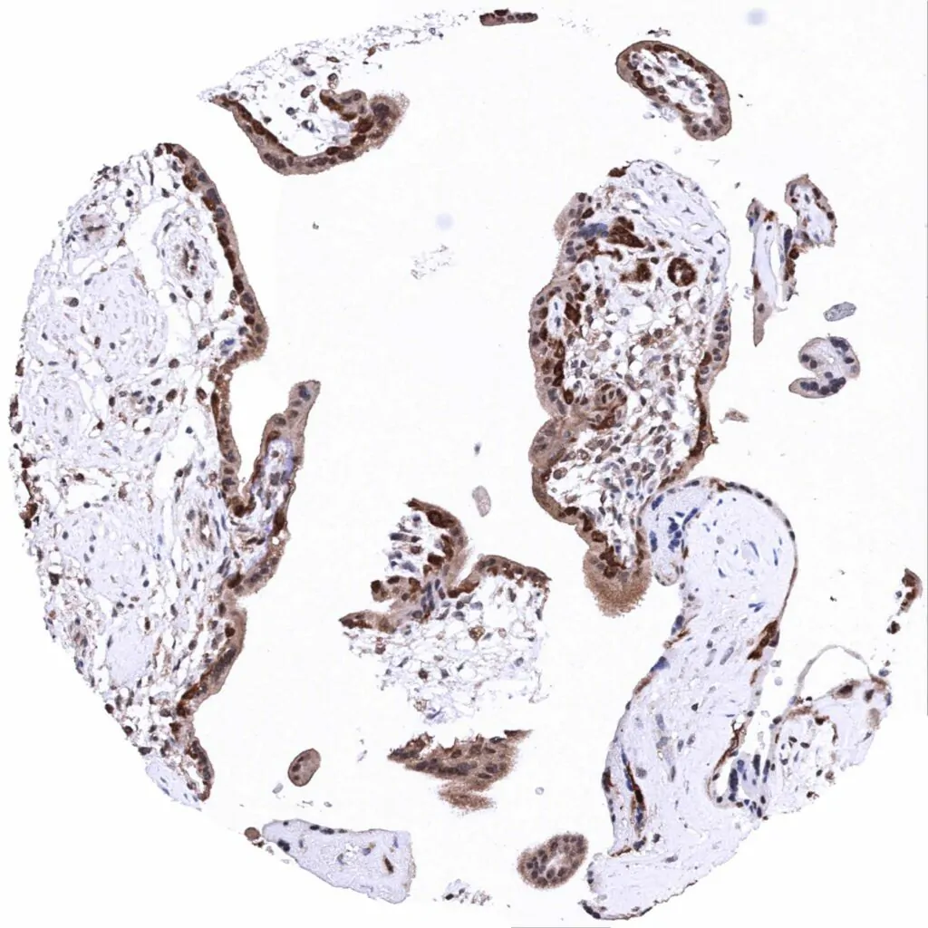
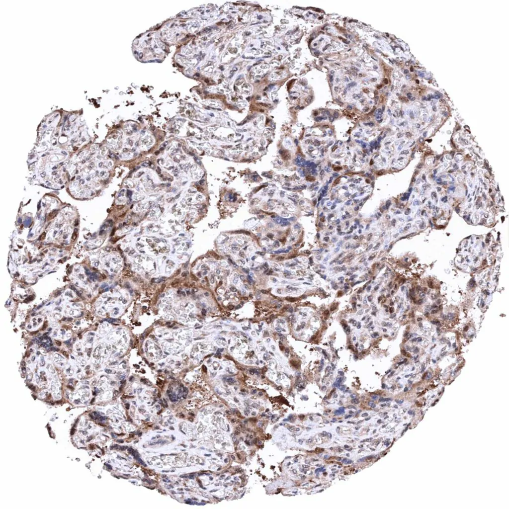
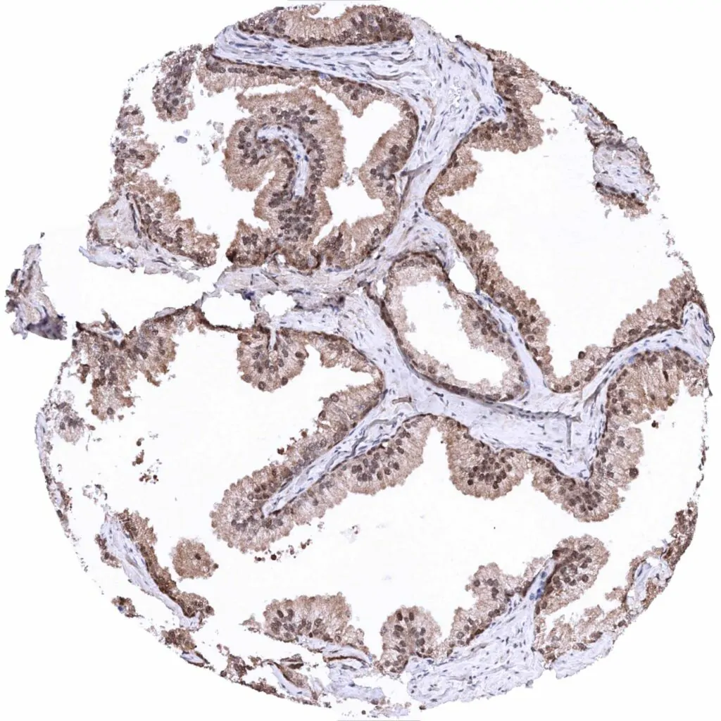
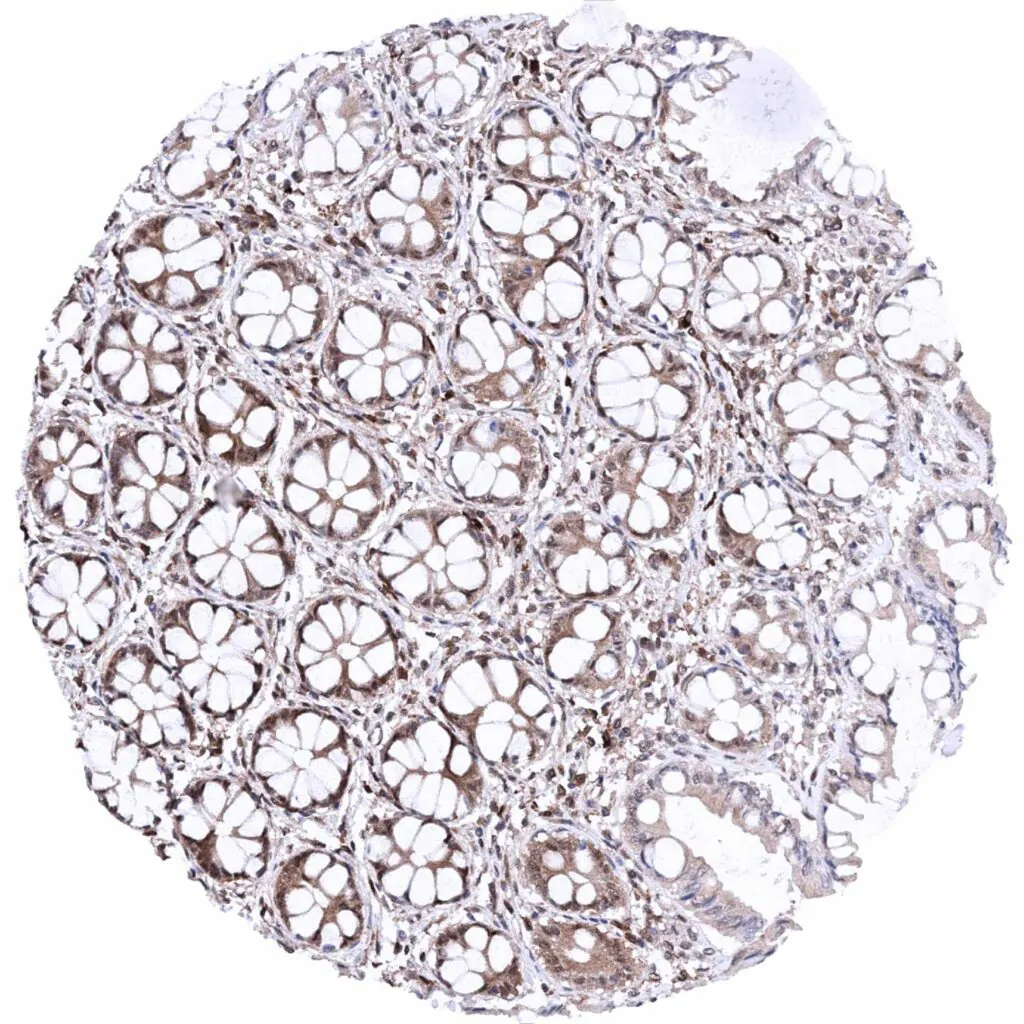
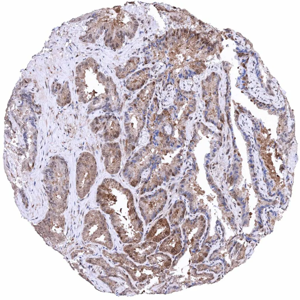
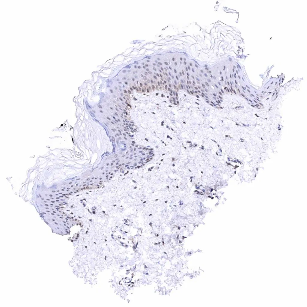
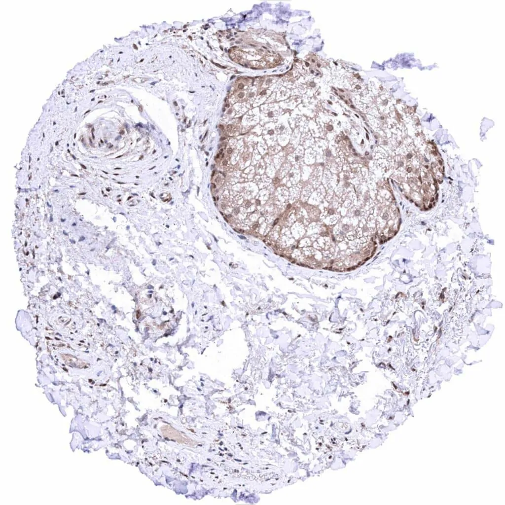
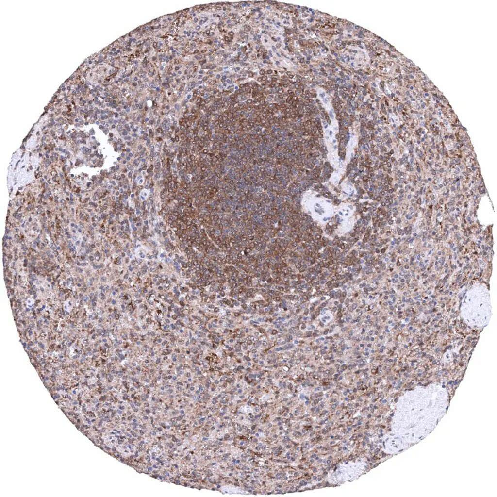
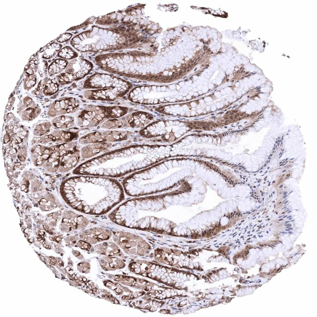
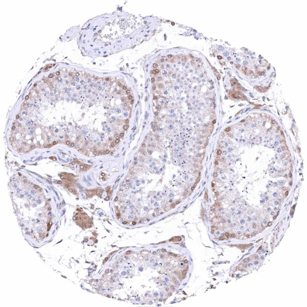
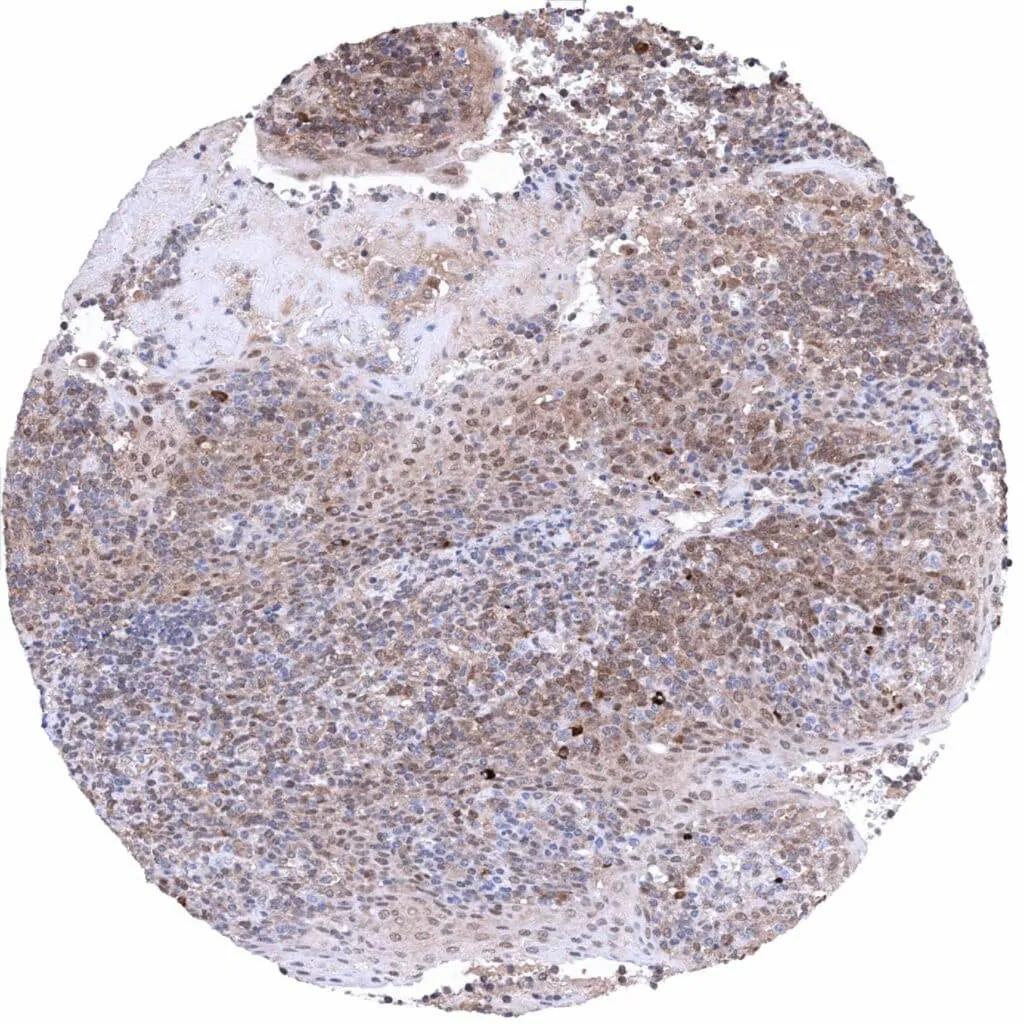
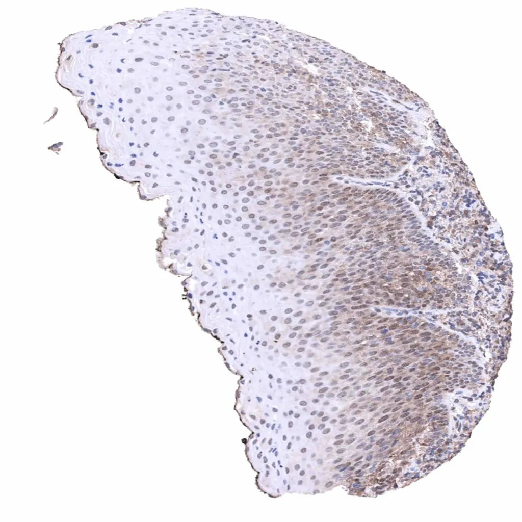
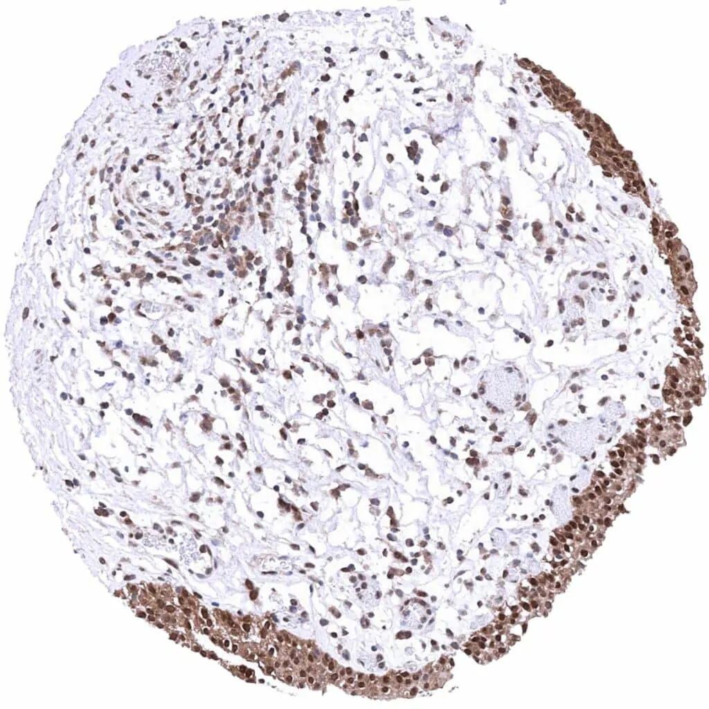
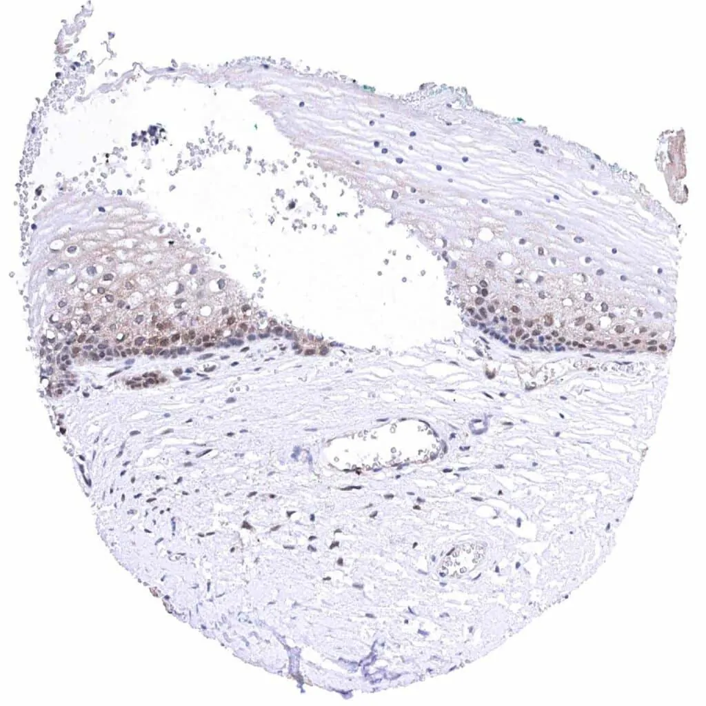
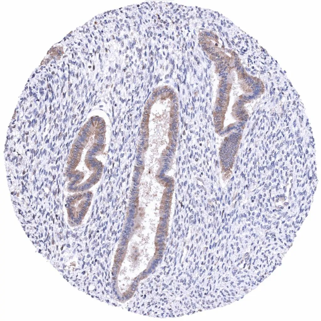
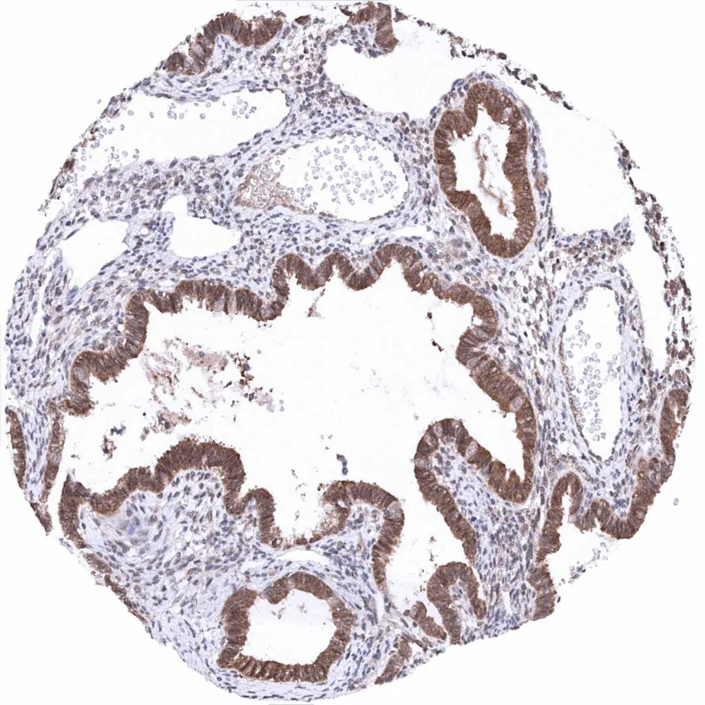
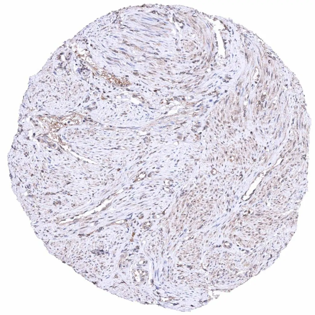
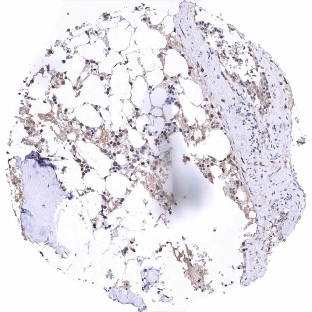
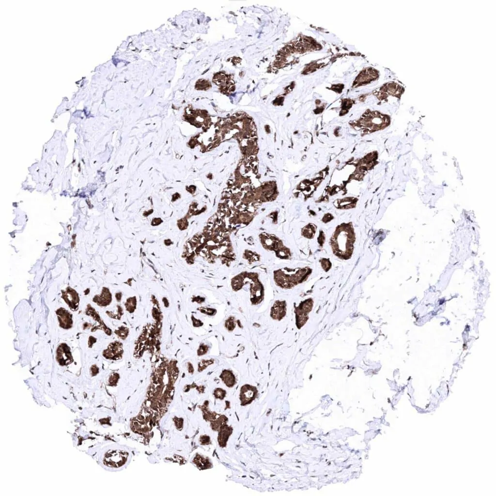
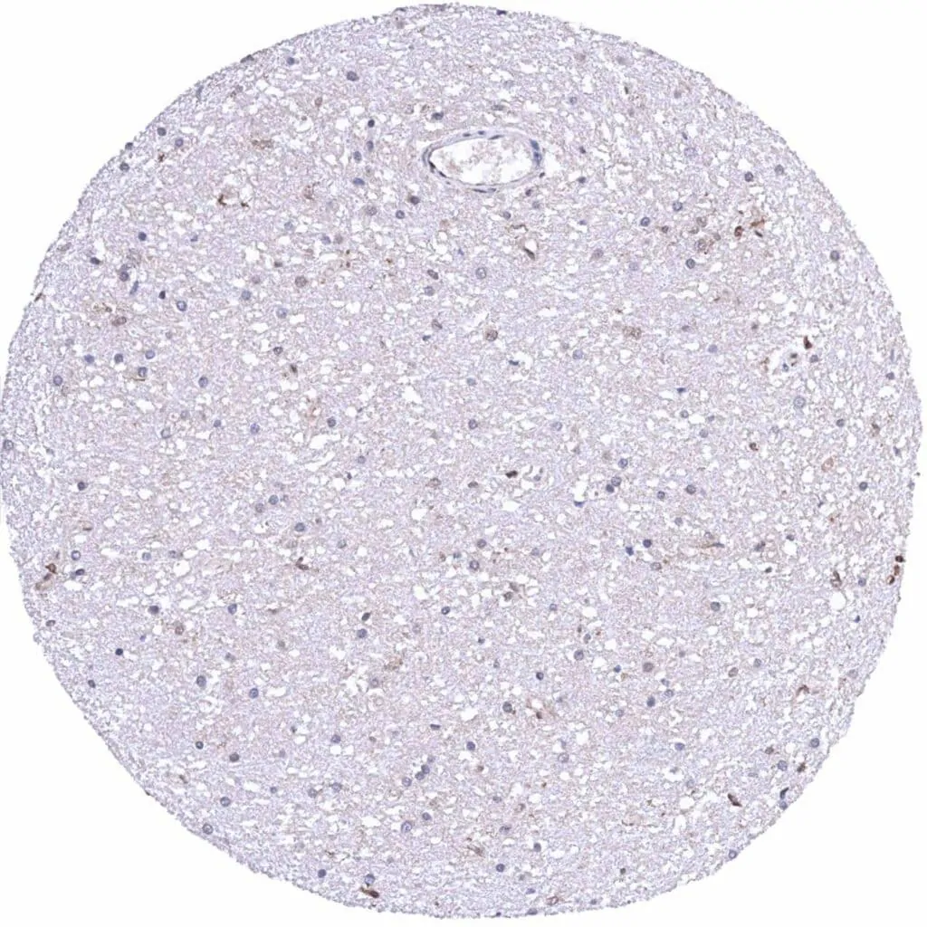
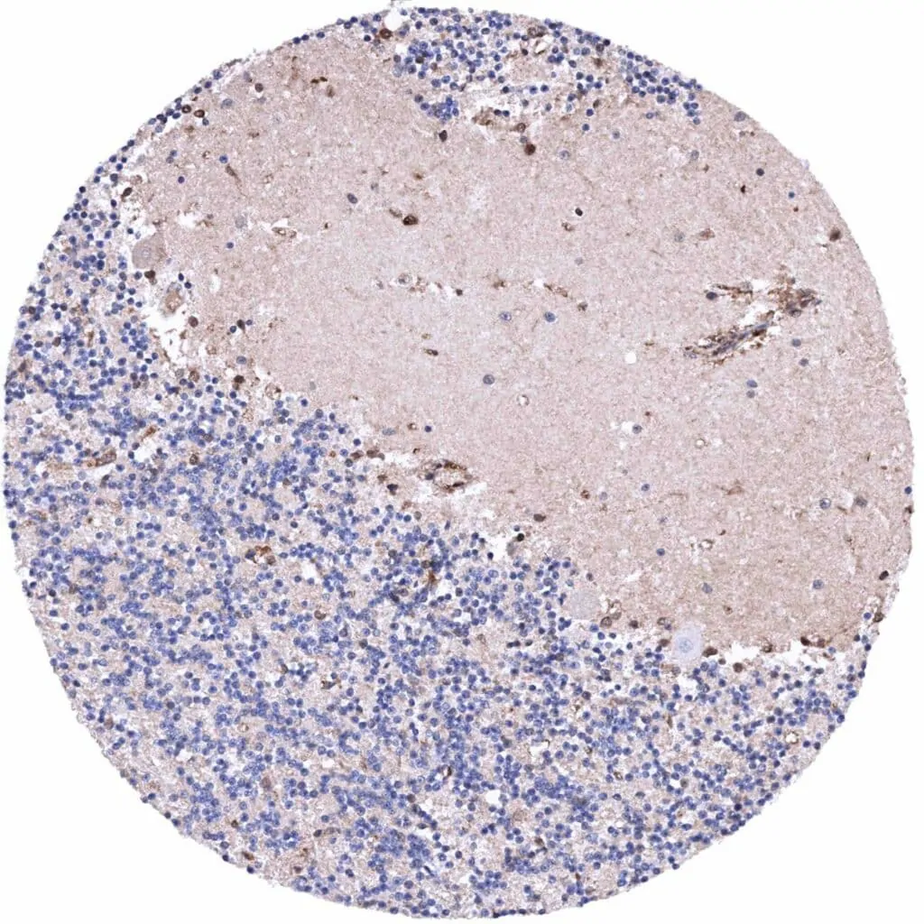
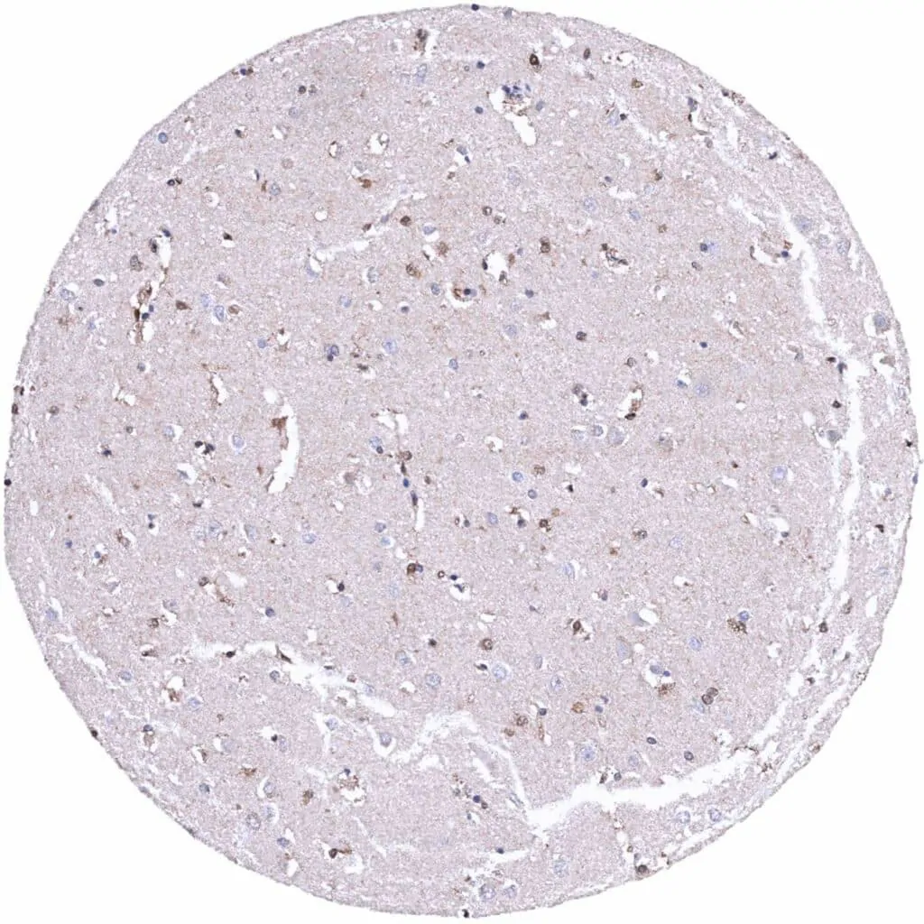
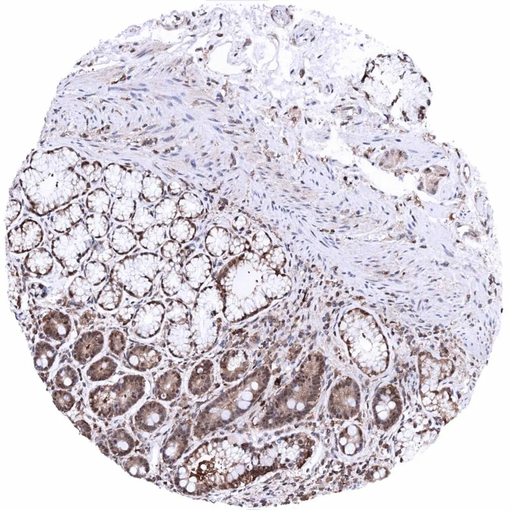
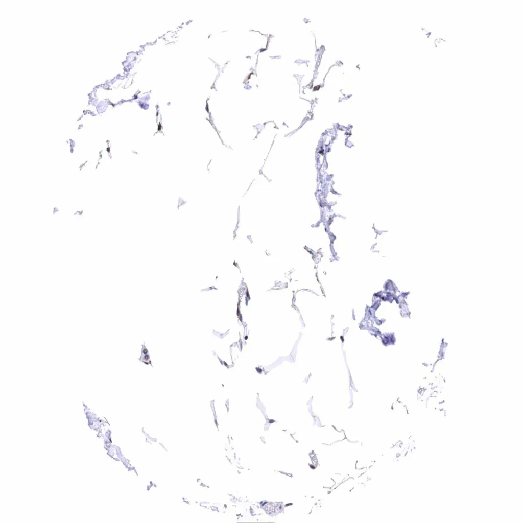
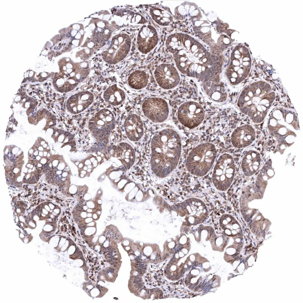
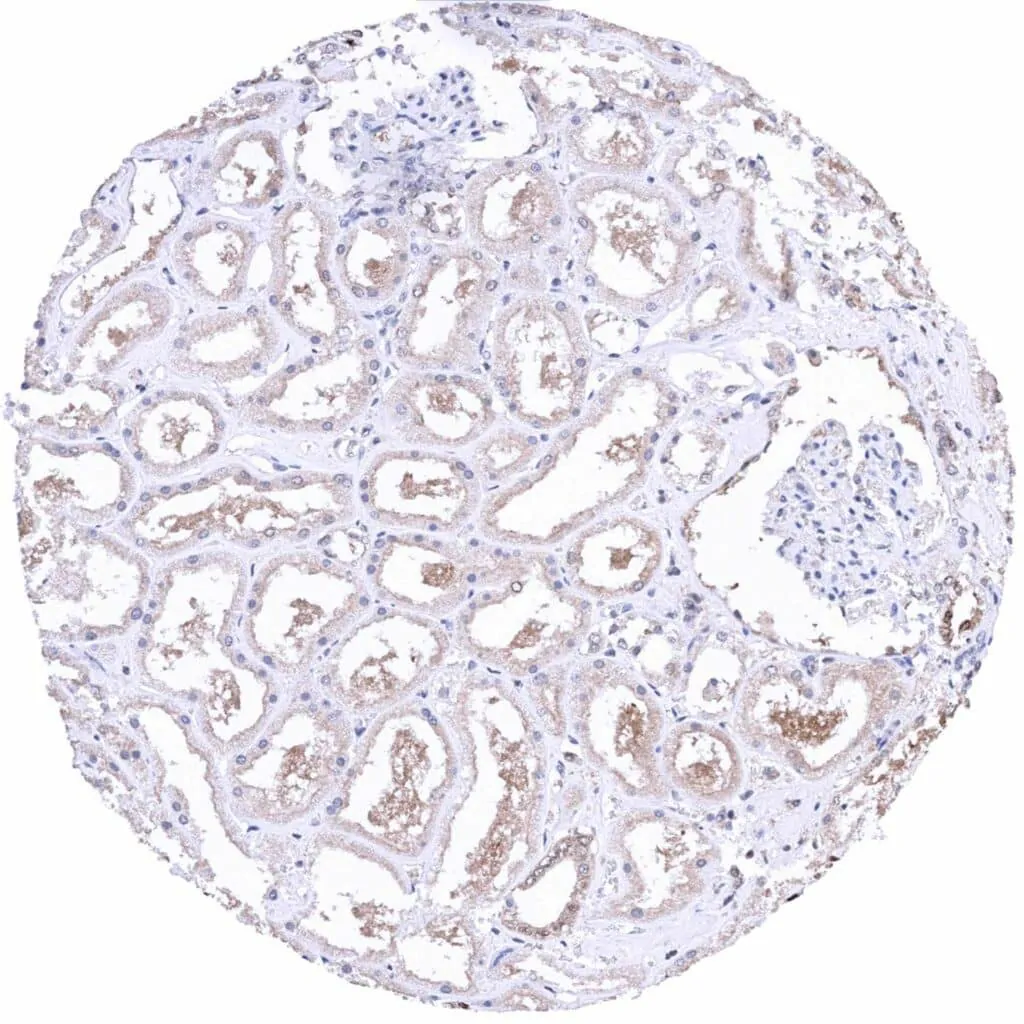
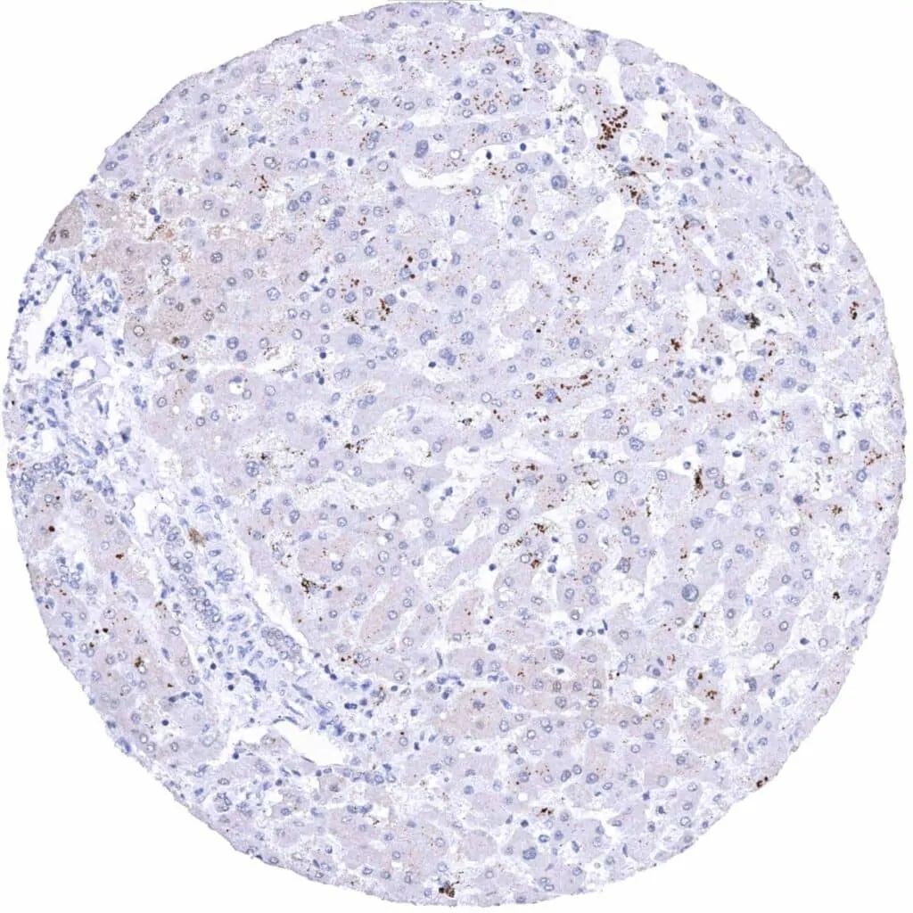
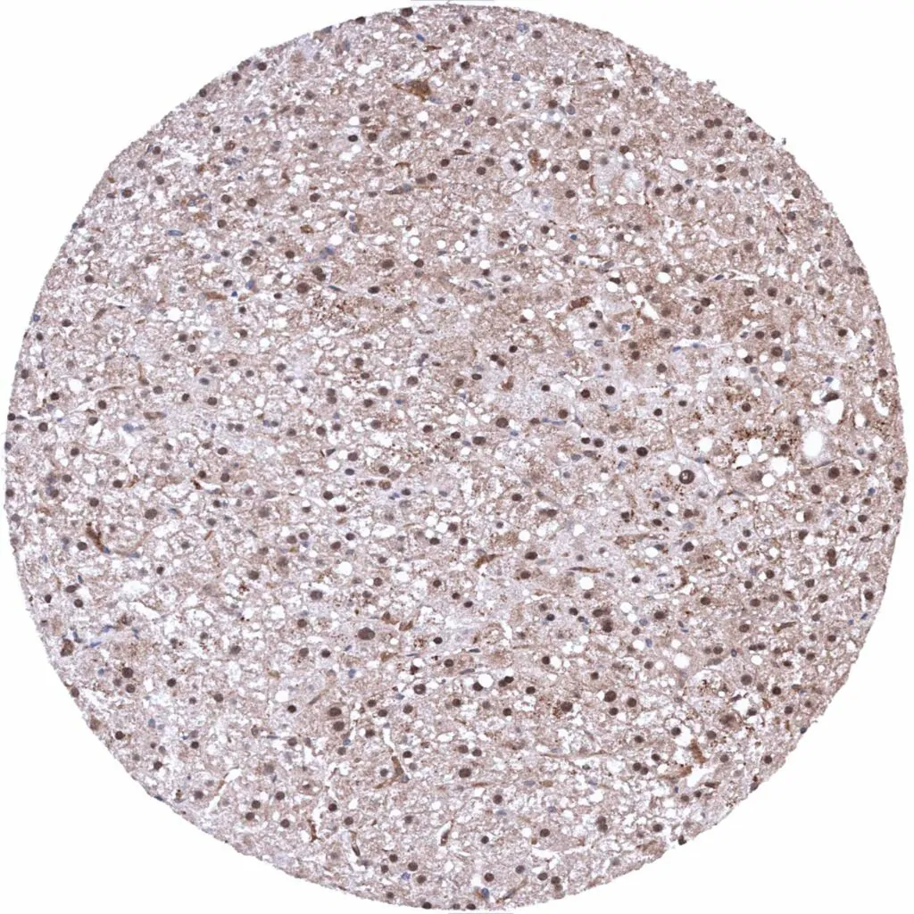
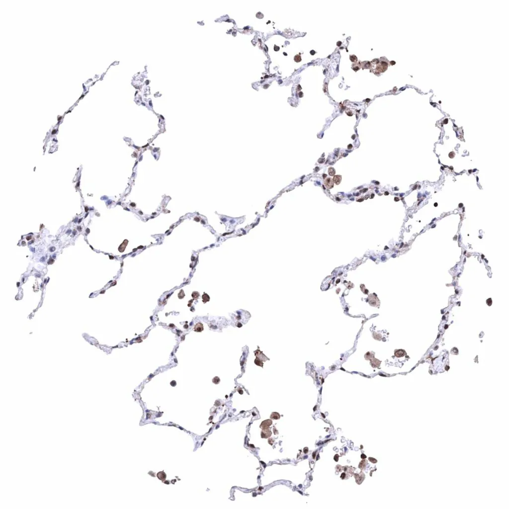
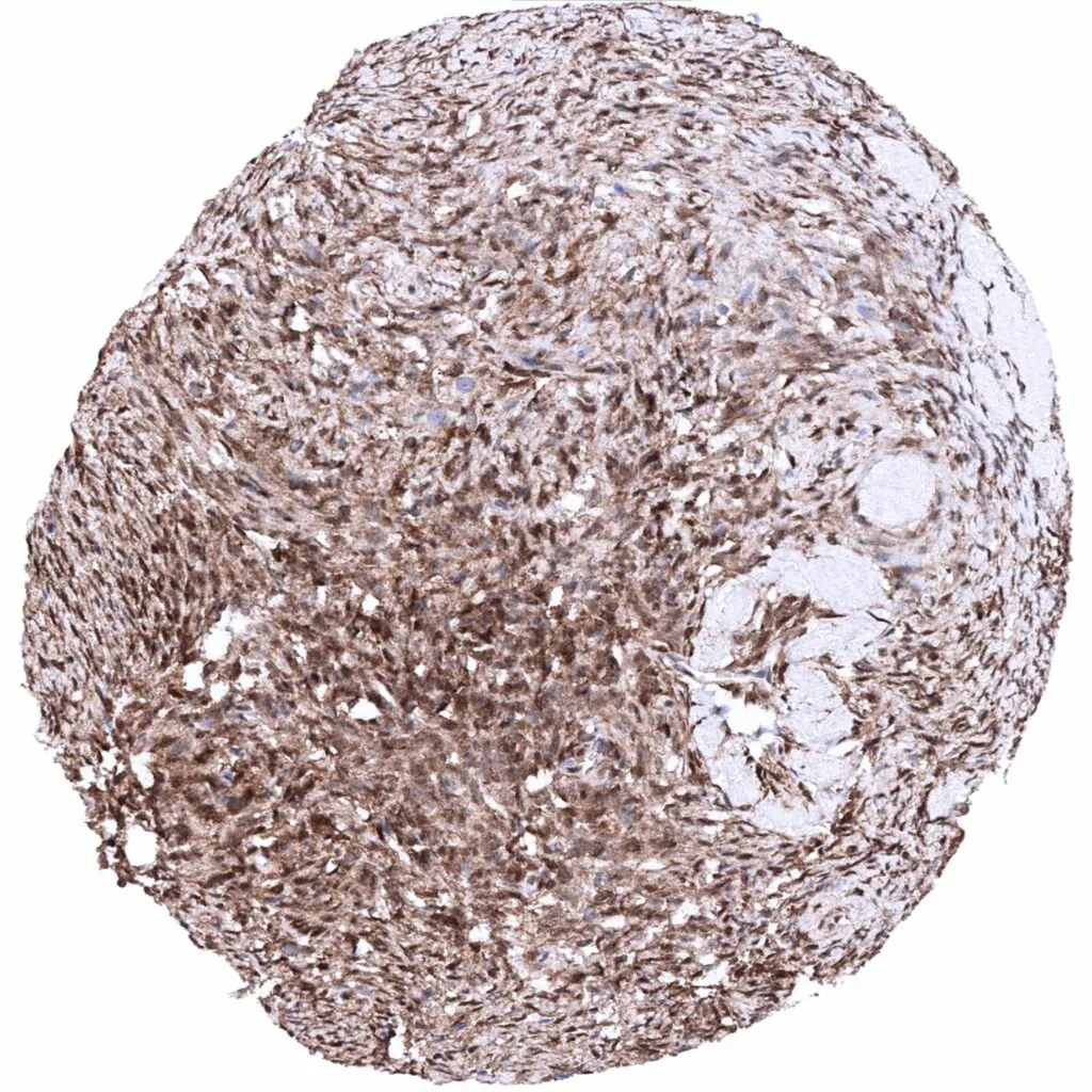
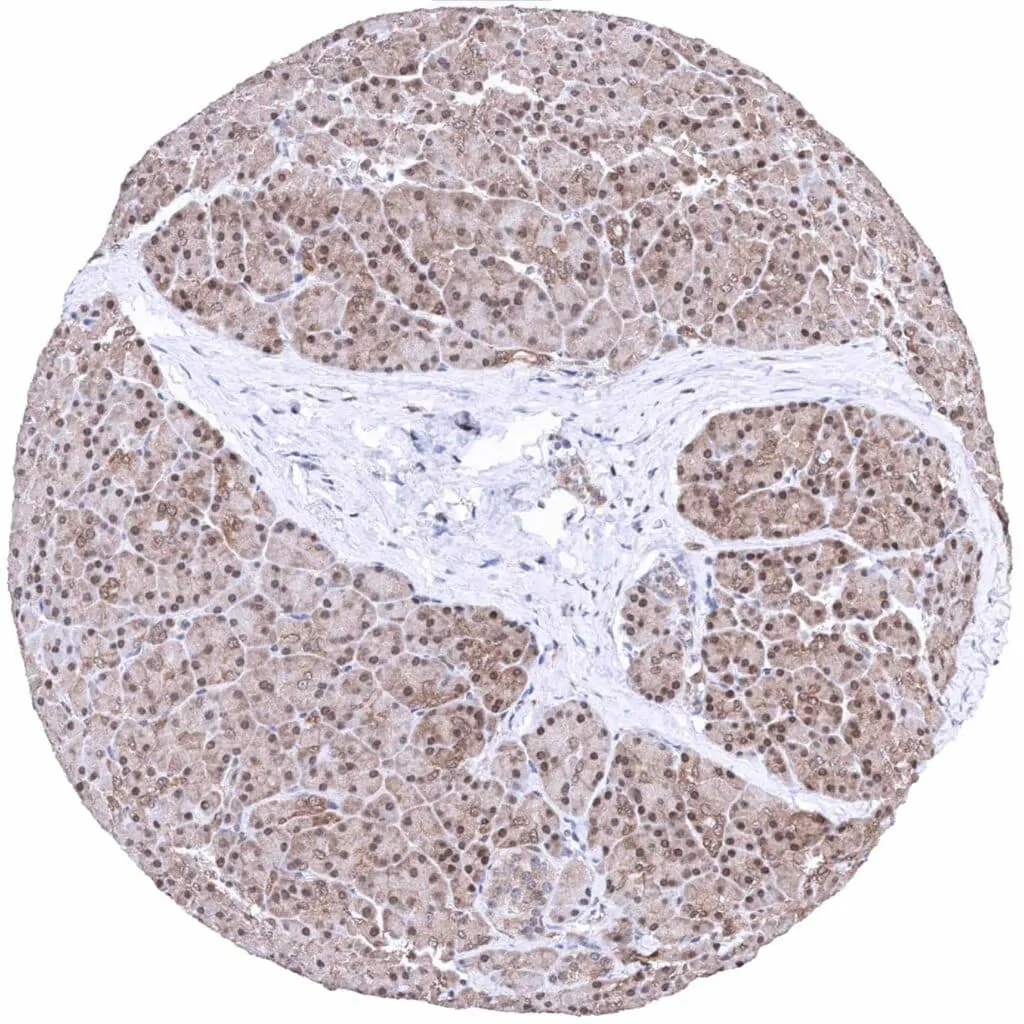
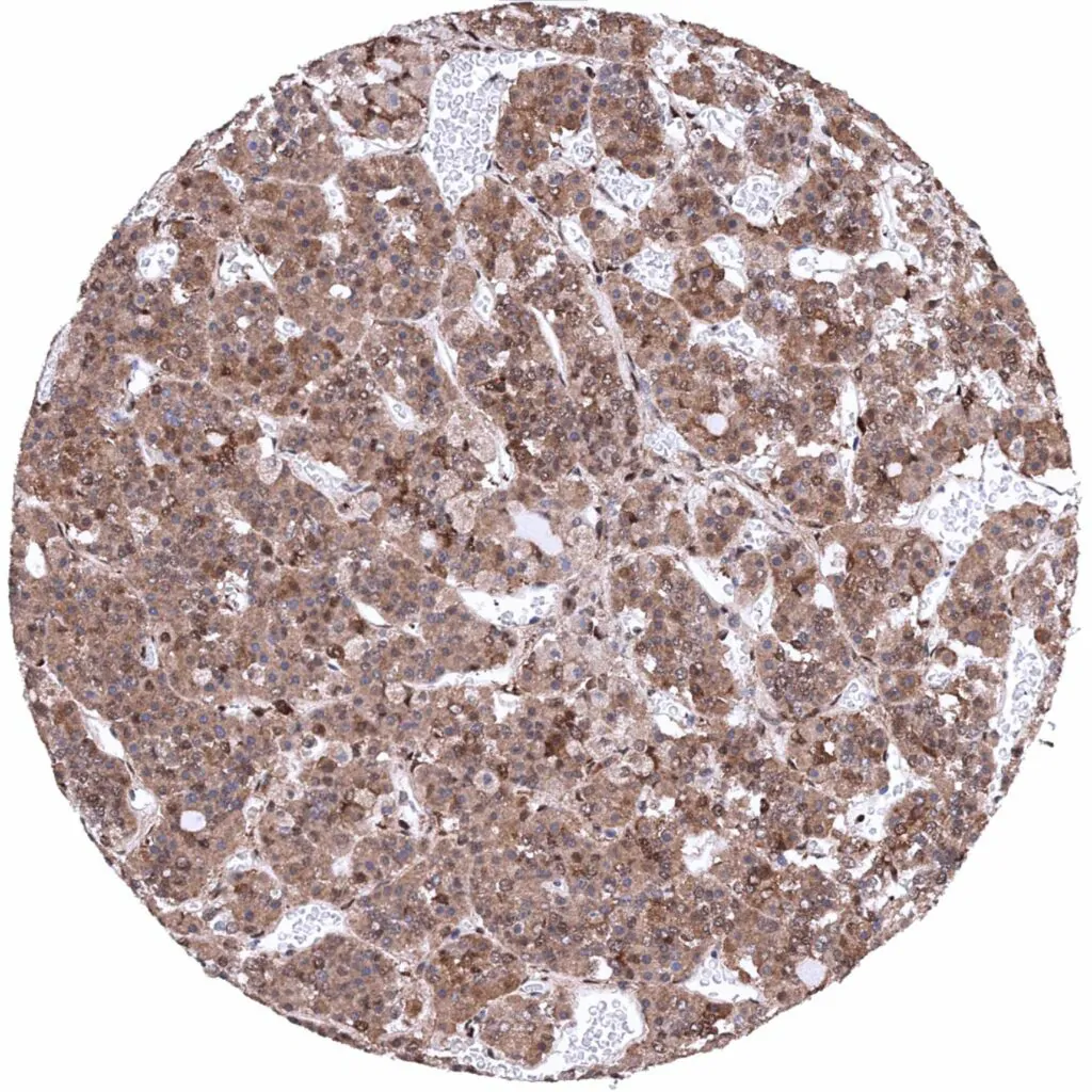
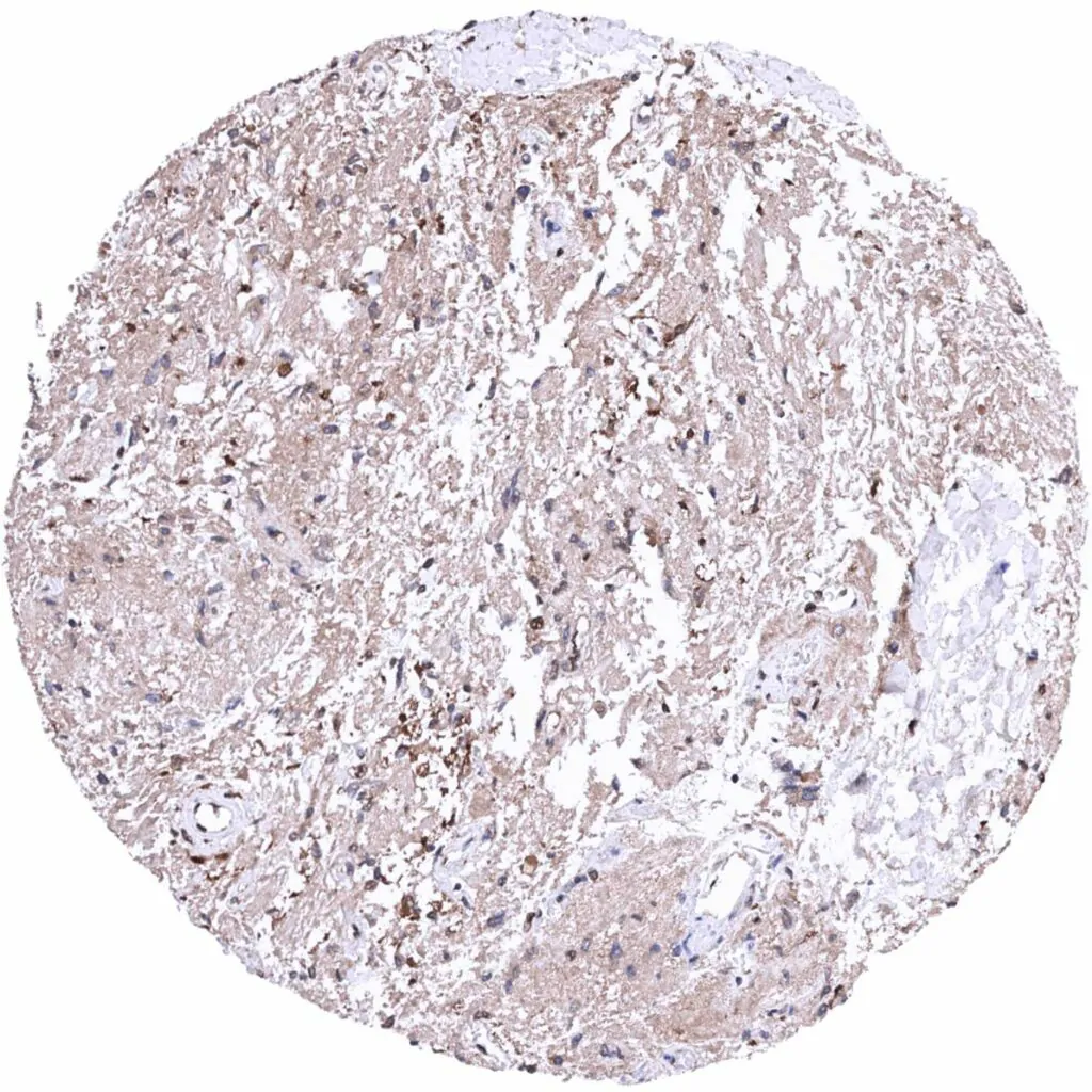
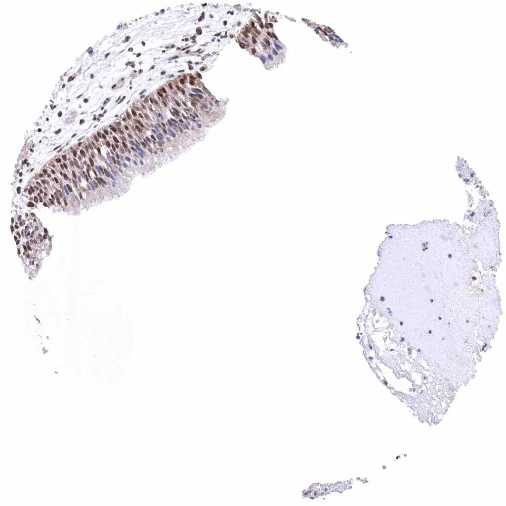
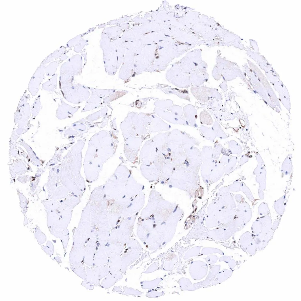
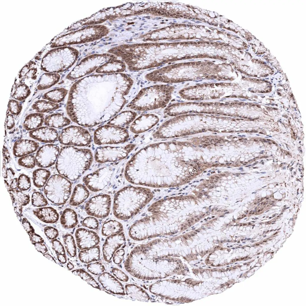
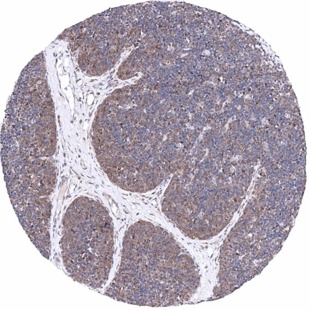
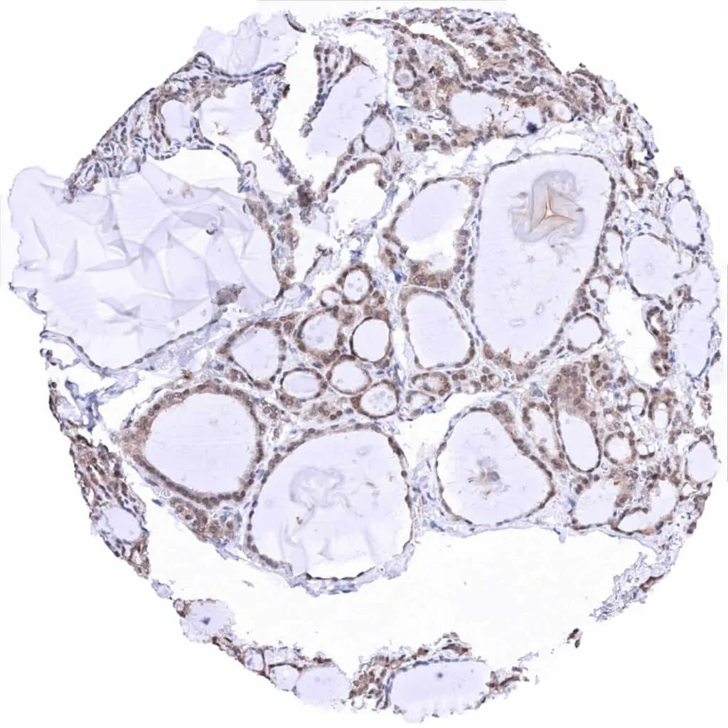
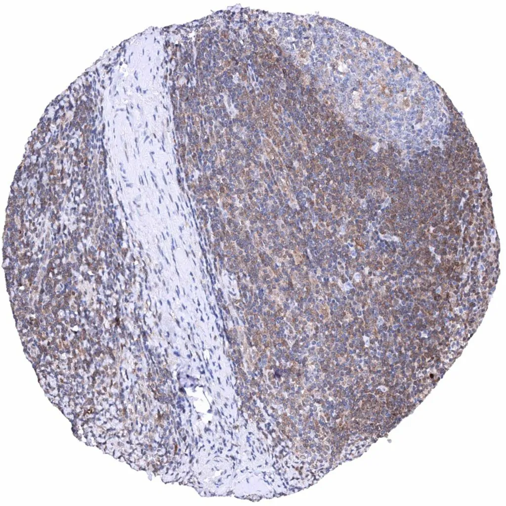
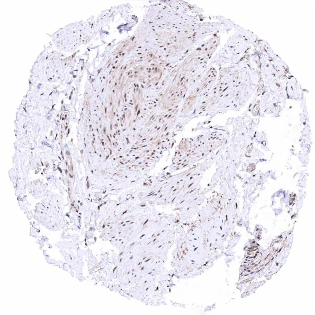
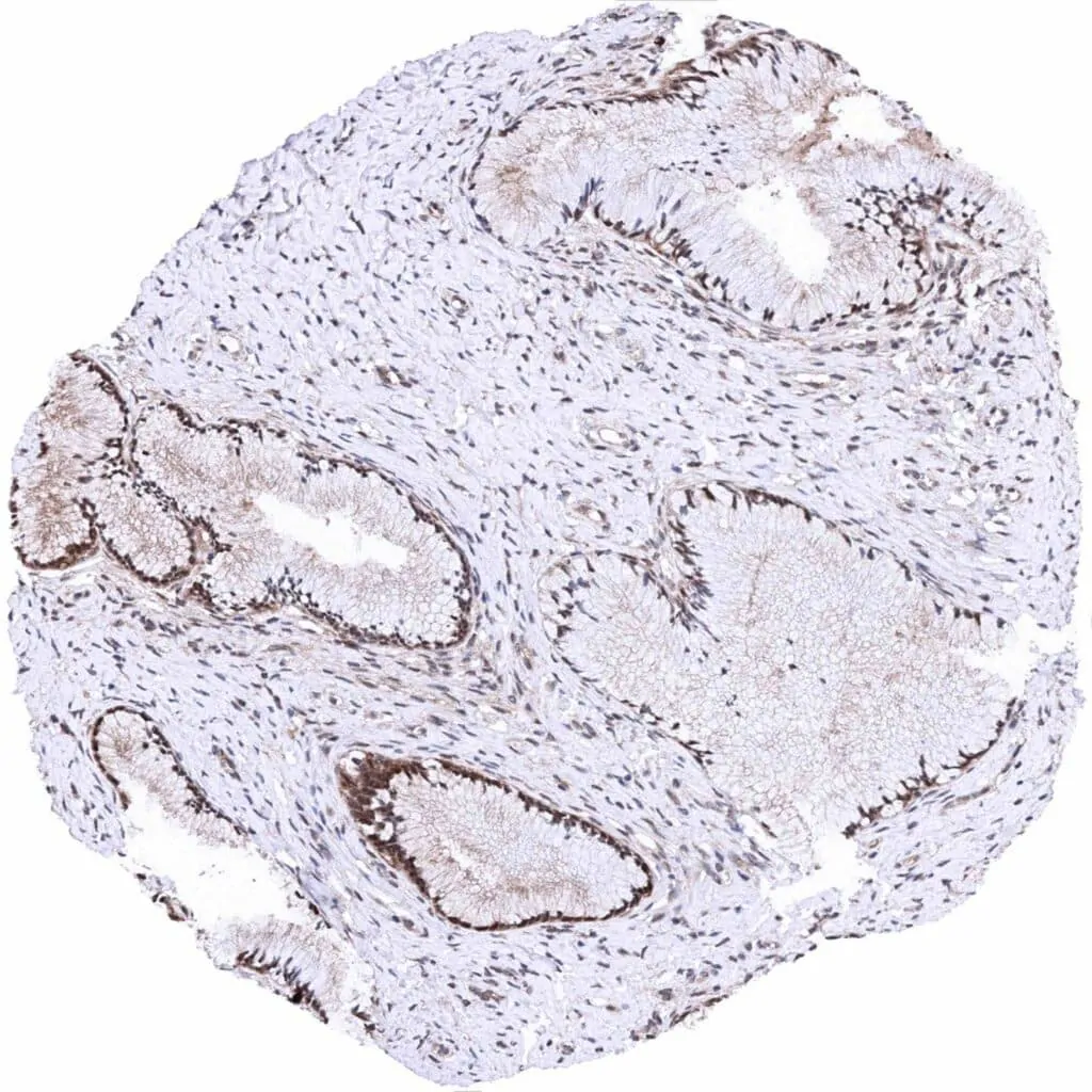
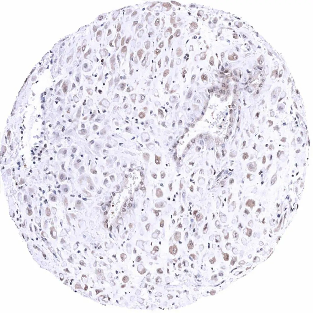
MTAP antibody [MSVA-741R] HistoMAX™


Go to Cancer Tissue Gallery
  
 Go back to product page
  

Go to HistoMAX page


Adrenal gland – Moderate to strong, nuclear and cytoplasmic MTAP positivity of epithelial cells

Aorta, endothelium

Appendix, muscular wall

Bone marrow


Breast – Intense, nuclear and cytoplasmic MTAP positivity of epithelial cells

Bronchus, mucosa

Cerebellum (white matter) – Faint to moderate staining in some glia cells

Cerebellum, cortex (molecular layer, Purkinje cell layer, granule cell layer) – Faint to moderate staining in small blood vessels and in some glia cells


Cerebrum, grey matter – Weak to moderate staining in small blood vessels and in some glia cells

Cerebrum, white matter

Colon descendens, mucosa

Colon descendens, muscular wall


Duodenum, Brunner gland

Duodenum, mucosa – Weak to moderate, predominantly cytoplasmic MTAP positivity of epithelial cells. Note- Mucus in goblet cells is MTAP negative

Epididymis (cauda) – Strong, predominantly cytoplasmic MTAP positivity of epithelial cells

Fallopian tube, mucosa – Moderate to strong, nuclear and cytoplasmic MTAP positivity of a subset of epithelial cells


Fat

Gallbladder, epithelium – Weak to moderate, predominantly nuclear MTAP positivity of epithelial cells

Heart muscle – Faint cytoplasmic and sometimes also nuclear MTAP positivity of heart muscle cells

Ileum, mucosa


Kidney, cortex – Weak nuclear and cytoplasmic MTAP positivity of tubular cells

Kidney, medulla – Moderate to strong, predominantly cytoplasmic MTAP positivity of tubular and collecting duct cells

Liver – MTAP staining can be rather weak in hepatocytes

Liver – Weak to moderate, cytoplasmic MTAP positivity hepatocytes in this sample


Lung

Lymph node – Moderate to strong, predominantly cytoplasmic MTAP positivity of virtually all lymphocytes. Staining is most intense in the interfollicular area and least in germinal centres

Ovary, corpus luteum

Ovary, stroma – Moderate to strong, nuclear and cytoplasmic MTAP positivity of stromal cells


Pancreas

Parathyroid gland – Moderate intensity, predominantly cytoplasmic MTAP positivity of epithelial cells

Parotid gland

Pituitary, anterior lobe – Moderate to strong, predominantly cytoplasmic MTAP positivity of epithelial cells


Pituitary, posterior lobe – Weak, predominantly cytoplasmic MTAP staining of pituicytes

Placenta (amnion and chorion) – Moderate to strong, nuclear and cytoplasmic MTAP positivity of chorion and amnion cells

Placenta, early – Strong, nuclear and cytoplasmic MTAP positivity of cytotrophoblast cells. MTAP staining is markedly less intense in the syncytiotrophoblast and in the stroma

Placenta, mature – Moderate to strong, nuclear and cytoplasmic MTAP positivity of cytotrophoblast cells. Staining is less intense in the syncytiotrophoblast and in the stroma


Prostate – Moderate intensity, predominantly cytoplasmic MTAP positivity of epithelial cells

Rectum, mucosa – Weak to moderate, predominantly cytoplasmic MTAP positivity of epithelial cells, especially seen at the base of crypts. Note- Mucus in goblet cells is MTAP negative

Seminal vesicle – Moderate intensity, predominantly cytoplasmic MTAP positivity of epithelial cells

Sinus paranasales


Skeletal muscle

Skin – Predominantly nuclear MTAP staining is strongest in the basal and suprabasal cell layers of the epidermis

Skin, hairfollicel and sebaceous glands

Spleen – Moderate to strong, predominantly cytoplasmic MTAP positivity of inflammatory cells, especially in the white pulp


Stomach, antrum – Moderate intensity, predominantly cytoplasmic MTAP positivity of gastric epithelium

Stomach, corpus – Moderate intensity, predominantly cytoplasmic MTAP positivity of gastric epithelium

Testis – Moderate to strong, nuclear and cytoplasmic MTAP positivity of spermatogonia. MTAP staining of other intratubular cells is markedly weaker or even absent. Strong positivity of Leydig cells

Thymus – Moderate, predominantly cytoplasmic MTAP positivity of the majority of lymphocytes


Thyroid gland – Moderate intensity nuclear and cytoplasmic MTAP positivity of epithelial cells

Tonsil – In the squamous epithelium, a variable, predominantly nuclear MTAP is strongest in the basal and suprabasal cell layers

Tonsil, surface epithelium – Cytoplasmic and nuclear MTAP staining is strongest in the basal and suprabasal epithelial cell layers

Tonsil


Urinary bladder, muscular wall

Urinary bladder, urothelium – Strong, nuclear and cytoplasmic MTAP positivity of all urothelial cells

Uterus, ectocervix – Cytoplasmic and nuclear MTAP staining predominantly occurs in in the basal and suprabasal epithelial cell layers

Uterus, endocervix


Uterus, endometrium (pregnancy)

Uterus, endometrium (proliferation) – Weak to moderate, predominantly cytoplasmic MTAP positivity of epithelial cells while staining is only faint in the stroma

Uterus, endometrium (secretion) – Intense, predominantly cytoplasmic MTAP positivity of epithelial cells while MTAP staining is markedly weaker in the stroma

Uterus, myometrium – Weak, predominantly cytoplasmic MTAP positivity of muscle cells


| TOP |