Normal Tissue Gallery- GTX643510

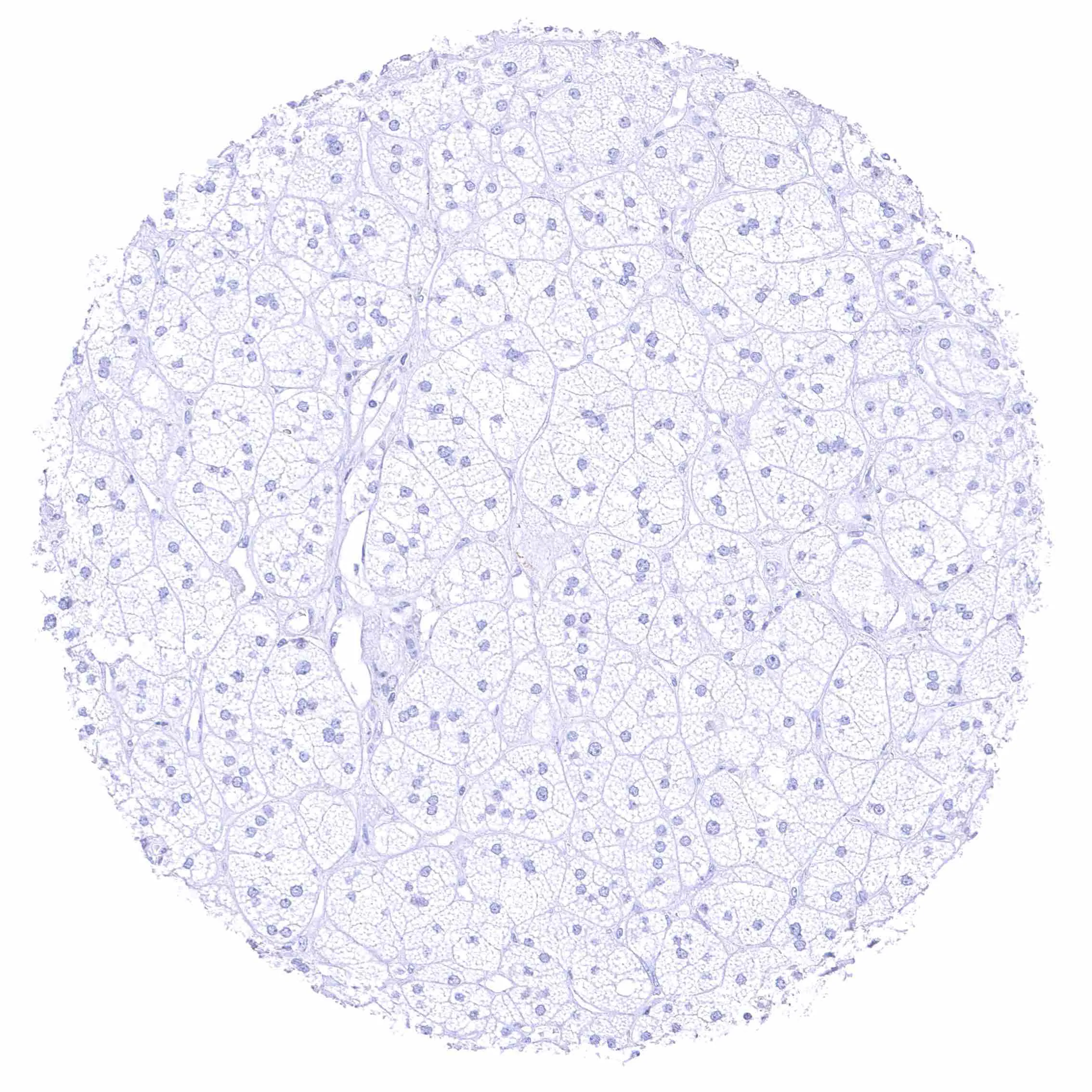
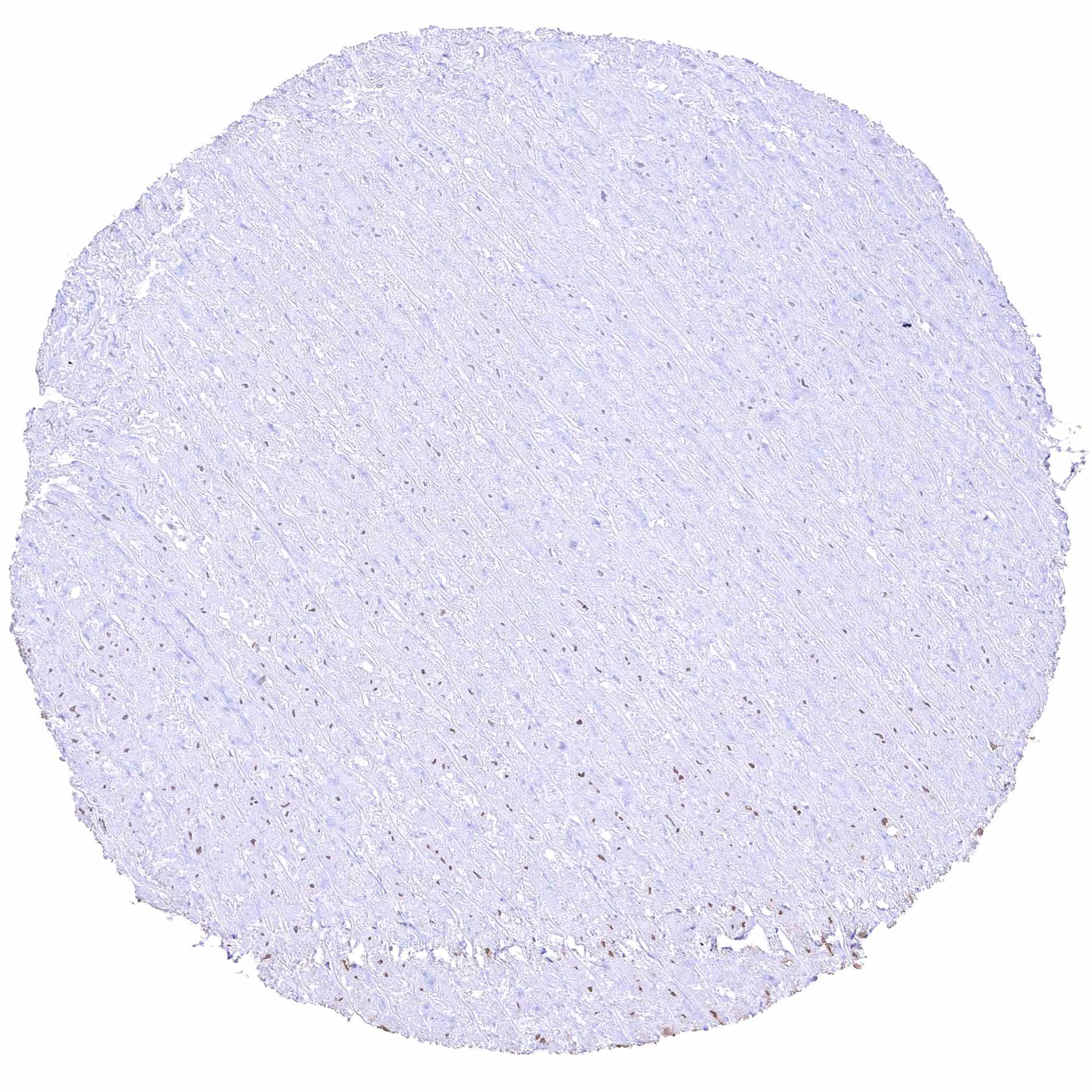
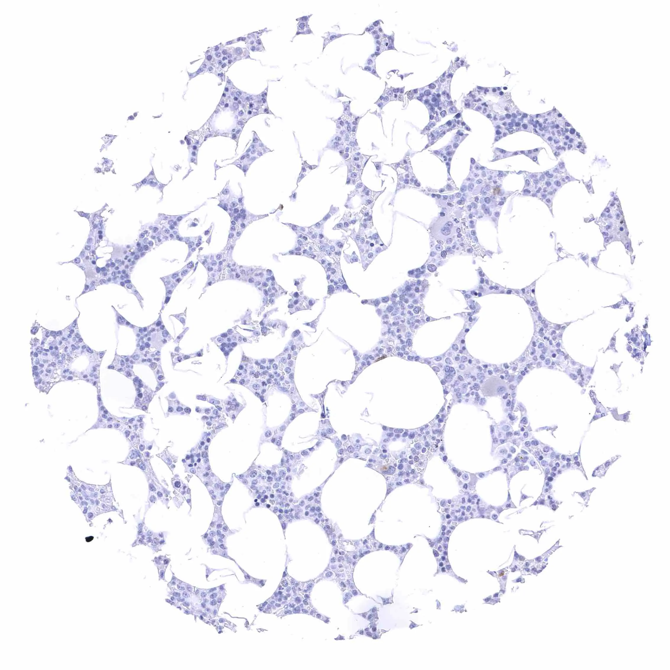
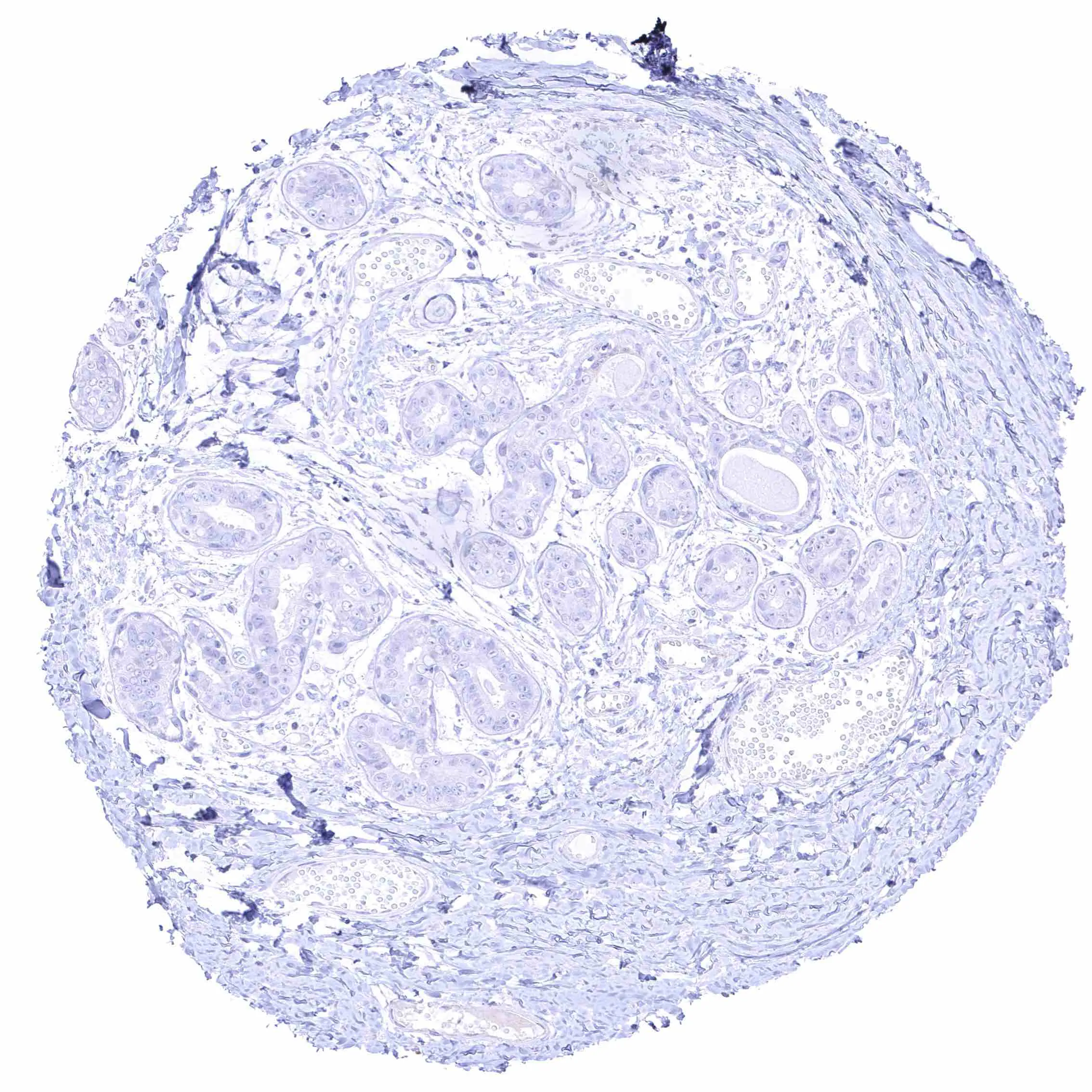
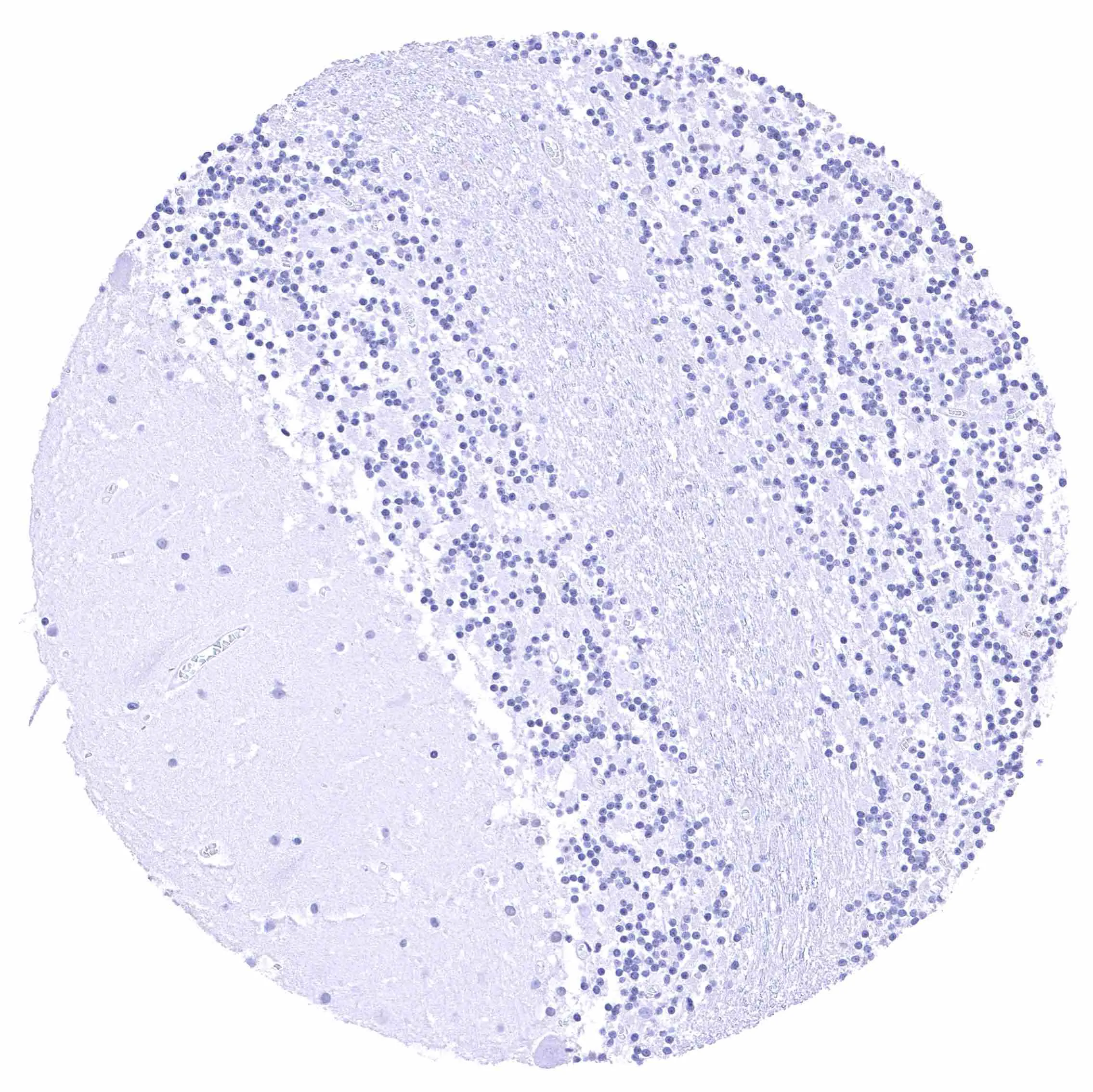
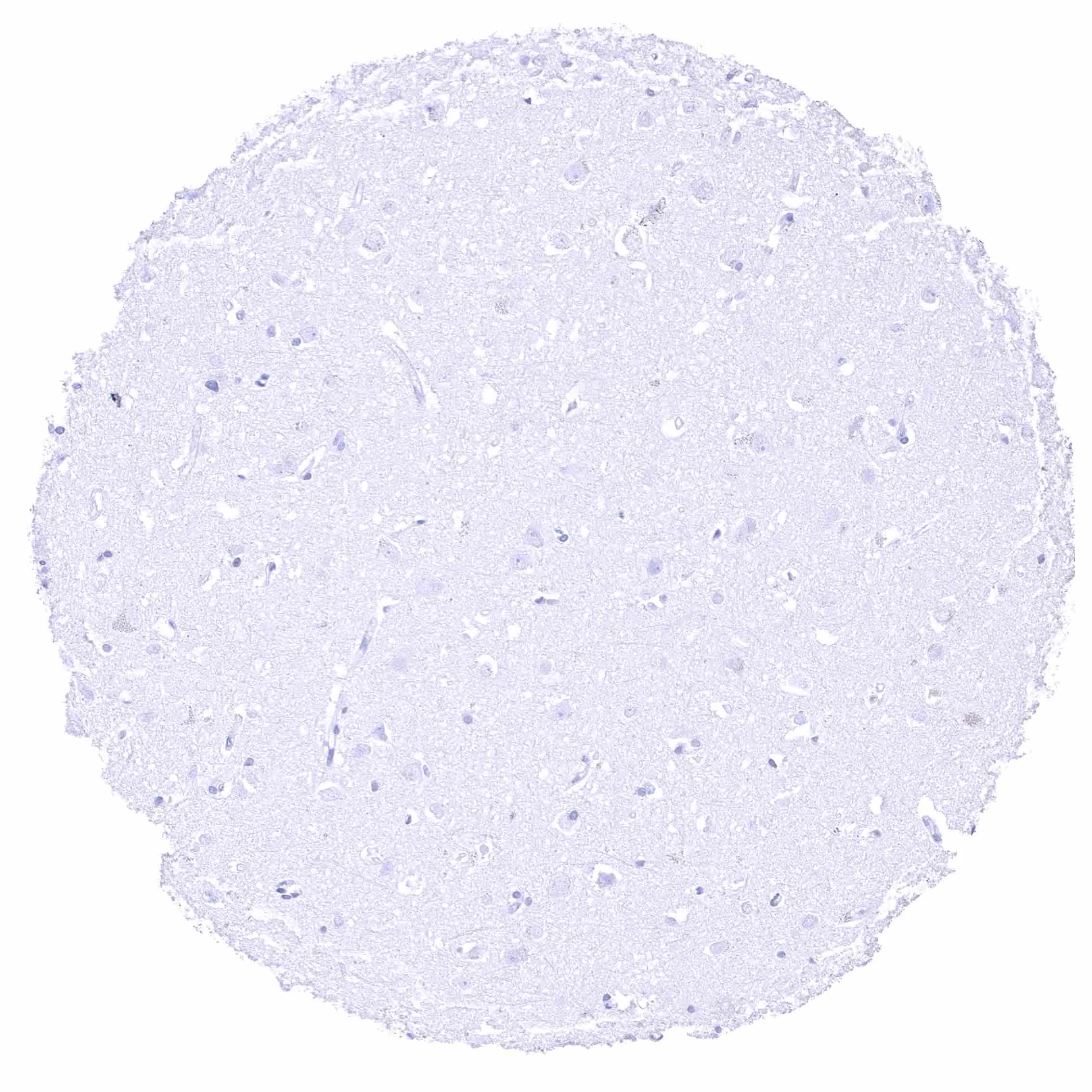
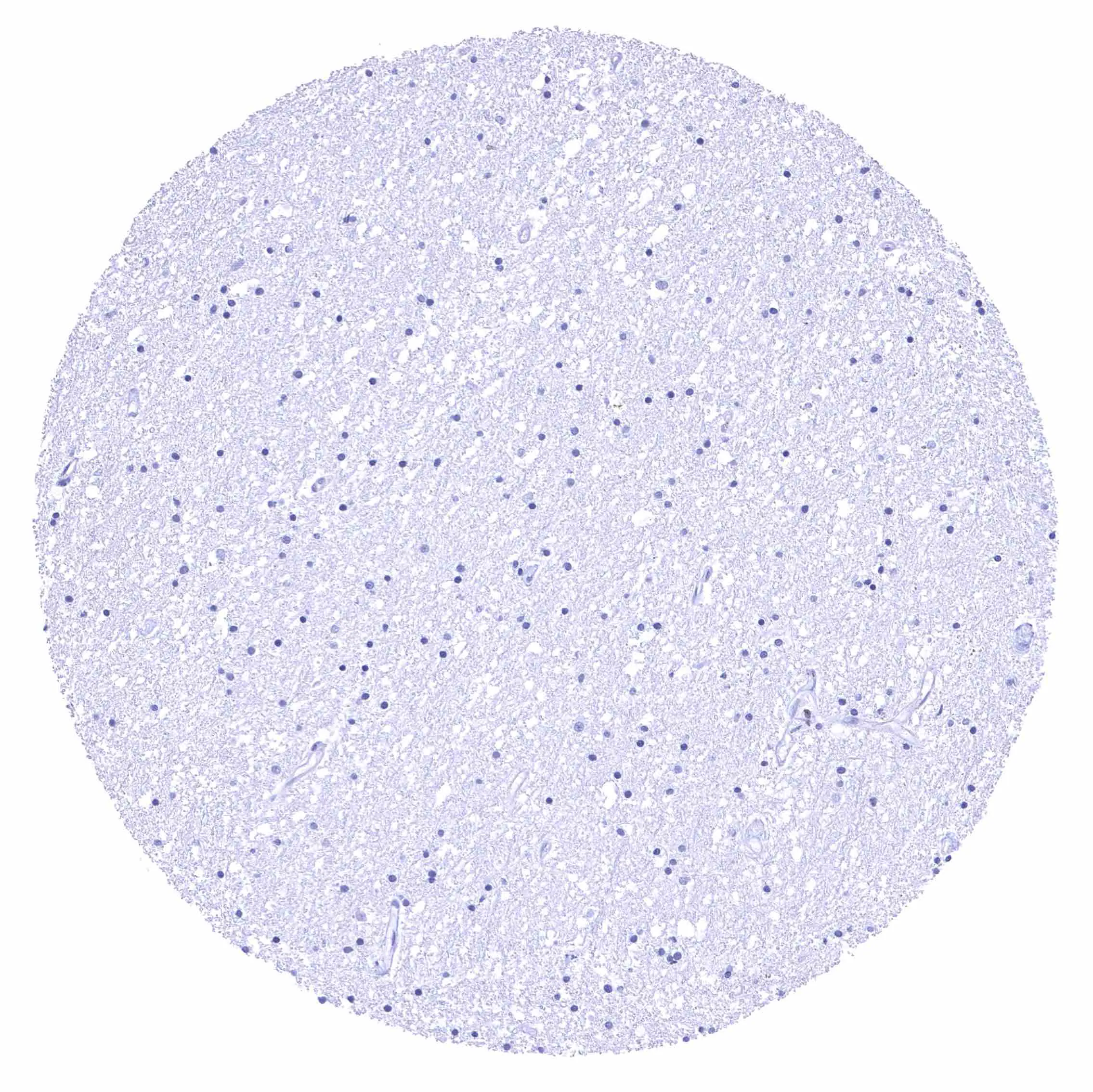
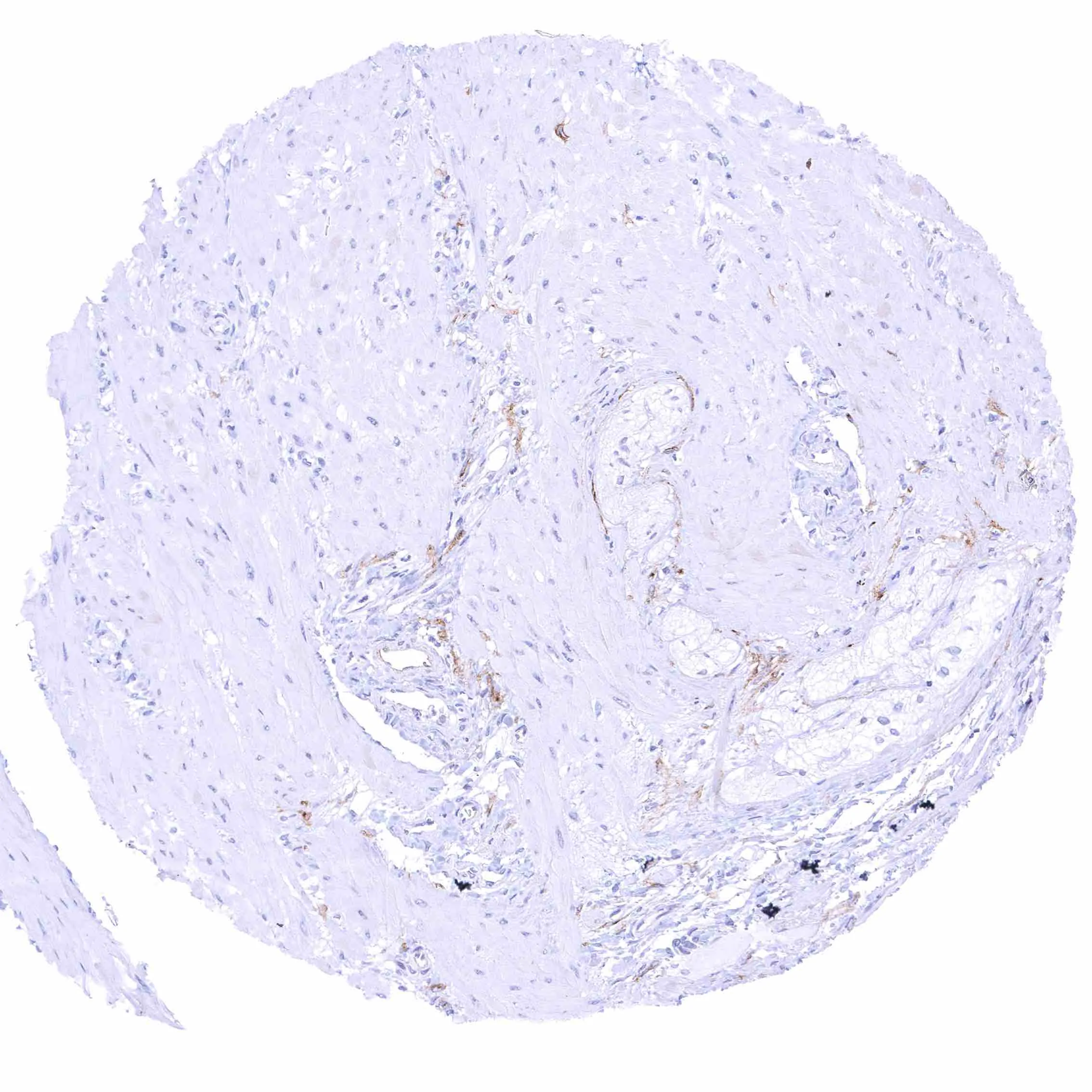
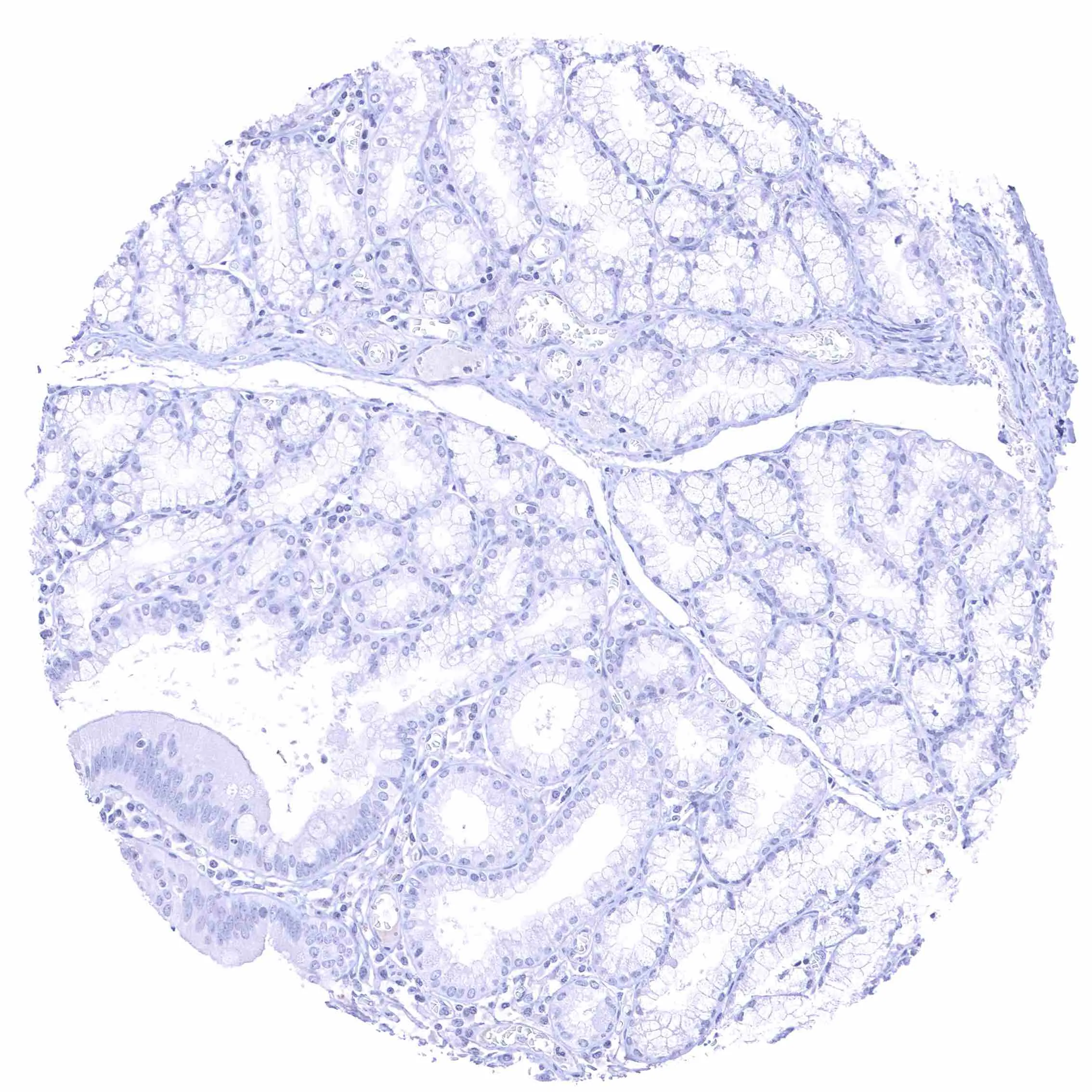
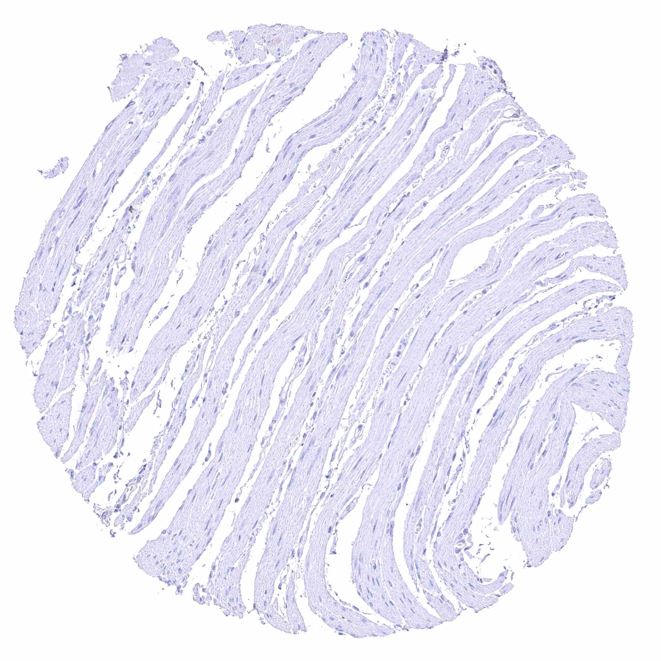
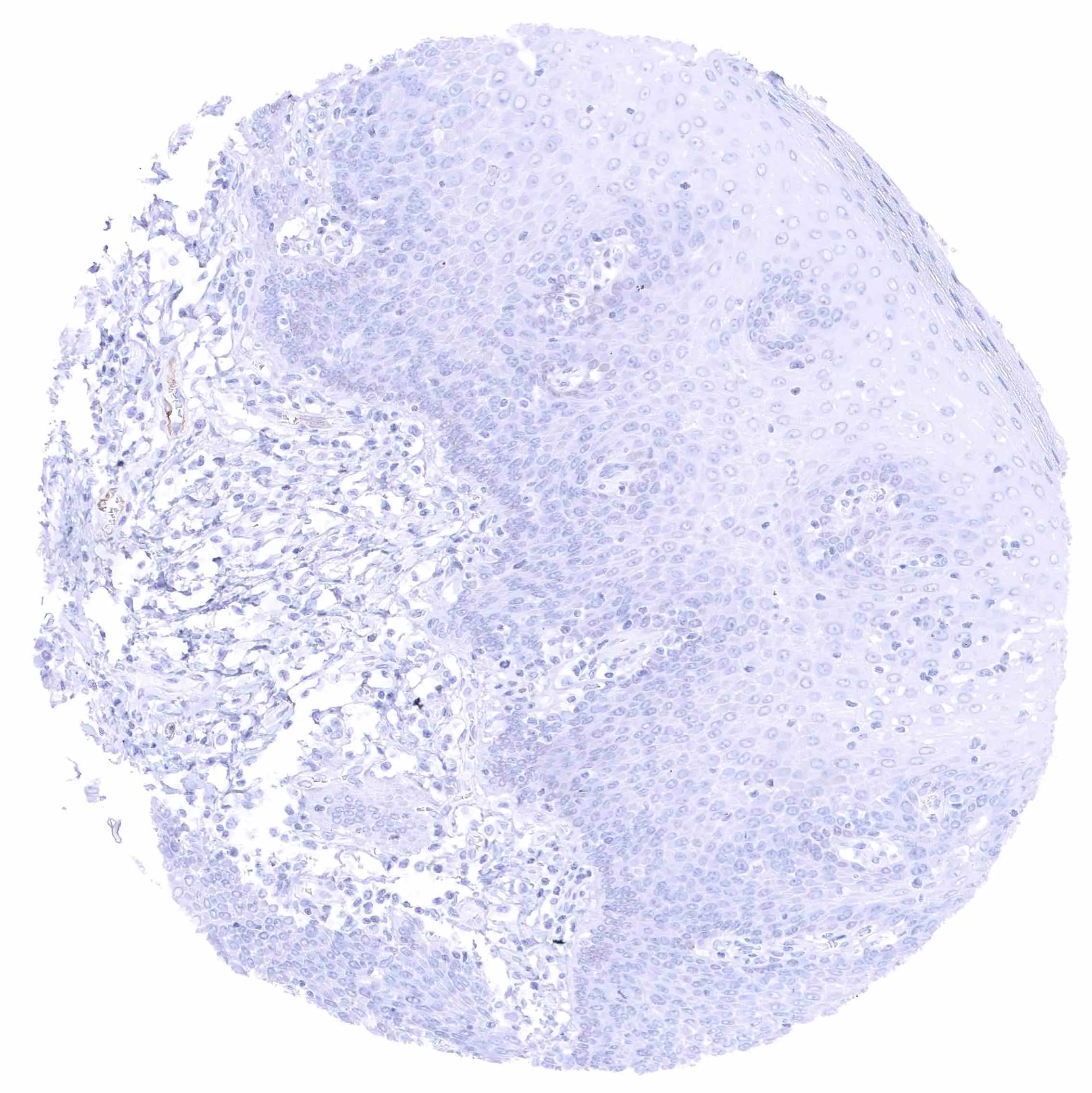
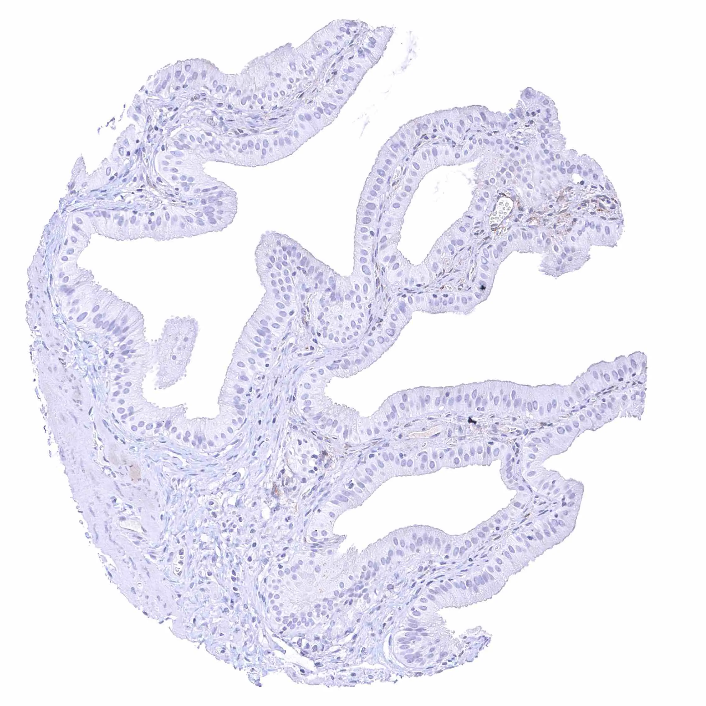
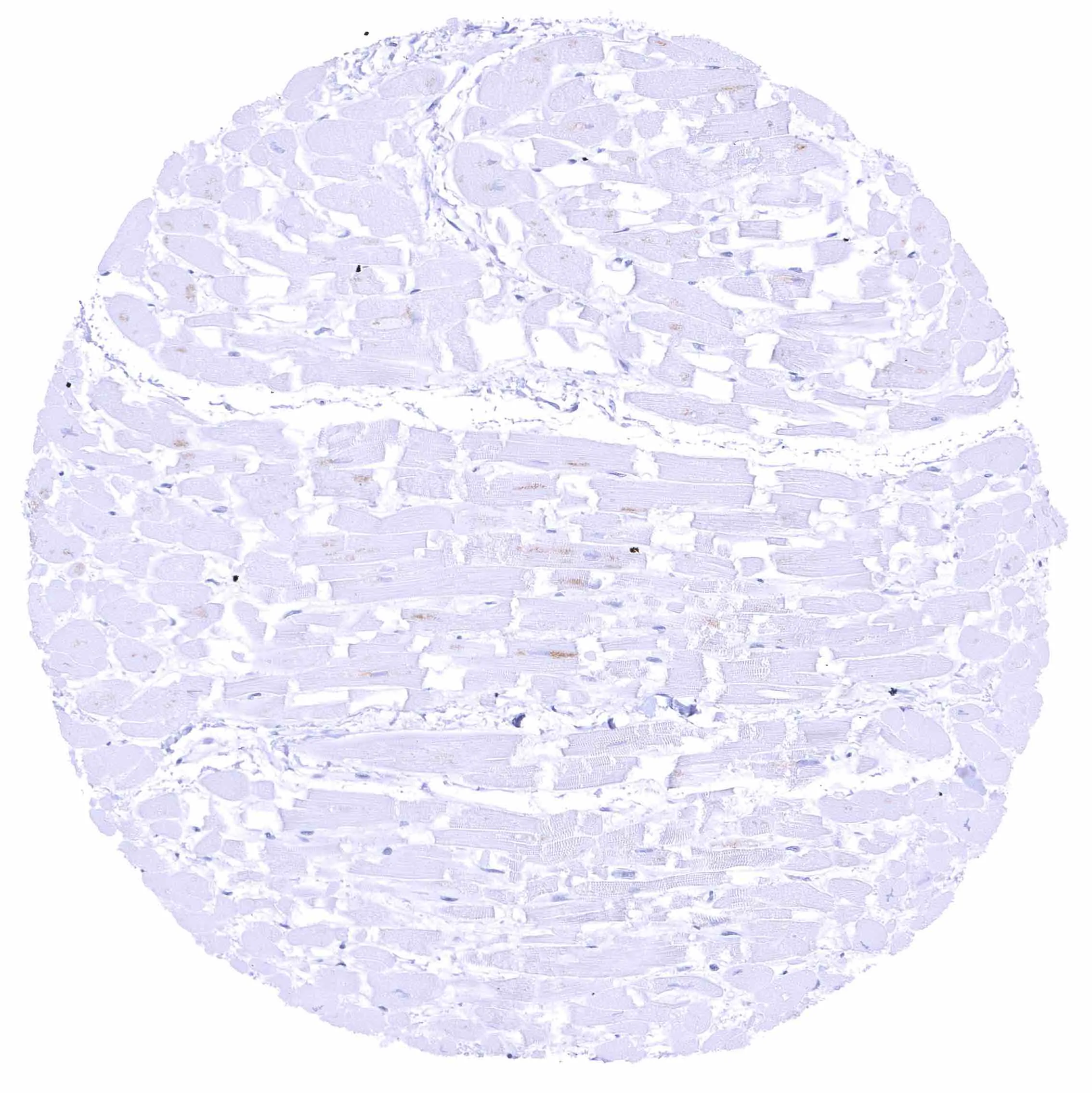
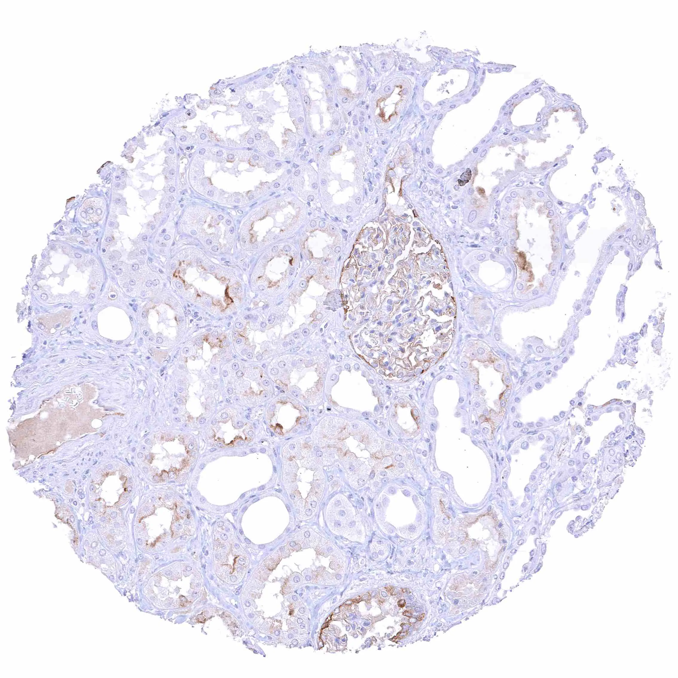
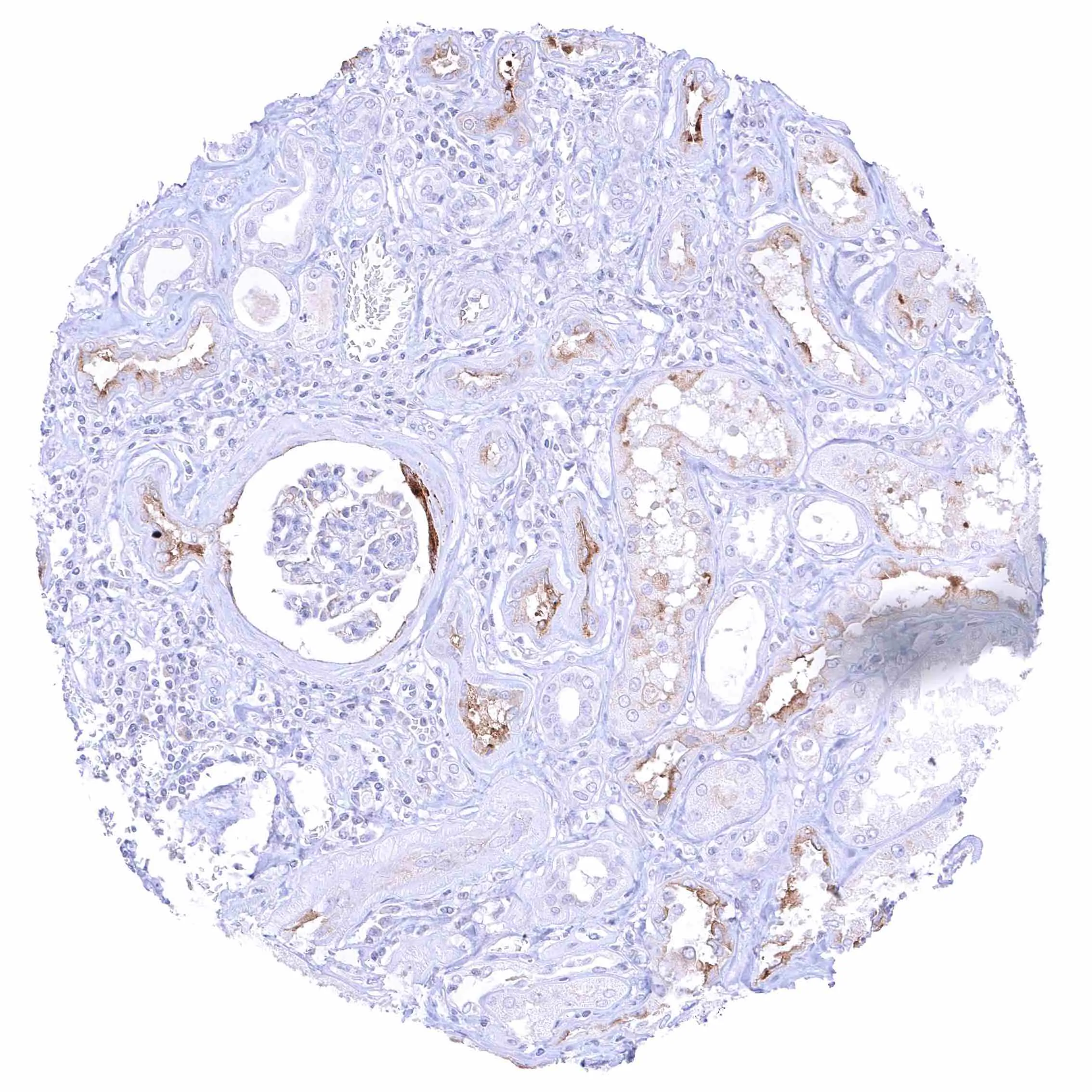
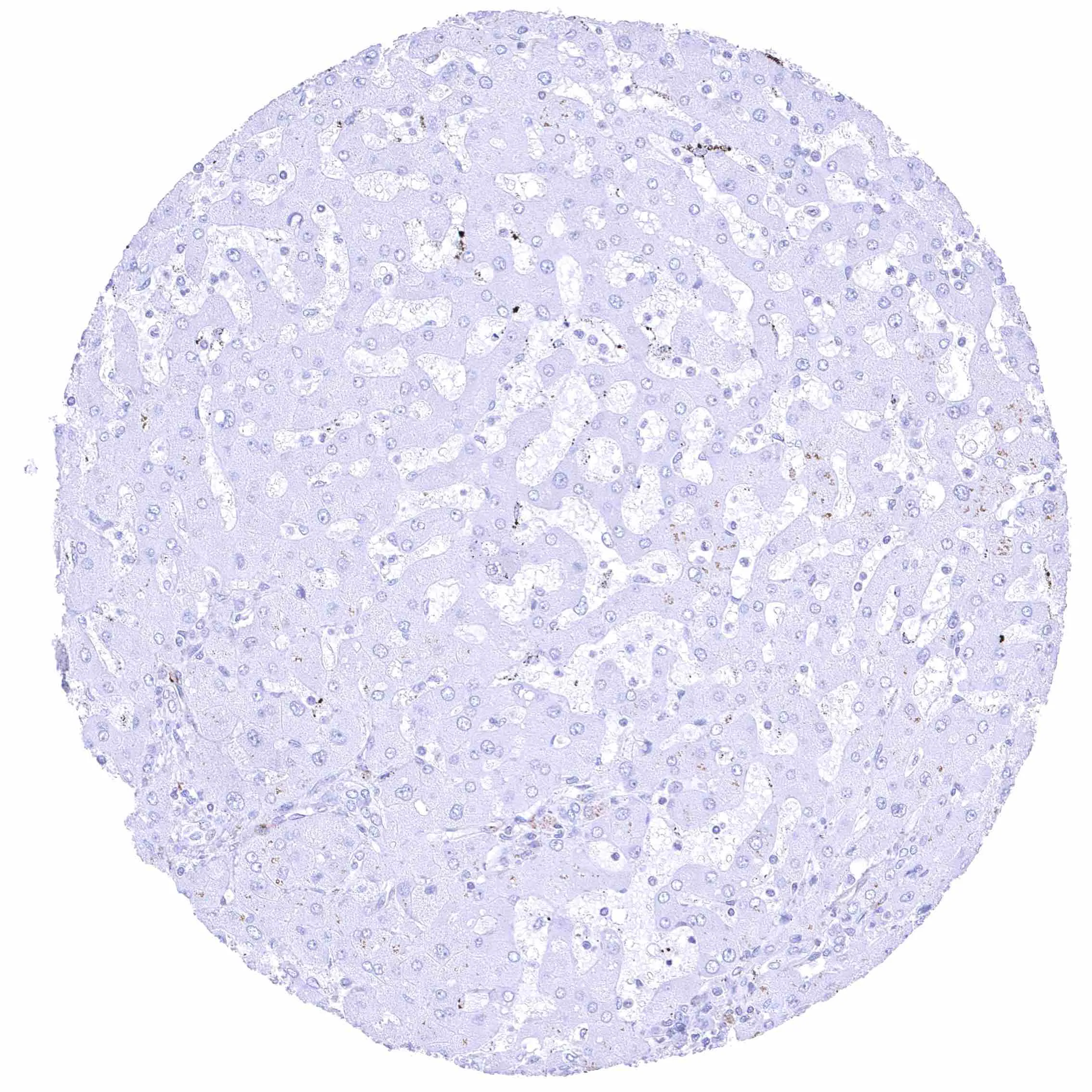
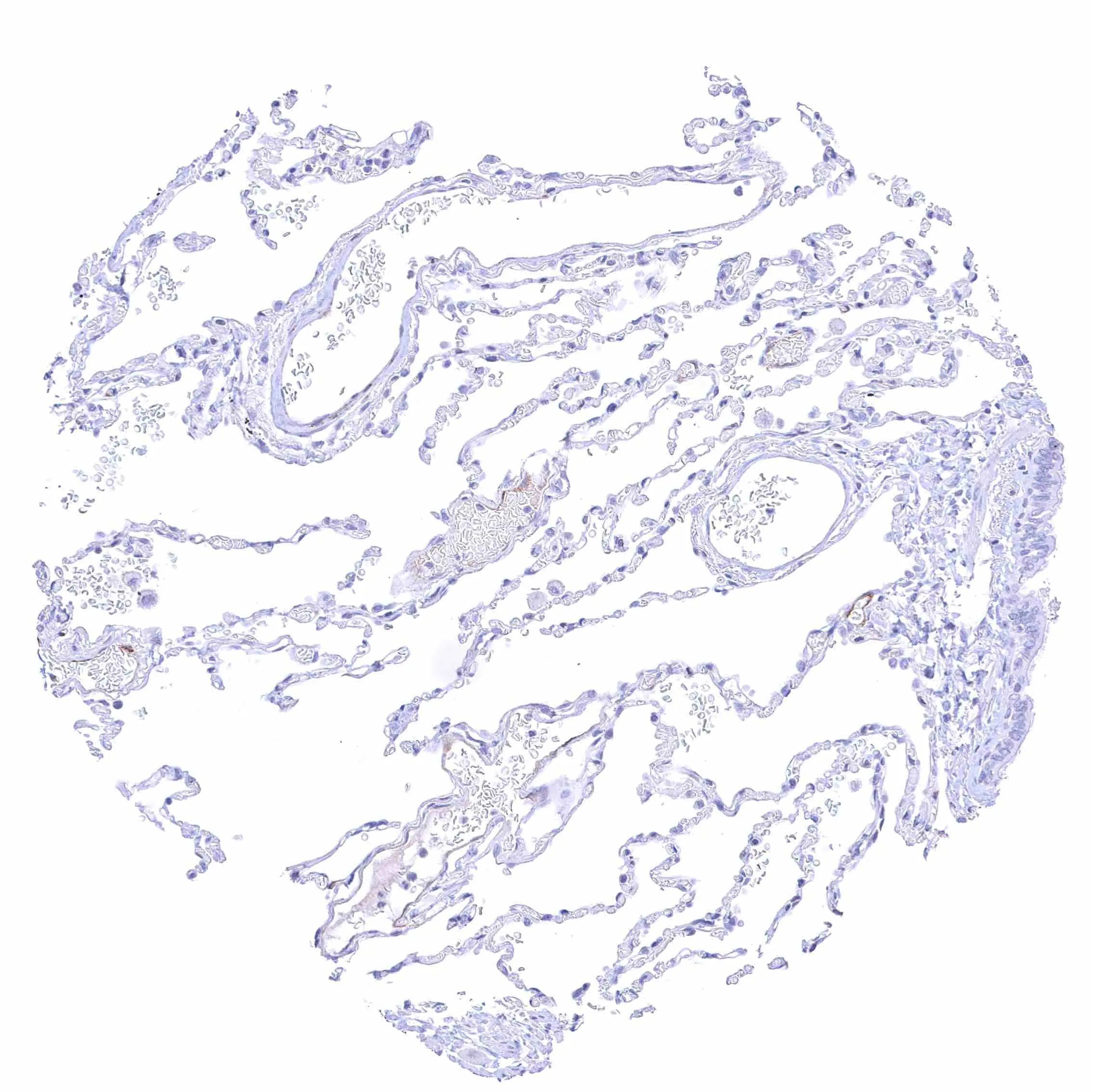
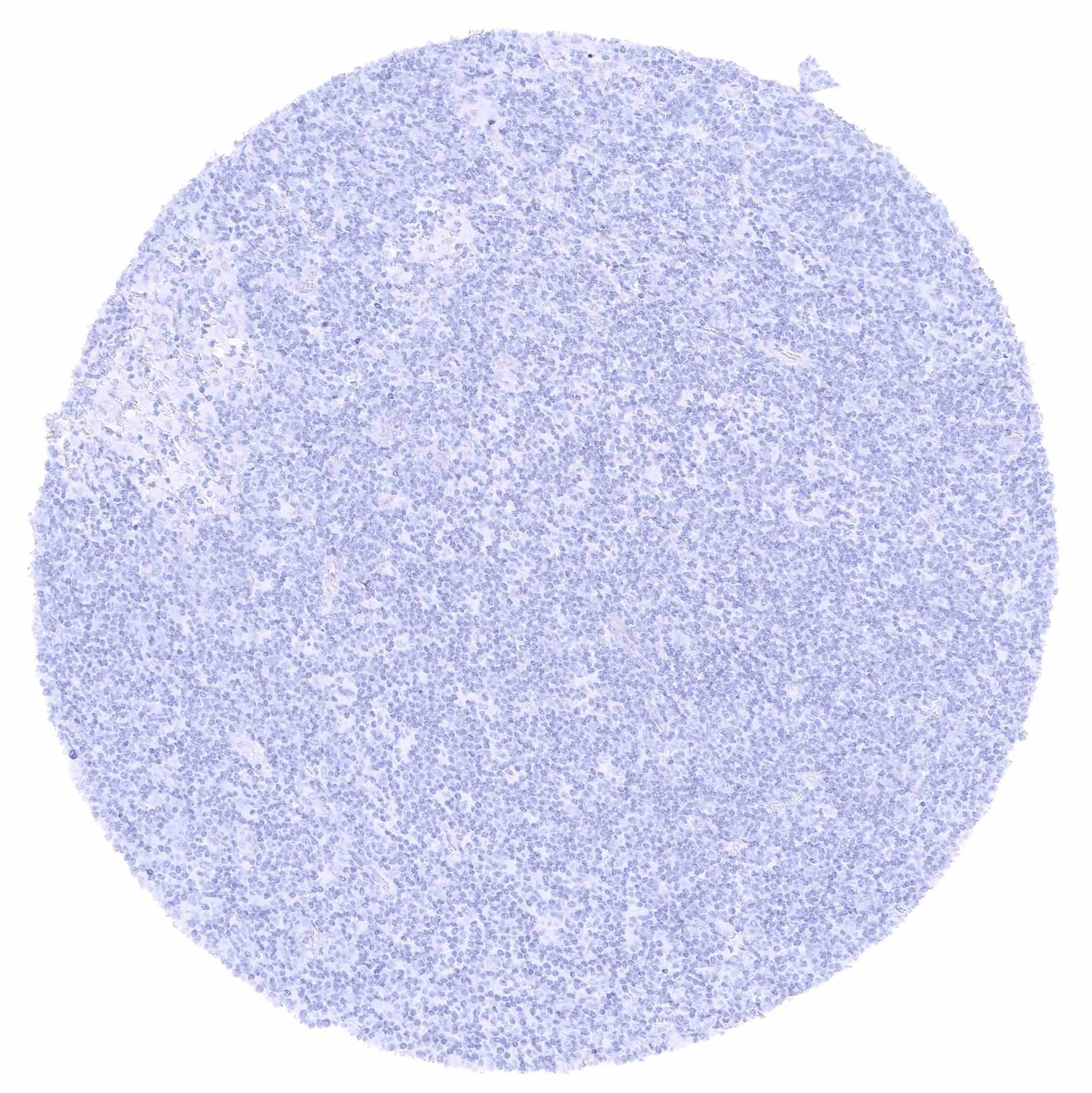
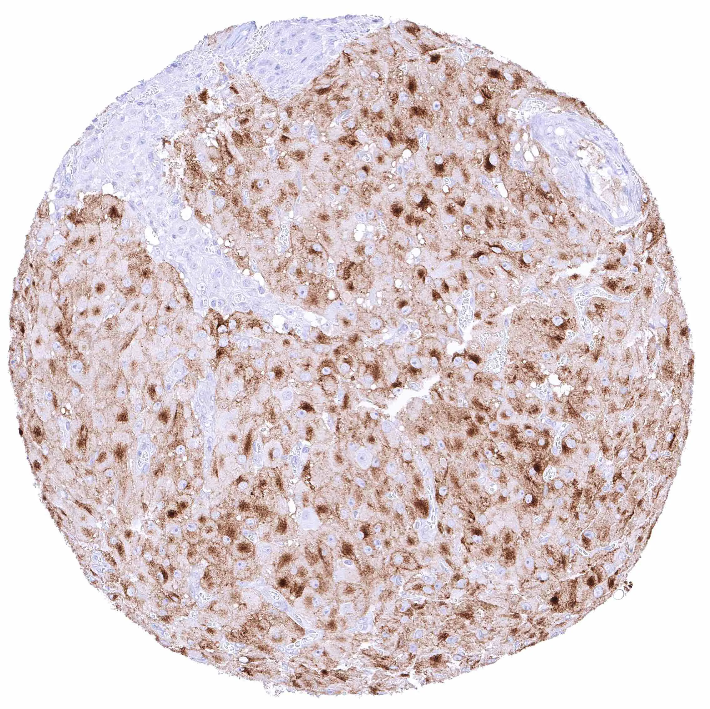
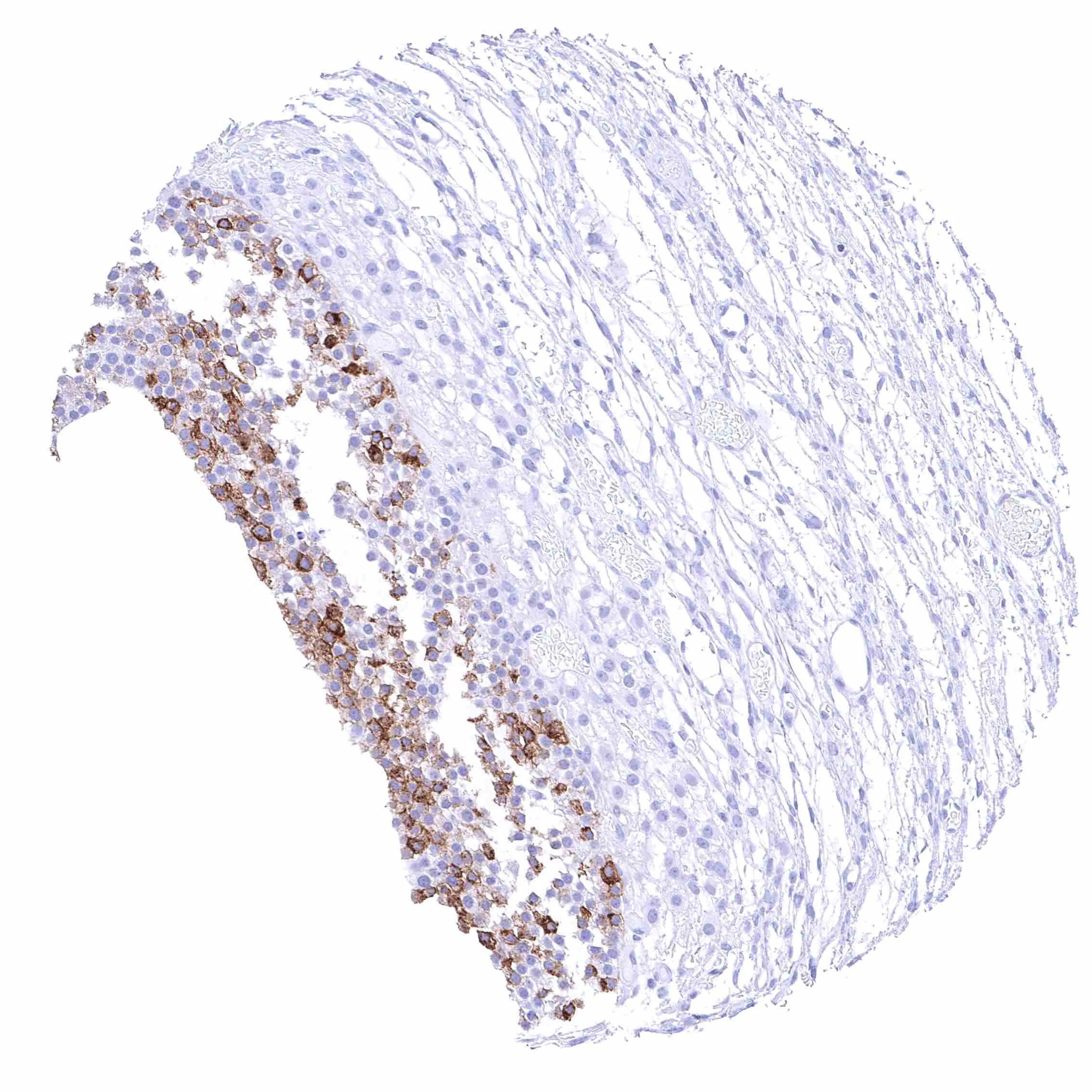
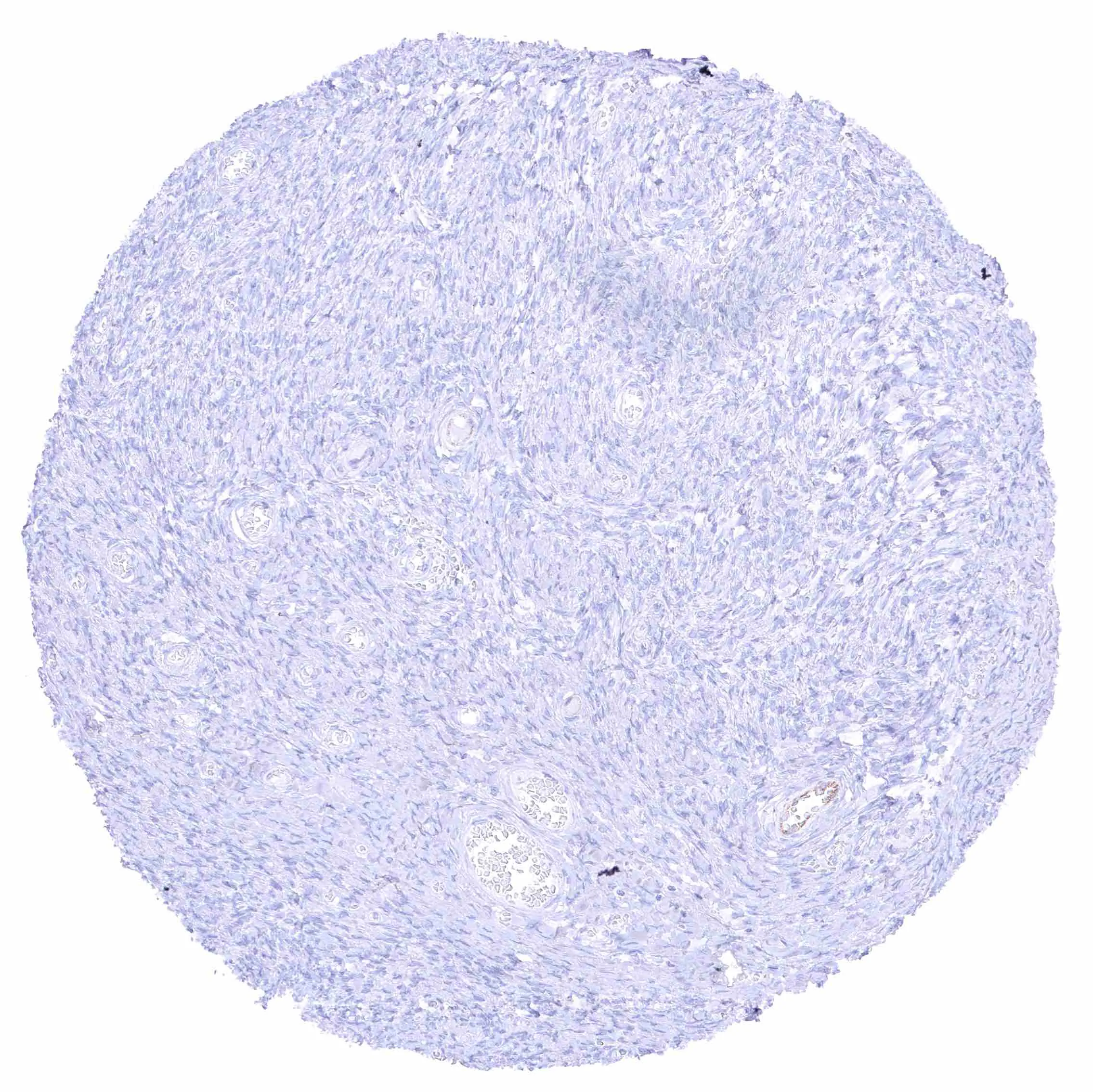
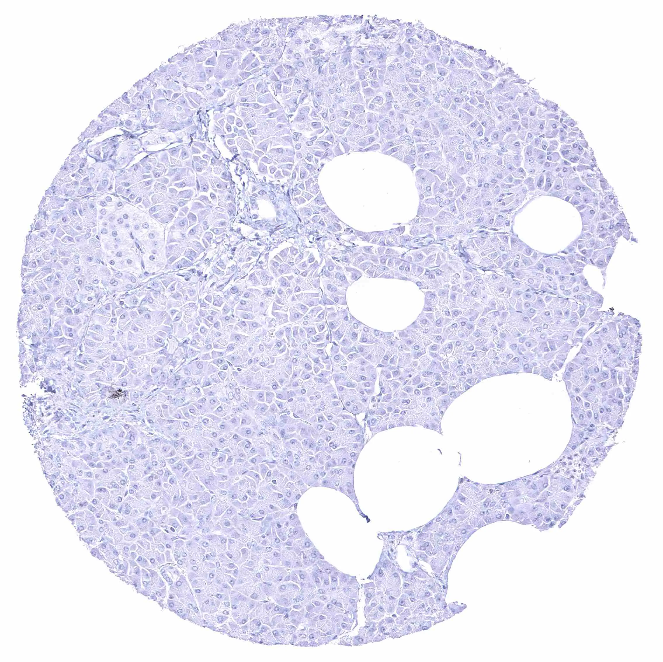
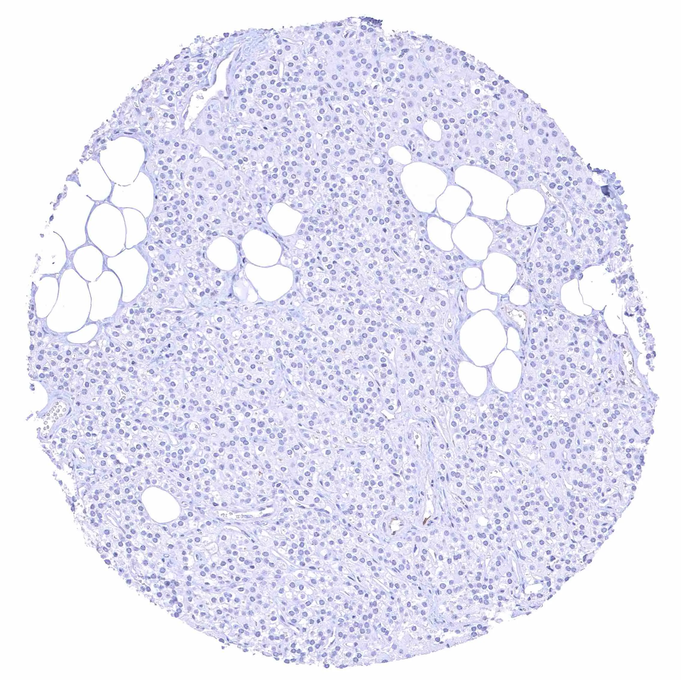
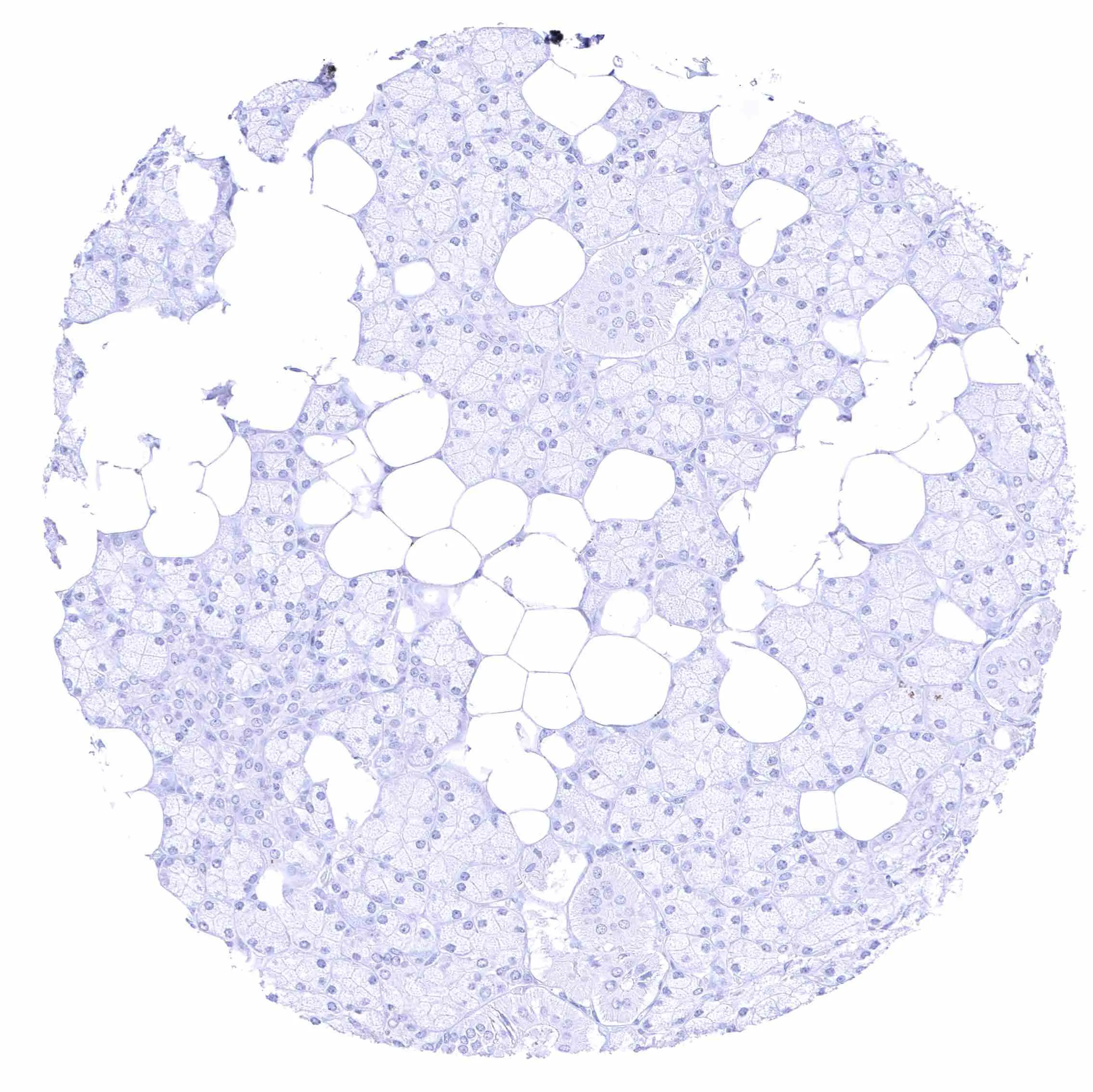
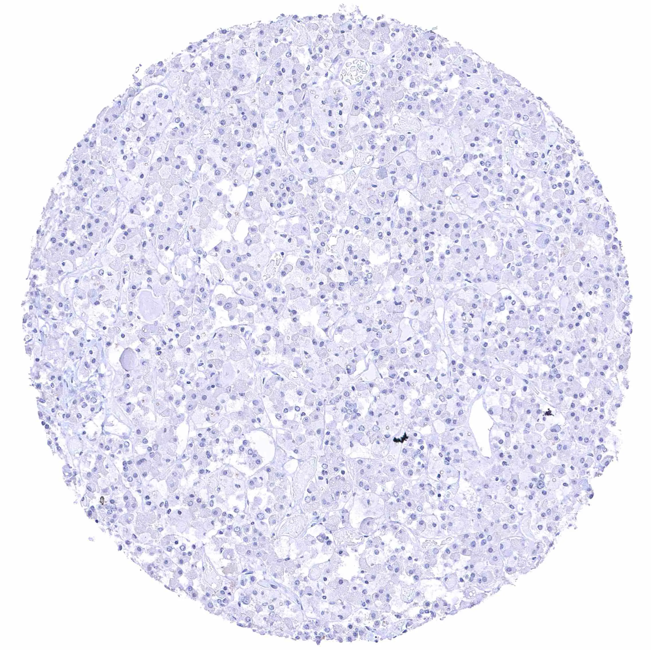
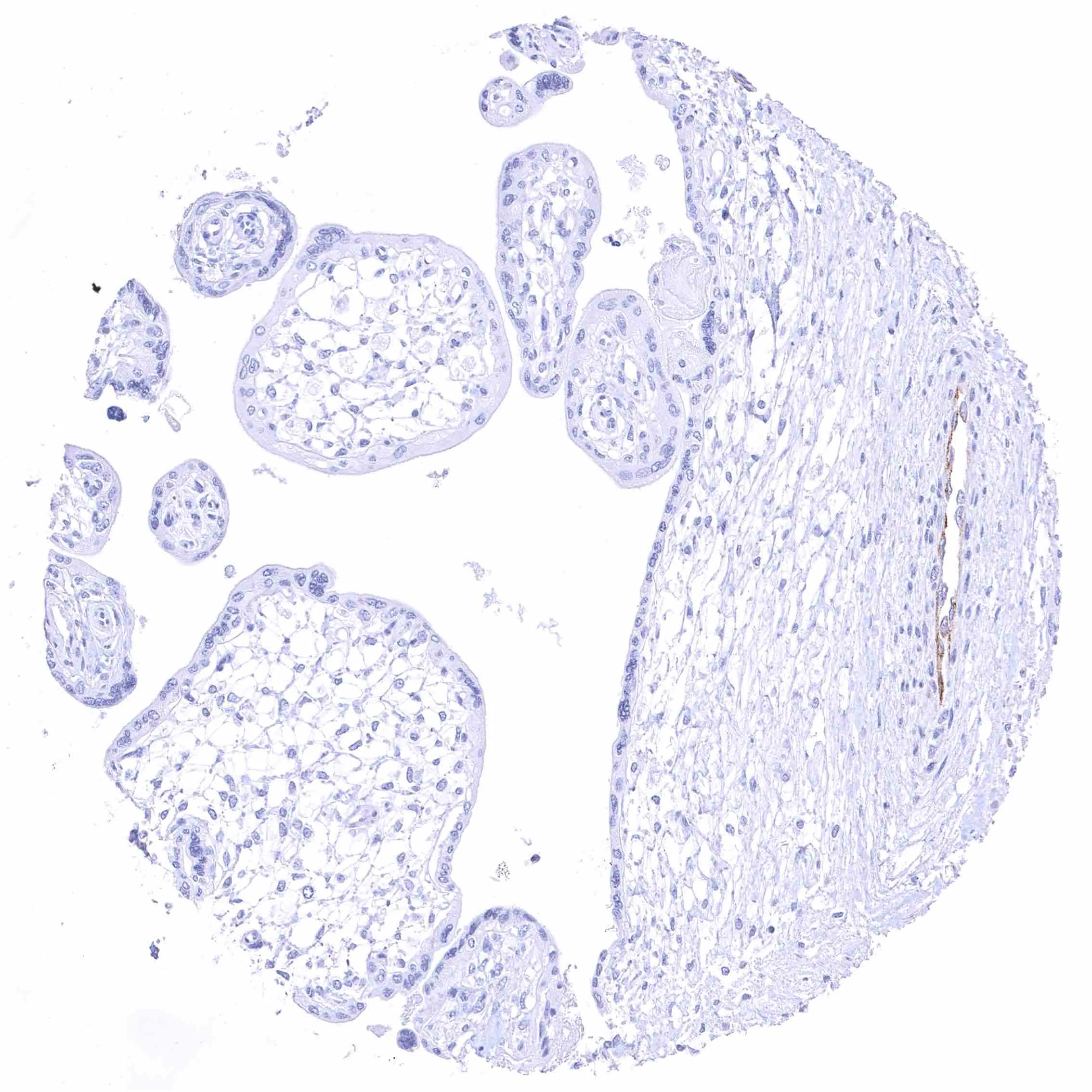
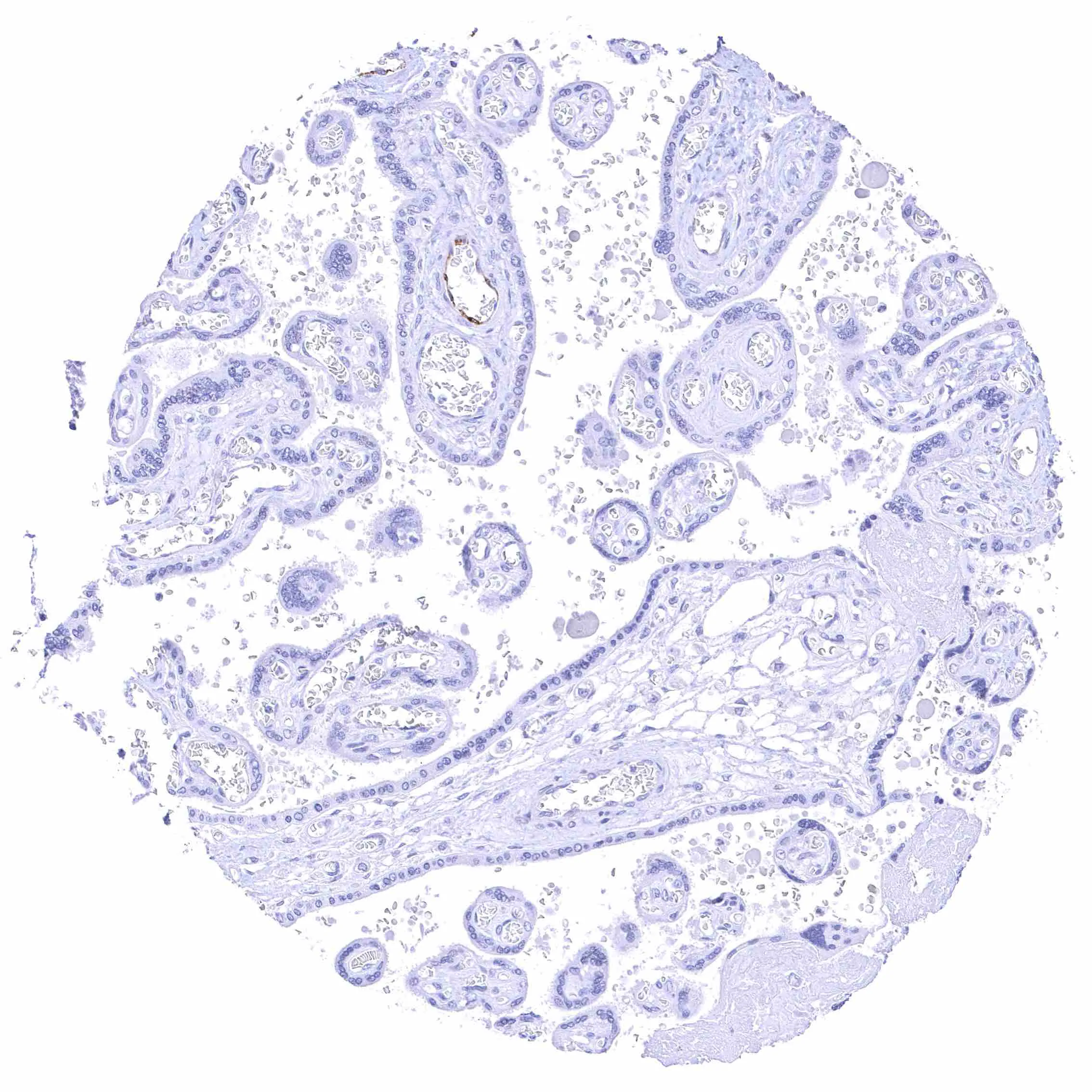
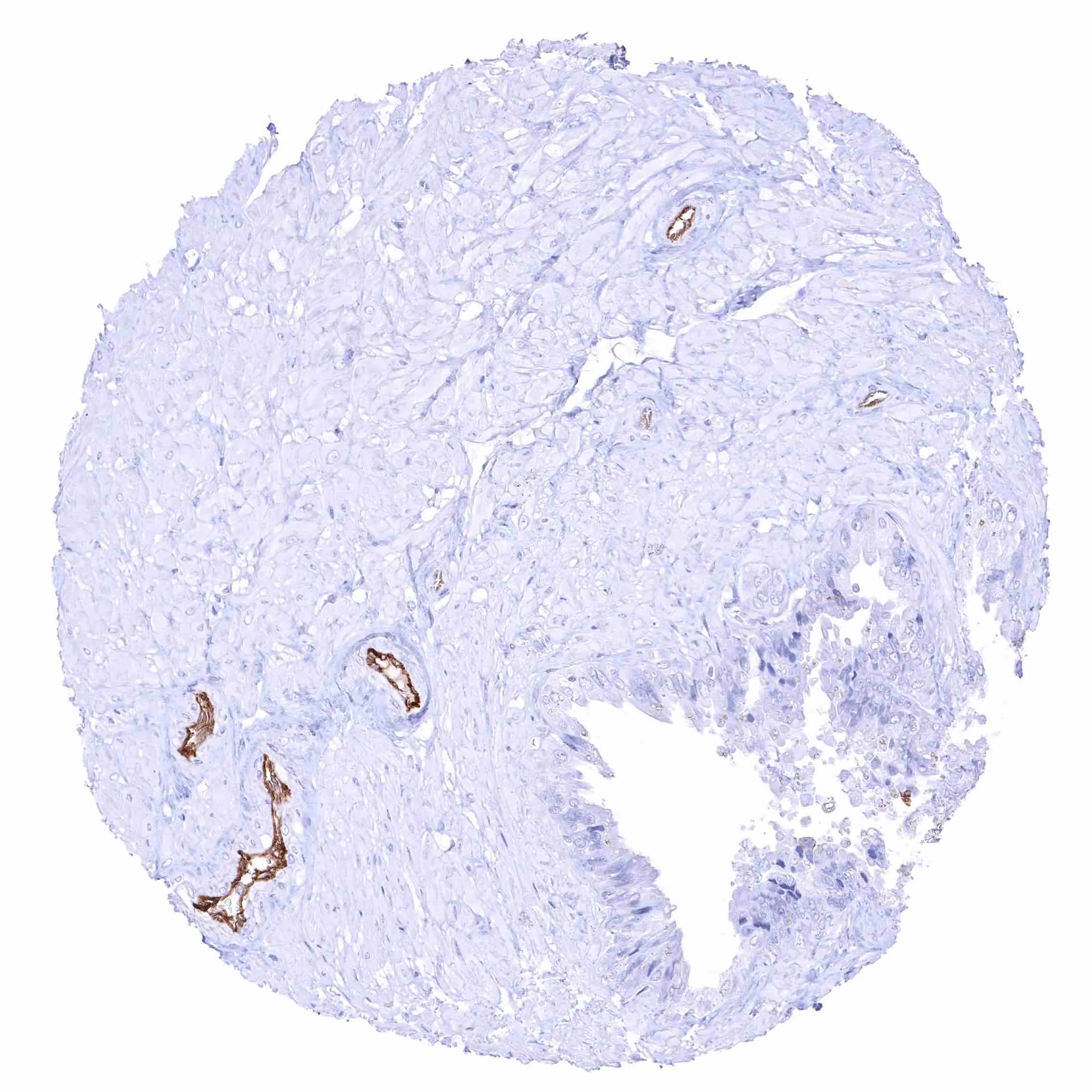
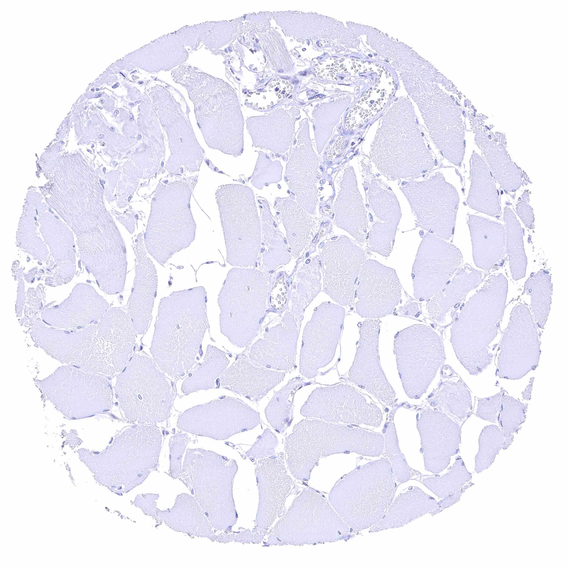
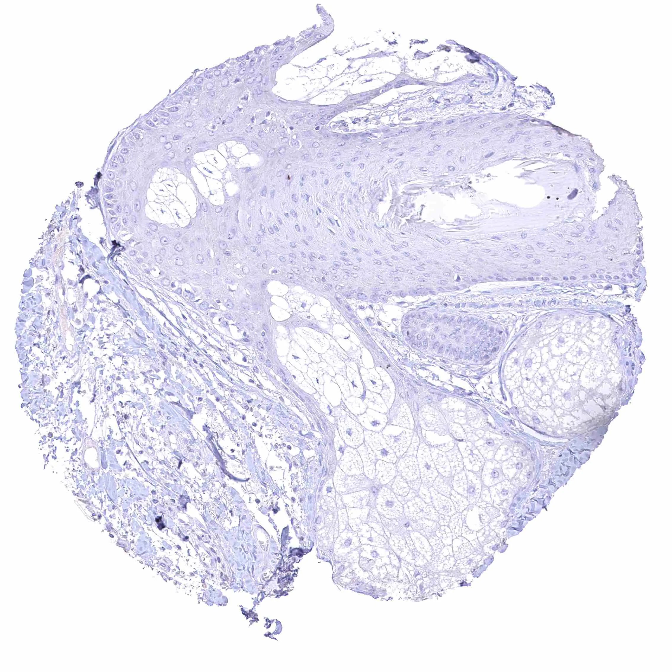
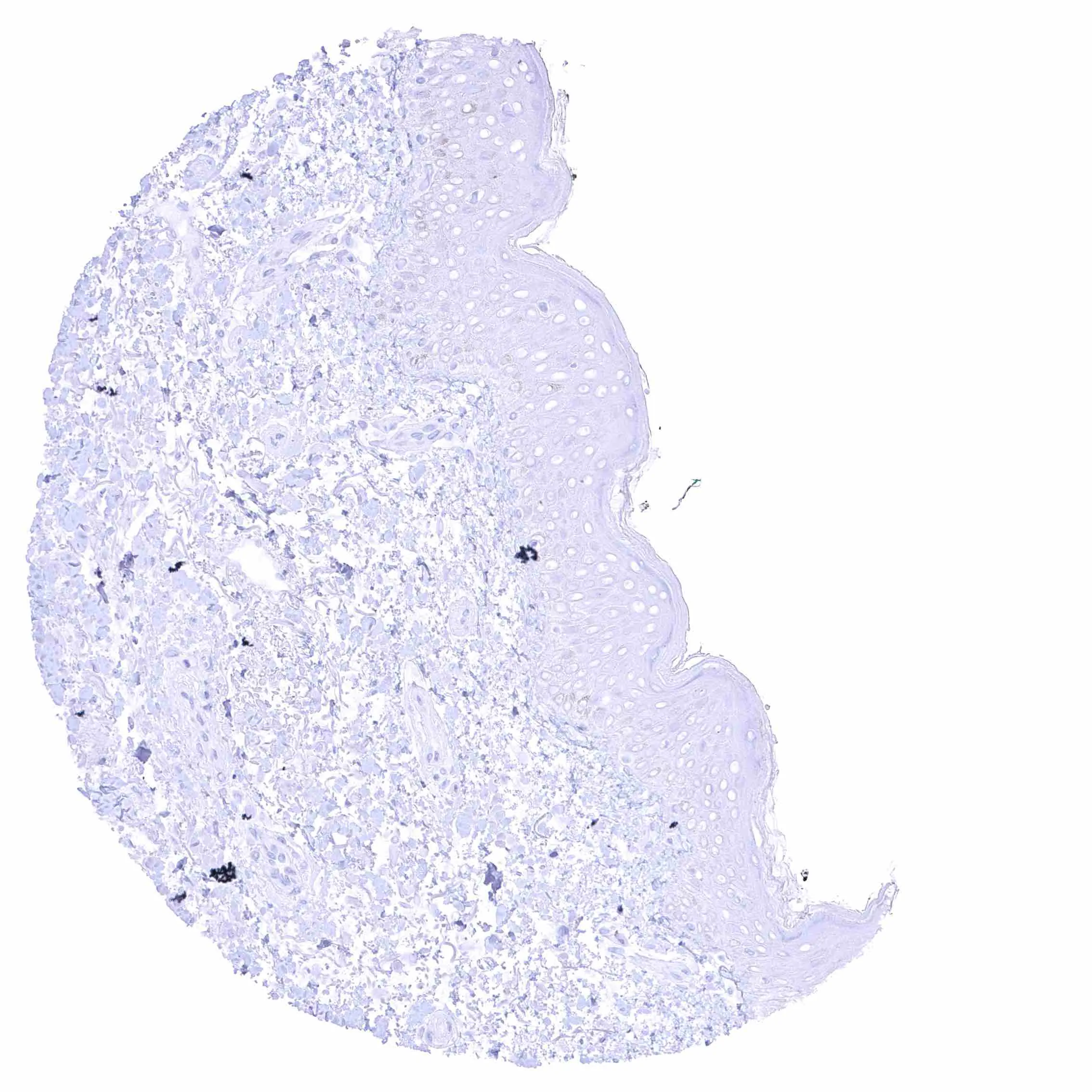
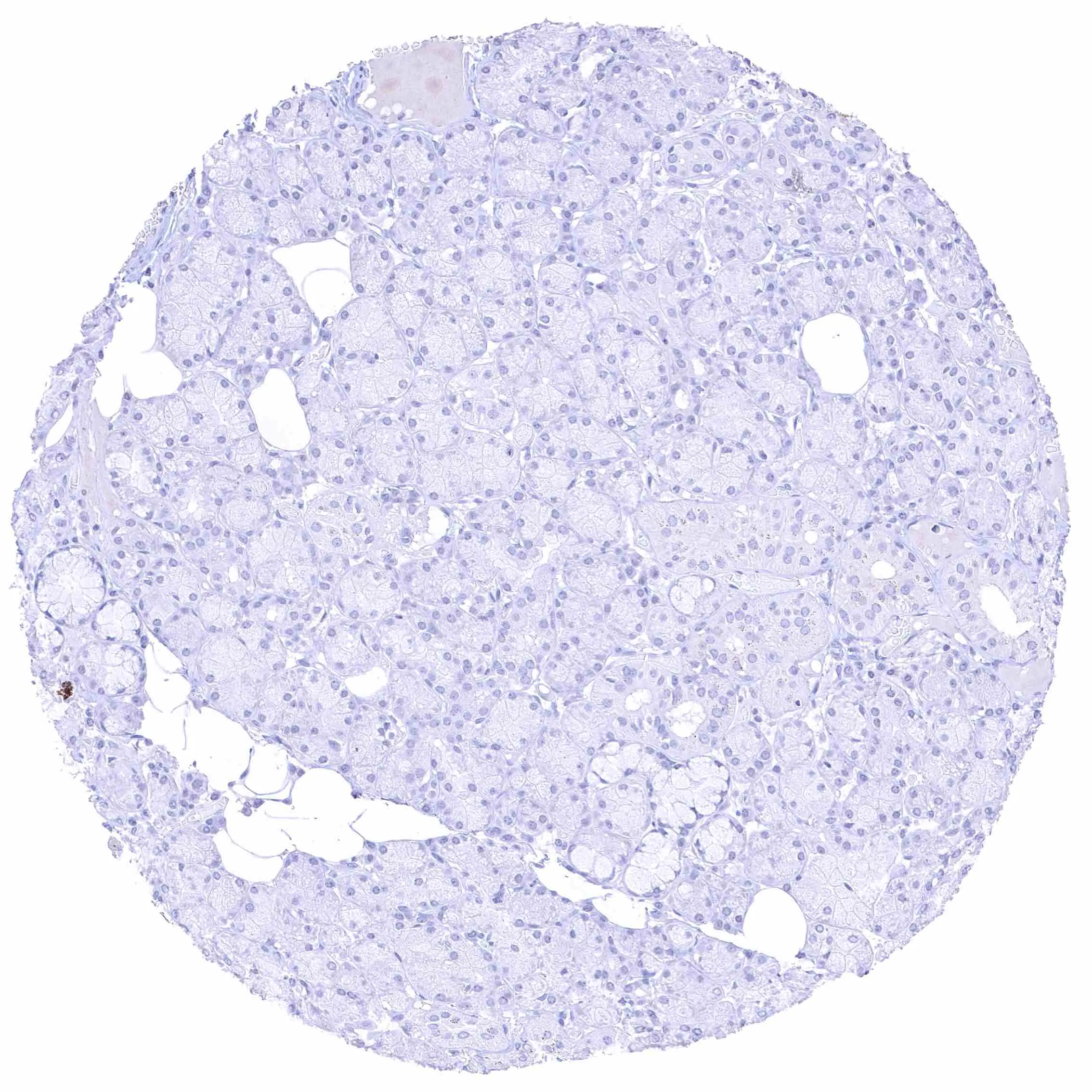
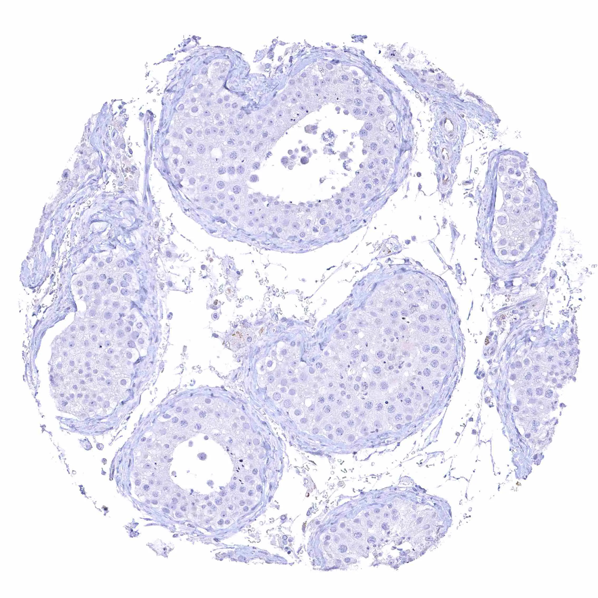
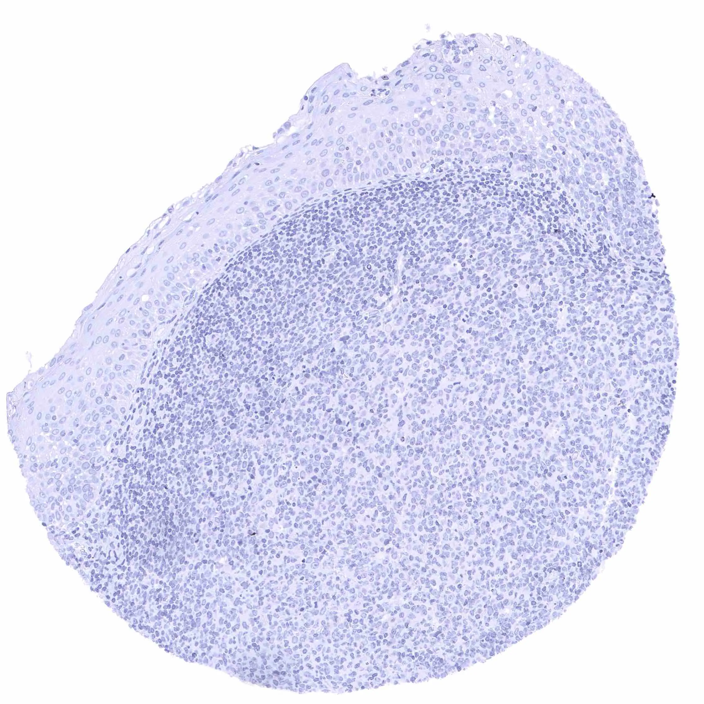
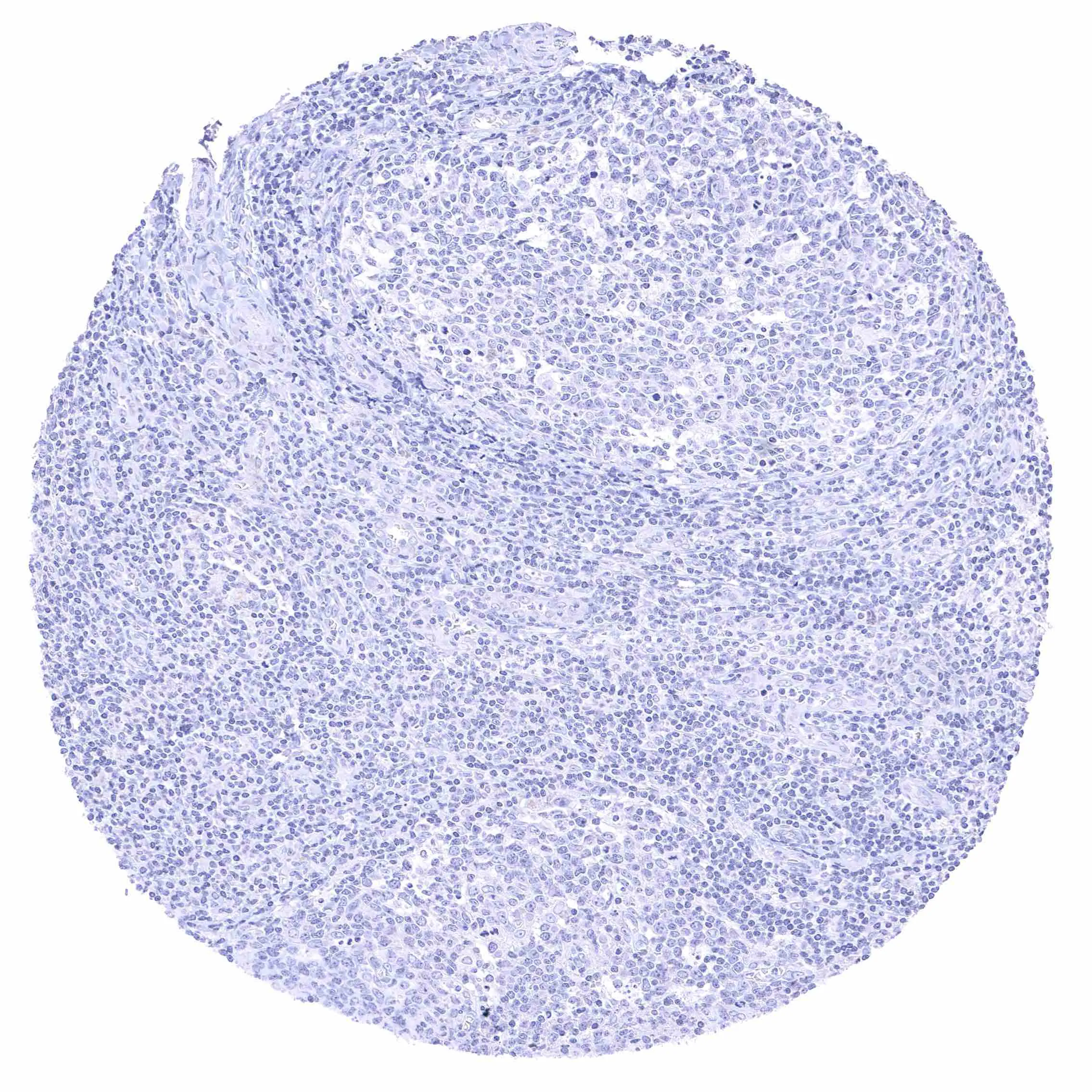
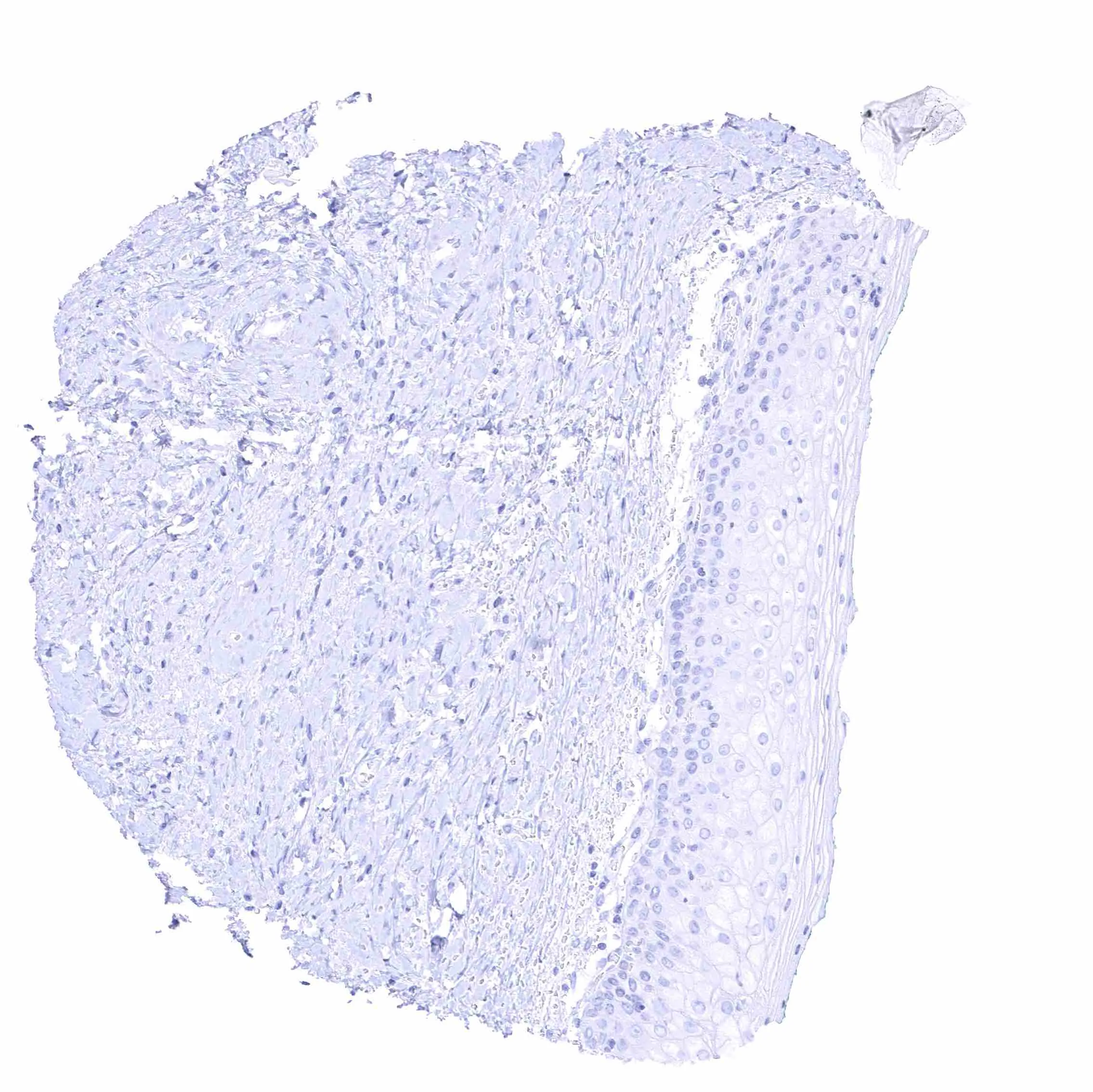
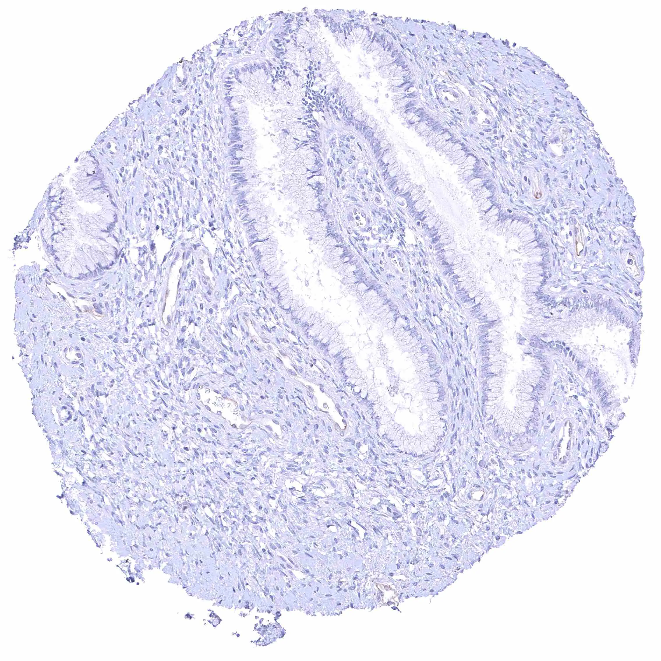
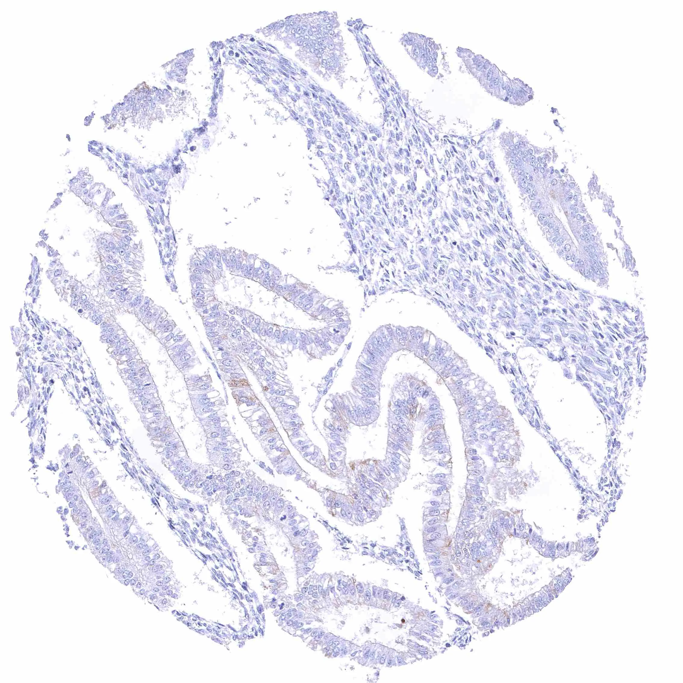
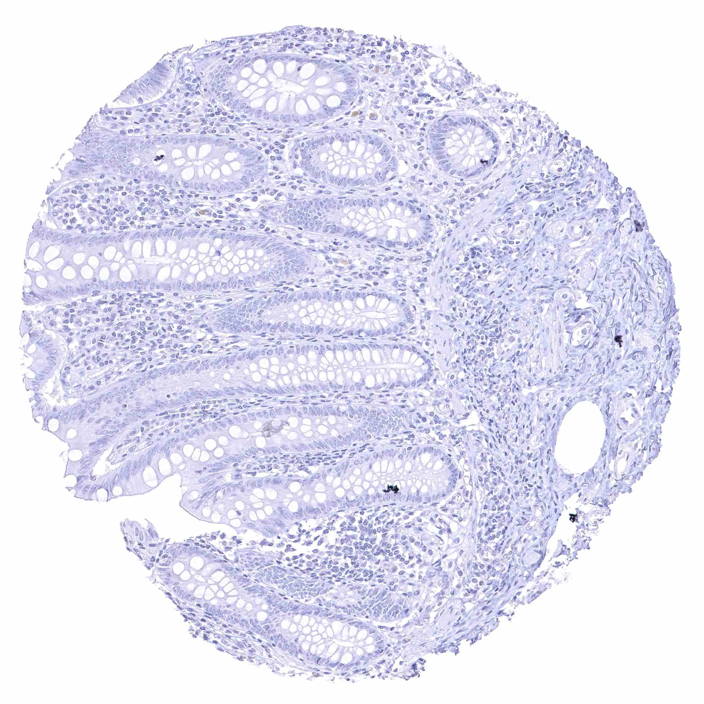
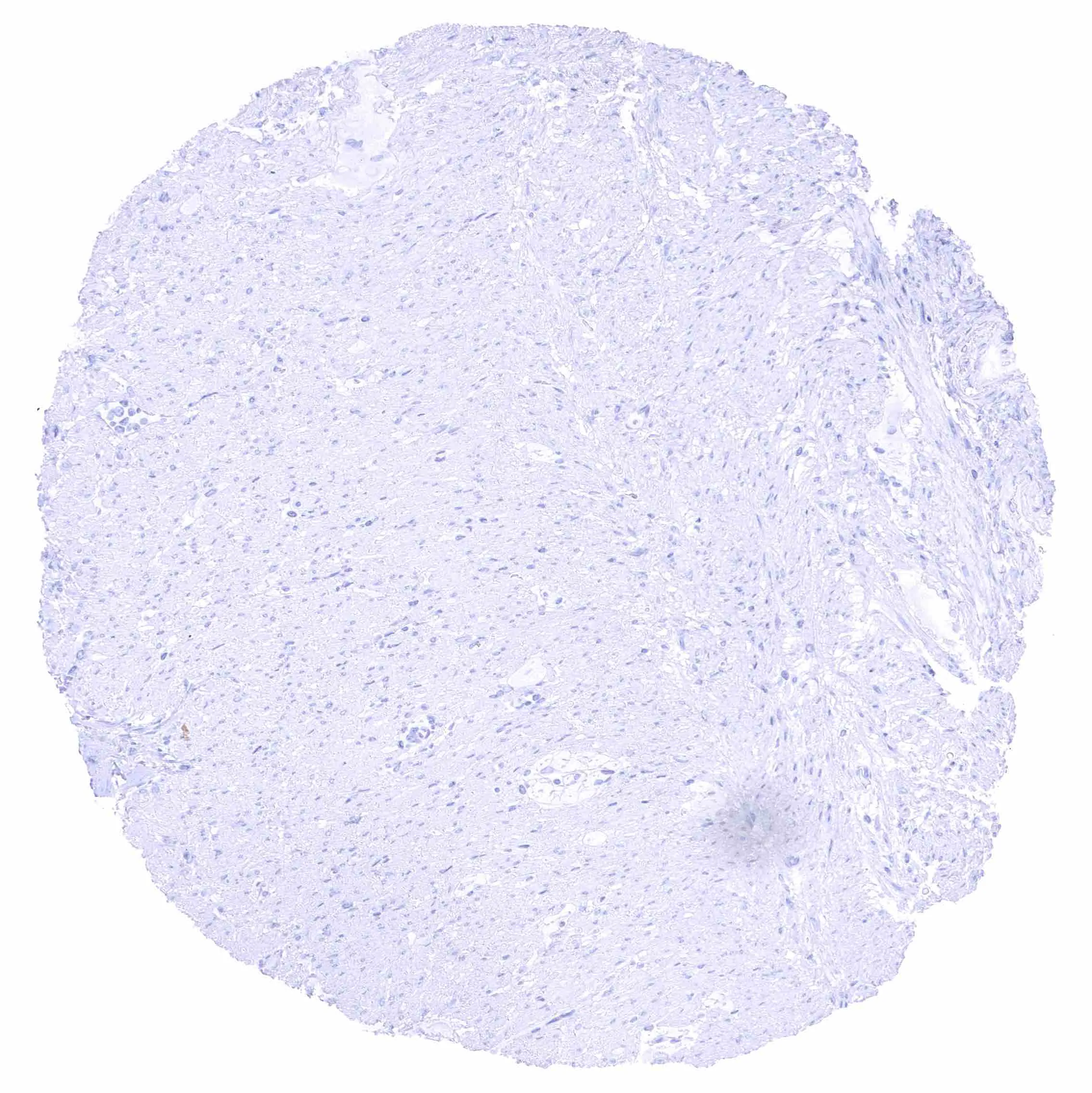
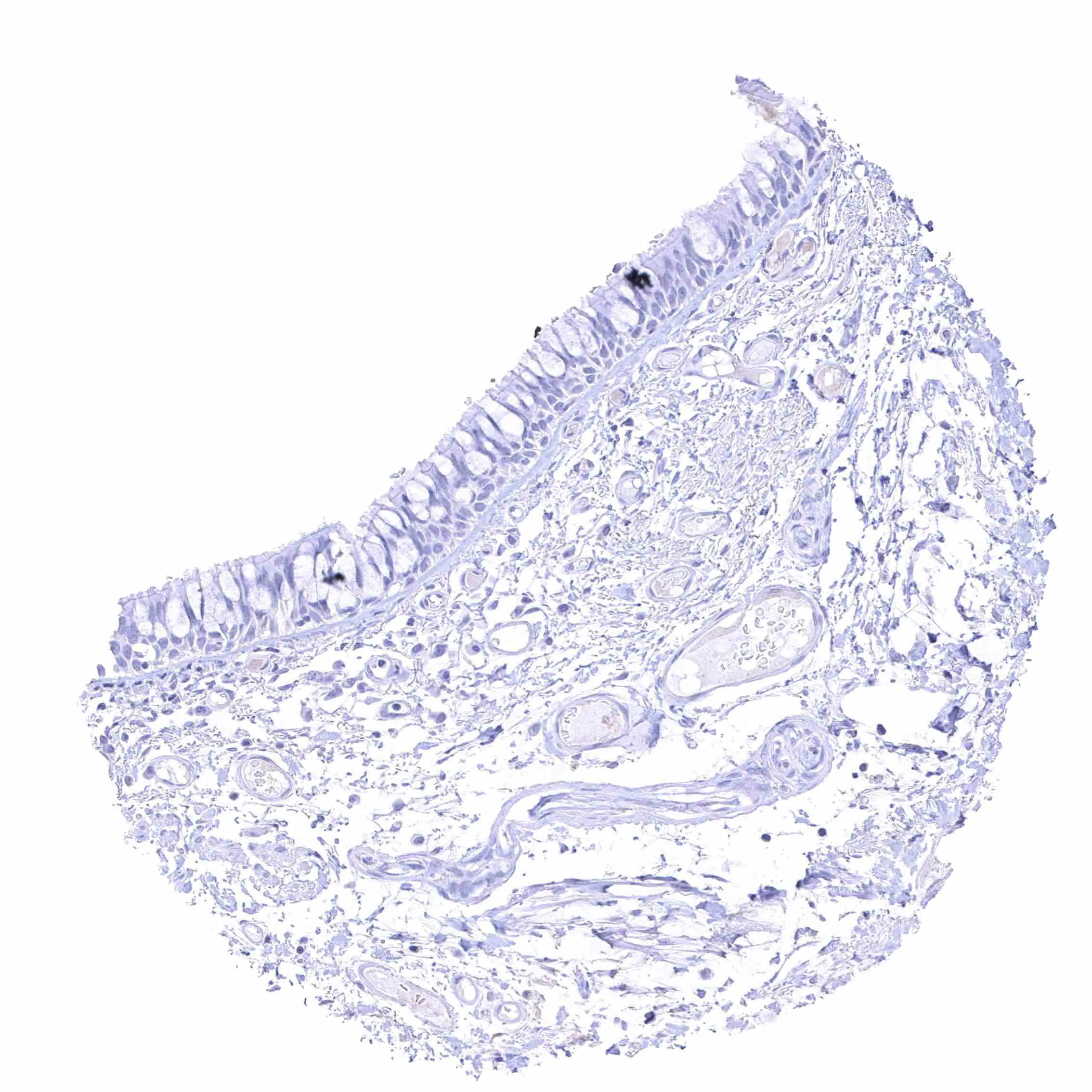
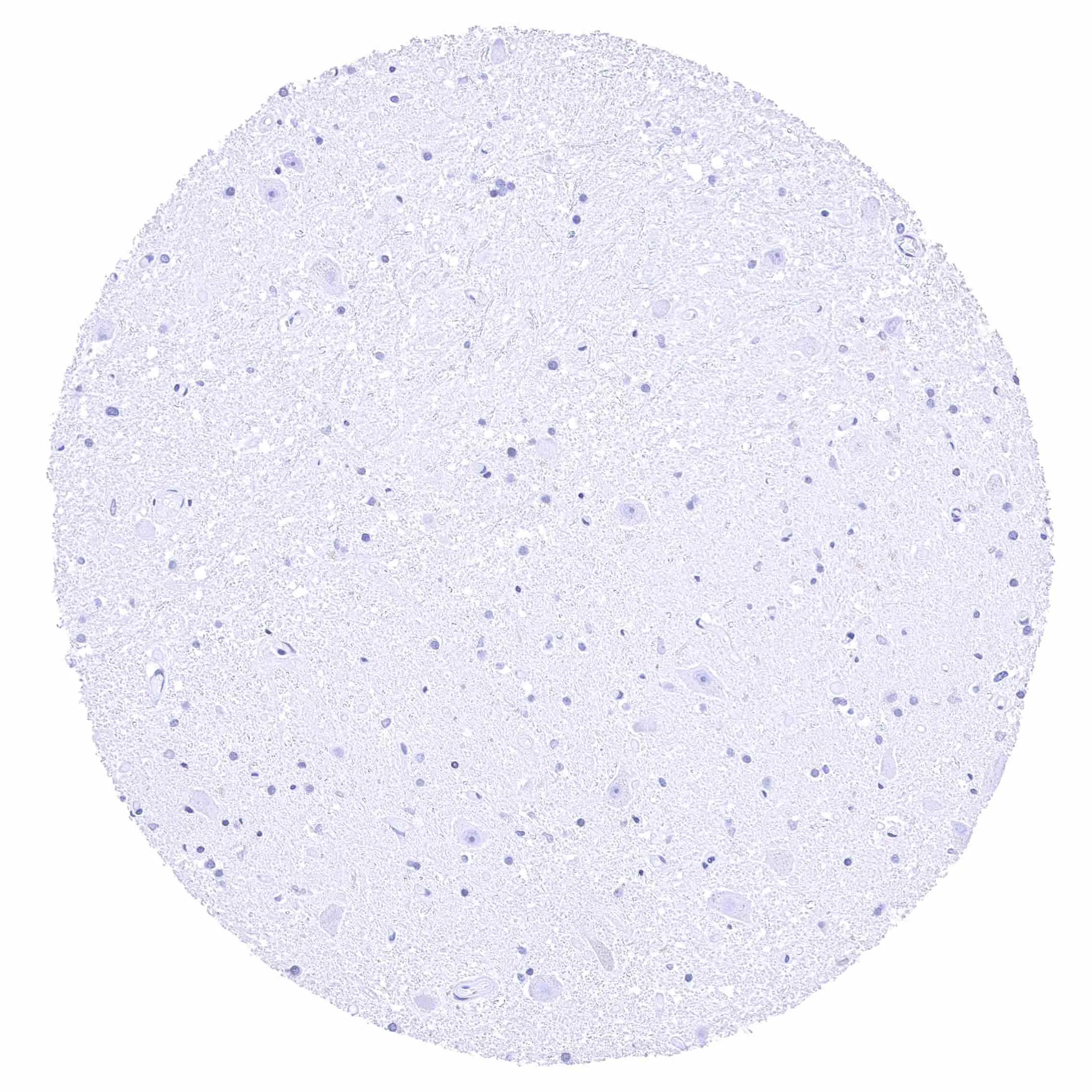
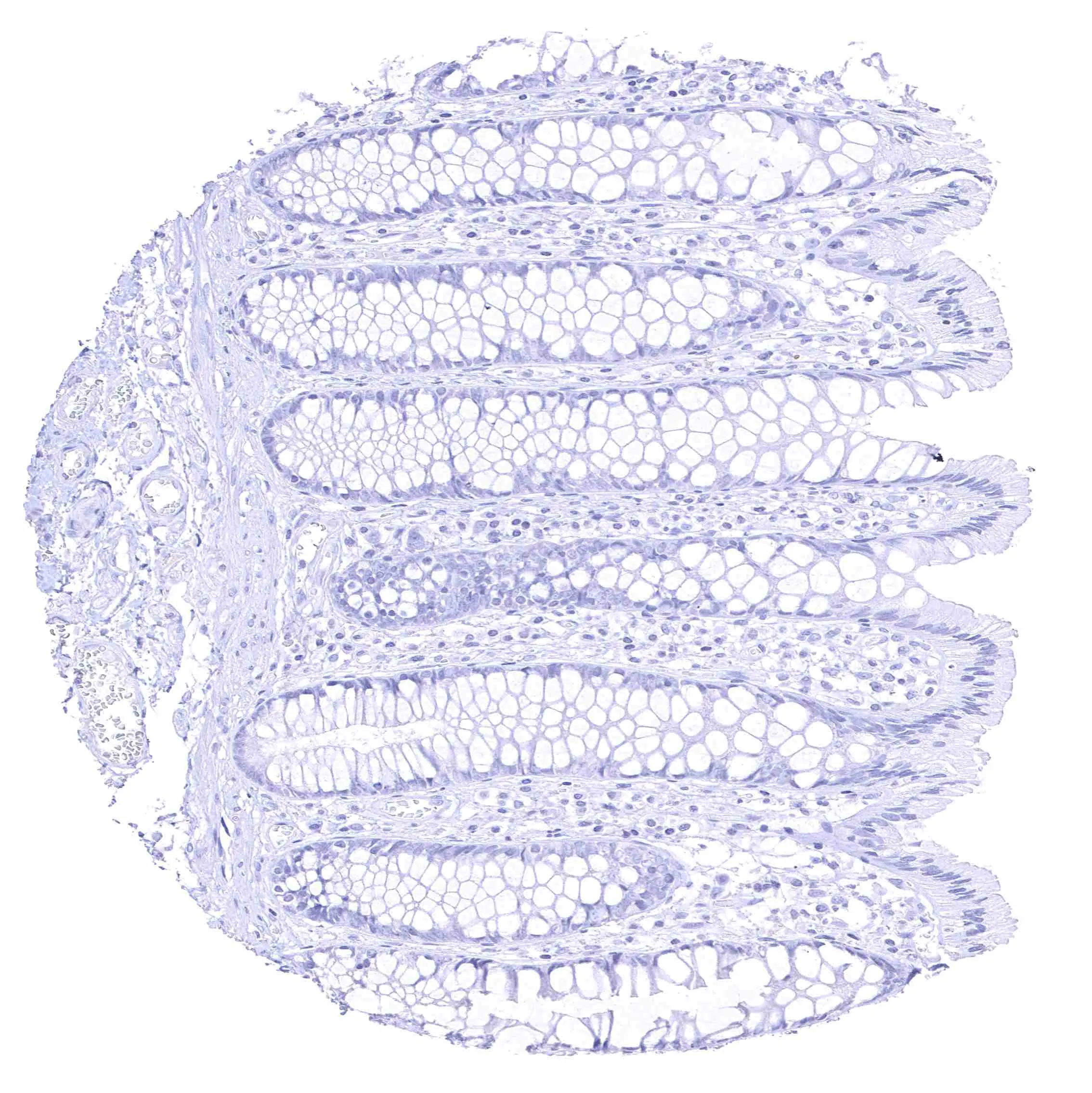
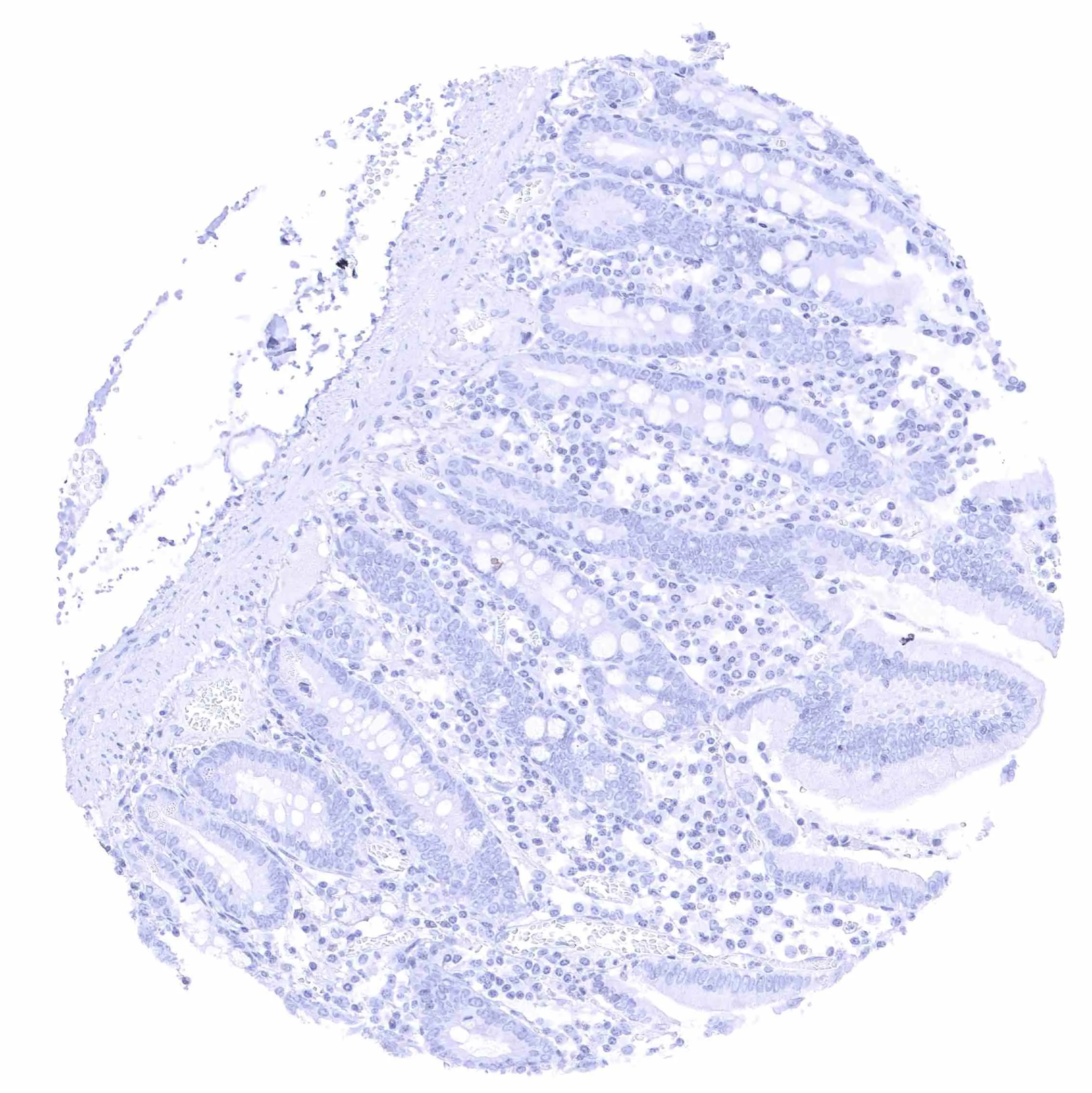
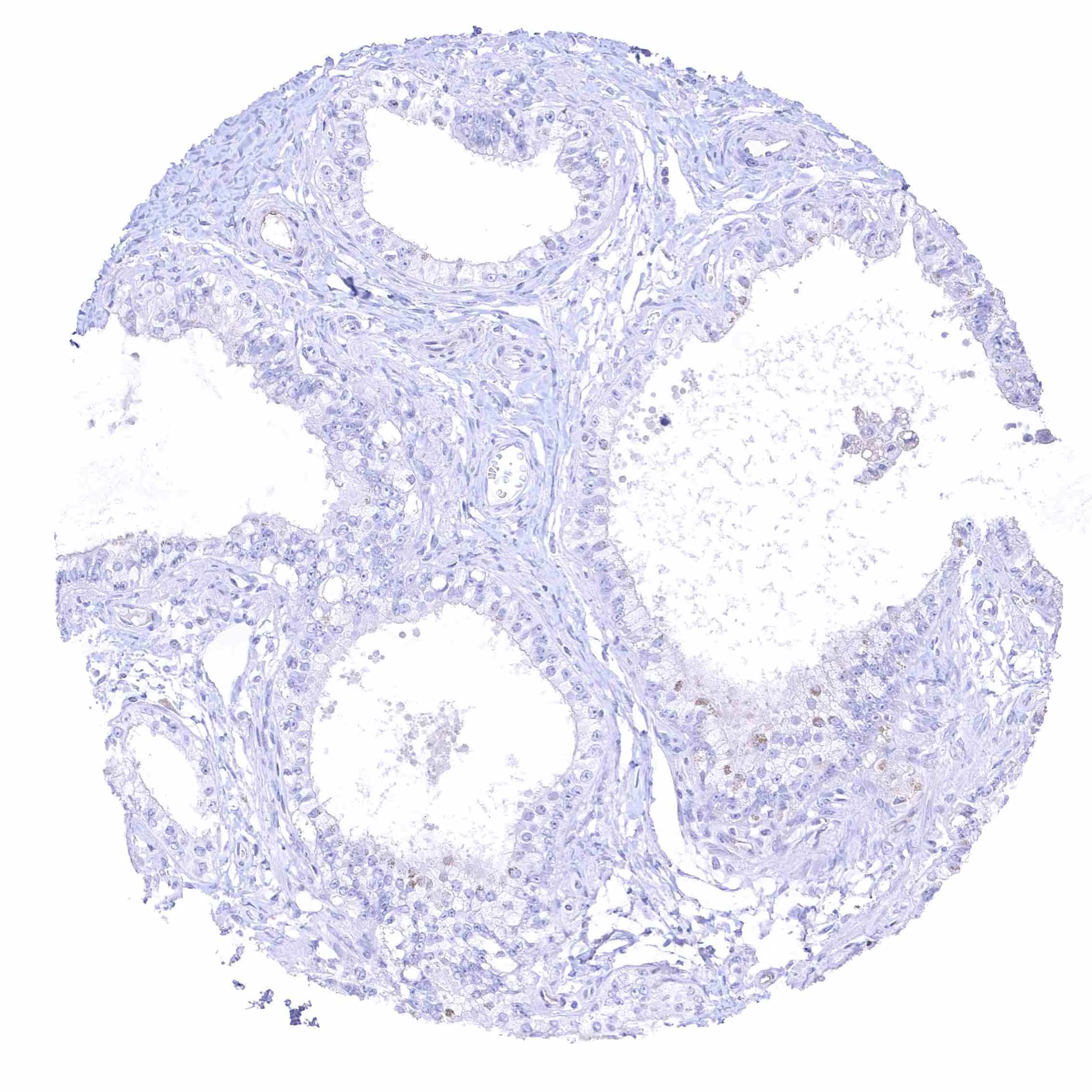
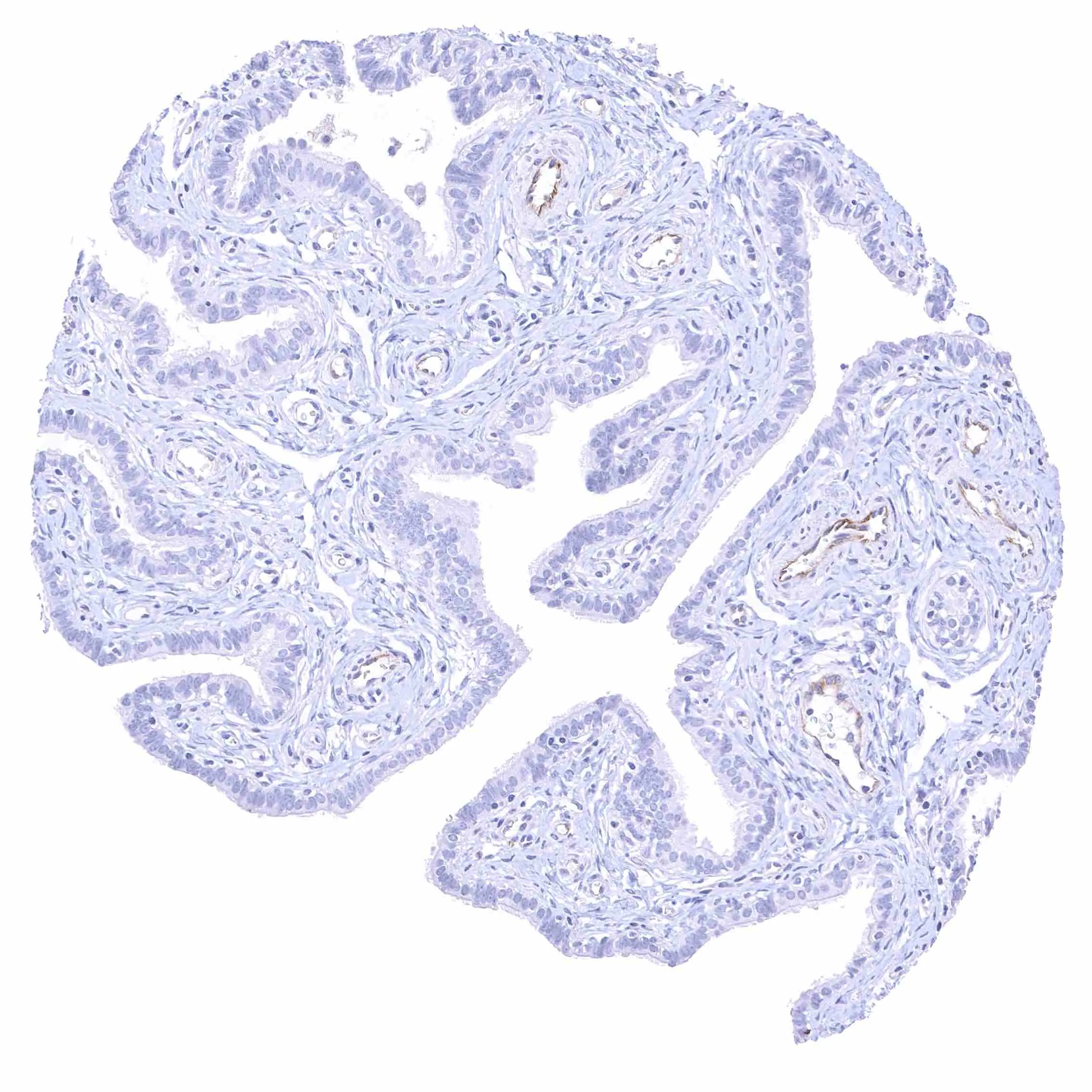
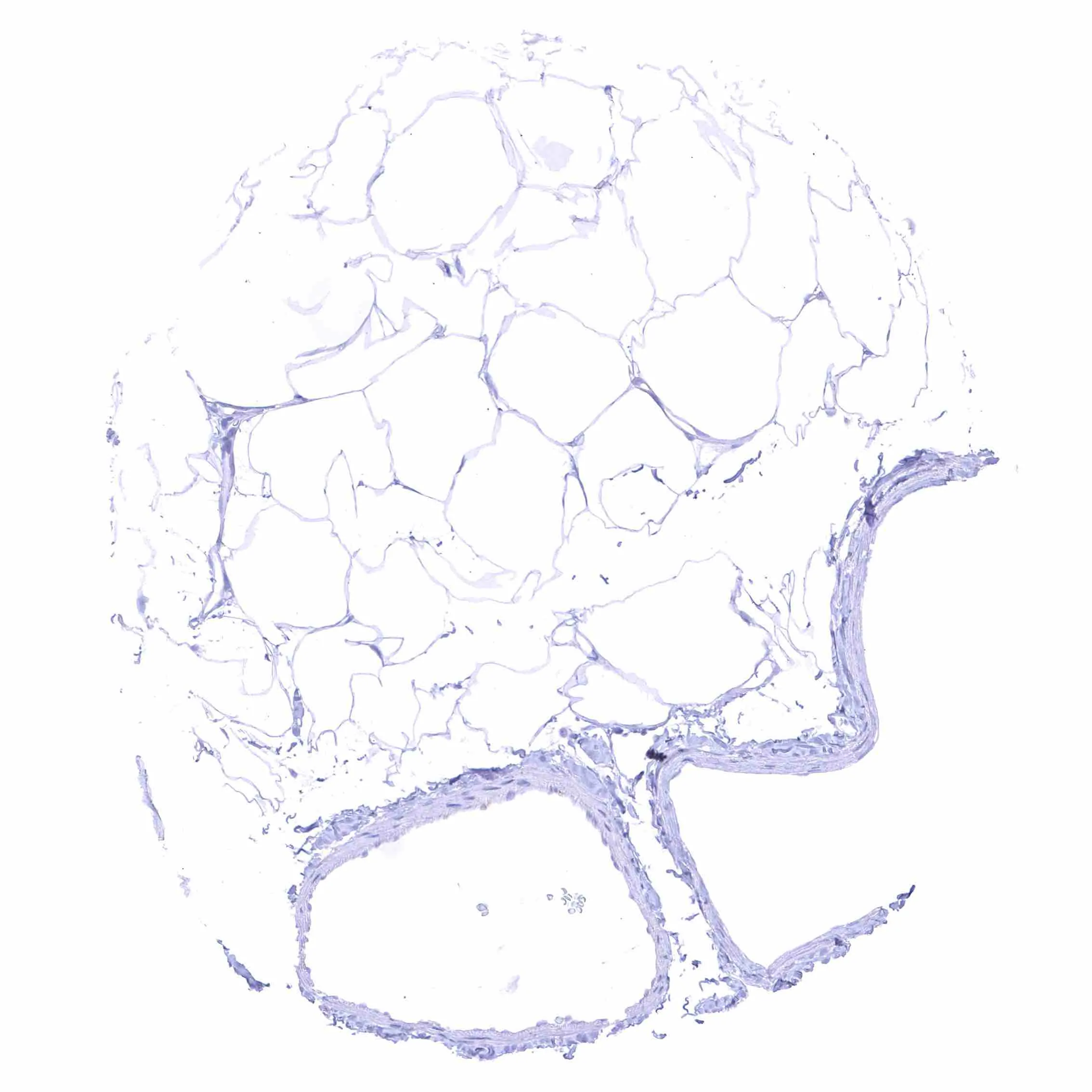
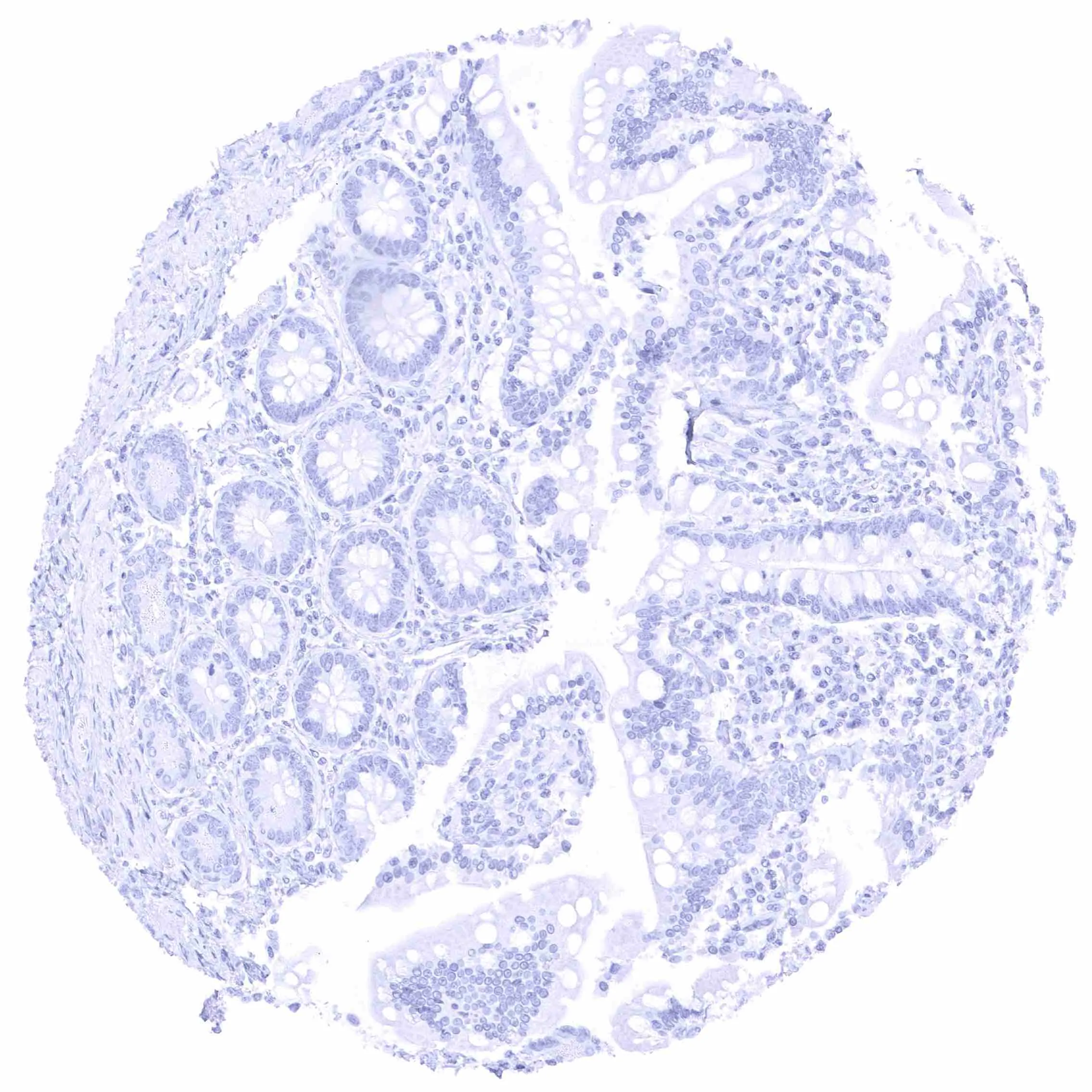
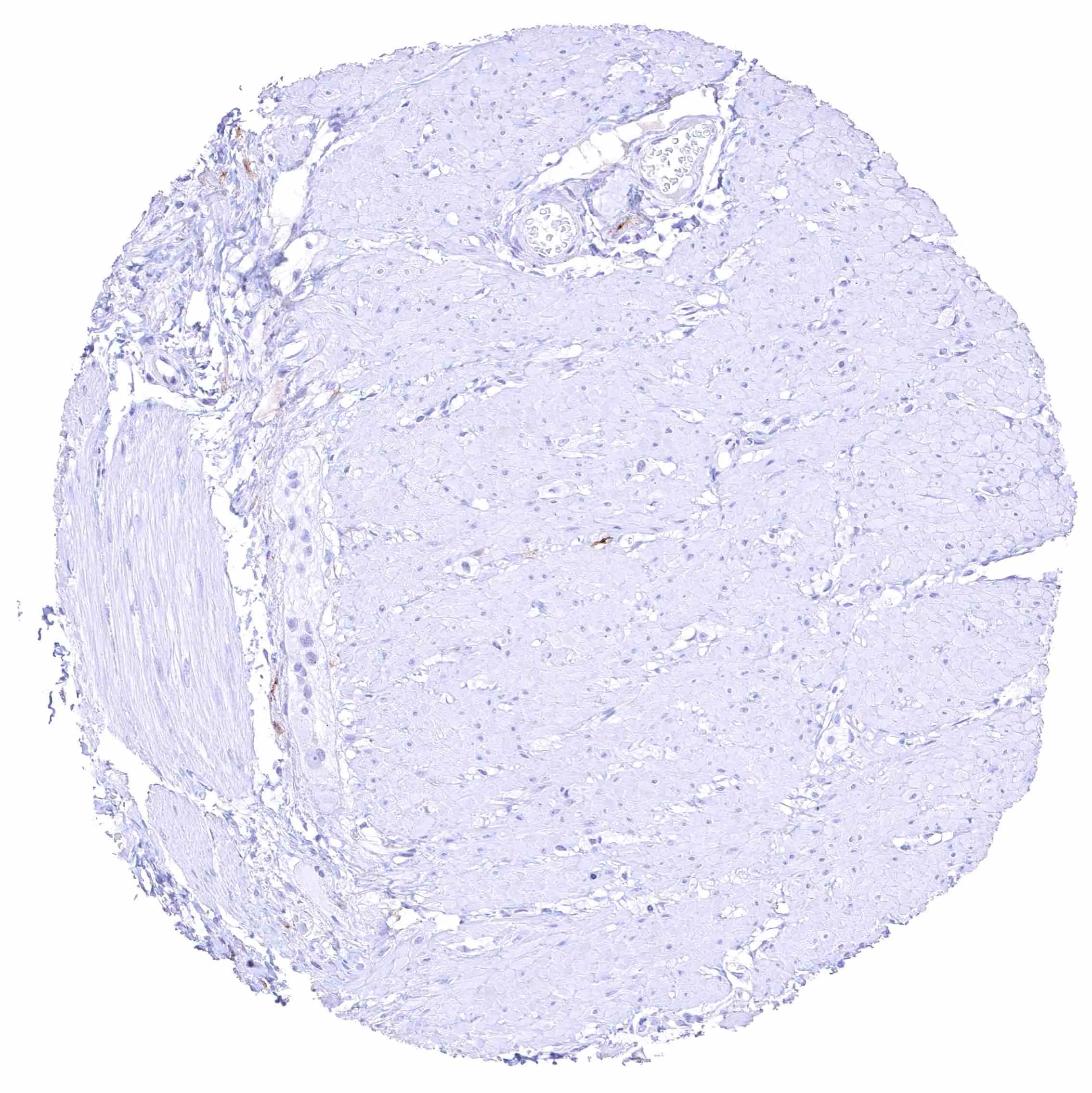
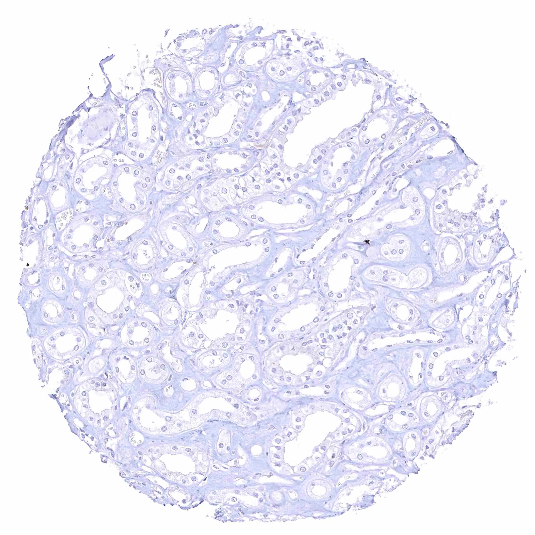
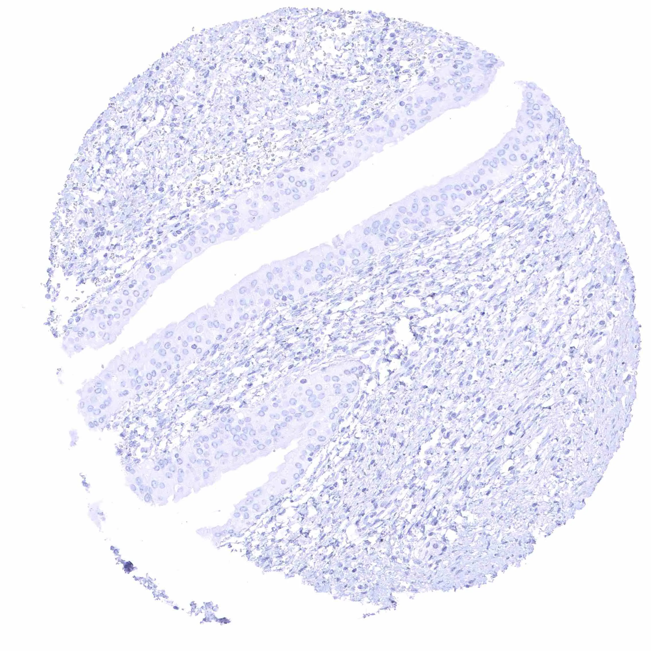
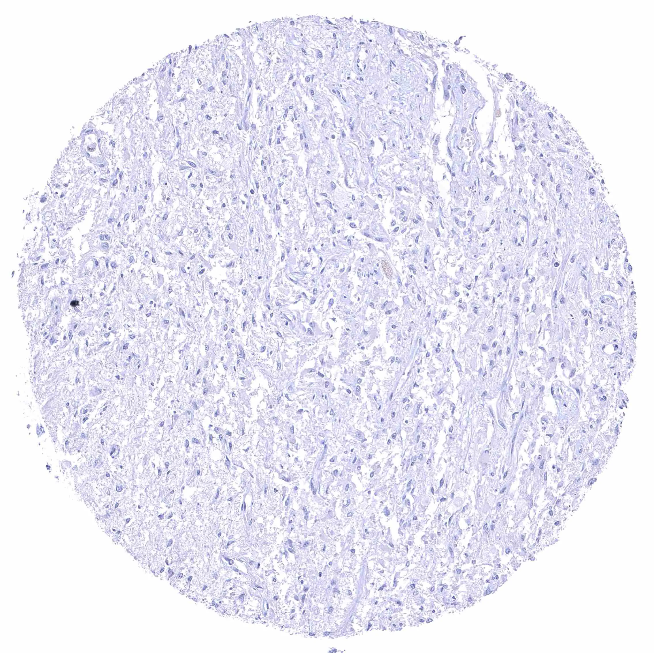
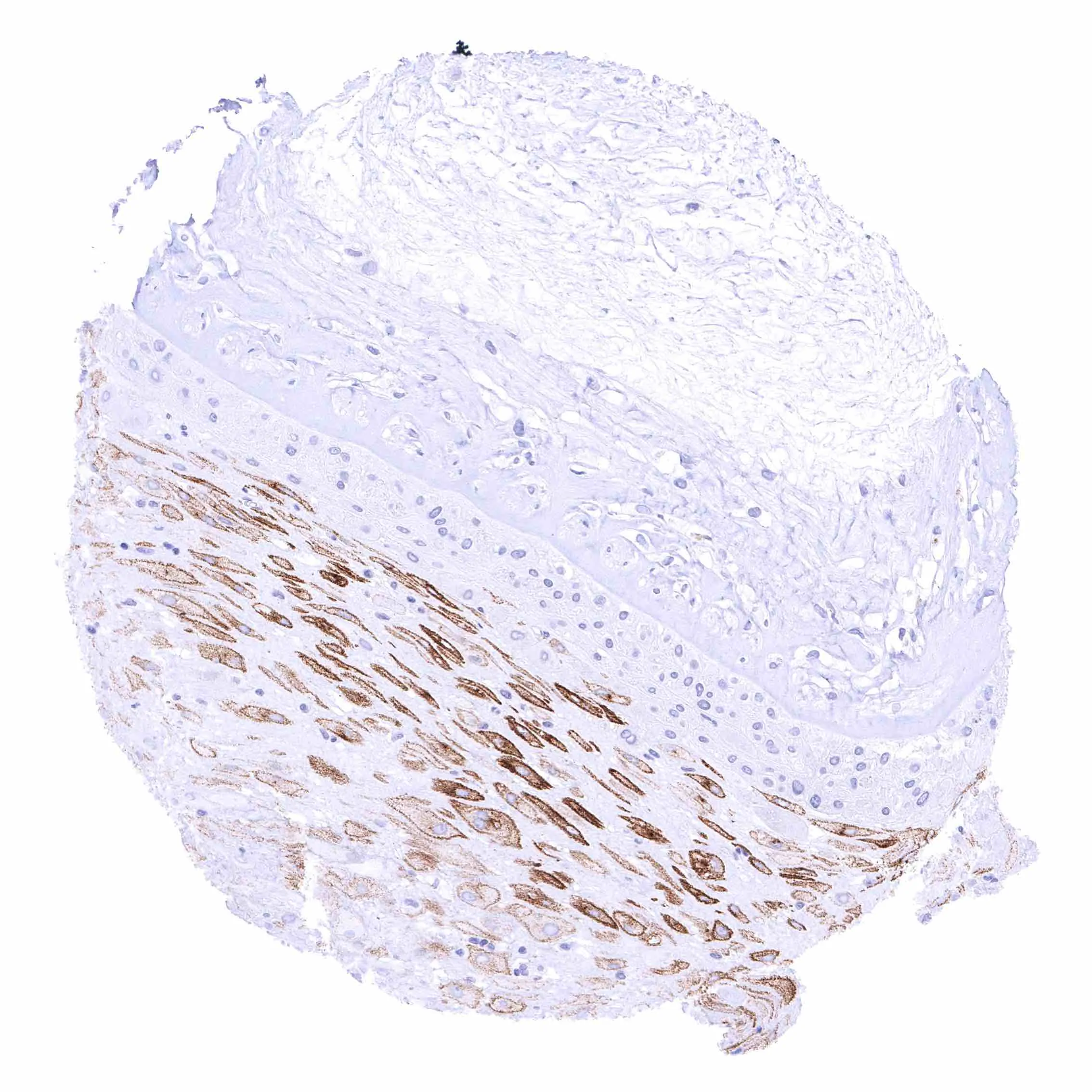
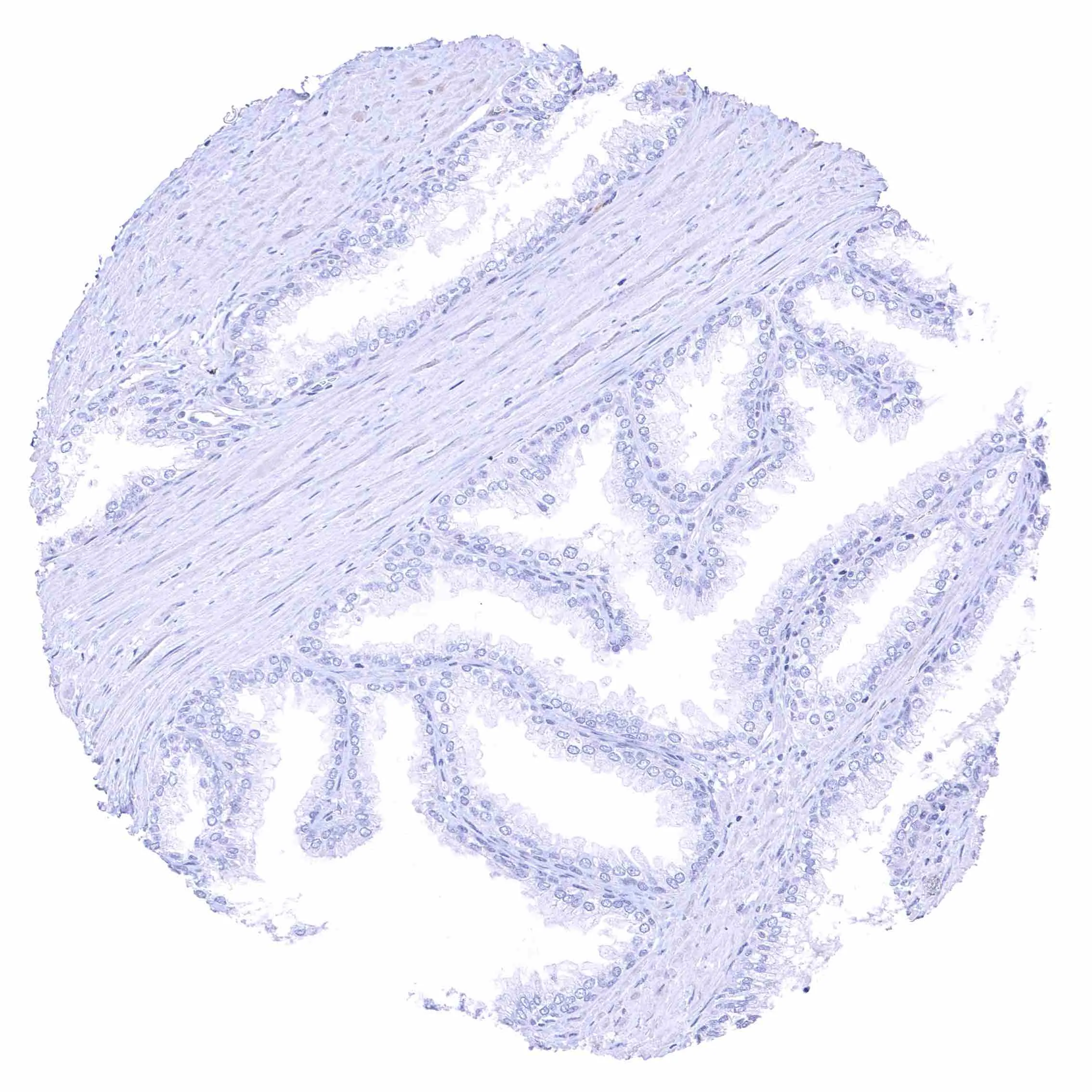
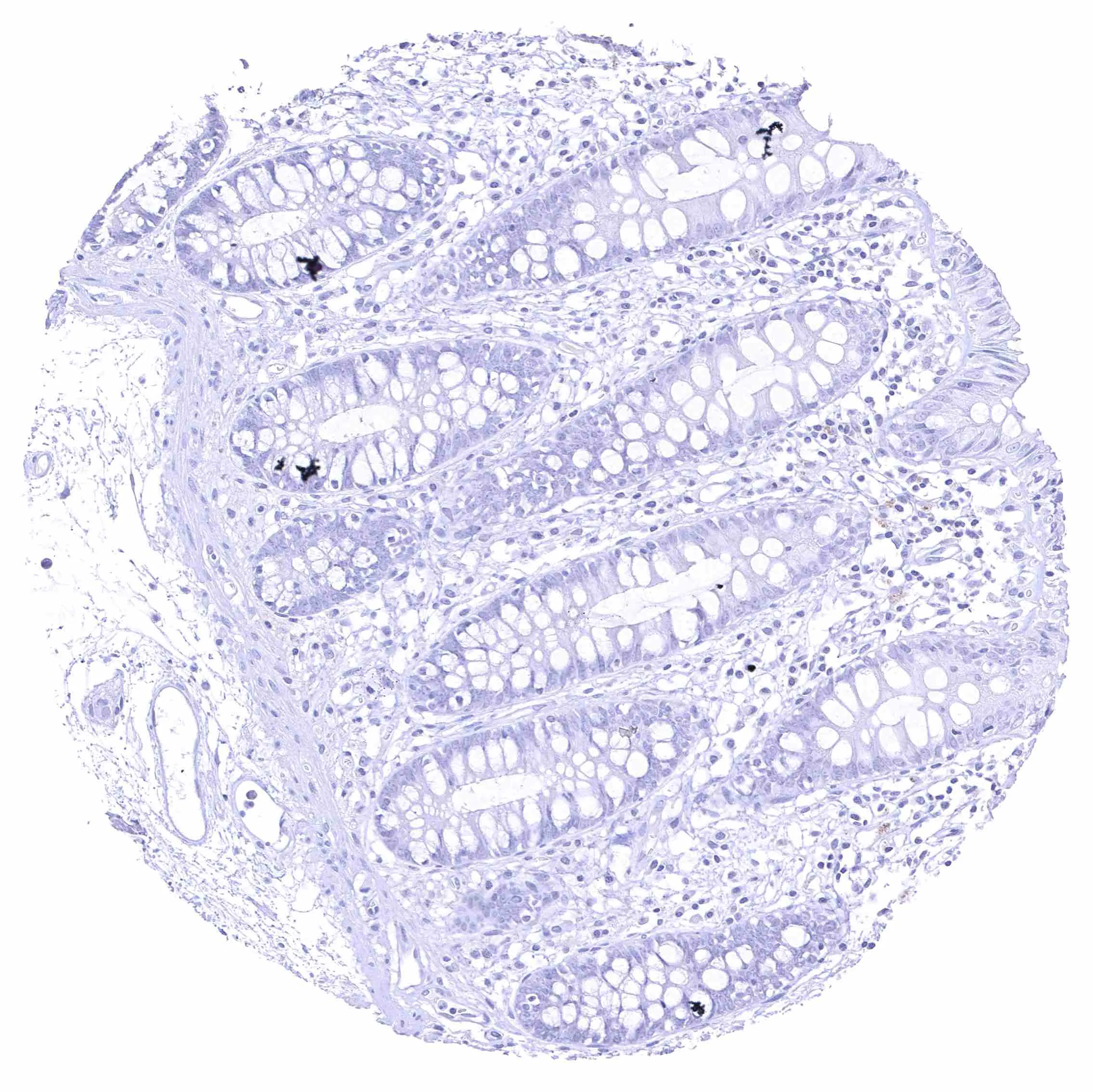
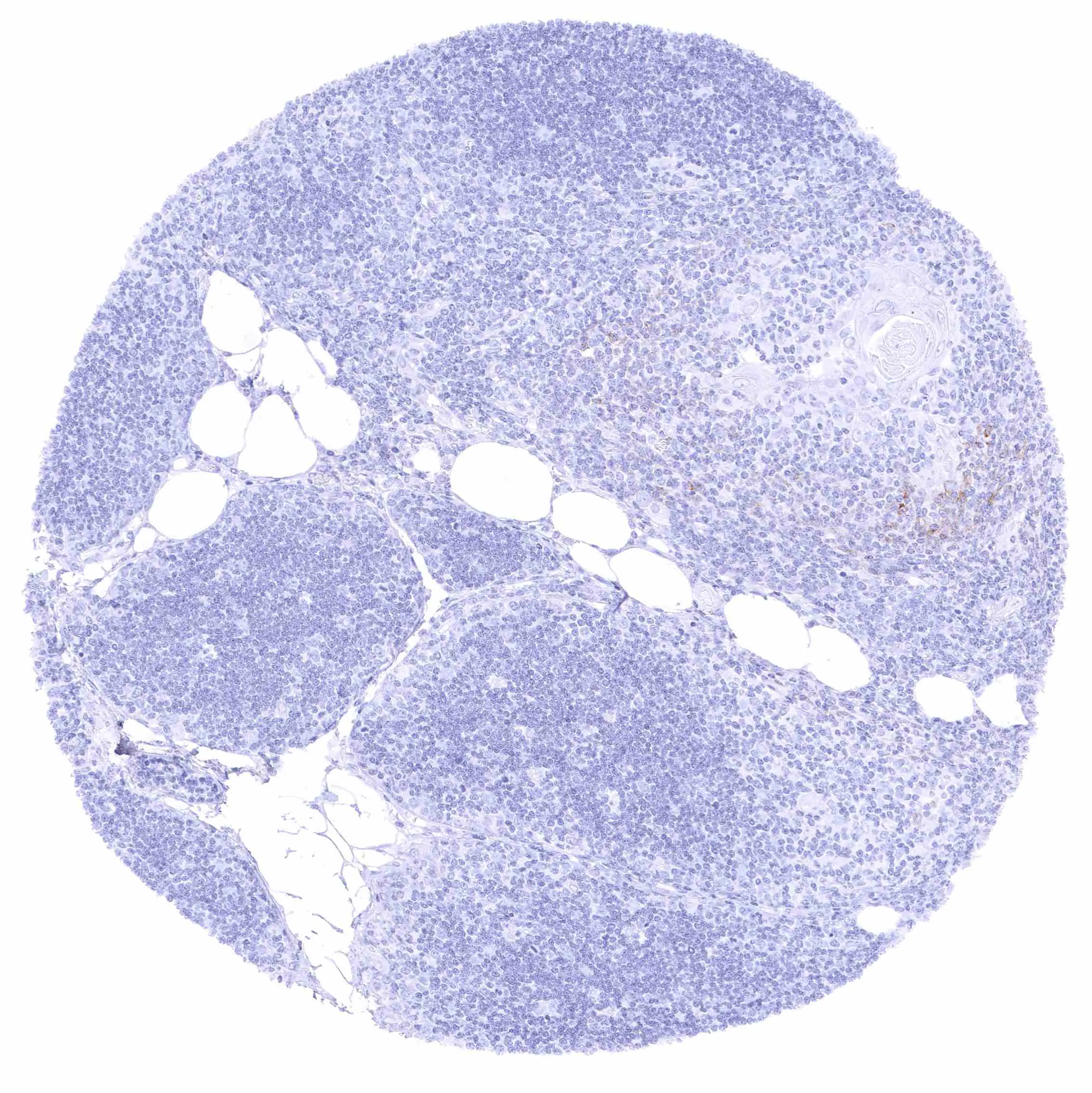
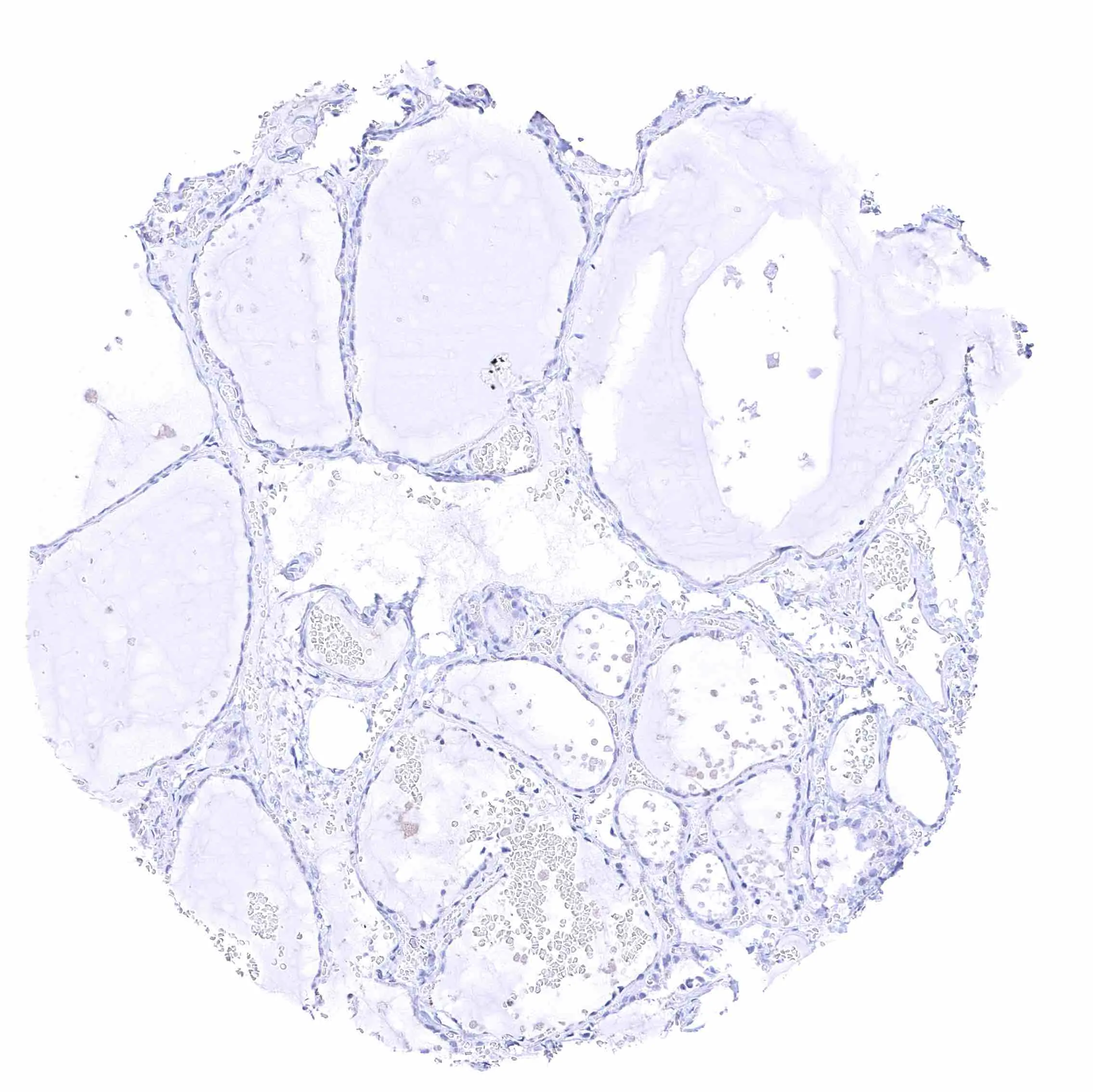
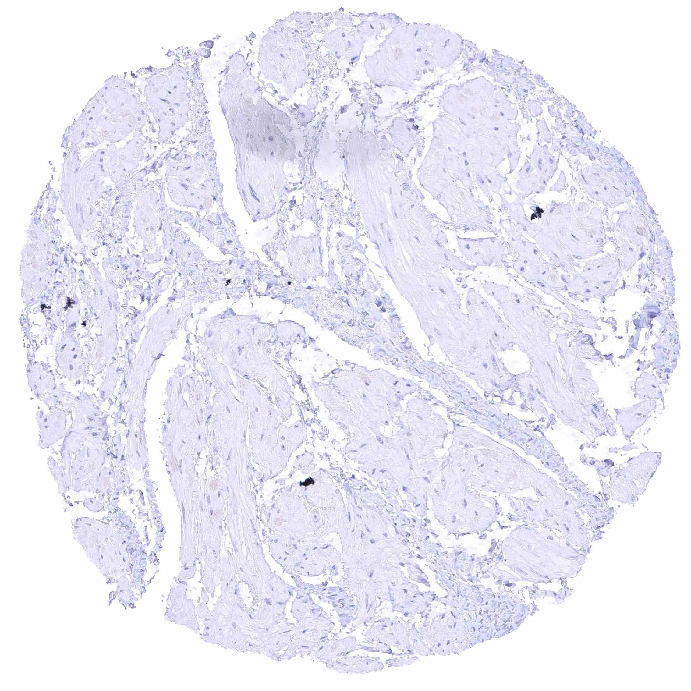
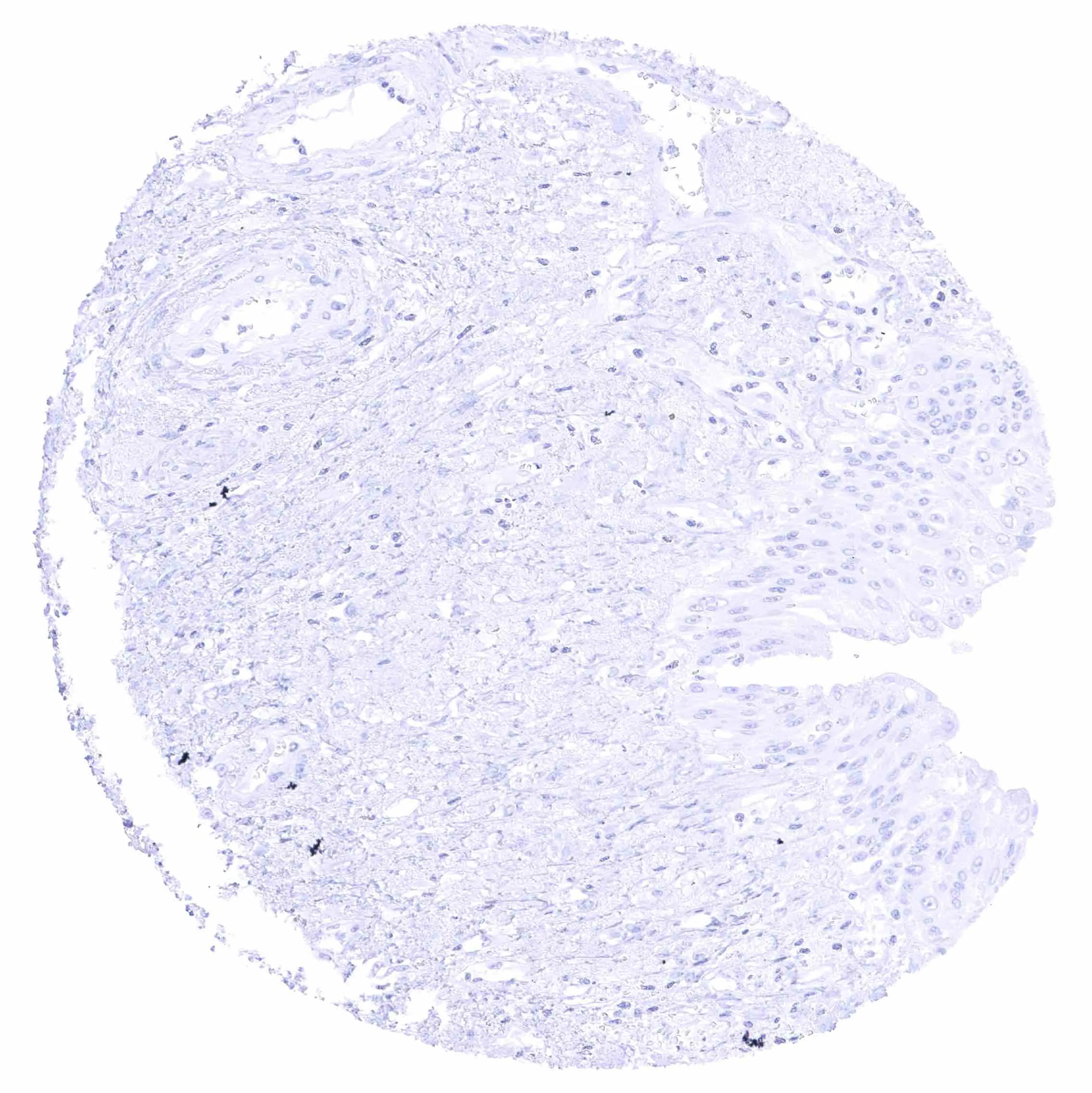
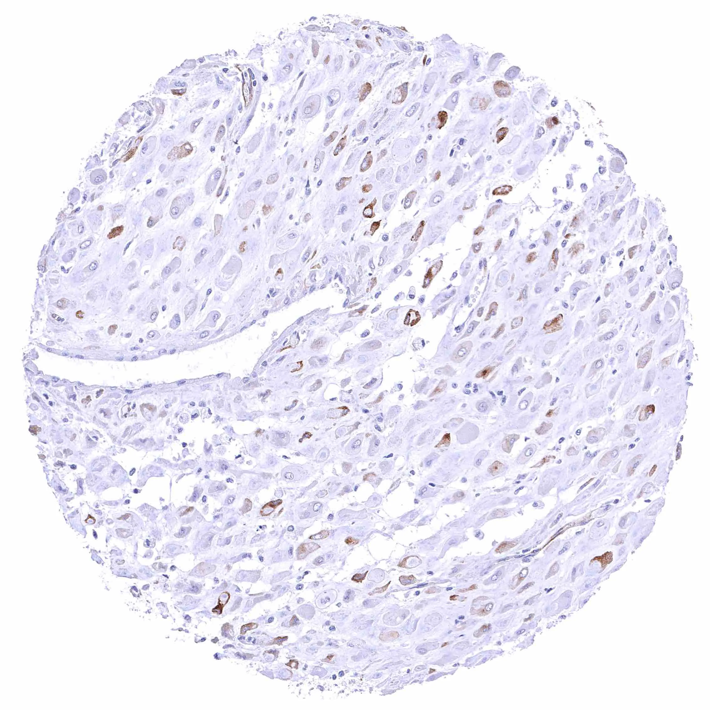
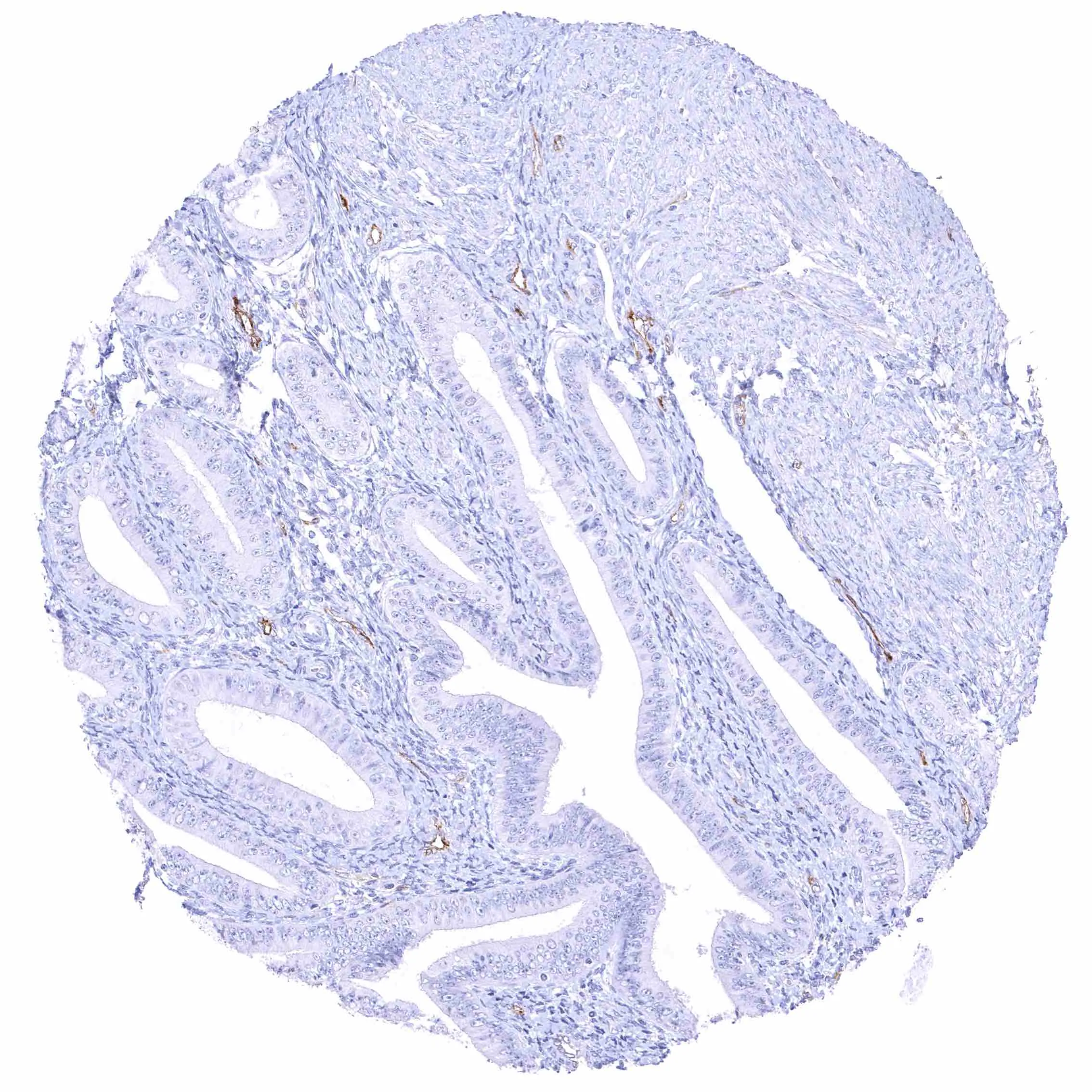
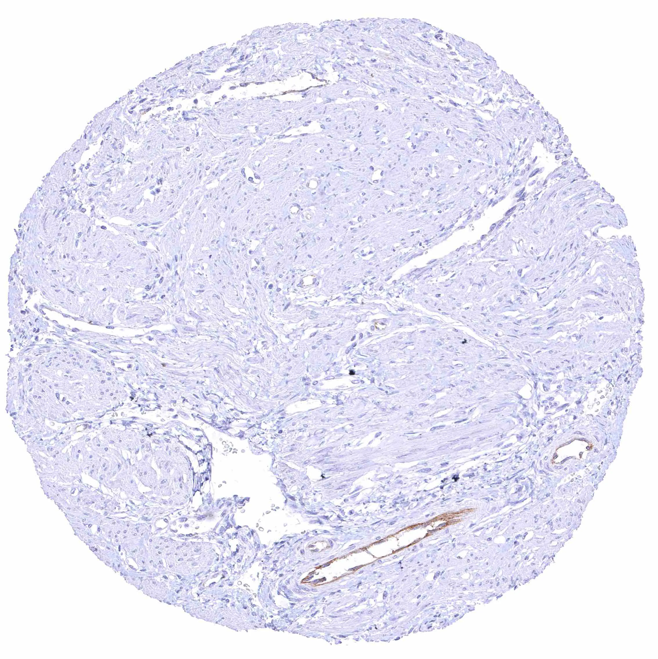
NPR-C antibody [HMV4274] HistoMAX™


Go to Cancer Tissue Gallery
  
 Go back to product page
  

Go to HistoMAX page


Adrenal gland

Aorta, media

Appendix, mucosa

Appendix, muscular wall


Bone marrow

Breast

Bronchus, mucosa

Cerebellum (white matter)


Cerebellum, cortex (grey)

Cerebrum (grey matter)

Cerebrum (white matter)

Colon descendens, mucosa


Colon descendens, muscular wall – Weak NPR-C staining of endothelial cells.

Duodenum, Brunner gland

Duodenum, mucosa

Epididymis (Cauda)


Esophagus, muscular wall

Esophagus, squamous epithelium

Fallopian tube, mucosa – Weak membranous NPR-C staining at the luminal cell border of endothelial cells of small blood vessels.

Fat


Gallbladder, epithelium

Heart muscle

Ileum, mucosa

Ileum, muscularis


Kidney, cortex – Distinct membranous NPR-C staining at the luminal cell border of a subset of tubuli, the parietal membrane of the Bowman capsule, and in glomerular endothelial cells.

Kidney, cortex – Distinct membranous NPR-C staining at the luminal cell border of a subset of tubuli, the parietal layer of the Bowman capsule, and in glomerular endothelial cells.

Kidney, medulla

Kidney, pelvis, urothelium


Liver

Lung – Faint membranous NPR-C staining at the luminal cell border of some endothelial cells.

Lymph node

Ovary, corpus luteum – Distinct membranous and perinuclear cytoplasmic NPR-C staining of corpus luteum cells.


Ovary, follicular cyst – Strong membranous NPR-C staining of a subset of granulosa cells.

Ovary, stroma – Weak NPR-C staining of endothelial cells in one blood vessel but not in others.

Pancreas

Parathyroid gland – Weak NPR-C staining of few endothelial cells.


Parotid gland

Pituitary gland, anterior lobe

Pituitary gland, posterior lobe

Placenta (chorion) – Distinct membranous and cytoplasmic NPR-C staining of decidua cells.


Placenta, early – Weak NPR-C staining of few endothelial cells.

Placenta, mature – Weak NPR-C staining of few endothelial cells.

Prostate

Rectum, mucosa


Seminal vesicle – Intense membranous NPR-C staining at the luminal cell border of endothelial cells of small blood vessels.

Skeletal muscle

Skin, hairfollicel and sebaceous glands

Skin


Spleen

Stomach, antrum

Stomach, corpus

Stomach, muscular wall


Submandibular gland

Testis

Thymus

Thyroid gland


Tonsil, surface epithelium

Tonsil

Urinary bladder, muscular wall

Urinary bladder, urothelium


Uterus, ectocervix

Uterus, endocervix

Uterus, endometrium (pregnancy) – Distinct membranous and cytoplasmic NPR-C staining of a subset of decidua cells.

Uterus, endometrium (proliferation)


Uterus, endometrium (secretion)

Uterus, myometrium – Distinct NPR-C staining of endothelial cells in some but not all blood vessels


| TOP |