Normal Tissue Gallery- GTX04965

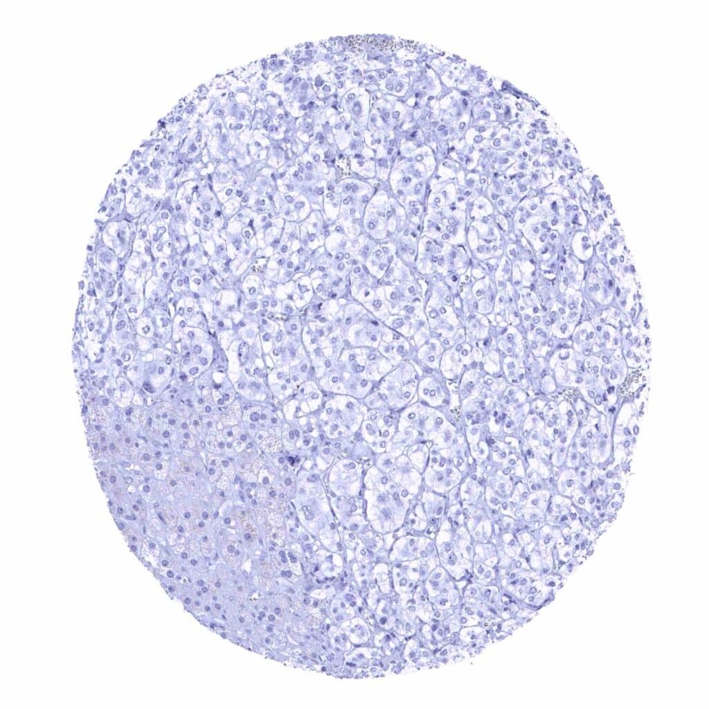
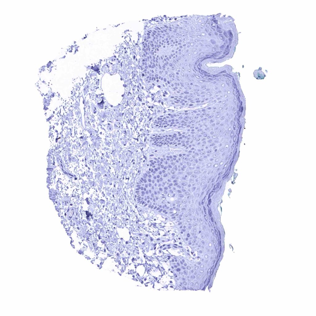
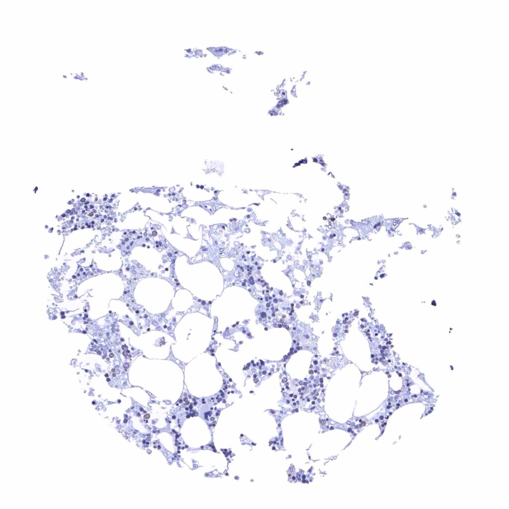
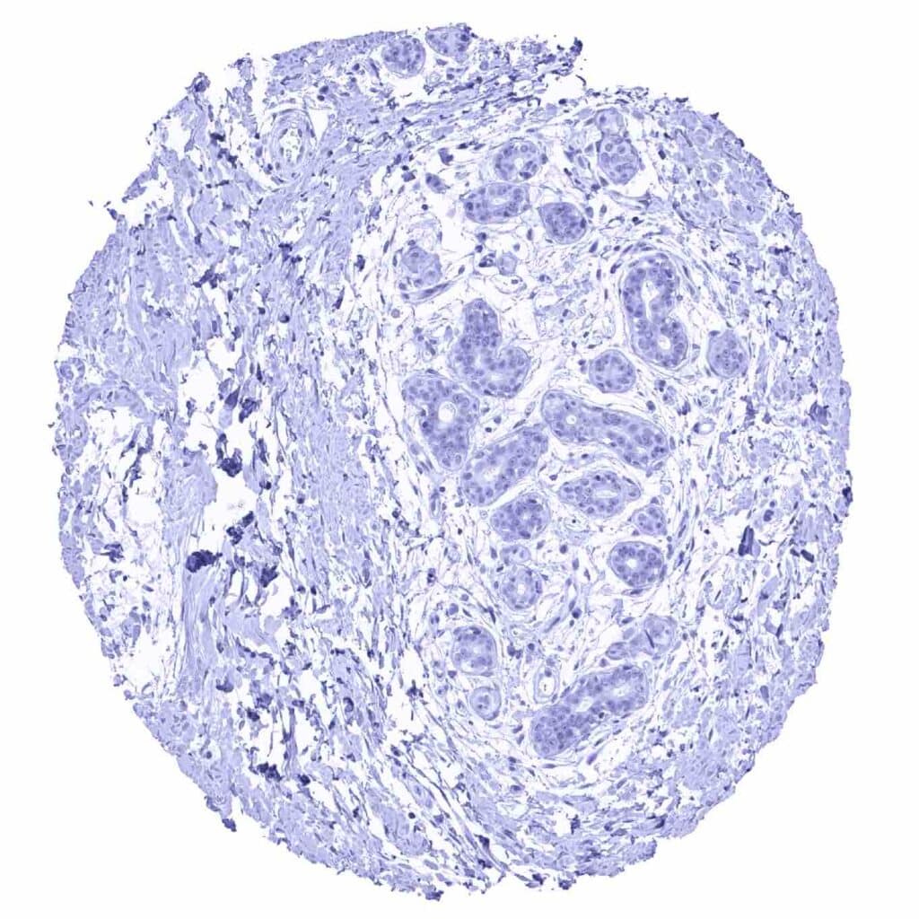
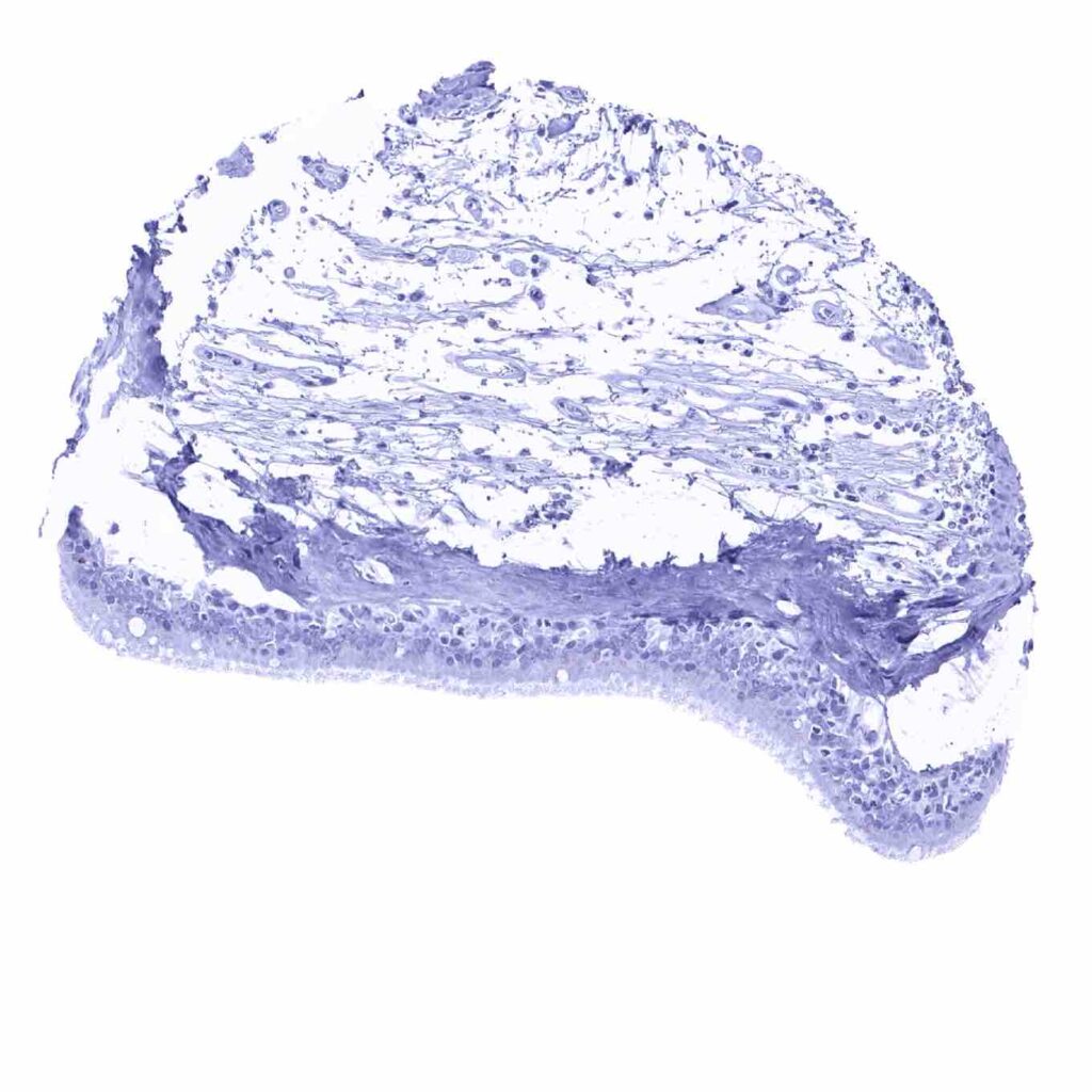
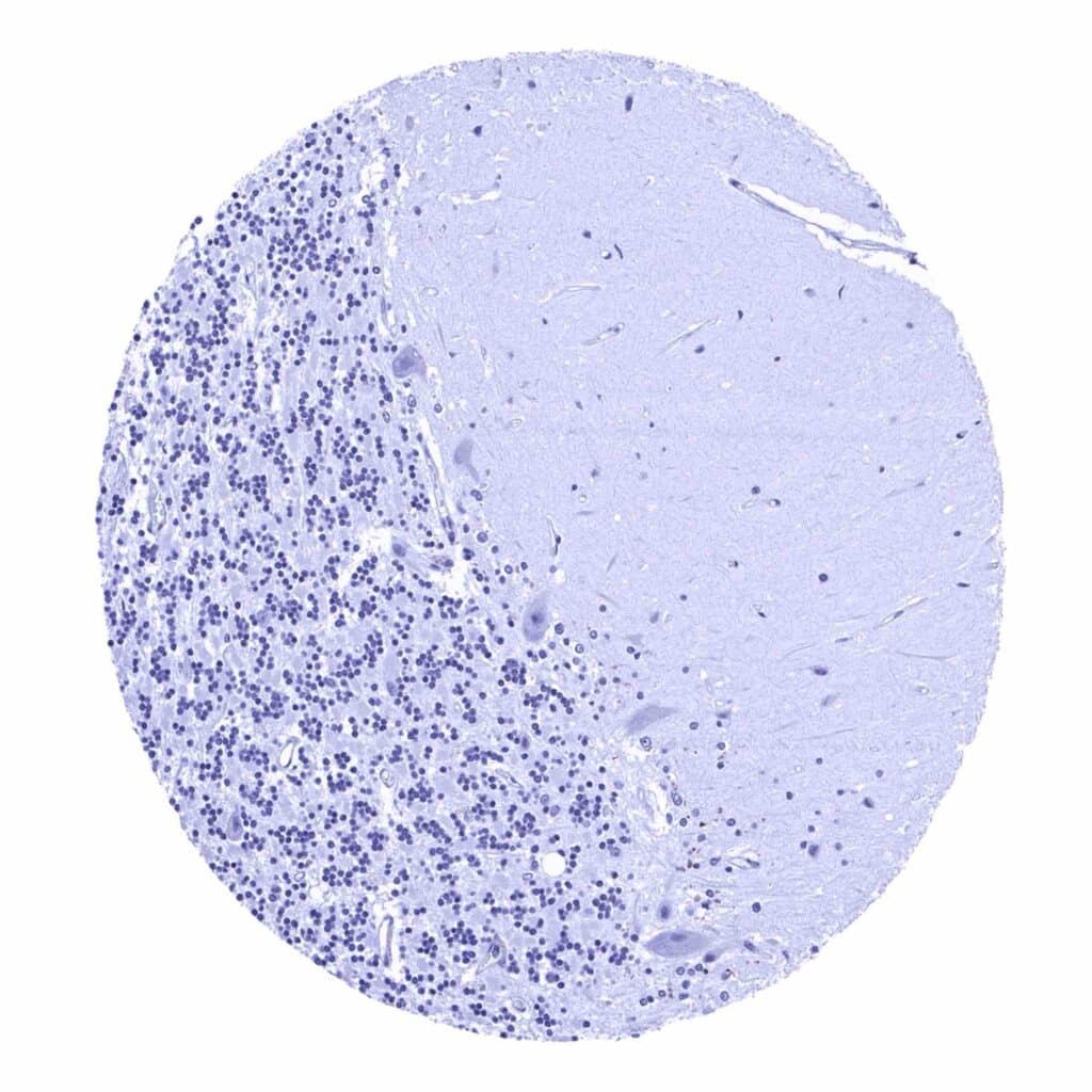
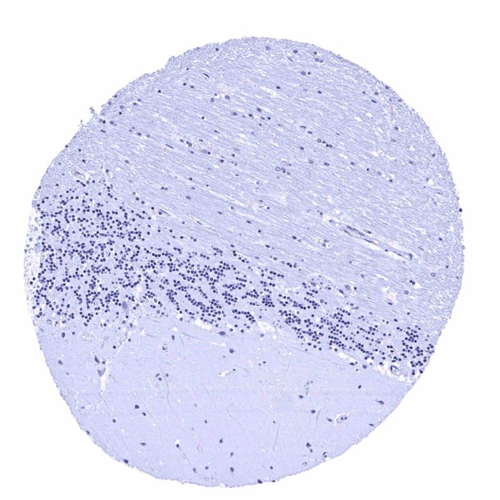
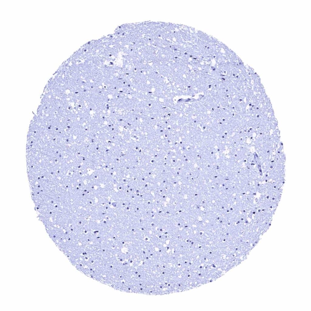
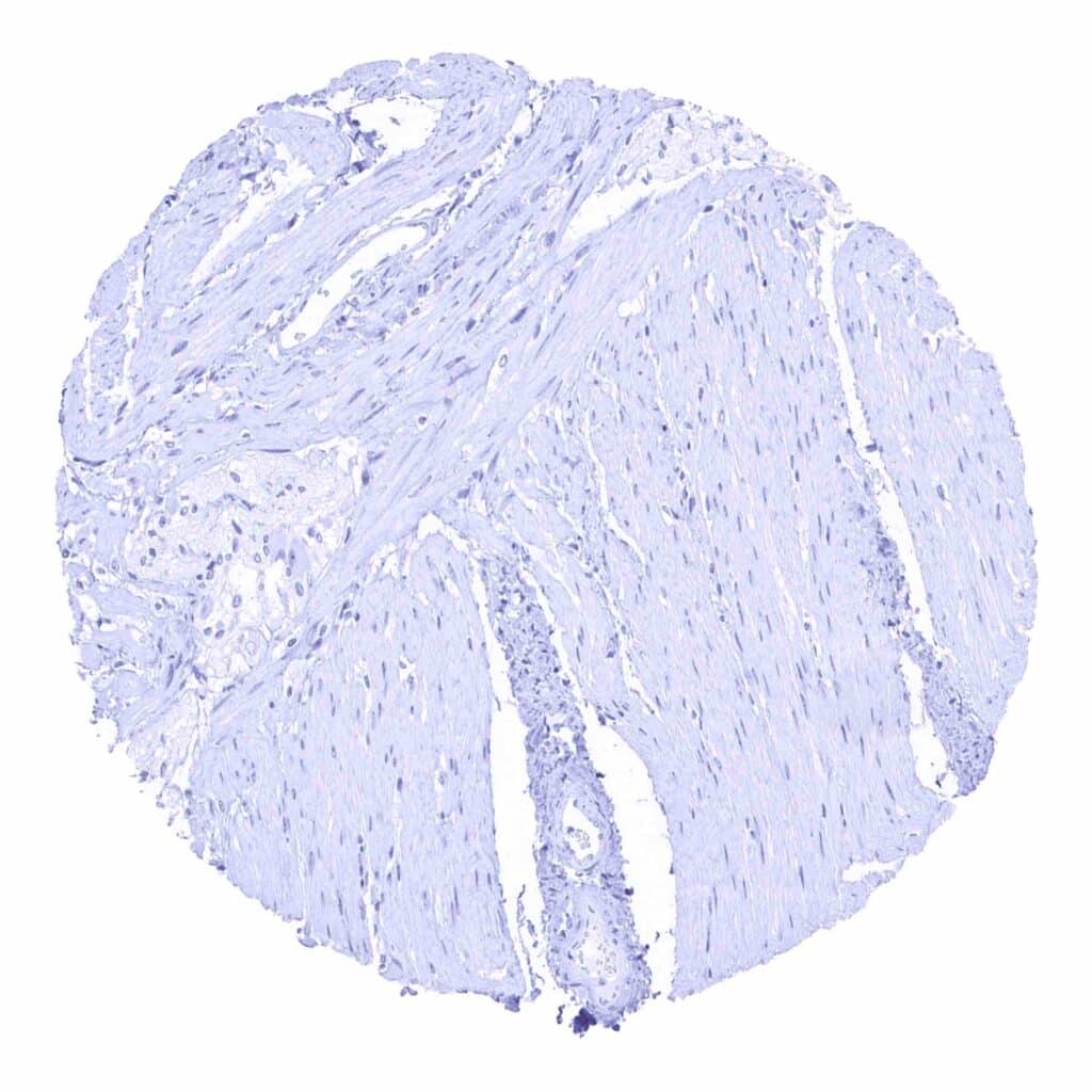
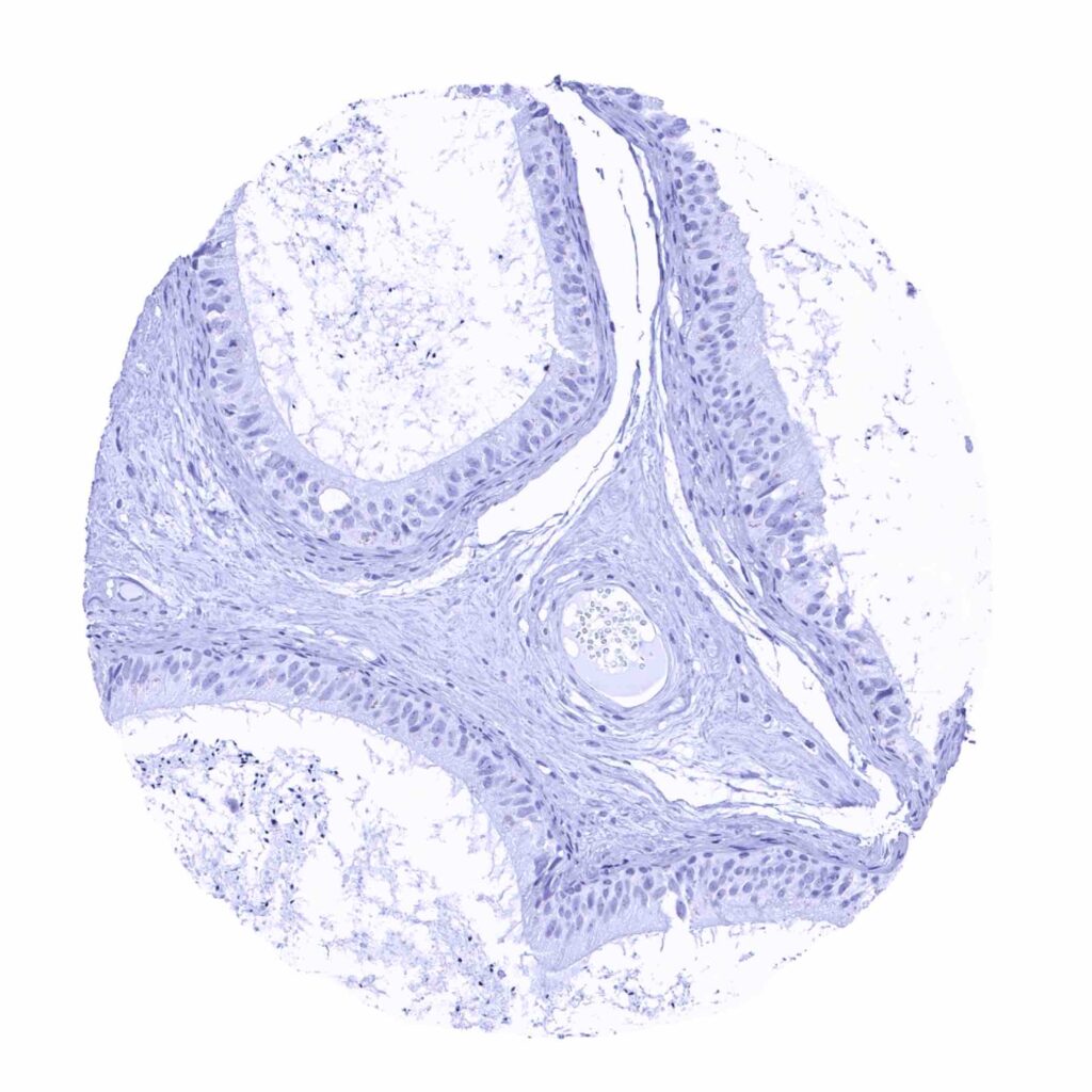
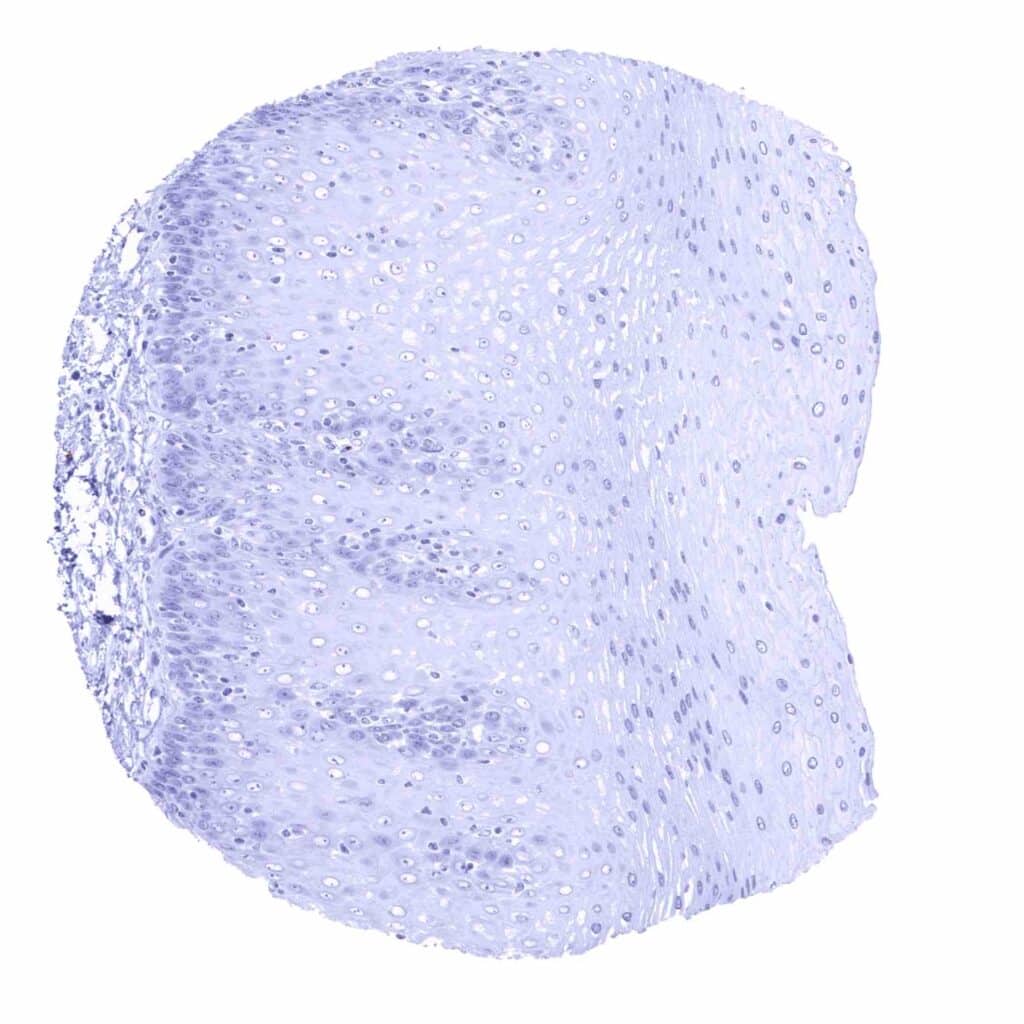
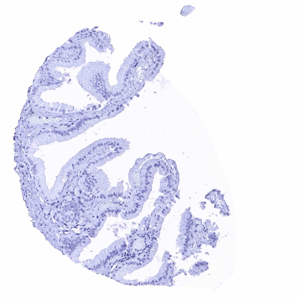
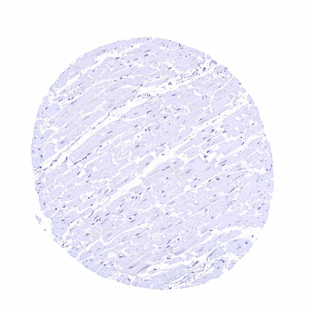
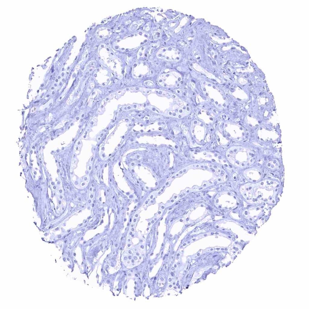
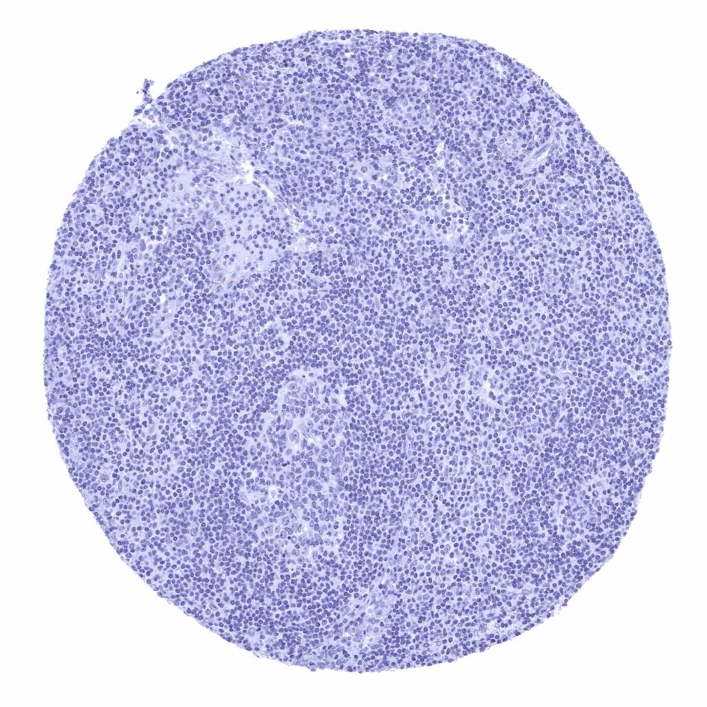
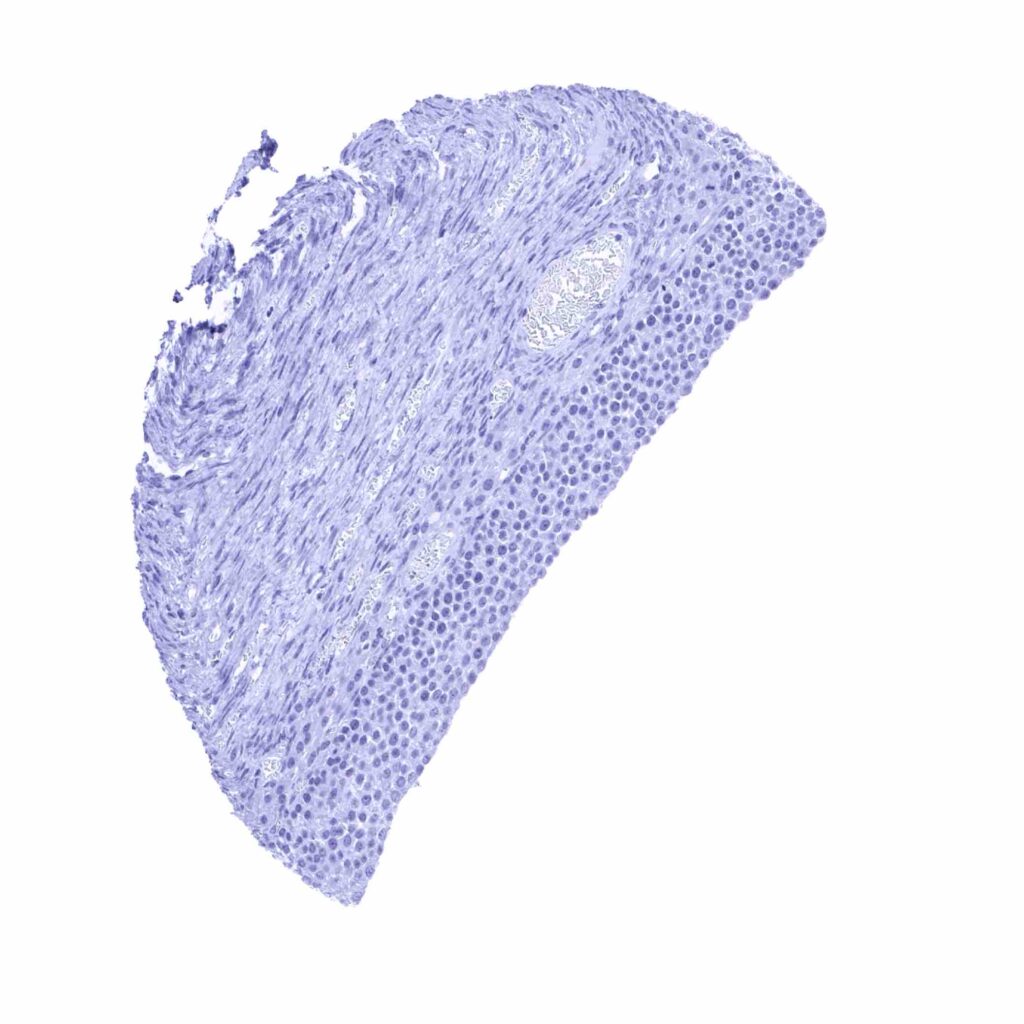
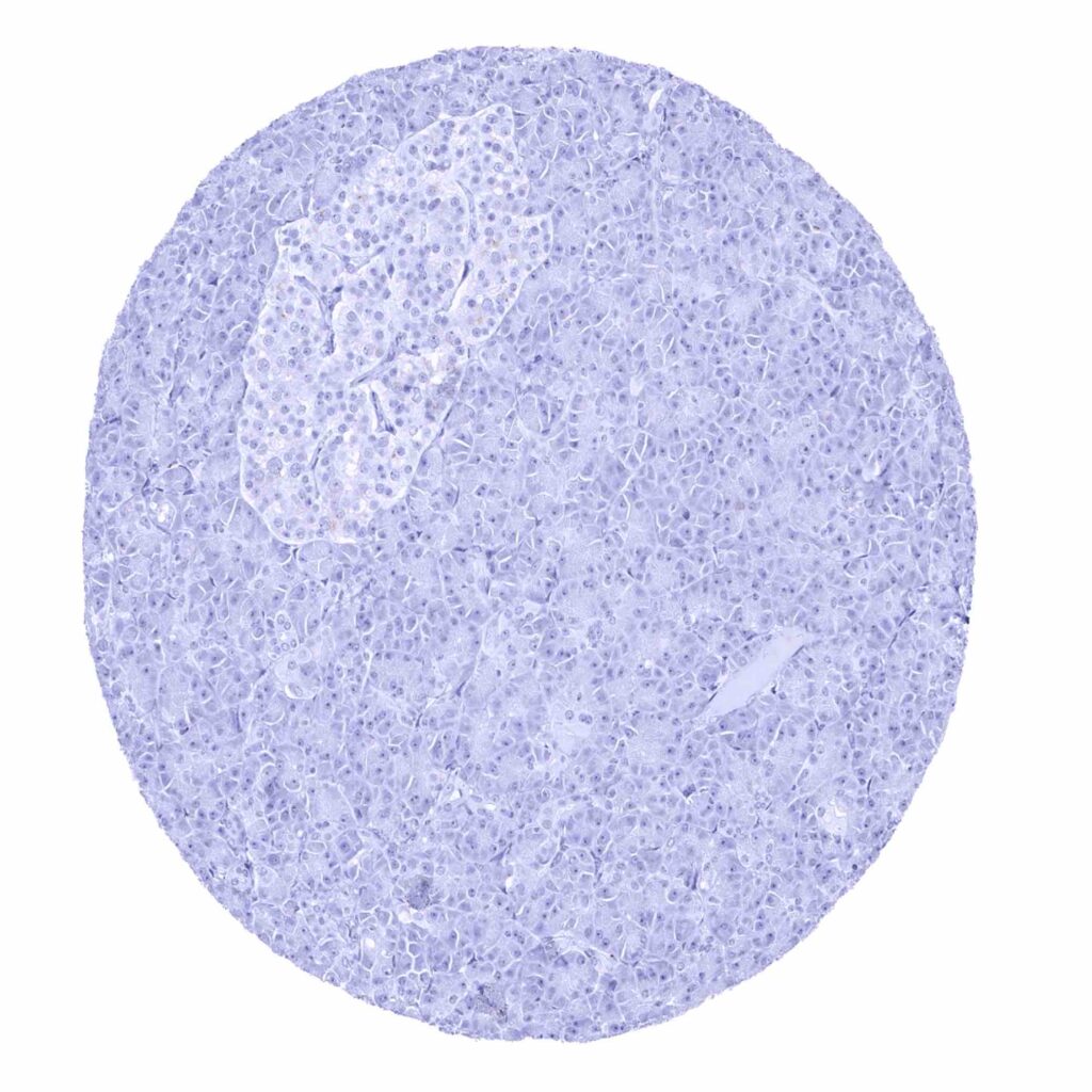
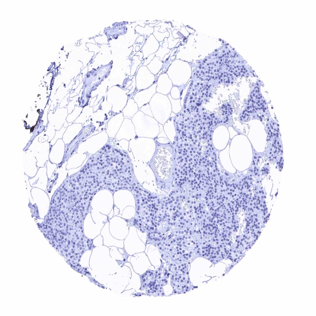
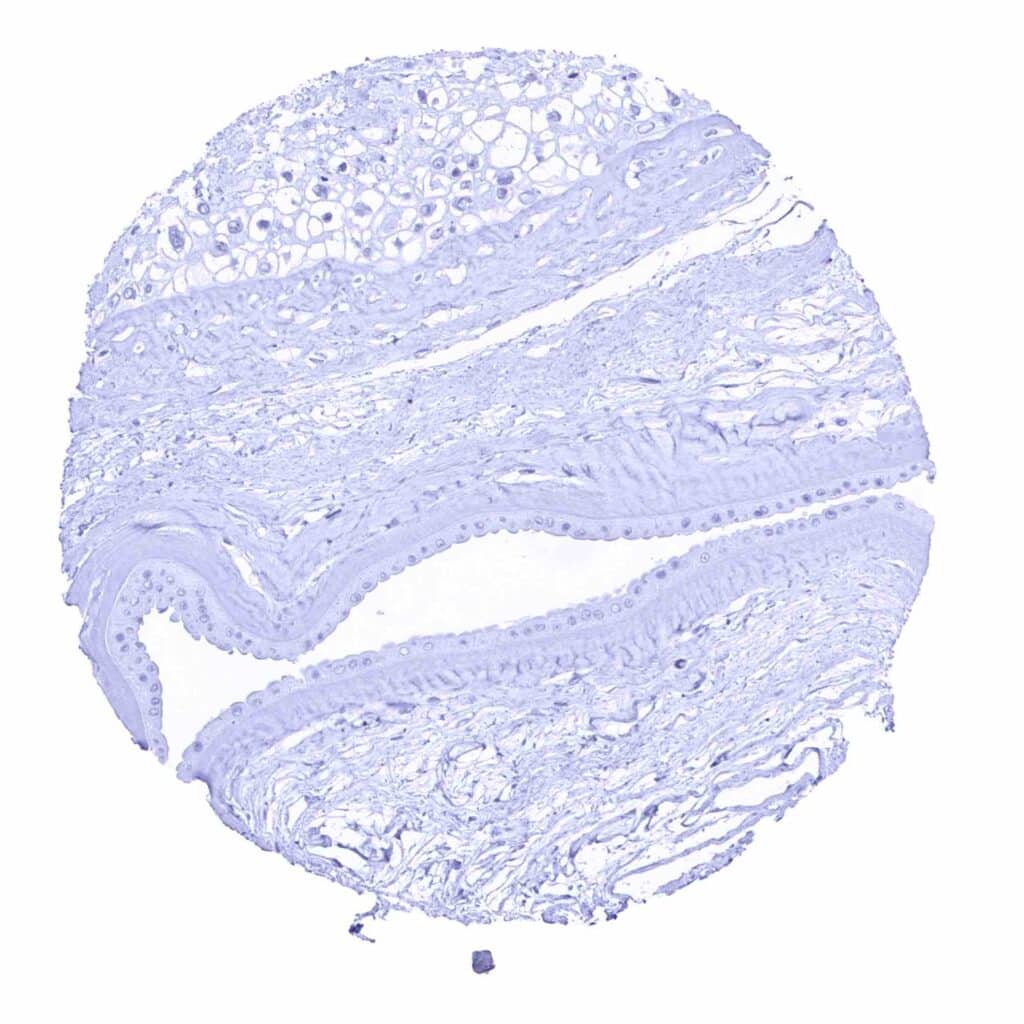
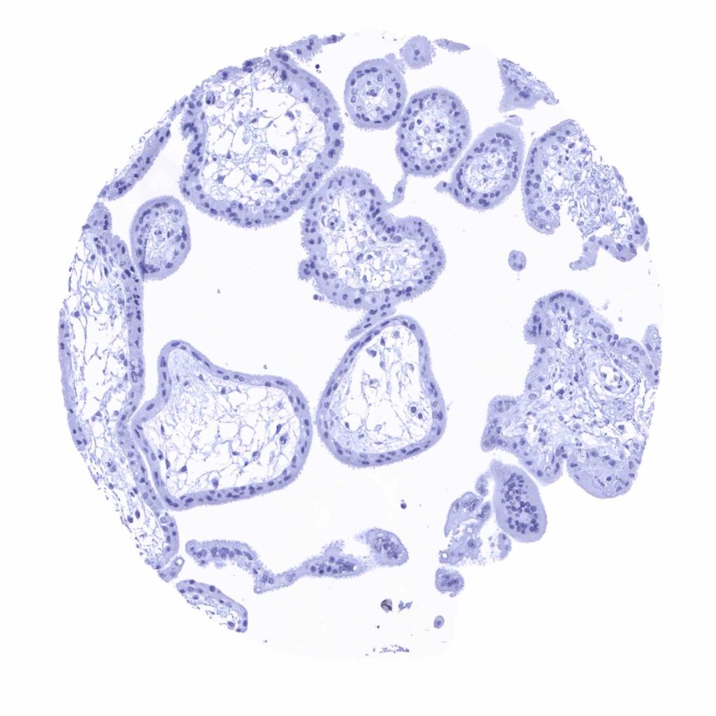
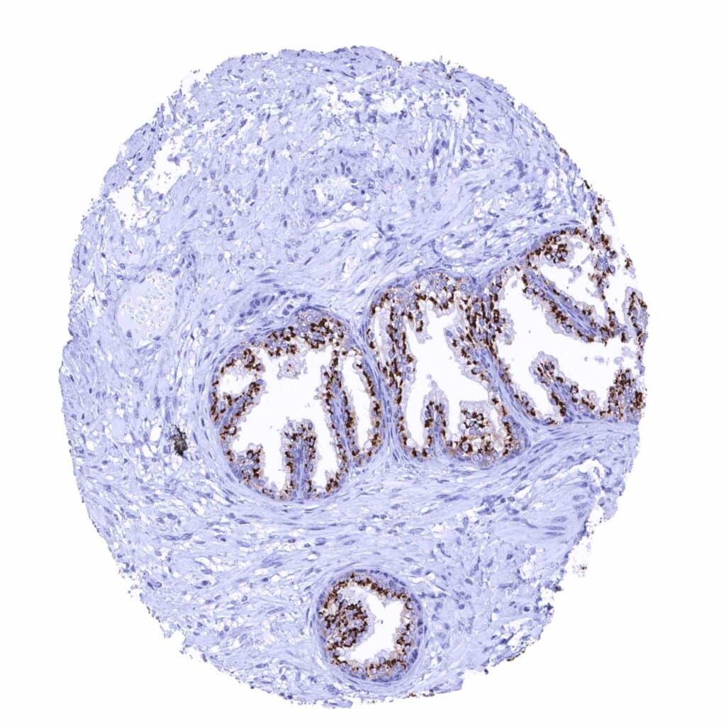
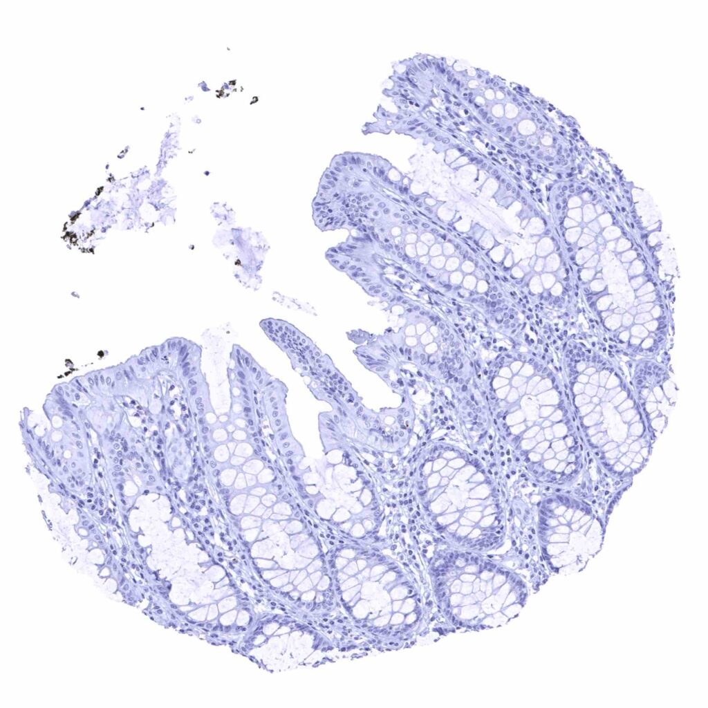
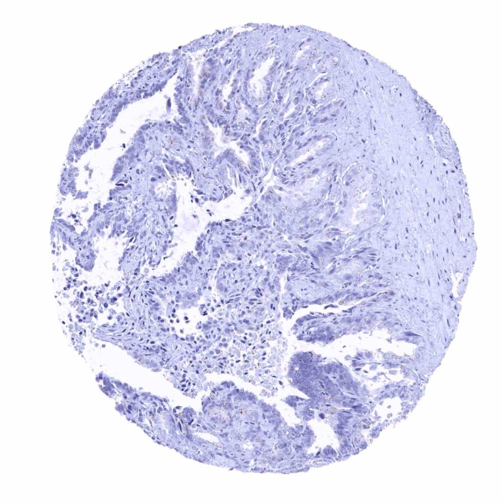
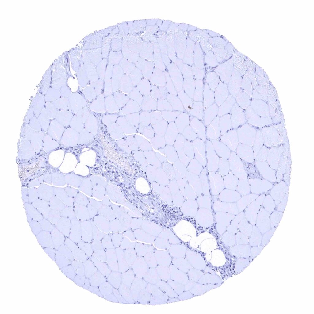
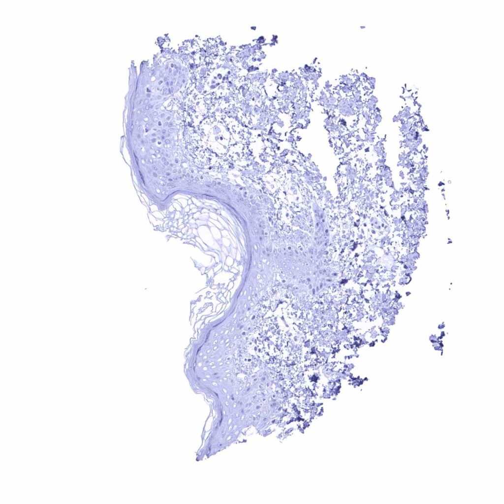
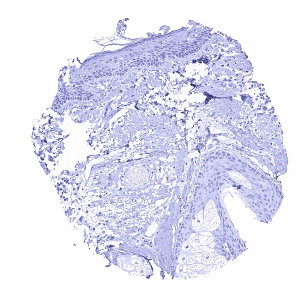
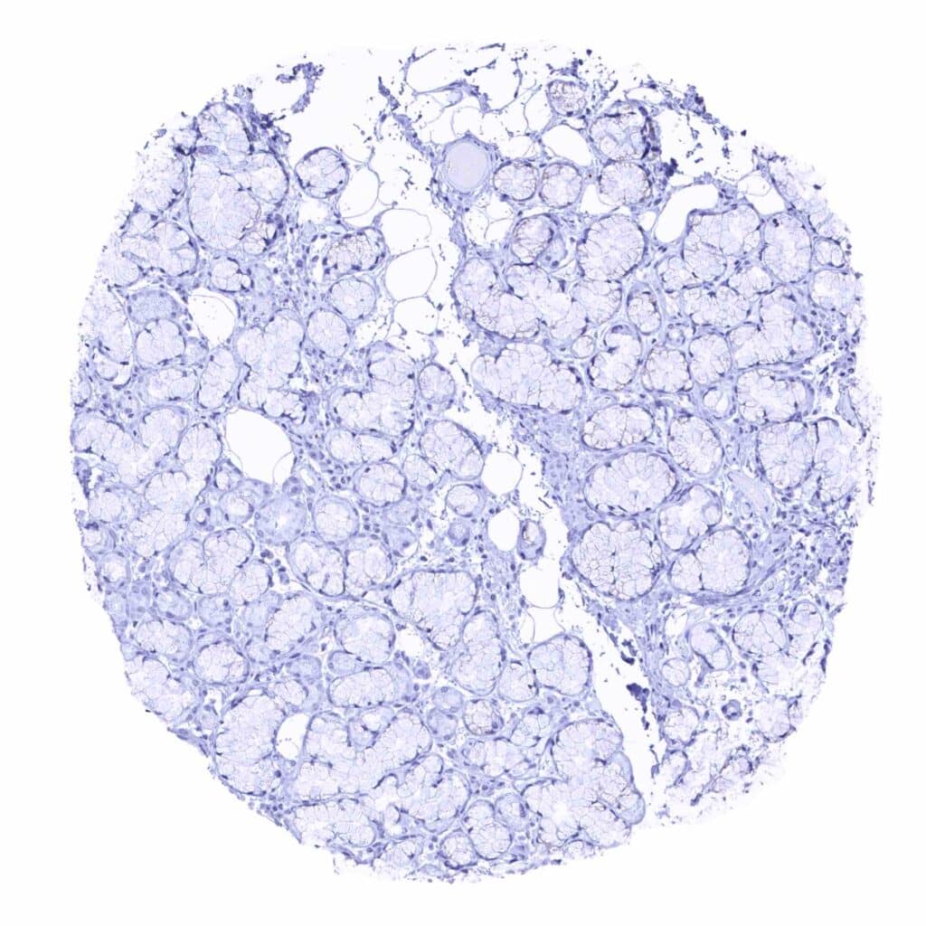
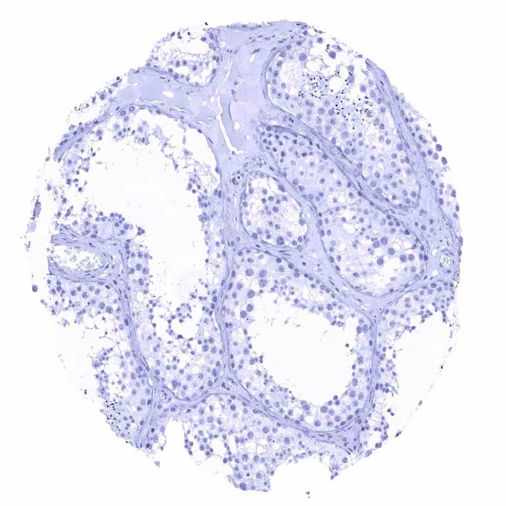
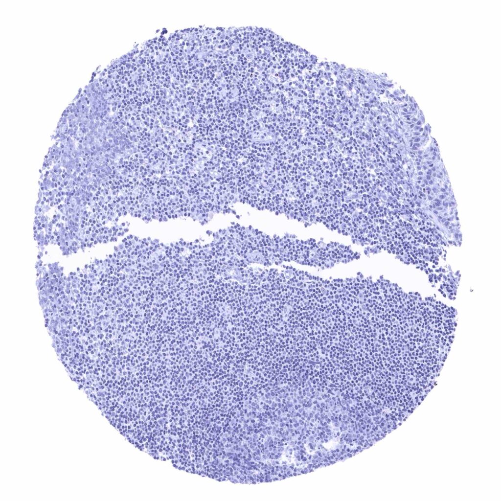
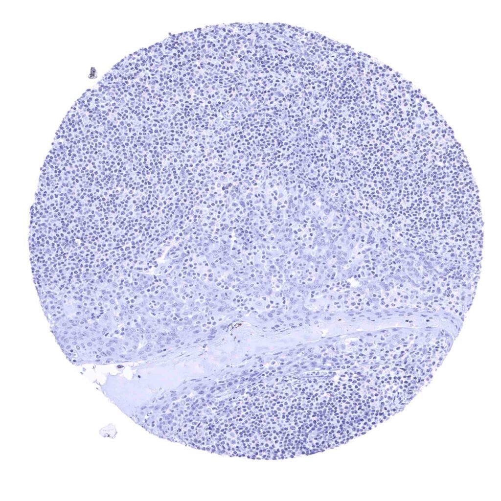
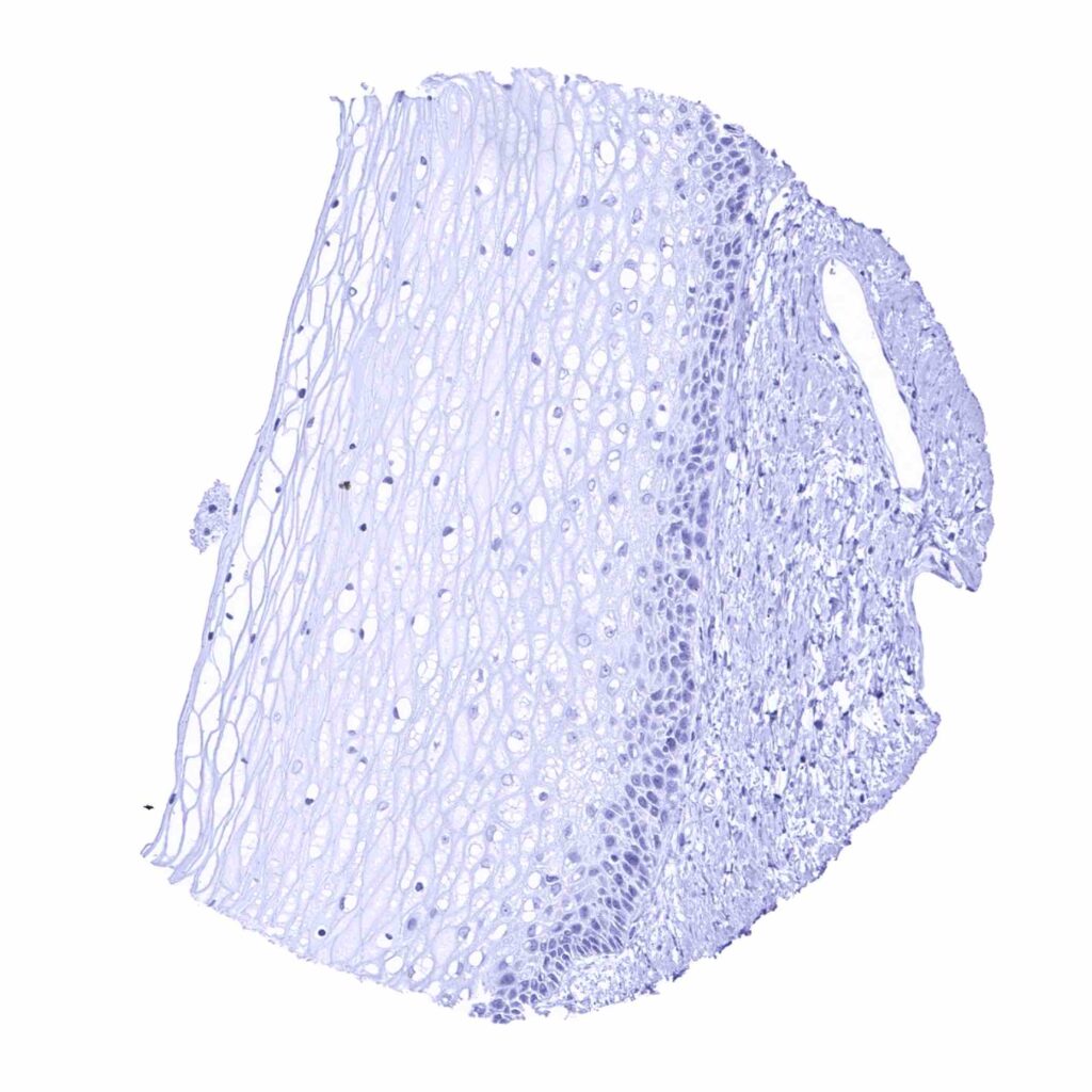
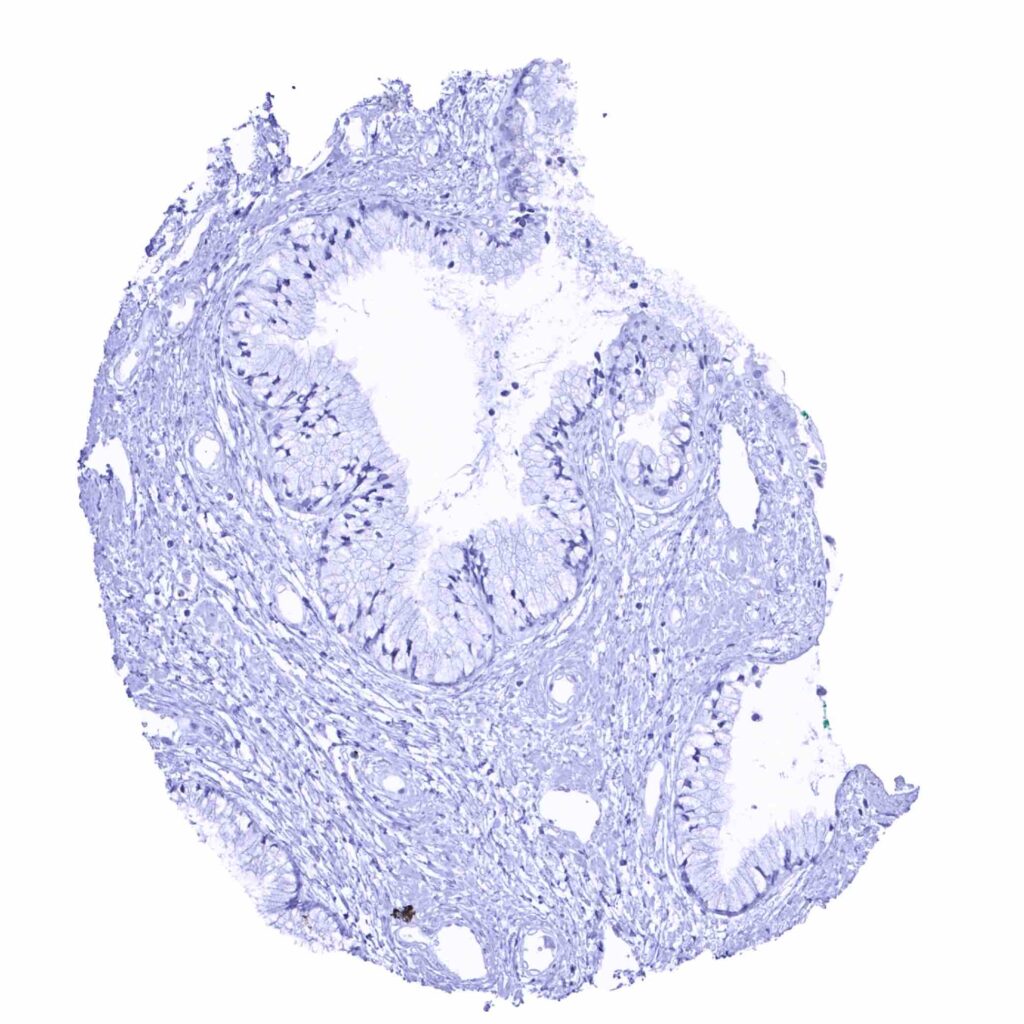
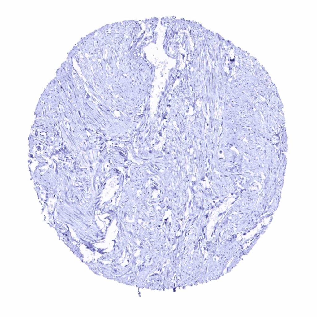
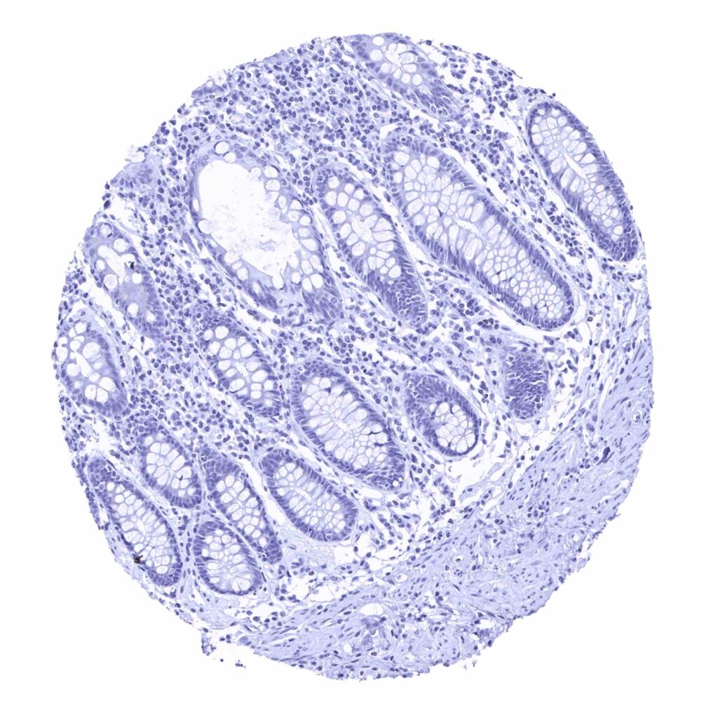
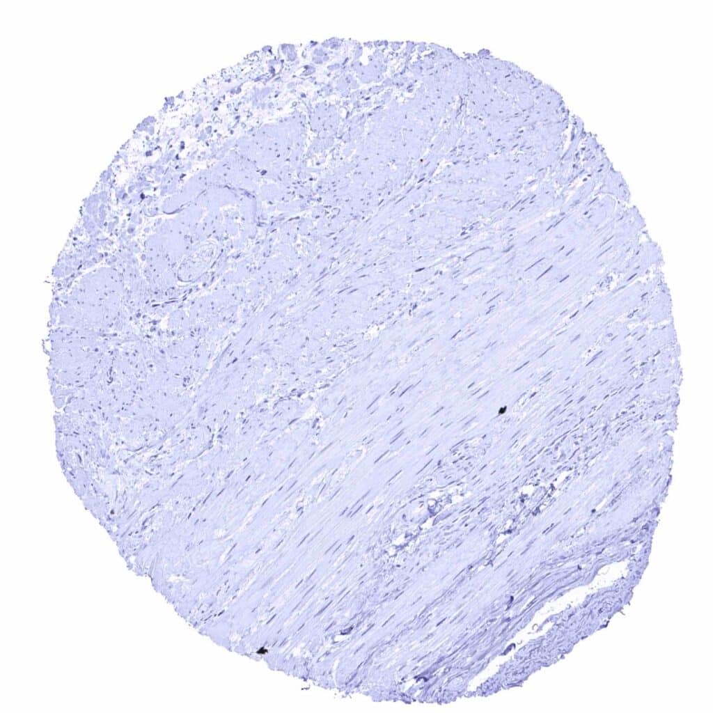
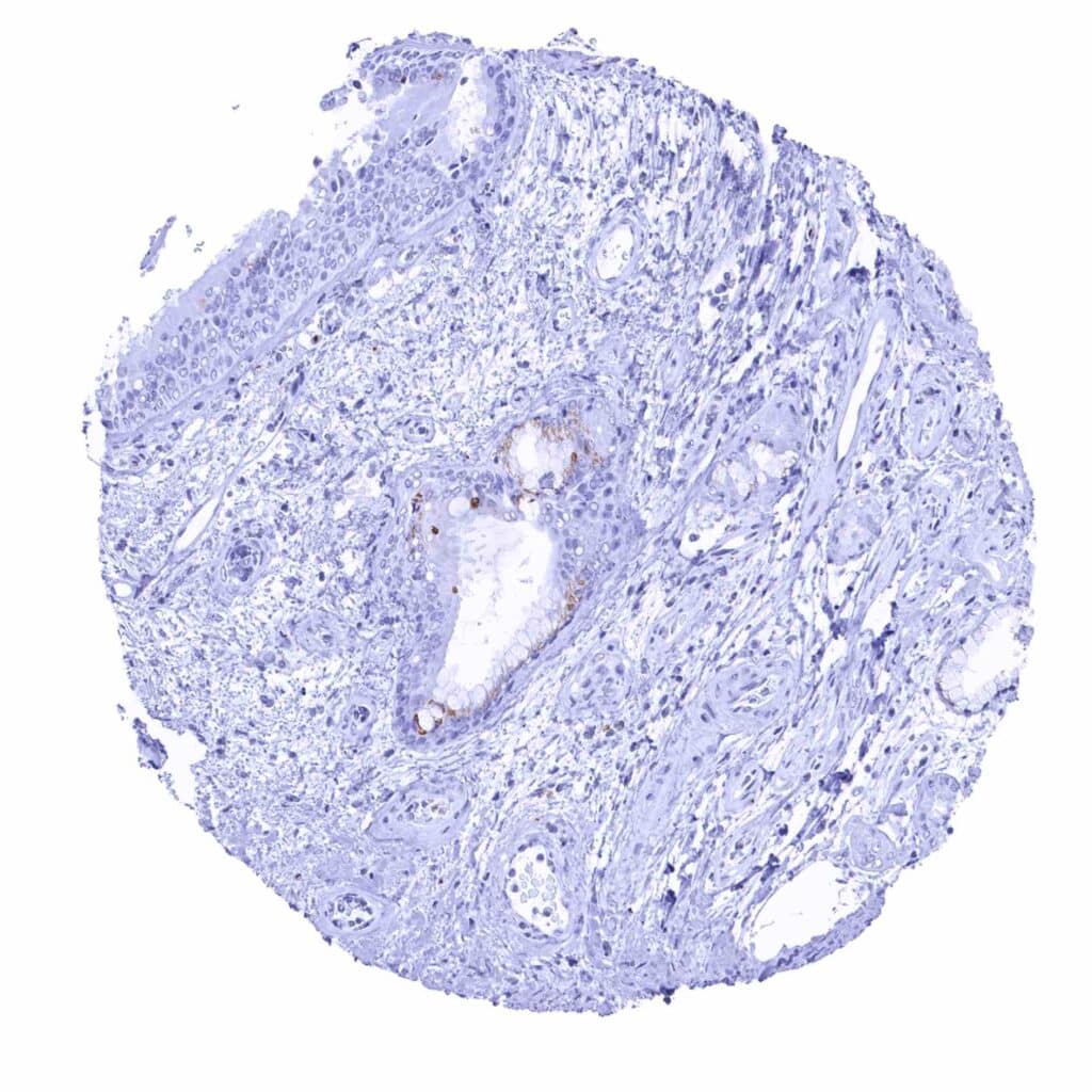
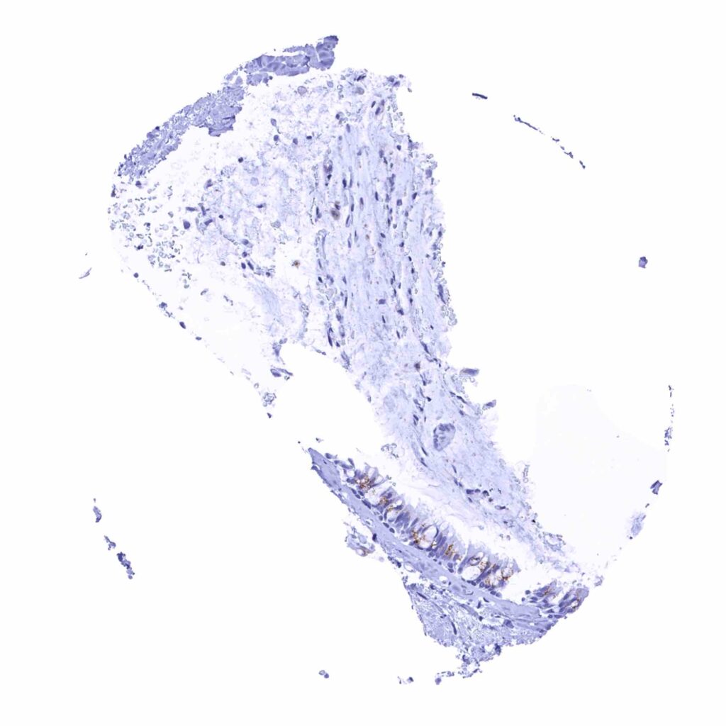
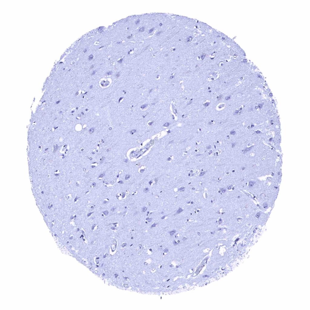
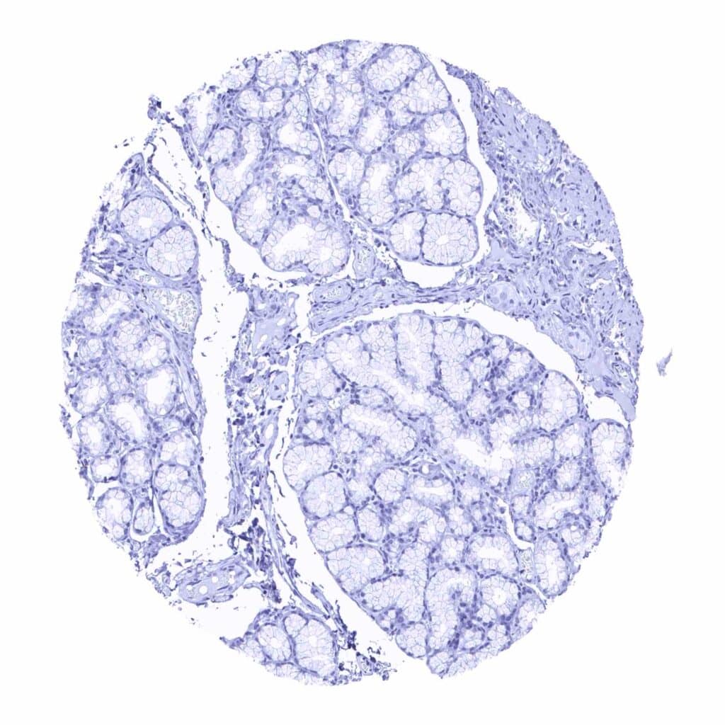
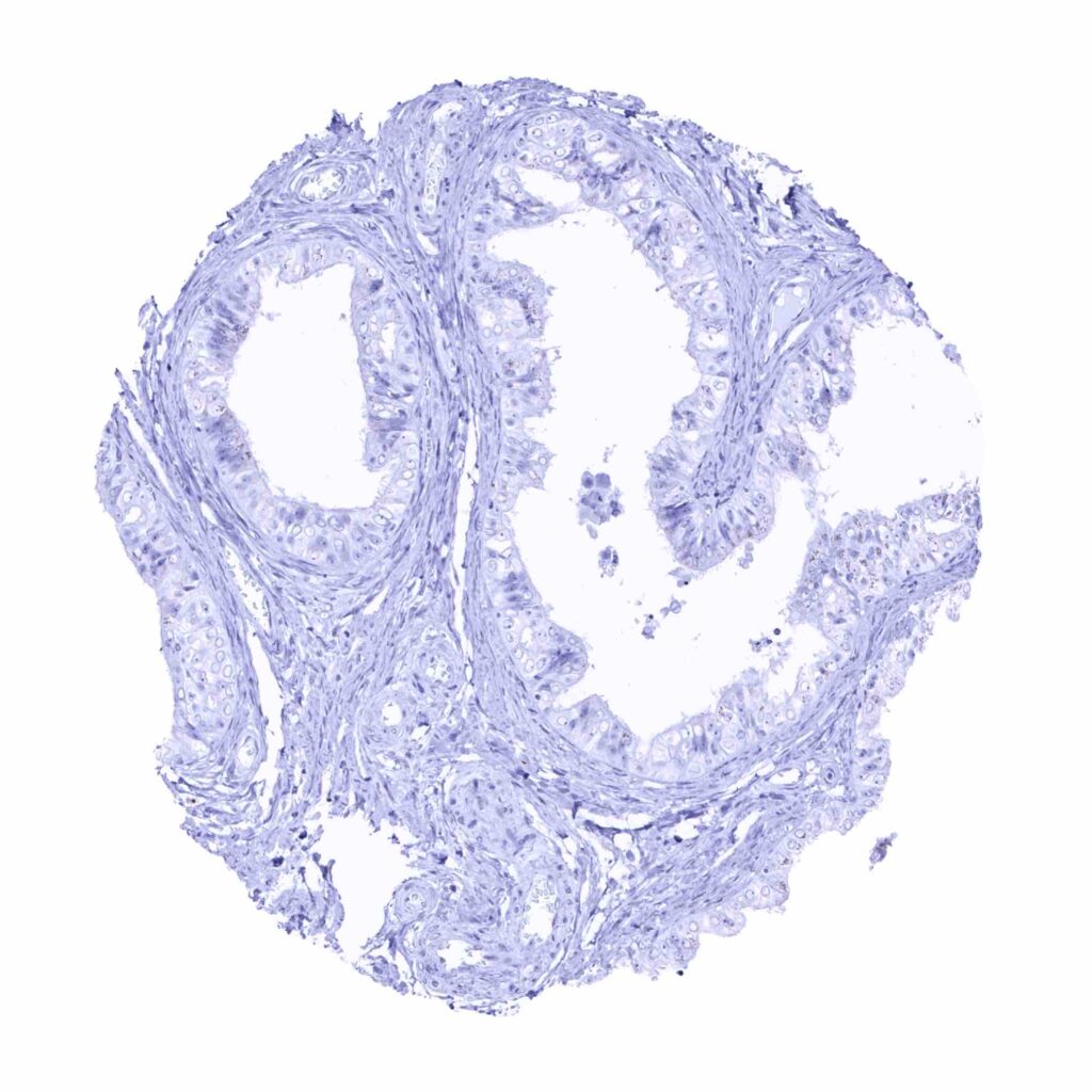
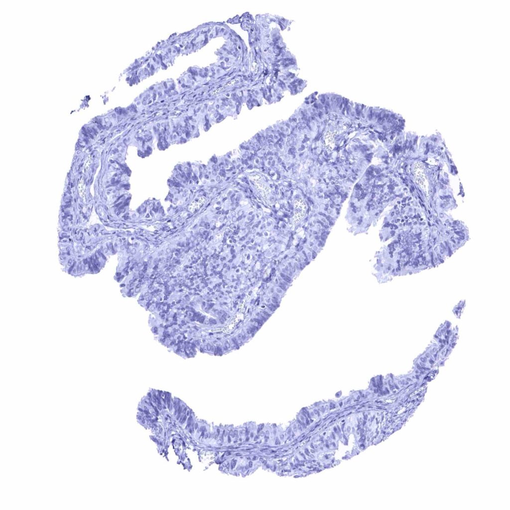
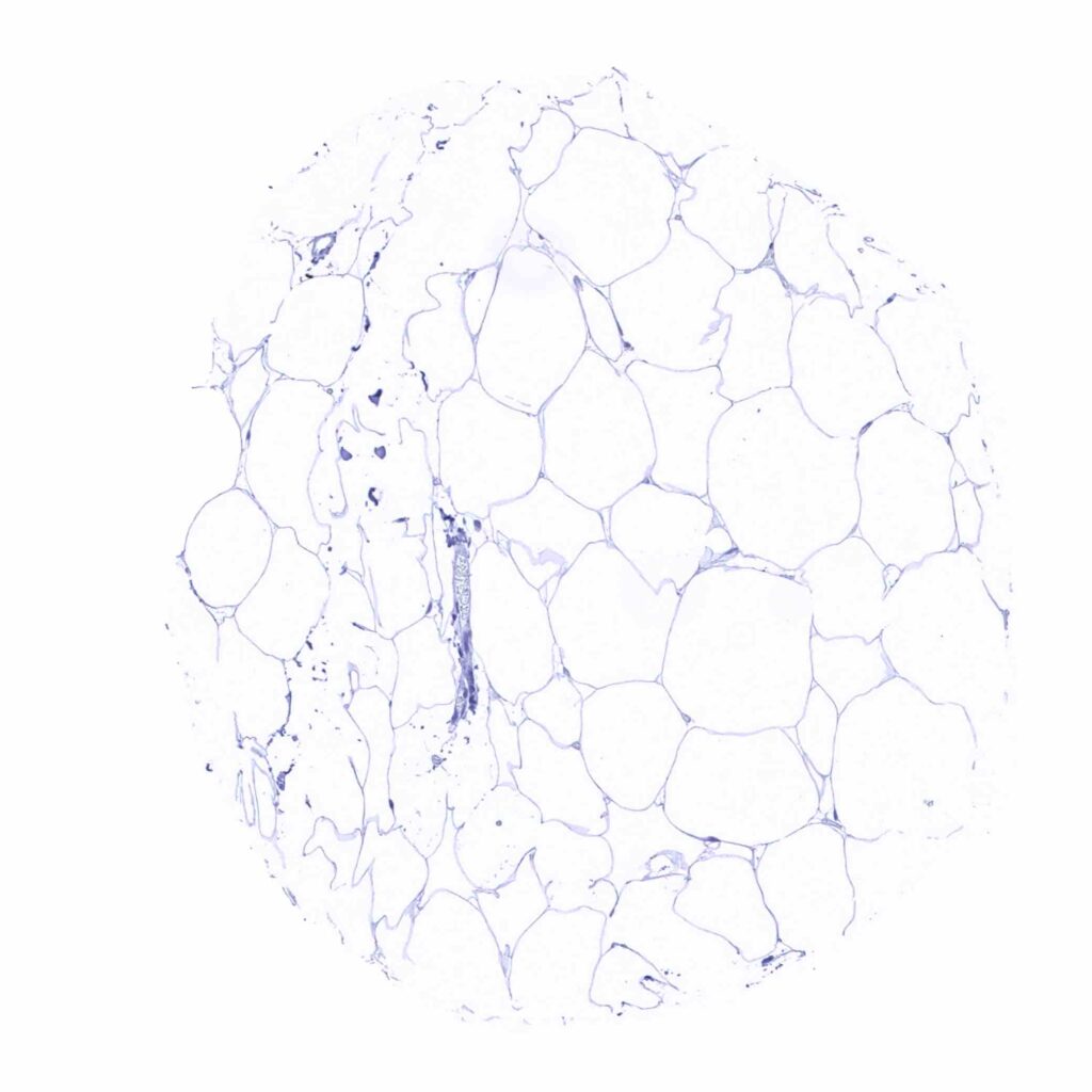
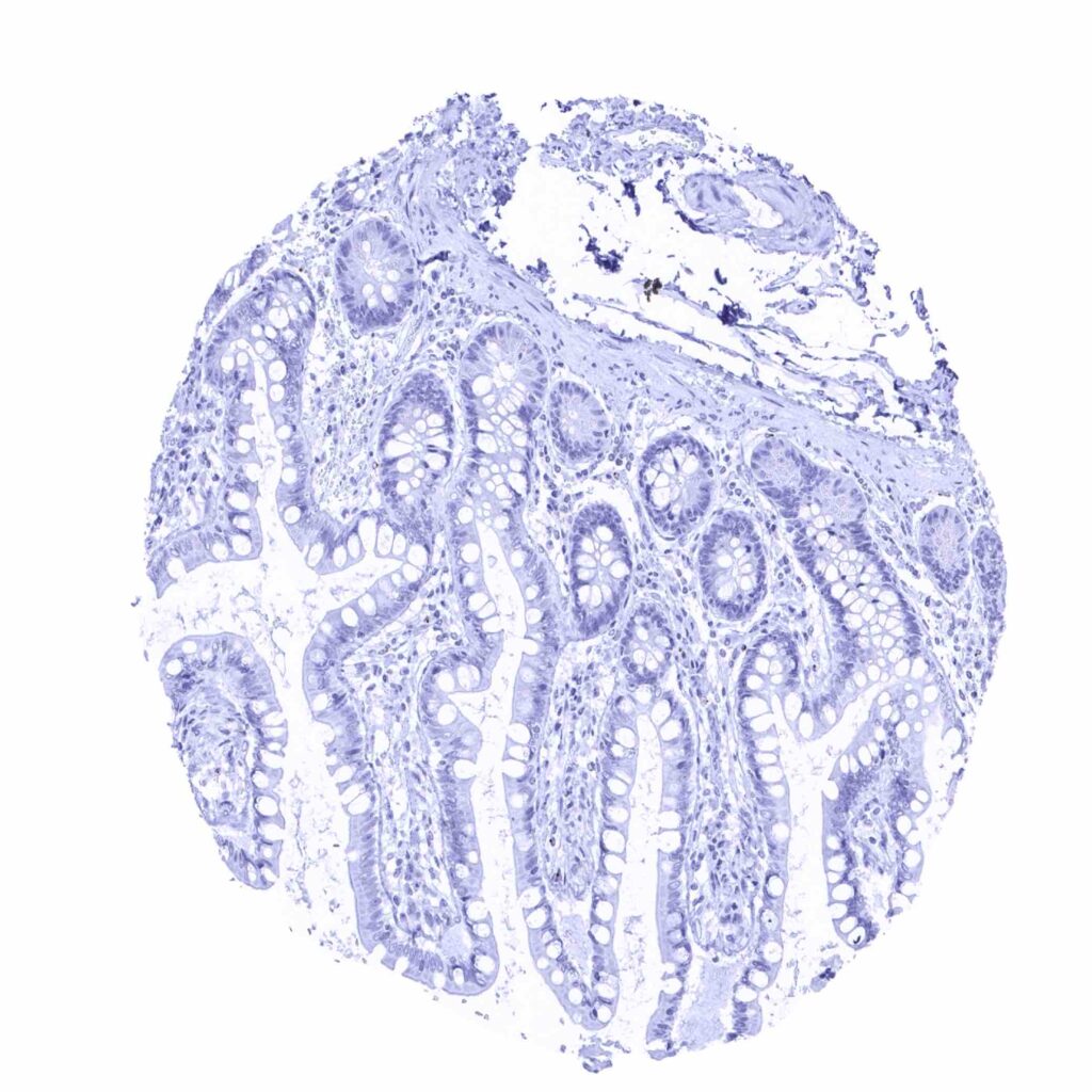
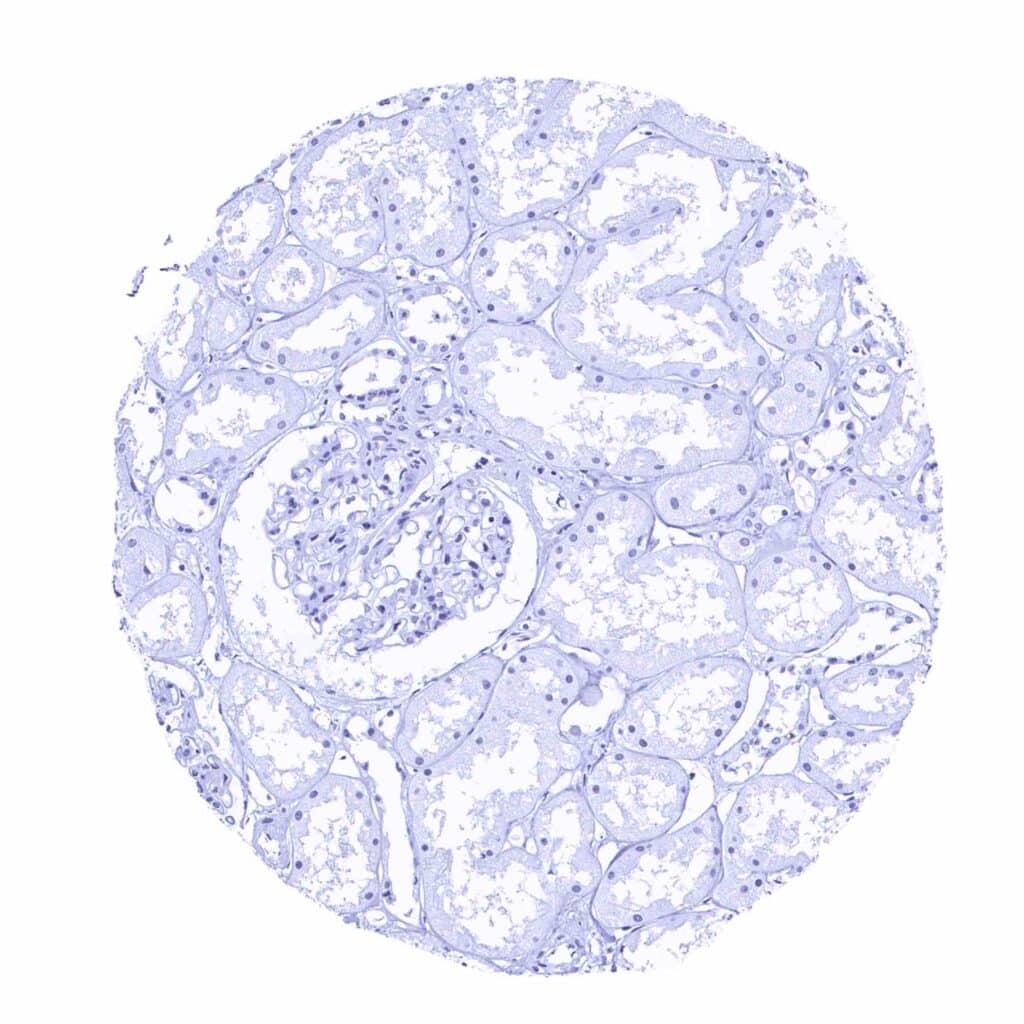
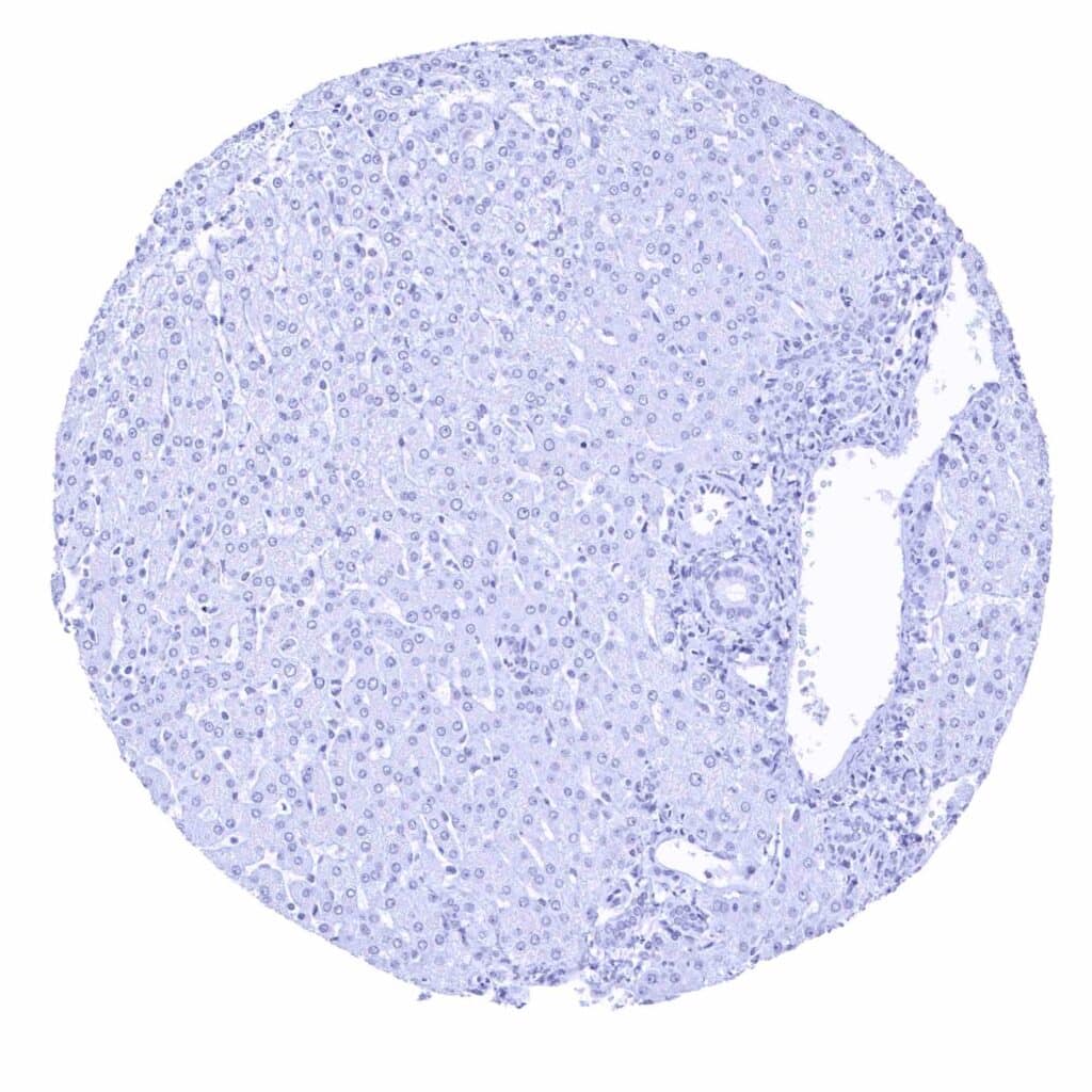
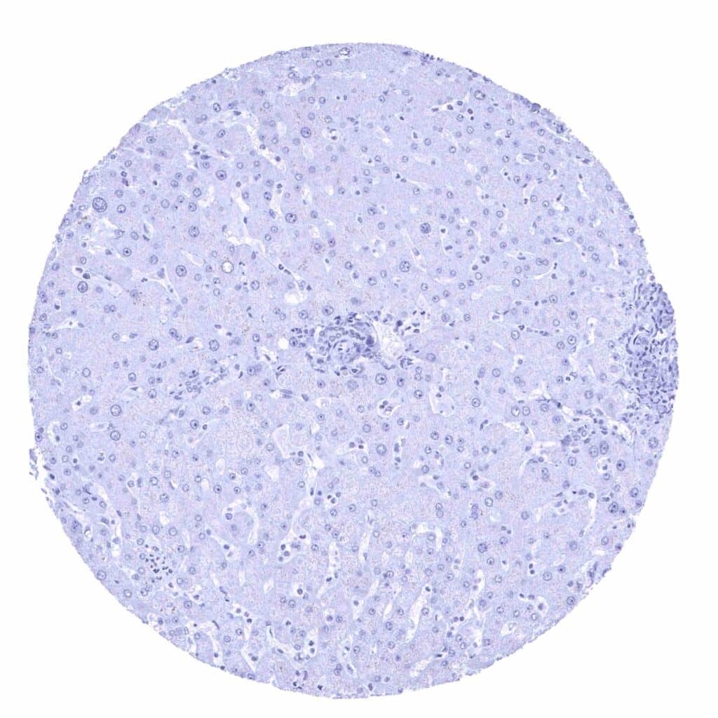
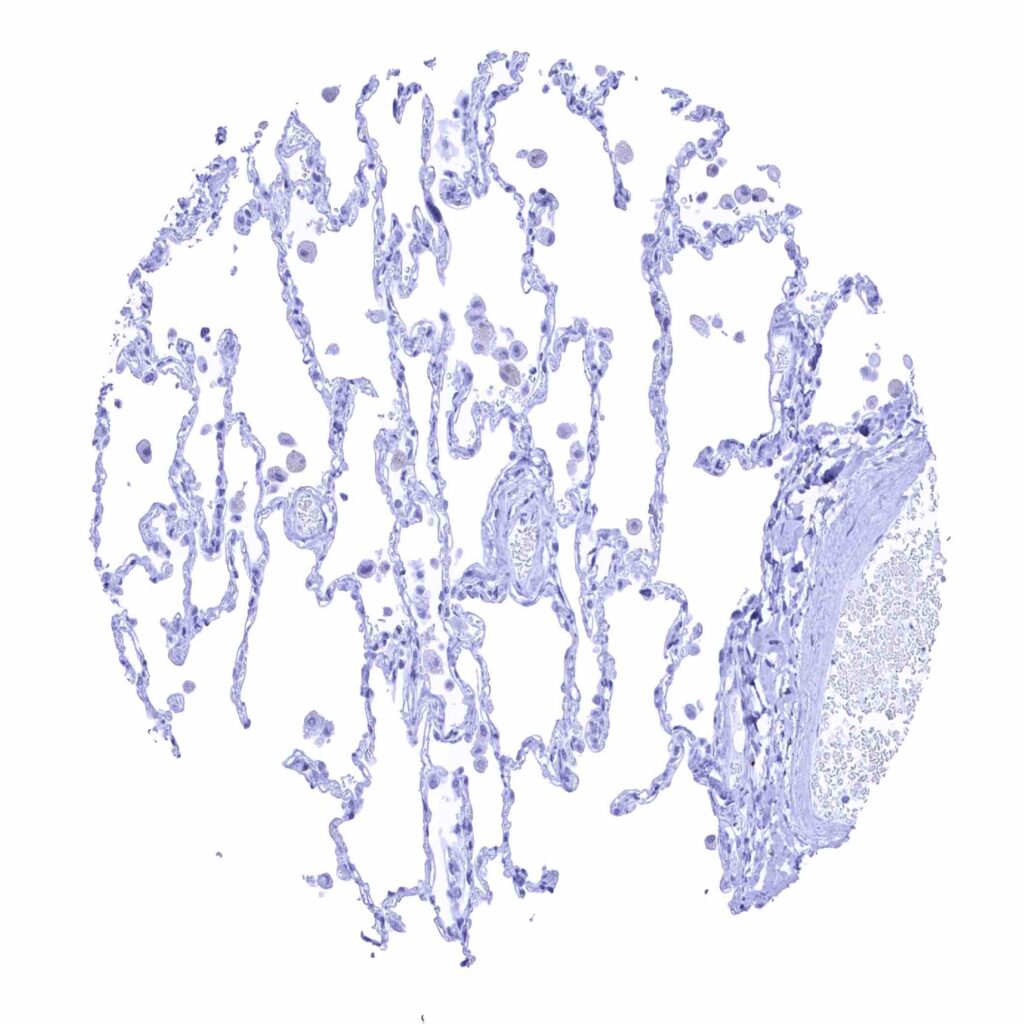
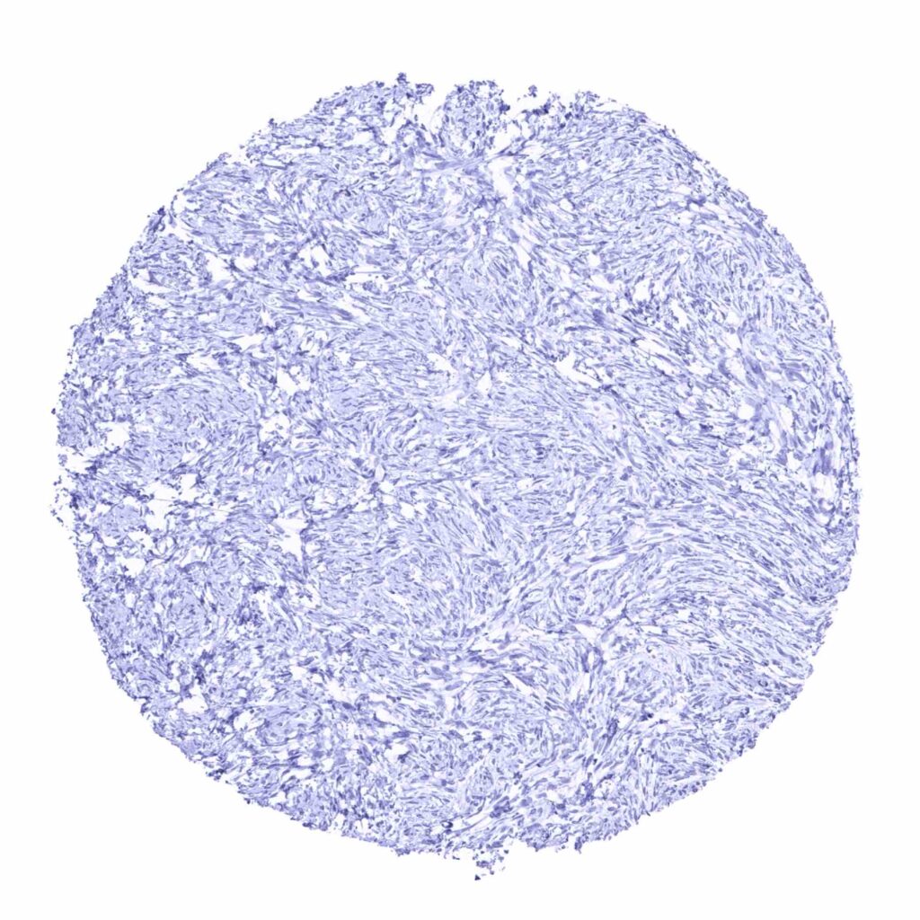
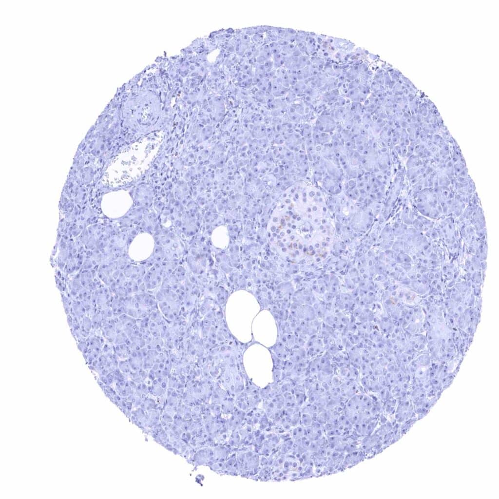
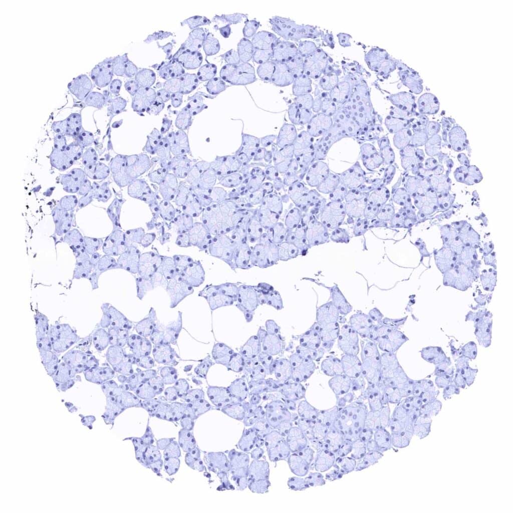
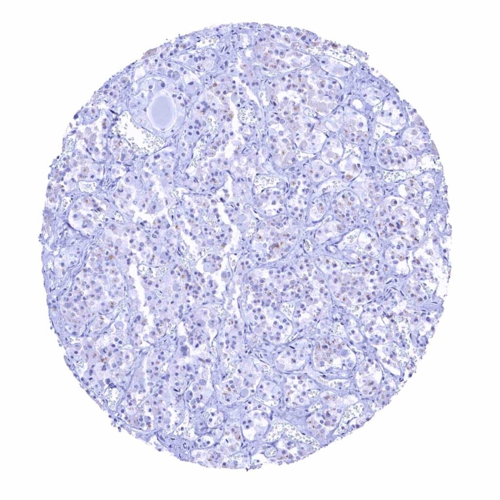
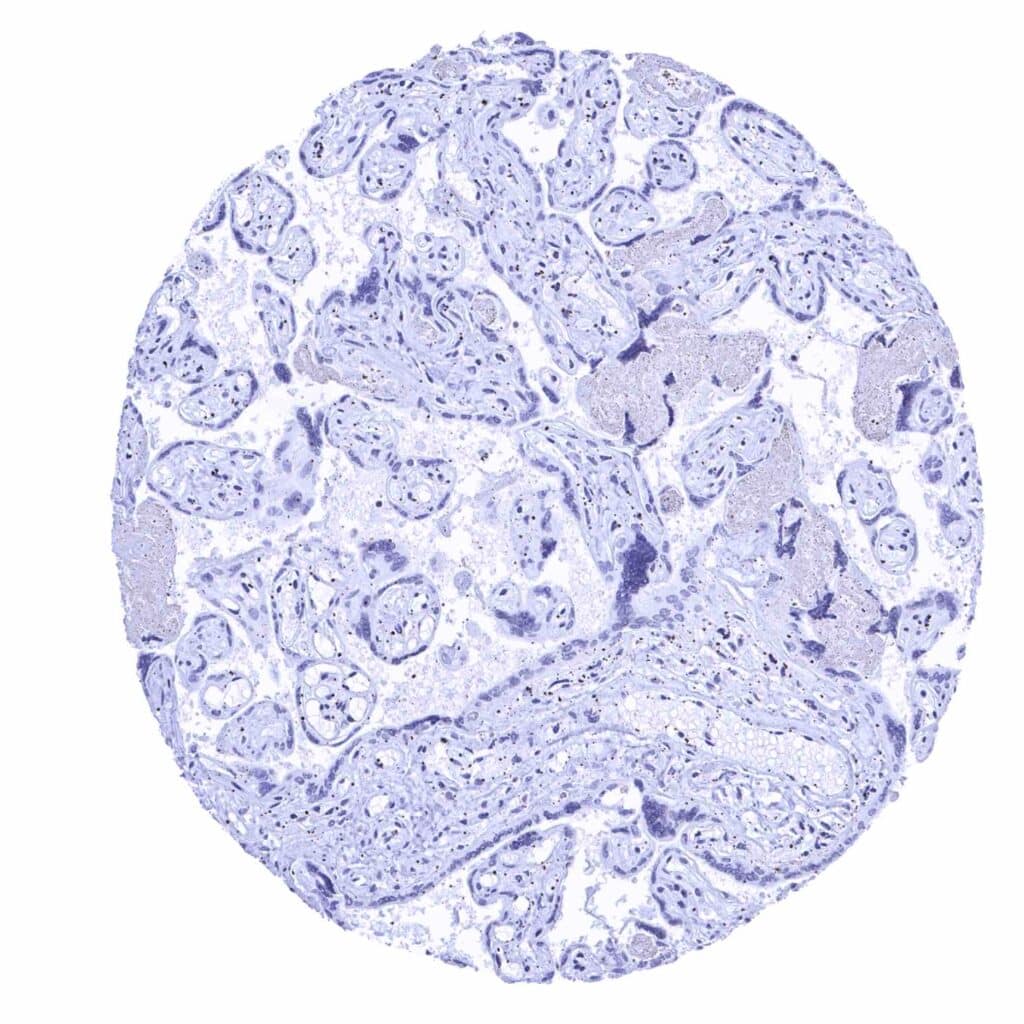
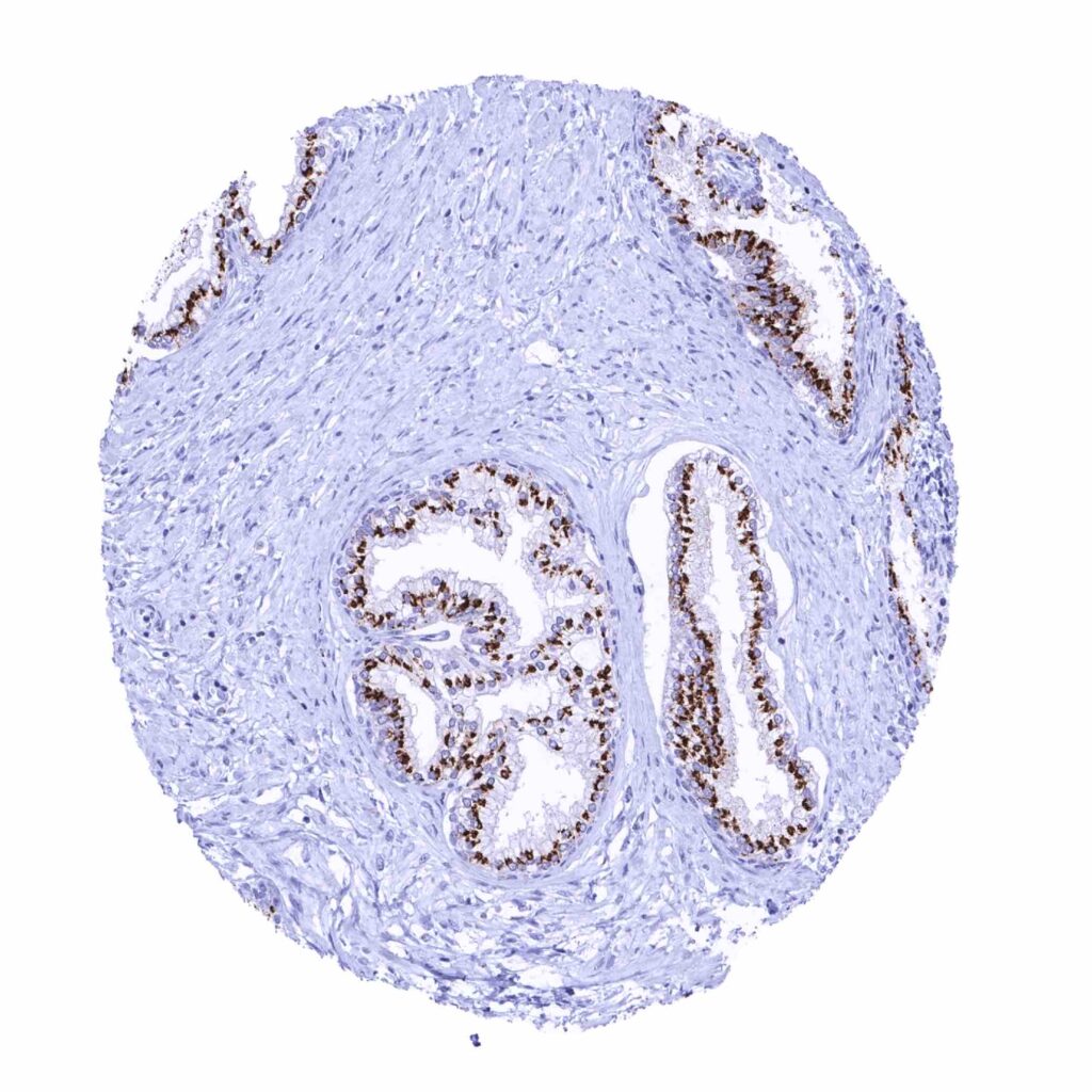
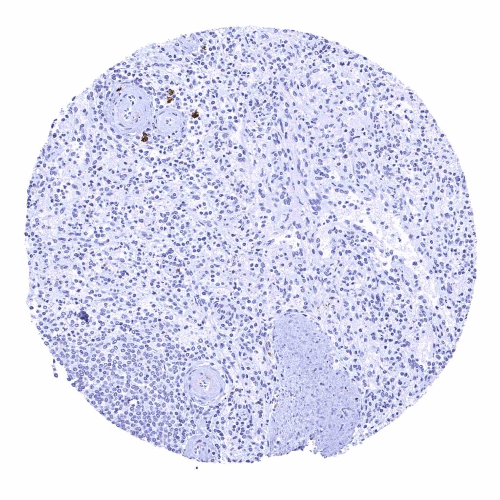
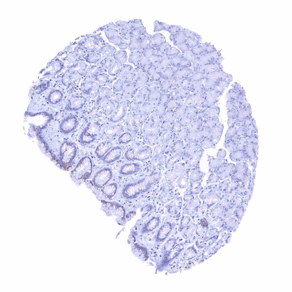
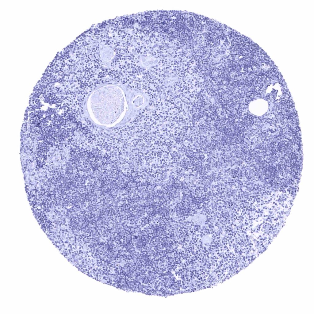
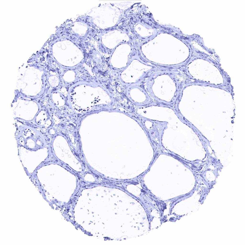
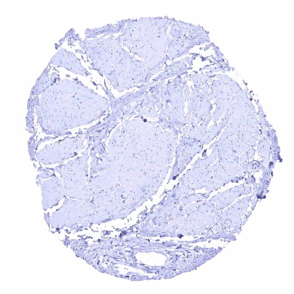
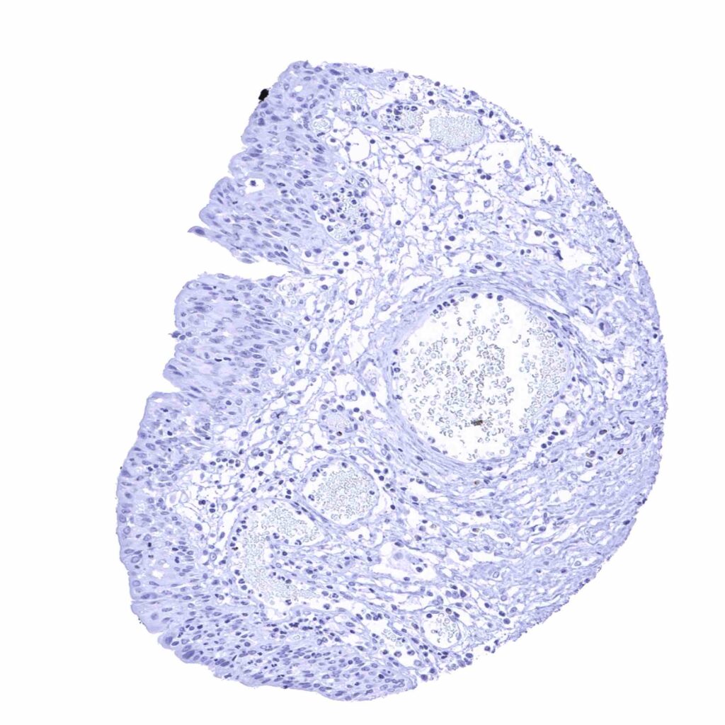
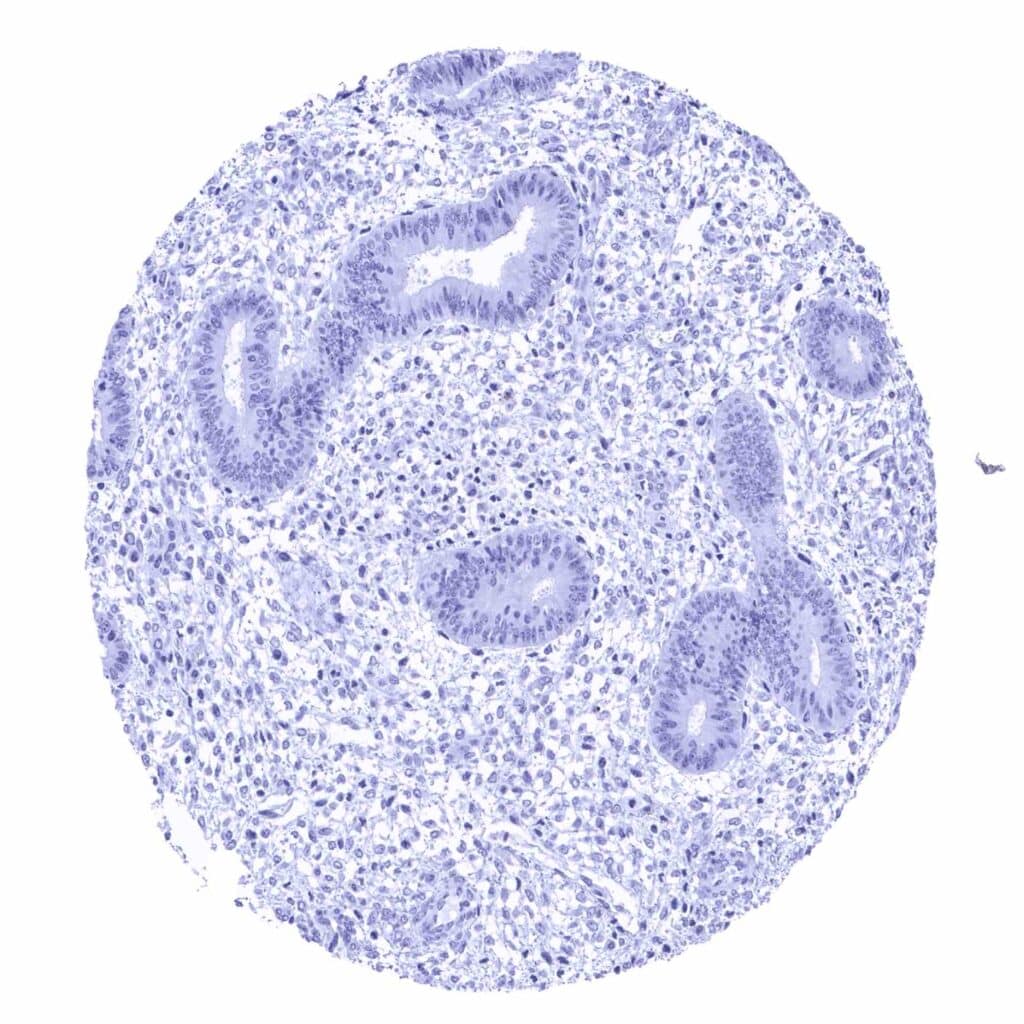
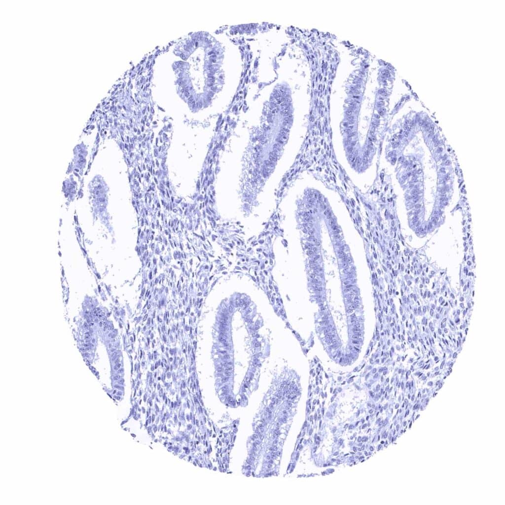
Prostein antibody [MSVA-460R] HistoMAX™


Go to Cancer Tissue Gallery
  
 Go back to product page
  

Go to HistoMAX page


Adrenal gland

Anal canal, skin

Aorta, media

Appendix, mucosa – Prostein staining is lacking


Appendix, muscular wall

Bone marrow

Breast

Bronchus, glands – Moderate granular, cytoplasmic and predominantly perinuclear prostein staining in goblet cells of respiratory epithelium


Bronchus, mucosa – A moderate granular, cytoplasmic and predominantly perinuclear prostein staining is seen in goblet cells of the respiratory epithelium

Bronchus, mucosa – Prostein staining is lacking in this respiratory epithelium which is not containing goblet cells

Cerebellum (molecular layer, Purkinje cell layer, granule cell layer, white matter)

Cerebellum (molecular layer, Purkinje cell layer, granule cell layer, white matter)


Cerebrum, grey matter

Cerebrum, white matter

Colon descendens, muscular wall

Duodenum, Brunner gland


Duodenum, mucosa – Absence of prostein staining

Epididymis

Epididymis

Esophagus, squamous epithelium


Fallopian tube, mucosa

Fat

Gallbladder, epithelium – Absence of prostein staining

Heart muscle


Ileum, mucosa – Absence of prostein staining

Kidney, cortex

Kidney, medulla

Liver


Liver

Lung

Lymph node

Ovary, follicular cyst


Ovary, stroma

Pancreas – A weak, granular cytoplasmic prostein staining can be seen in a subset of pancreatic islet cells

Pancreas – Pancreatic islet cells are prostein negative in his sample

Parathyroid gland


Parotid gland

Pituitary gland, anterior lobe

Pituitary gland, posterior lobe

Placenta (amnion and chorion)


Placenta, early

Placenta, mature

Prostate – Intense granular, cytoplasmic prostein staining of luminal cells of the prostate (prostein immunohistochemistry)

Prostate – Luminal epithelial cells of the prostate show a strong granular, perinuclear cytoplasmic prostein staining (prostein immunohistochemistry)


Prostate – Strong, granular, perinuclear cytoplasmic prostein staining of luminal cells of the prostate (prostein immunohistochemistry)

Rectum, mucosa – Absence of prostein staining

Seminal vesicle – Absence of prostein staining

Skeletal muscle


Skin

Skin, hairfollicel and sebaceous glands

Spleen

Stomach, antrum – Granular cytoplasmic prostein staining of moderate intensity in surface epithelial cells (prostein immunohistochemistry)


Stomach, corpus – Granular cytoplasmic prostein staining of moderate intensity in surface epithelial cells (prostein immunohistochemistry)

Sublingual gland

Testis

Thymus


Thyroid gland

Tonsil, surface epithelium

Tonsil

Urinary bladder, muscular wall


Urinary bladder, urothelium

Uterus, ectocervix

Uterus, endocervix

Uterus, endometrium (pregnancy)


Uterus, endometrium (proliferation)

Uterus, endometrium (secretion)

Uterus, myometrium


| TOP |