Normal Tissue Gallery- GTX639936

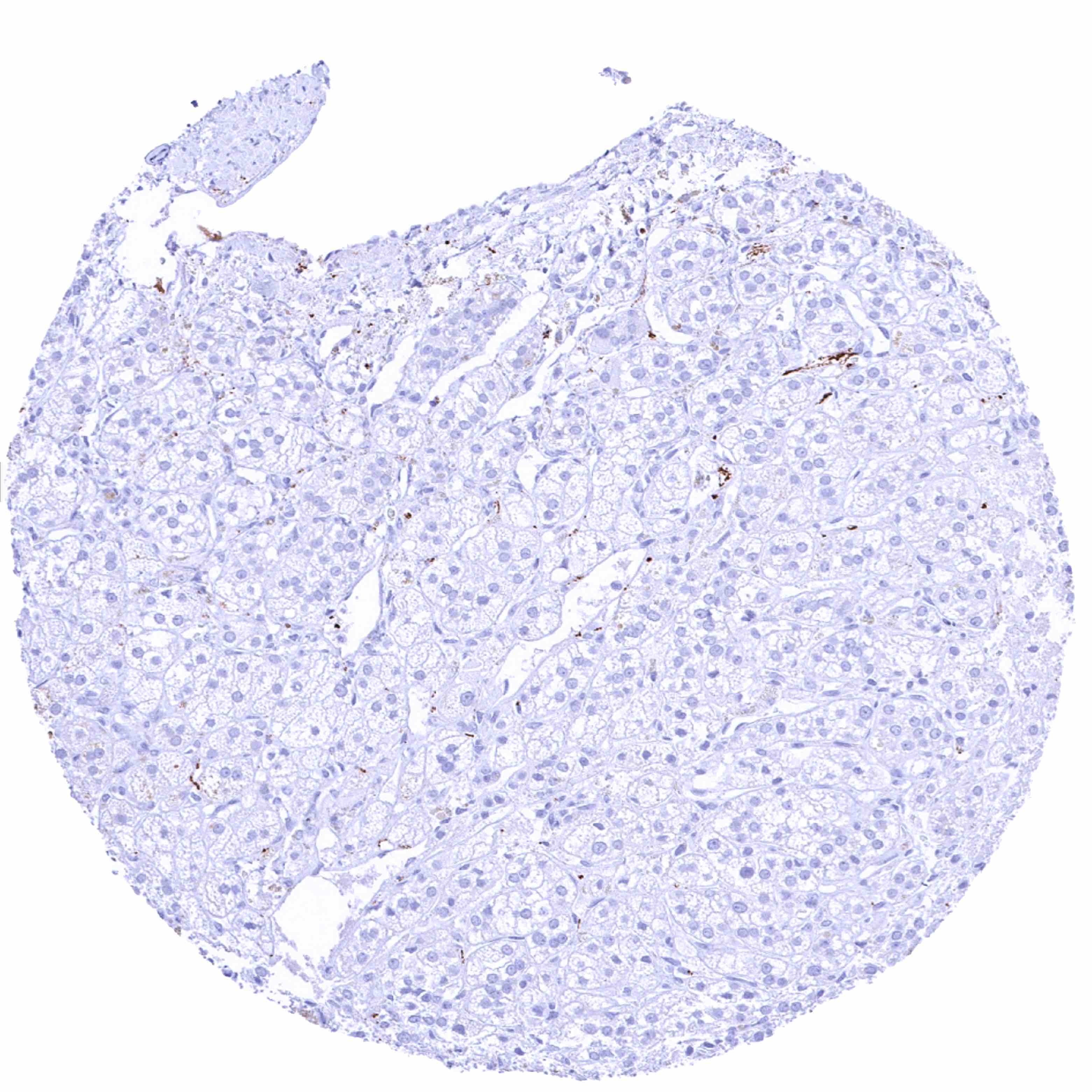
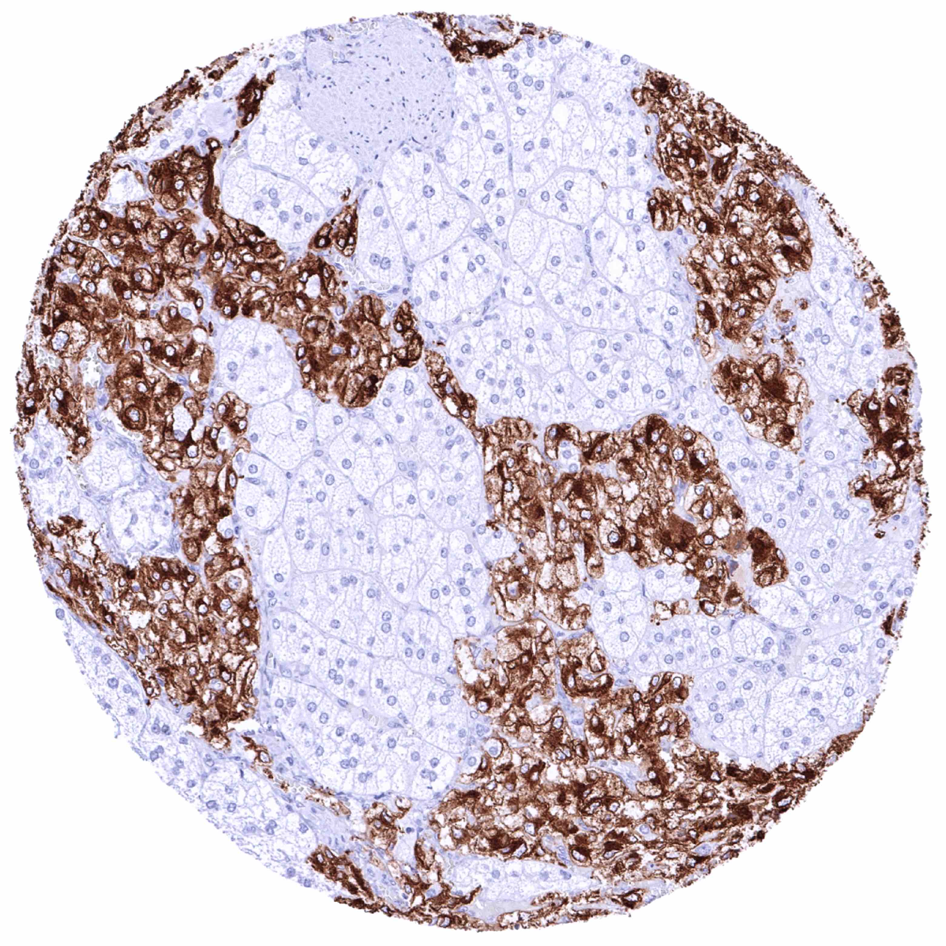
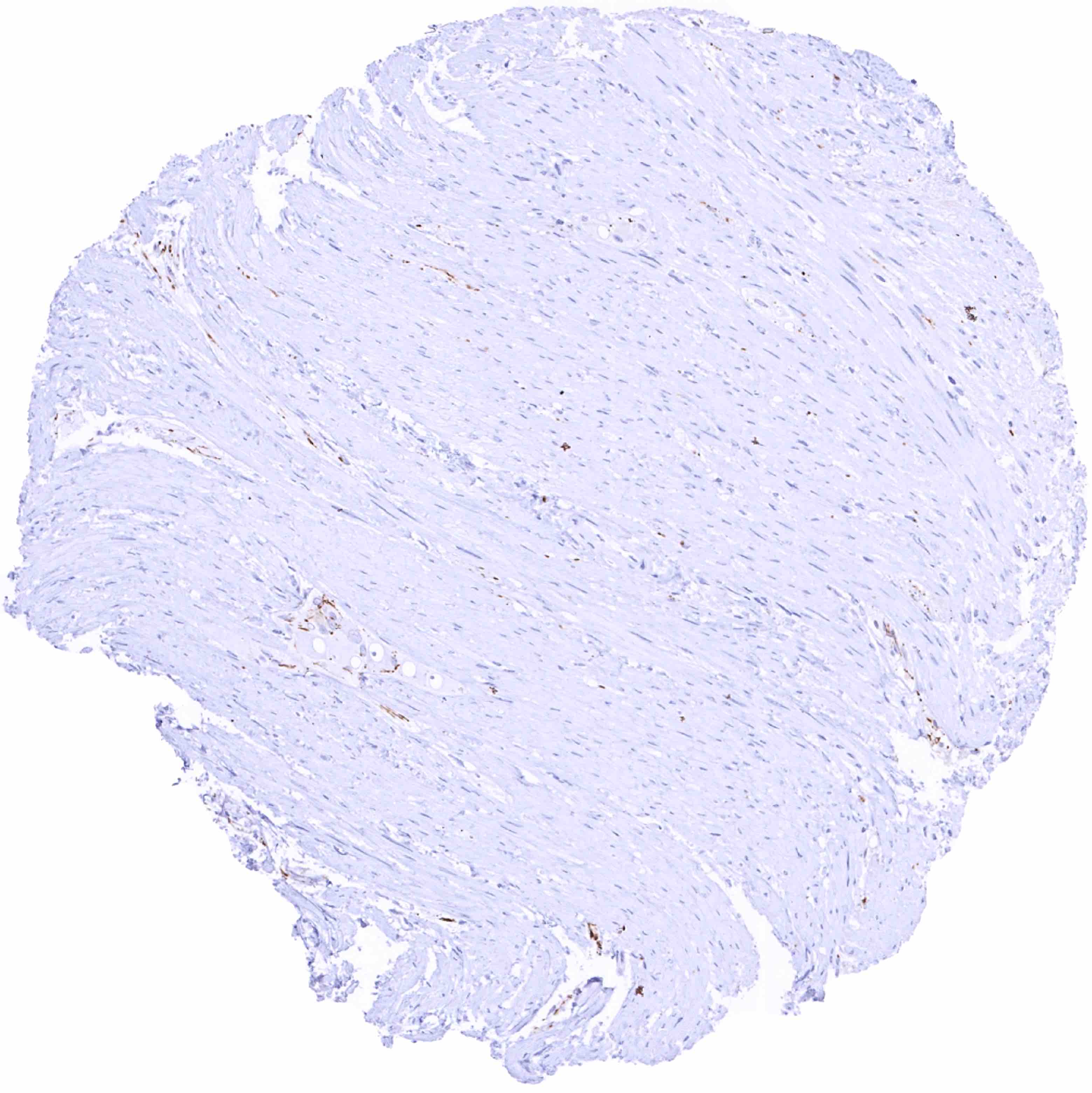
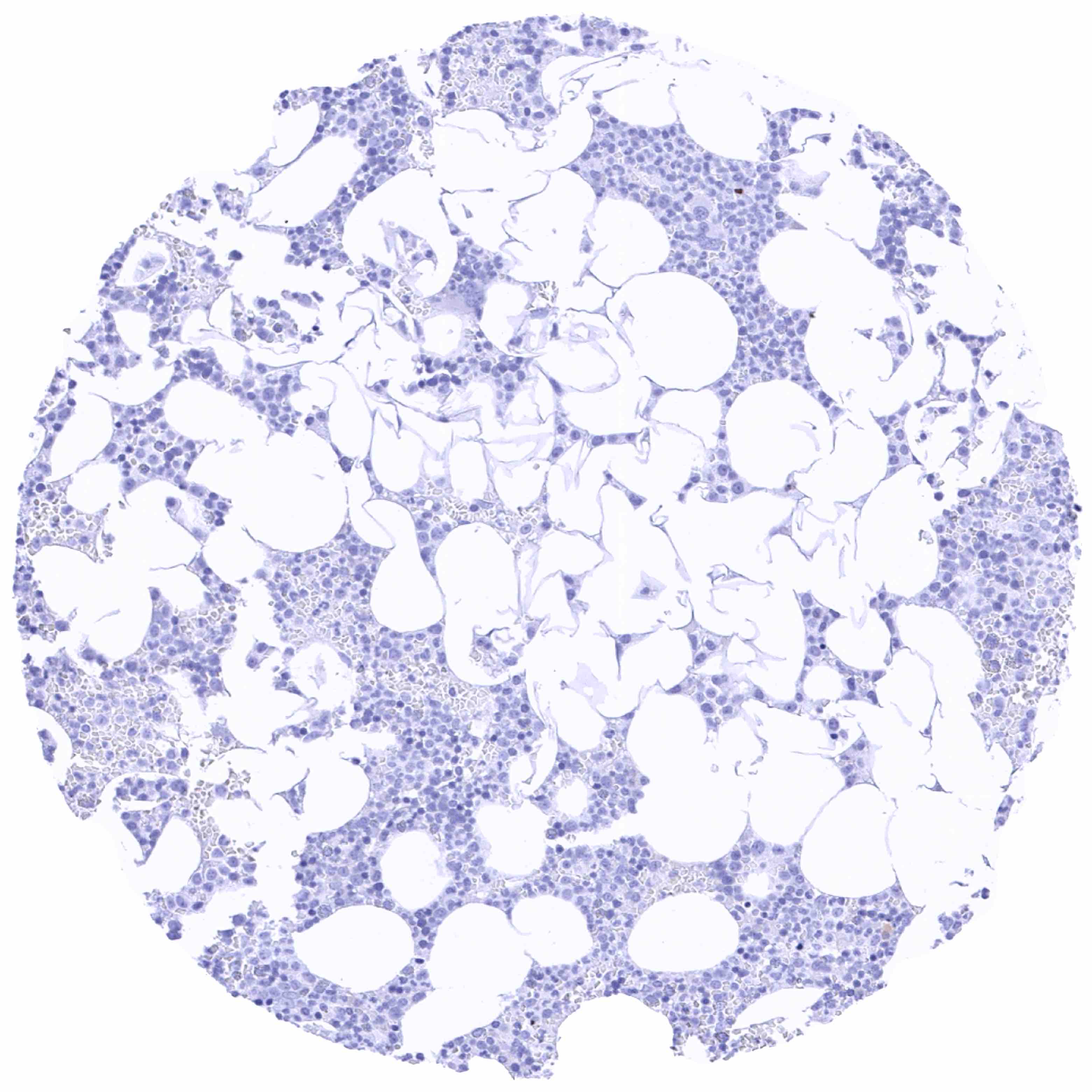
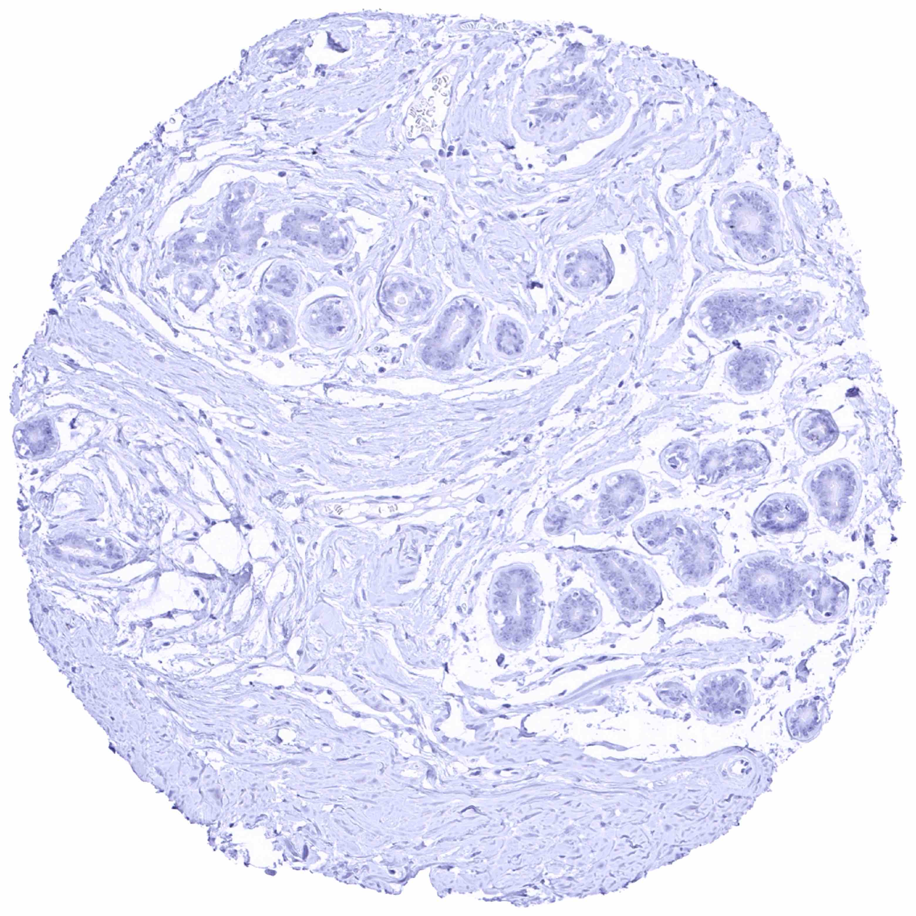
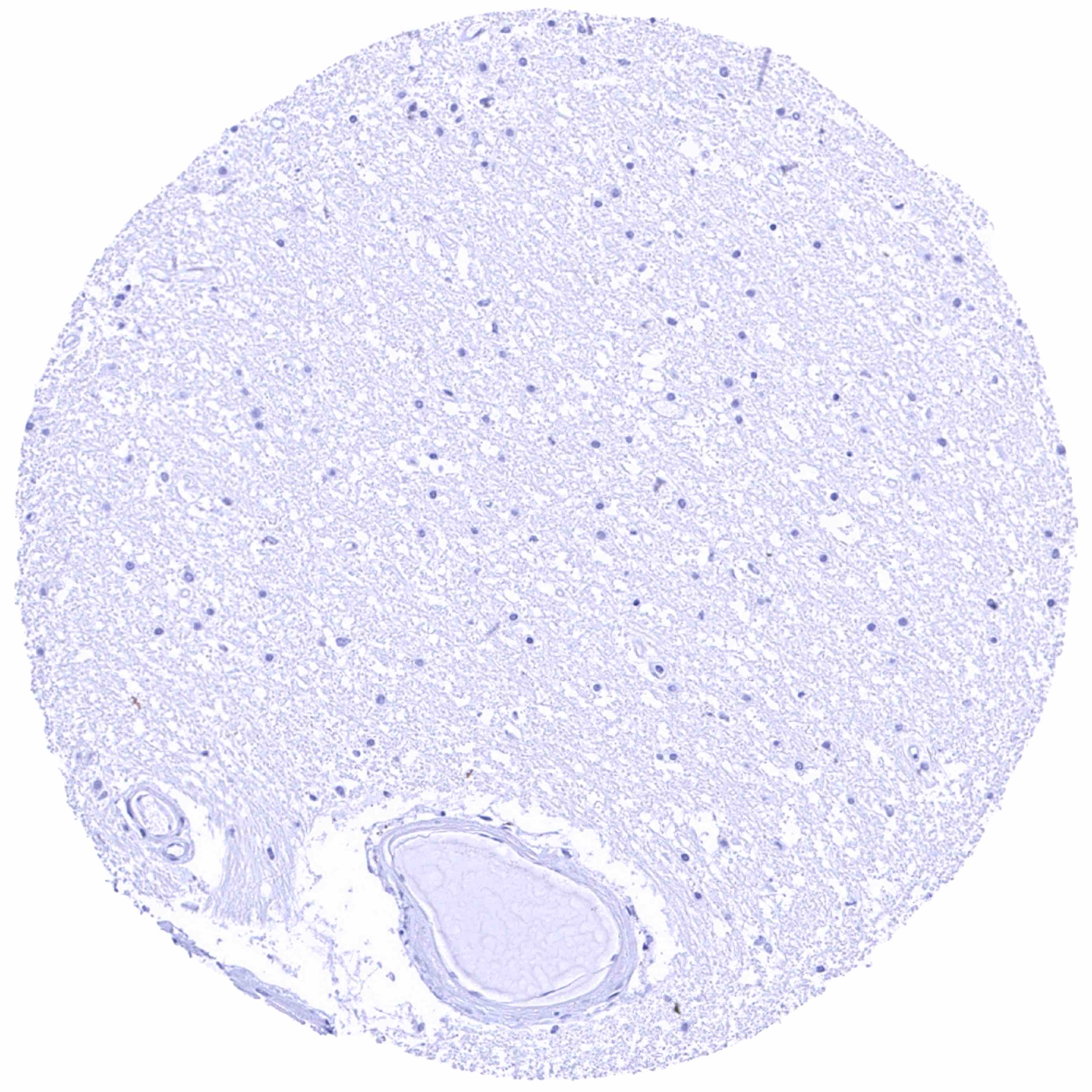
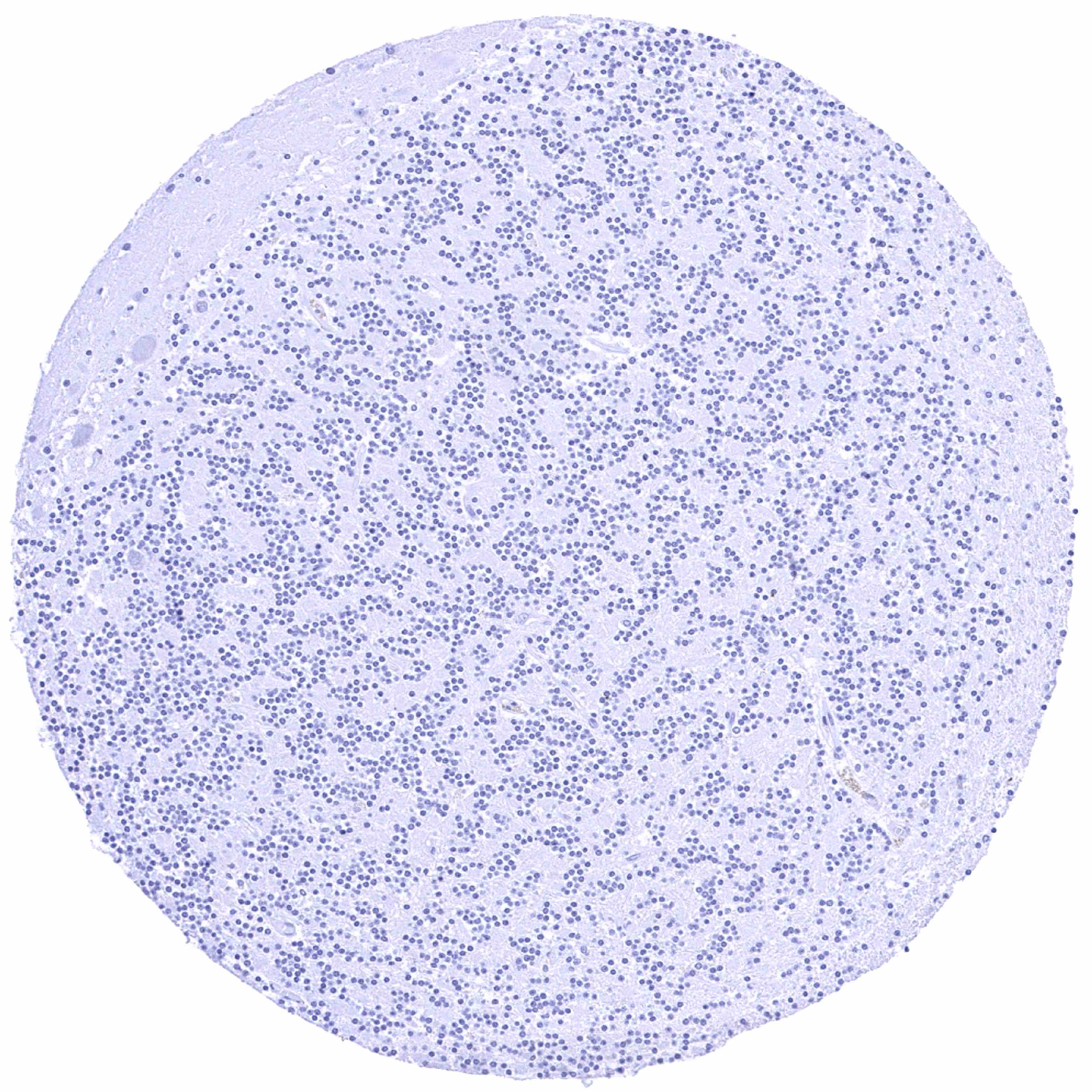
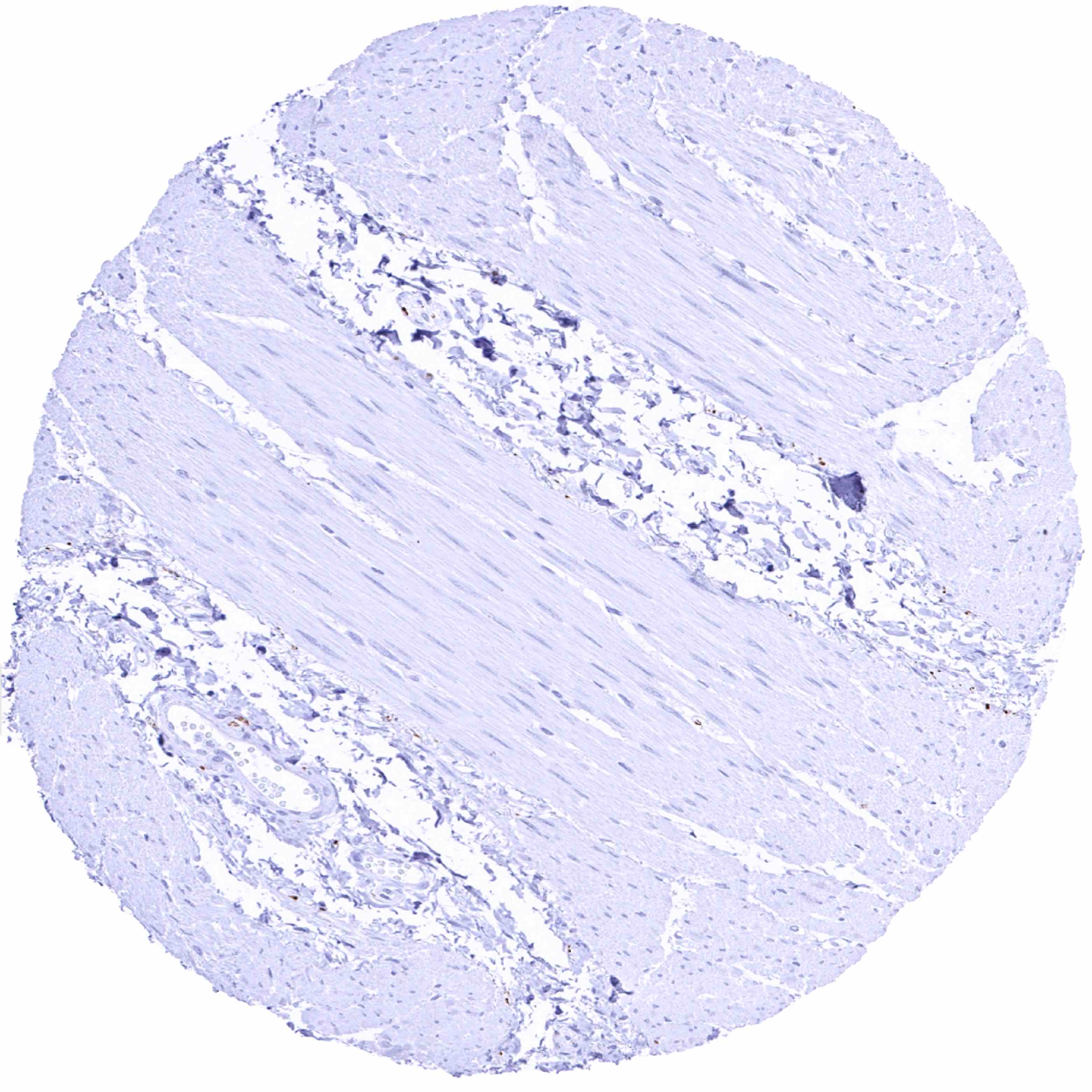
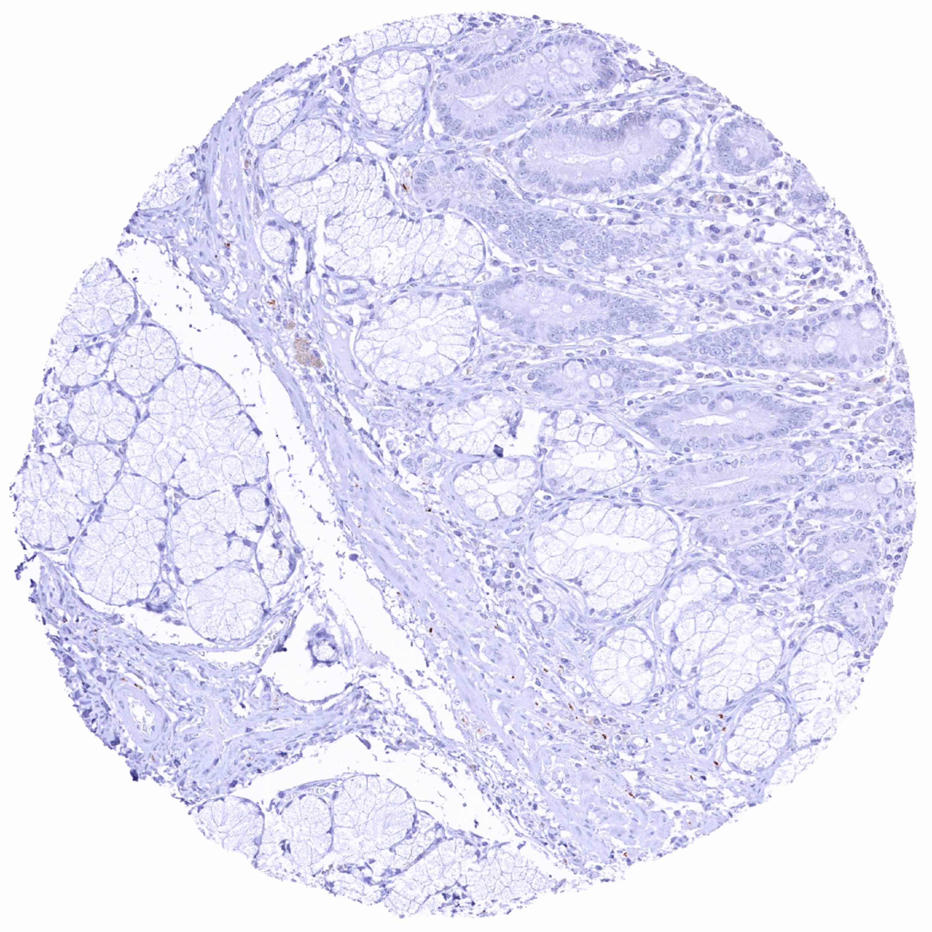
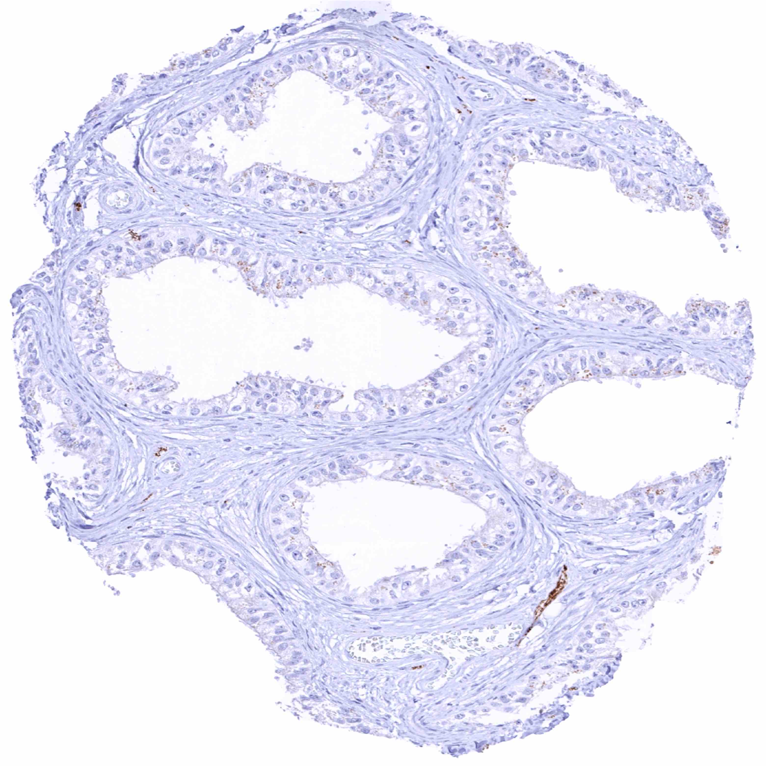
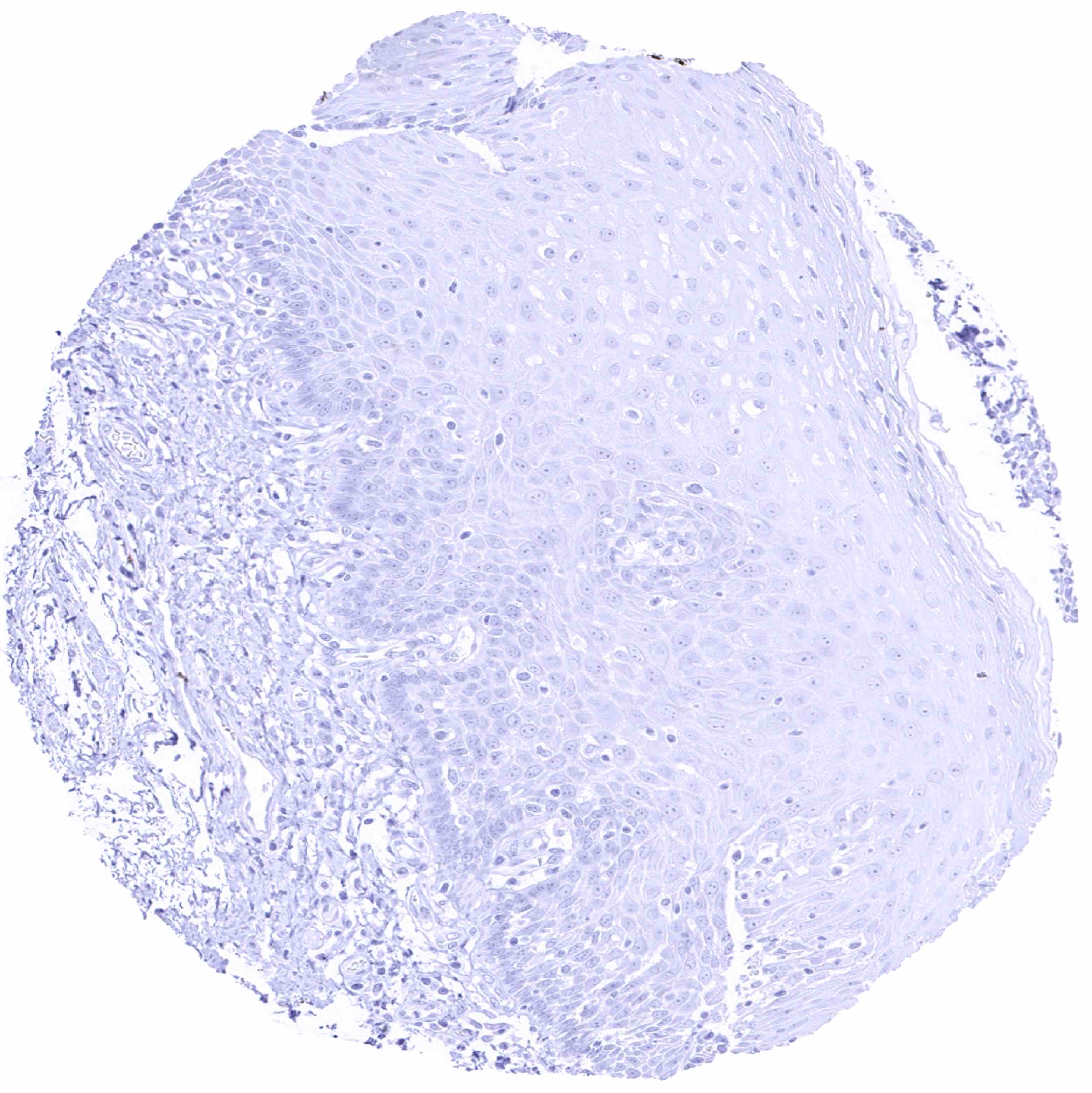
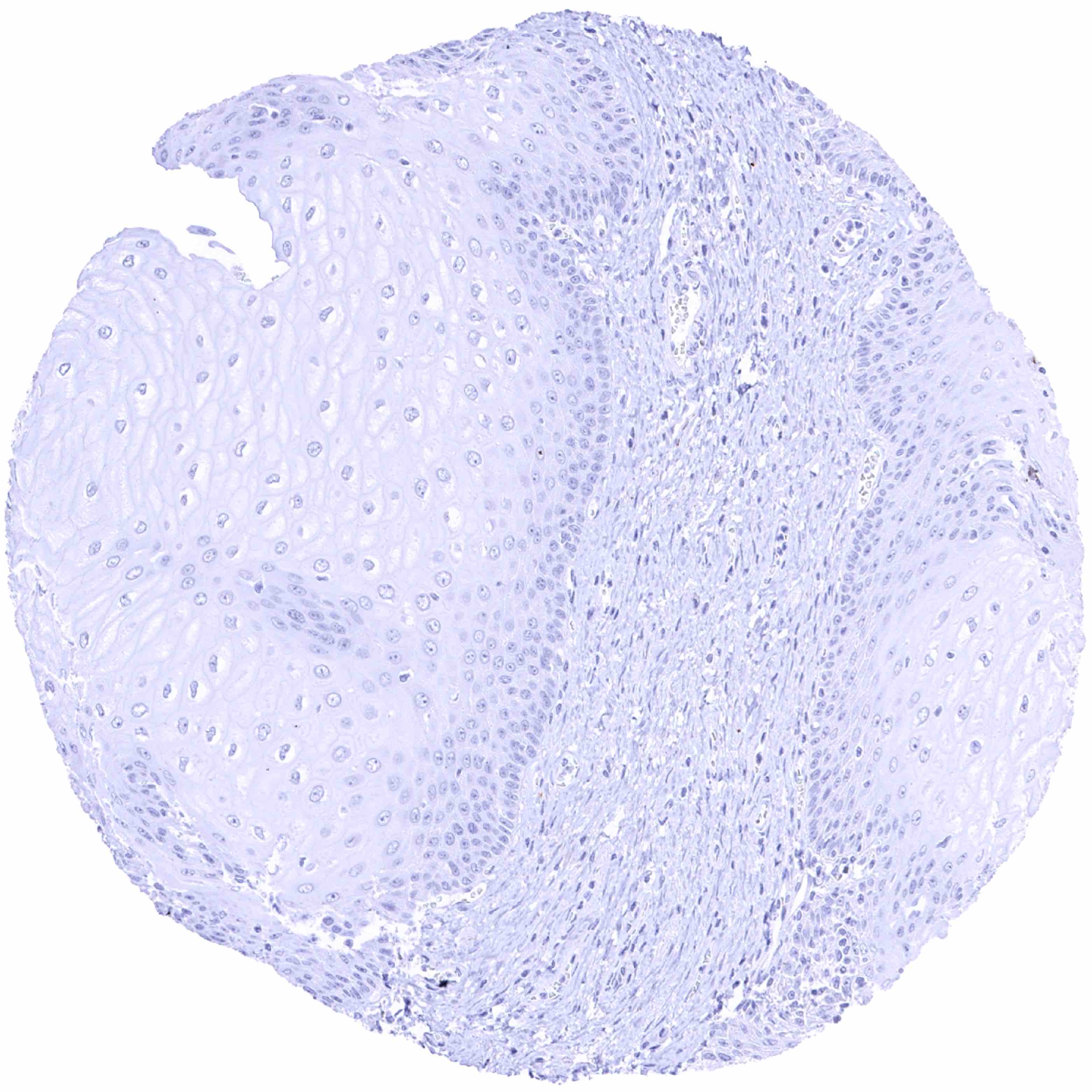
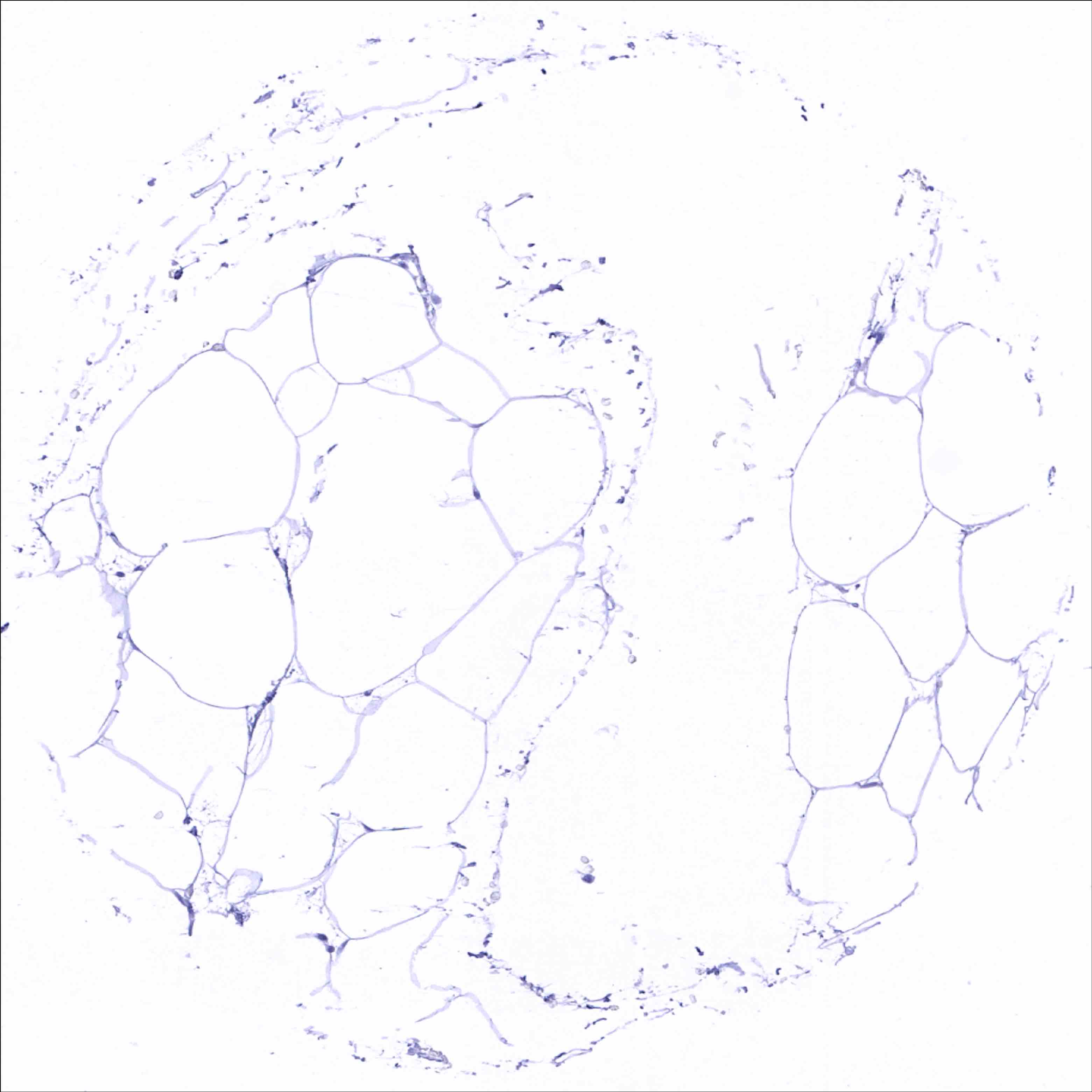
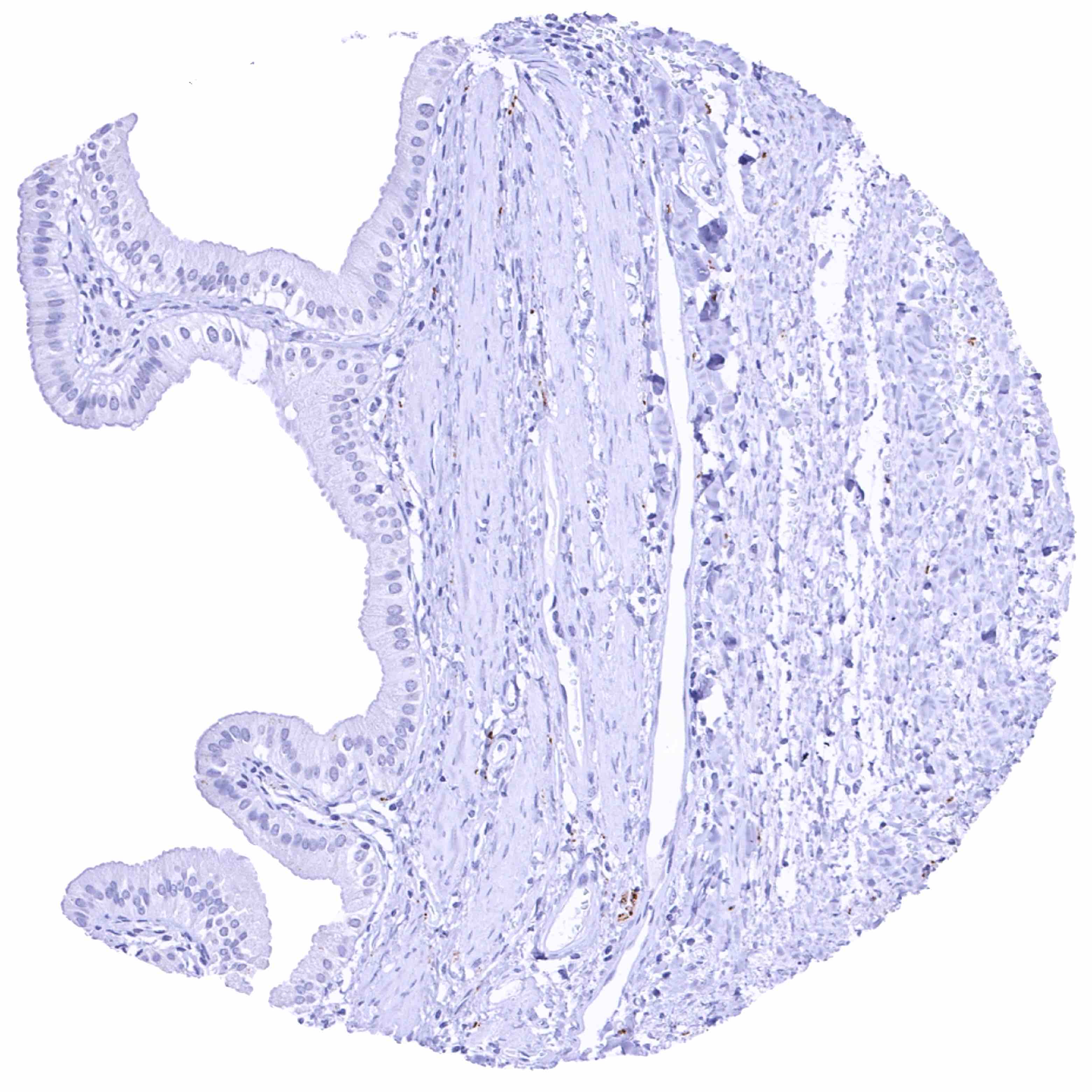
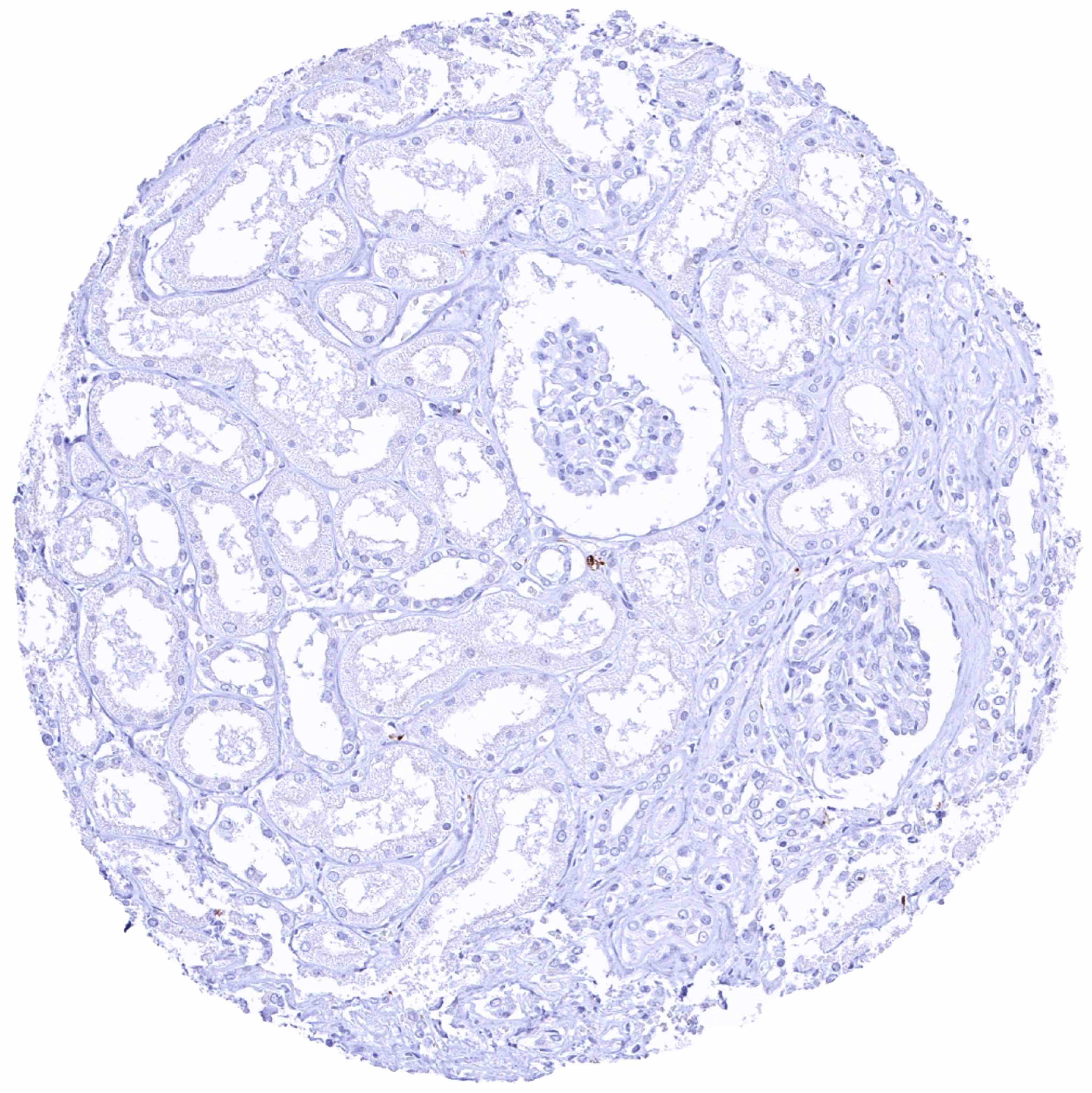
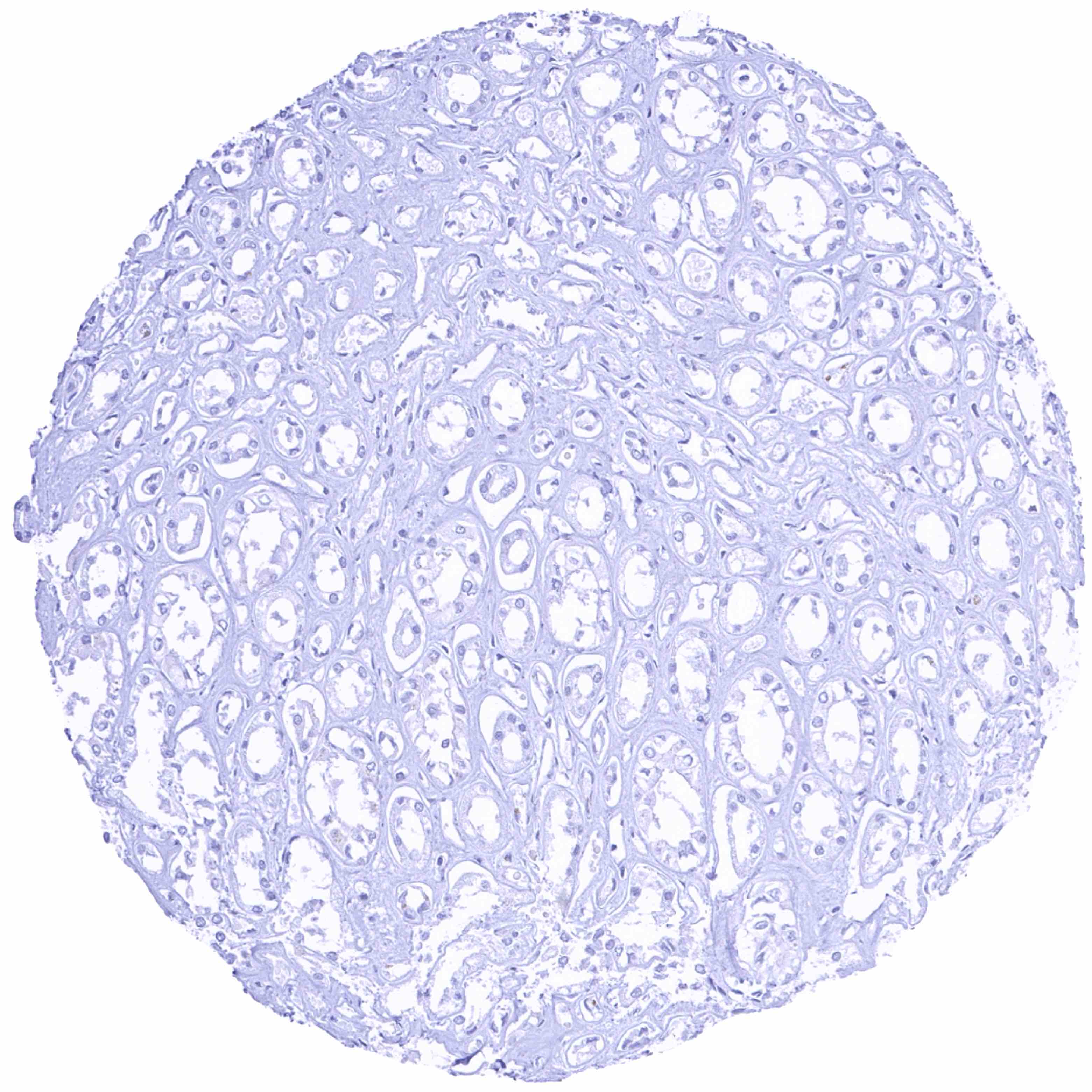
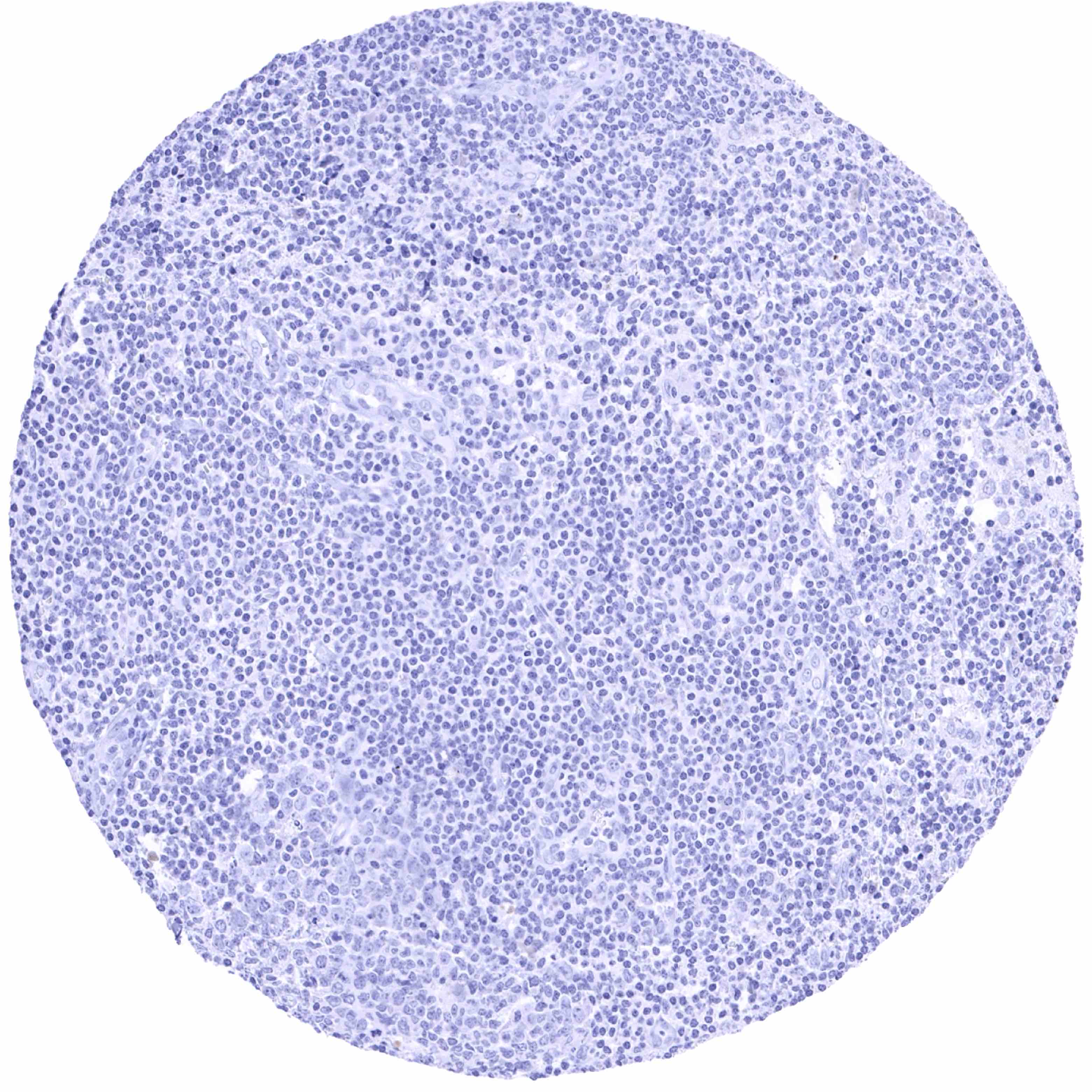
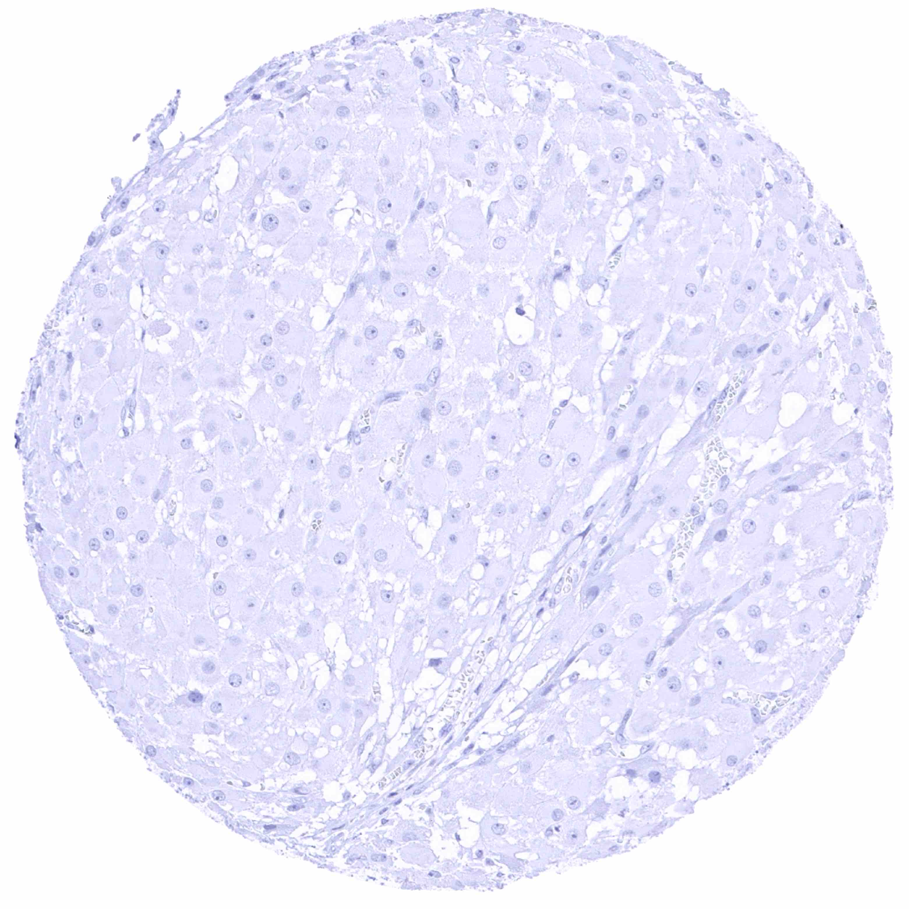
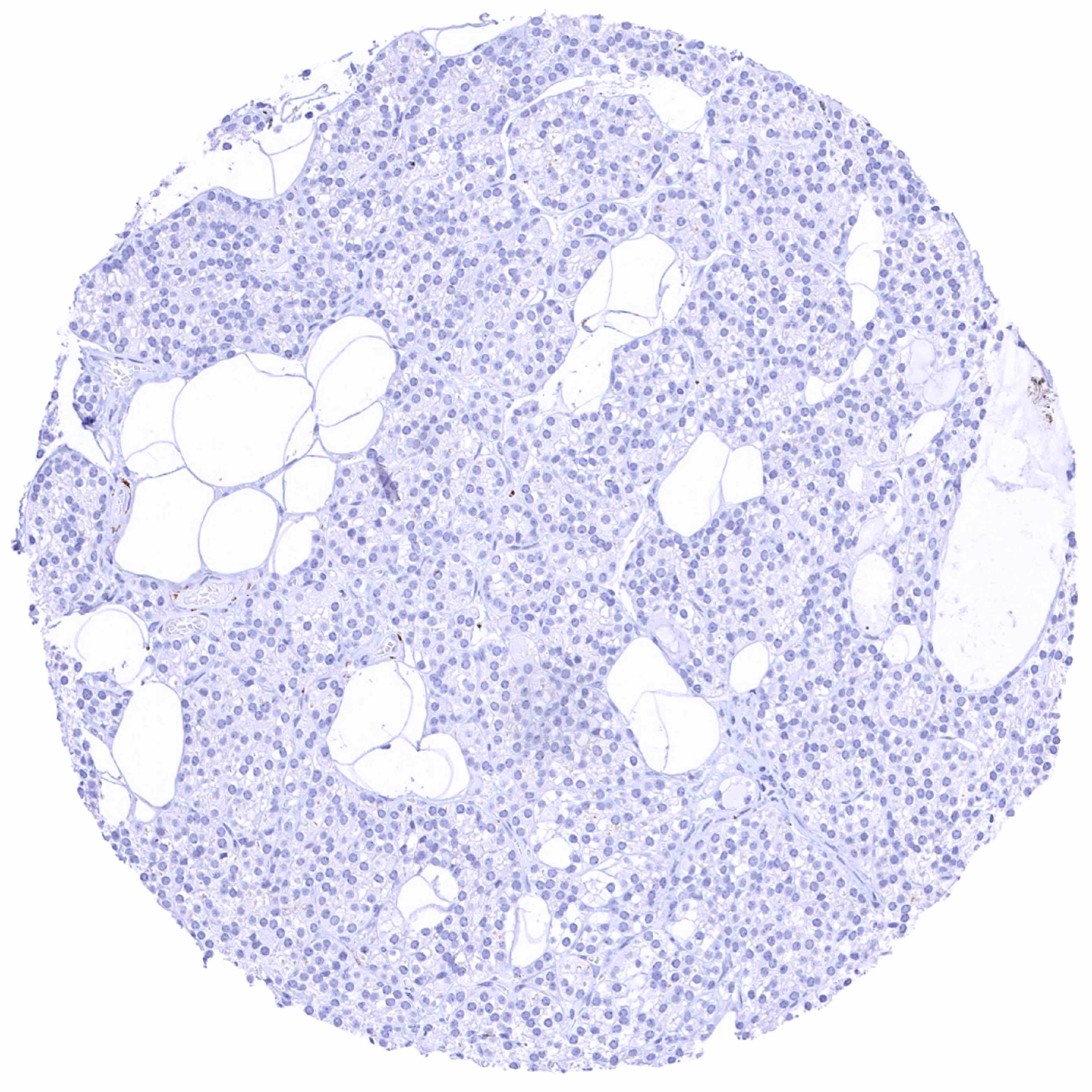
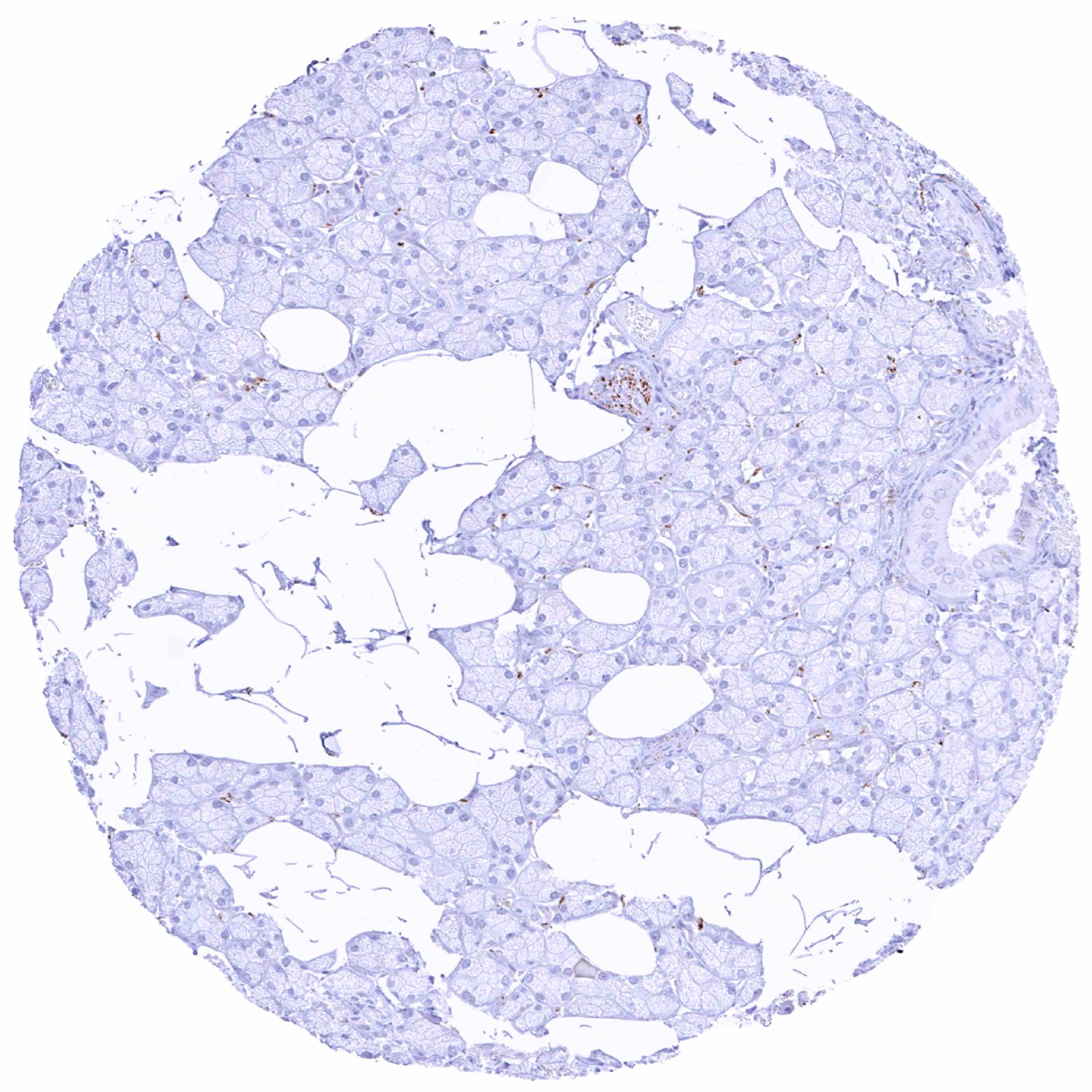
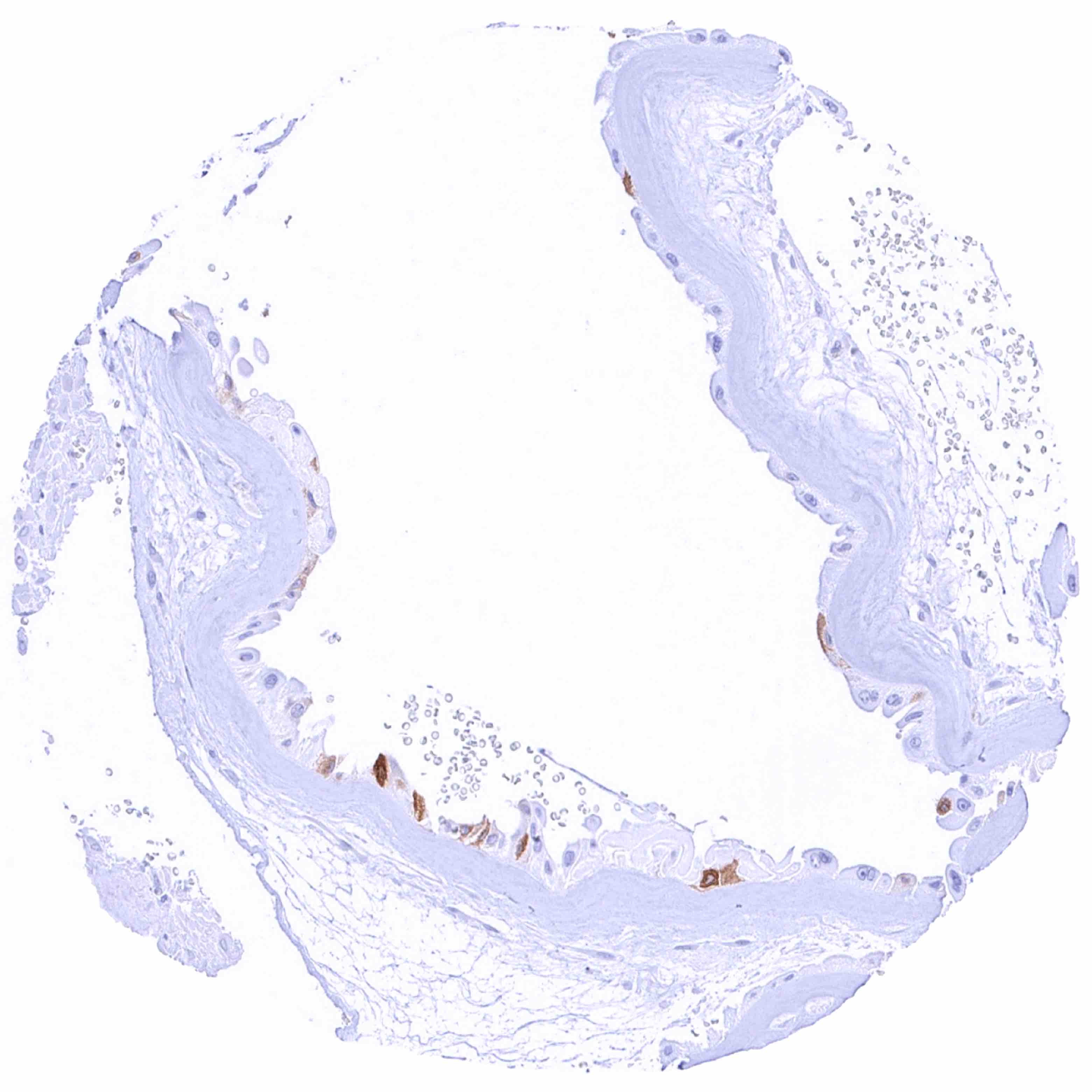
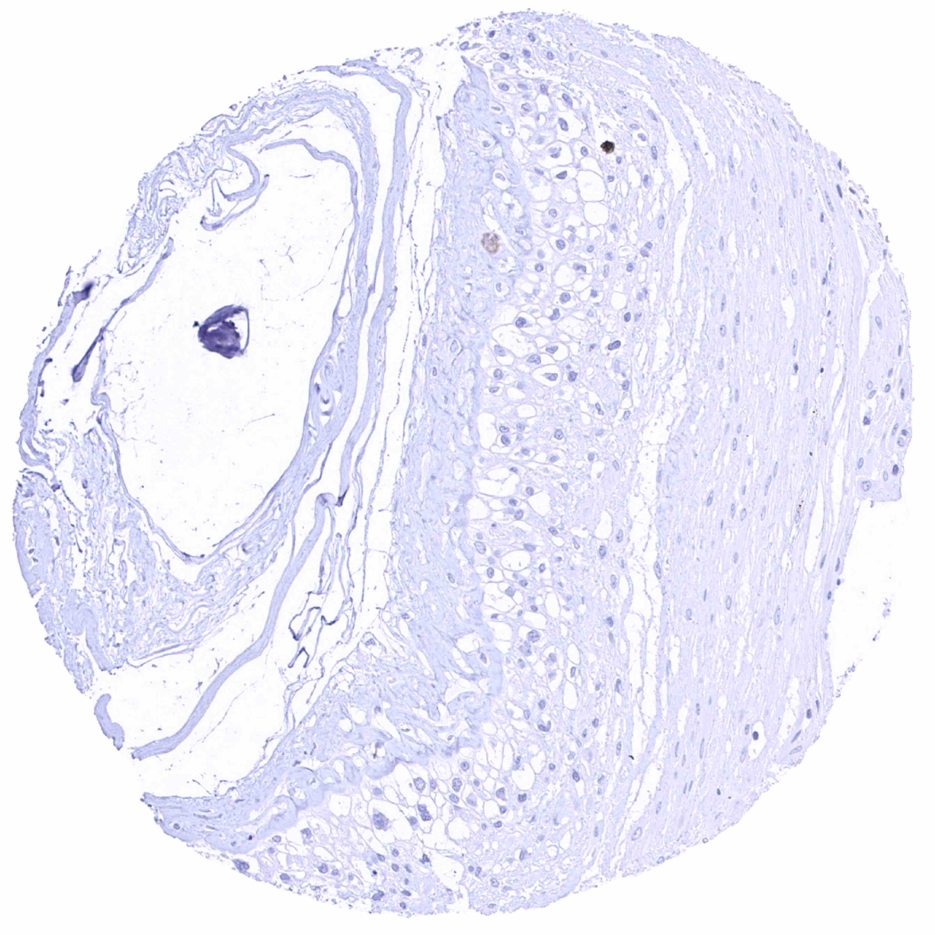
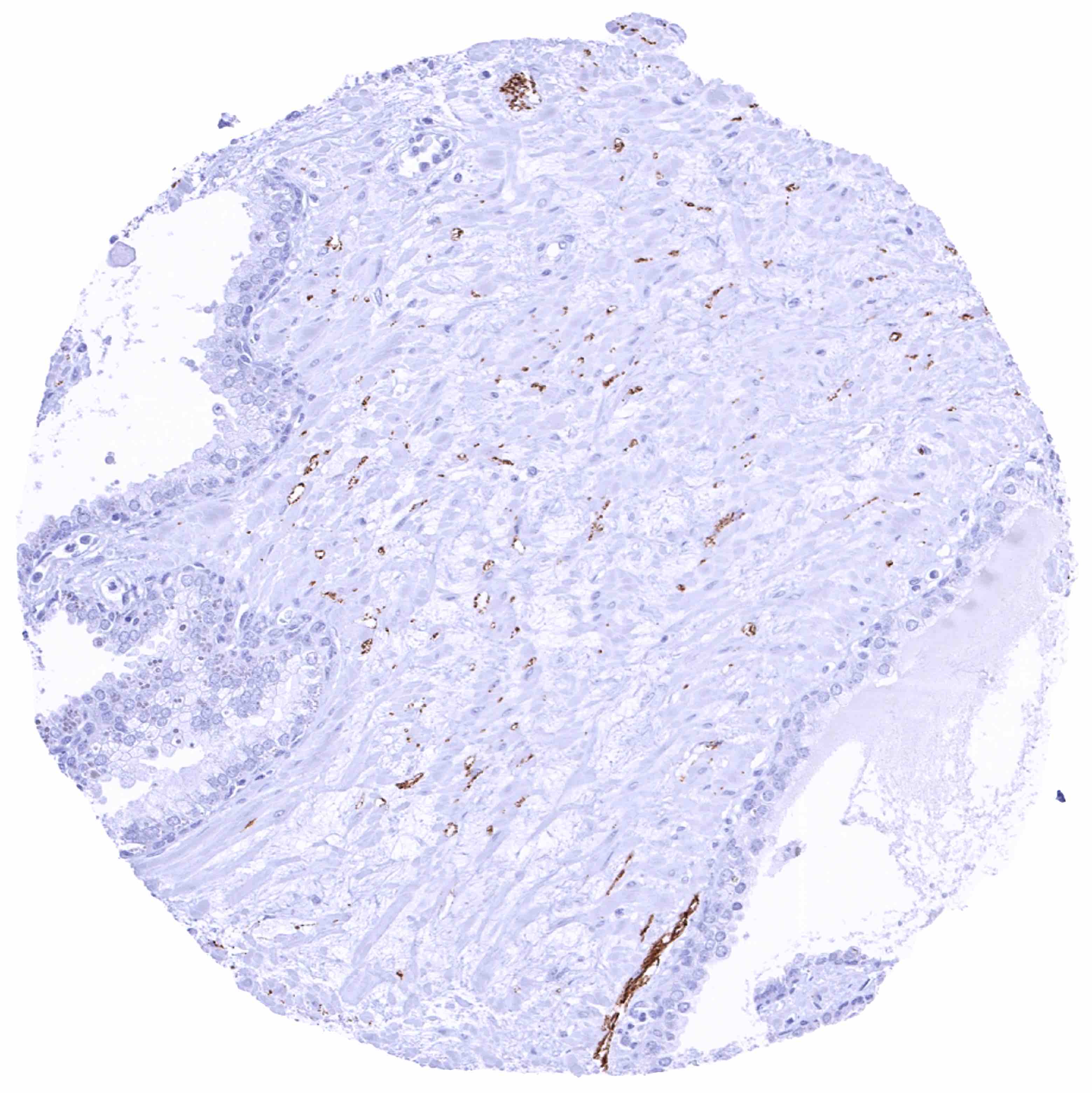
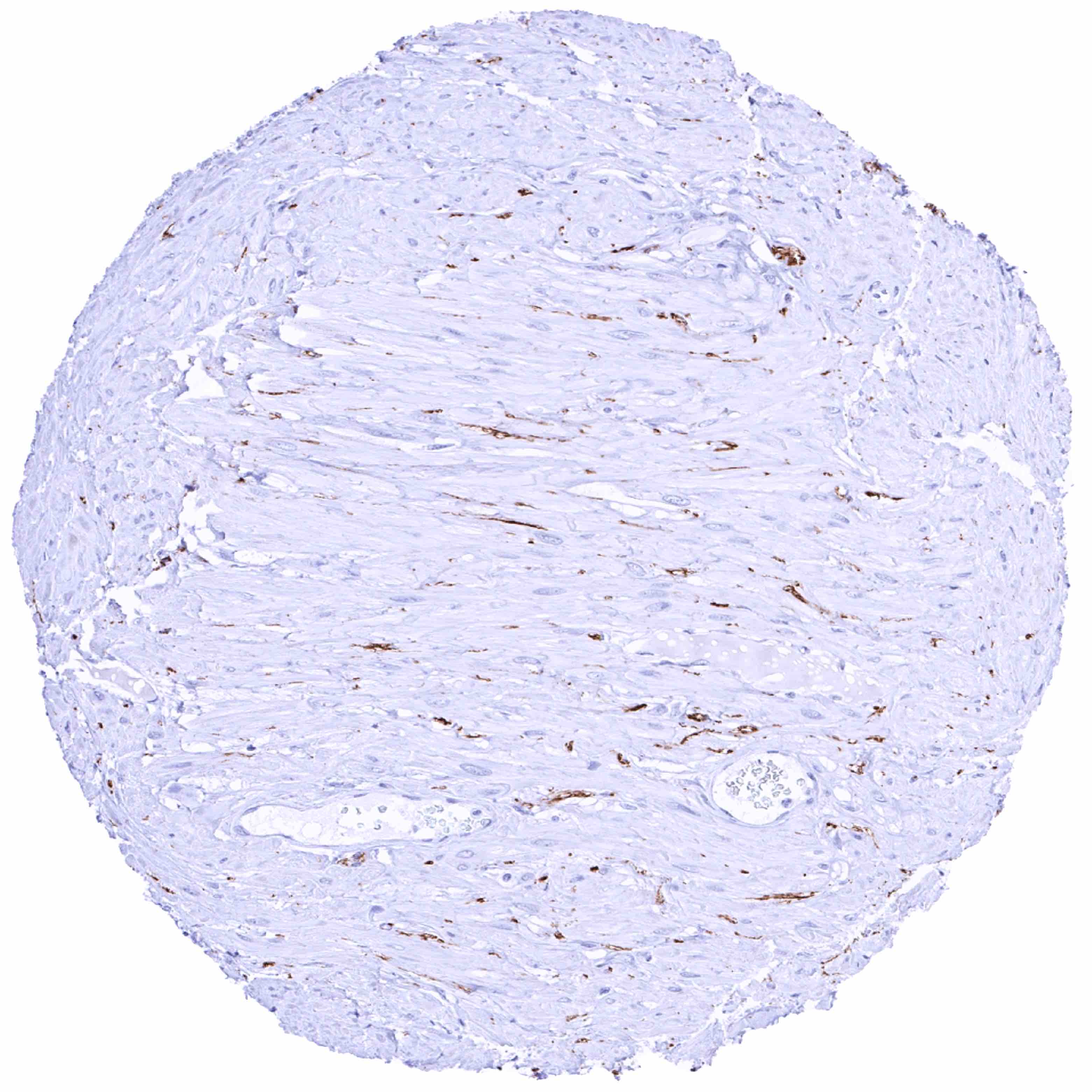
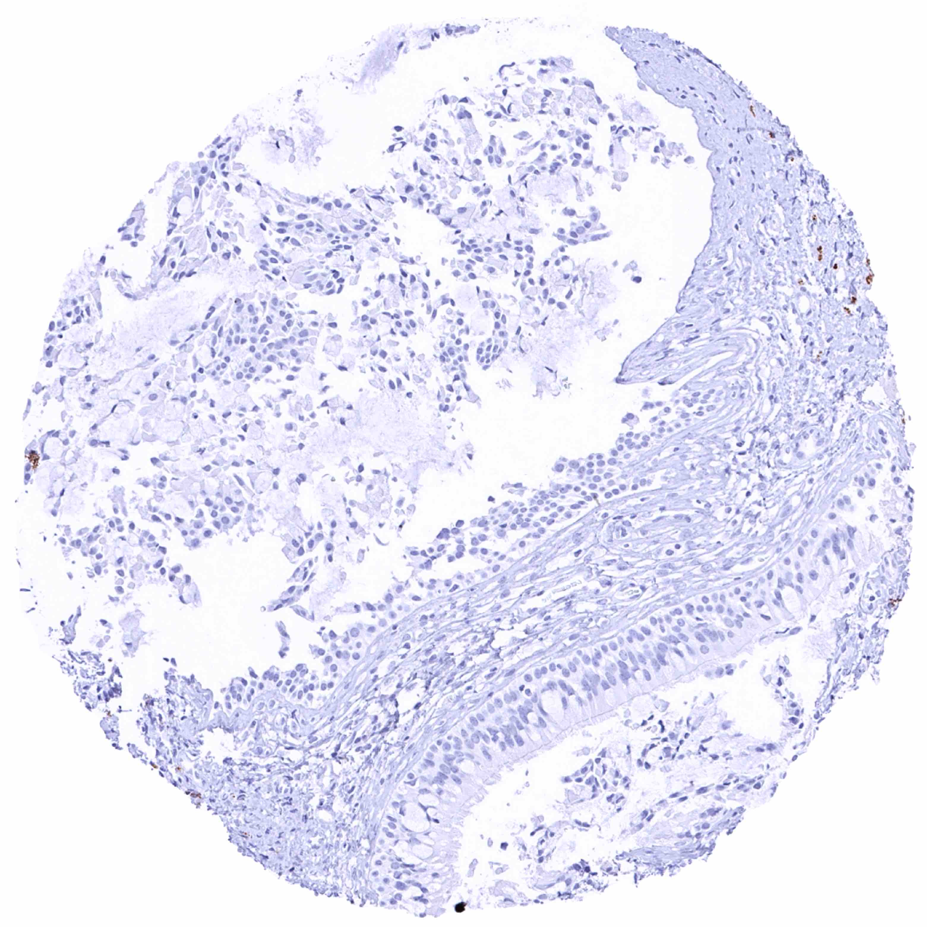
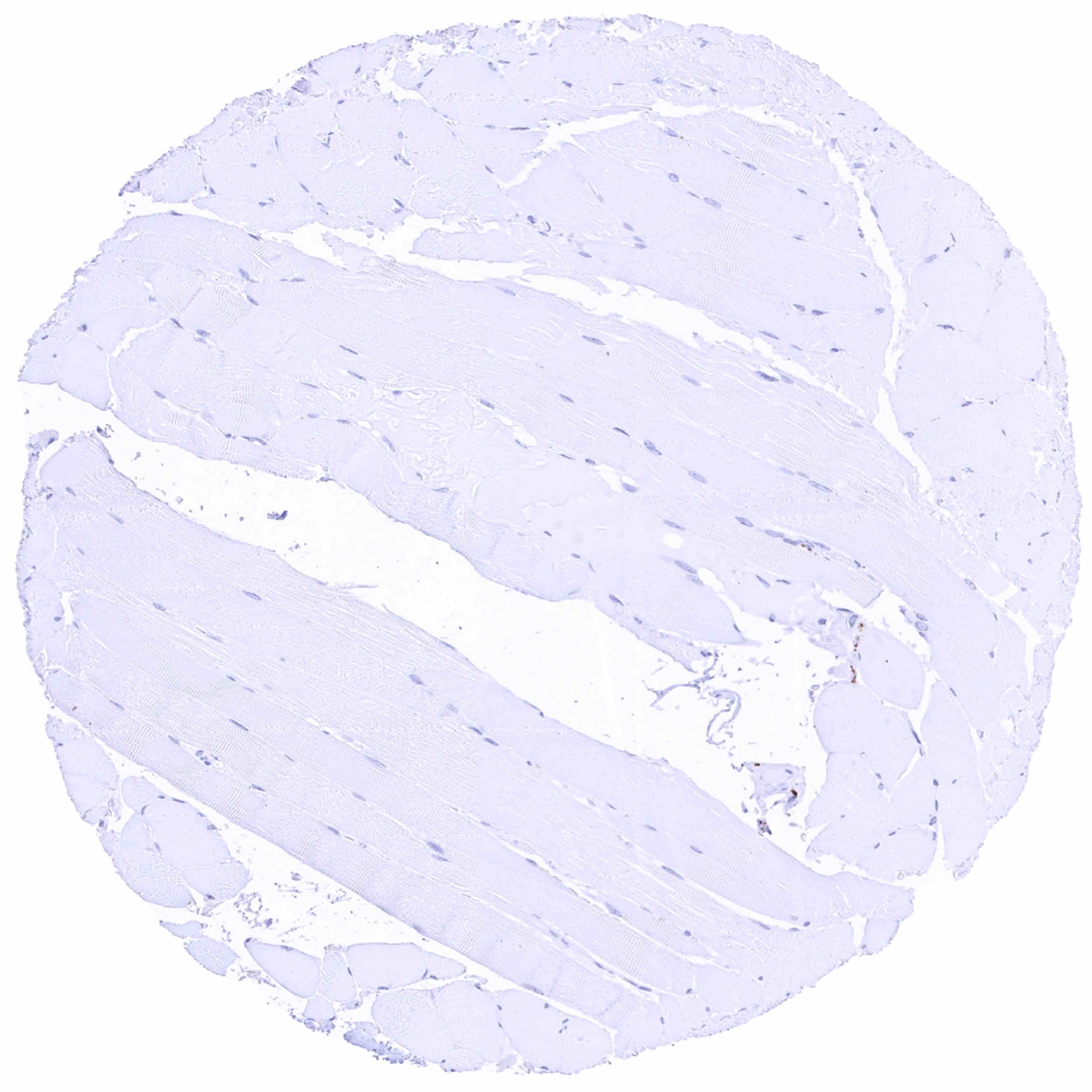
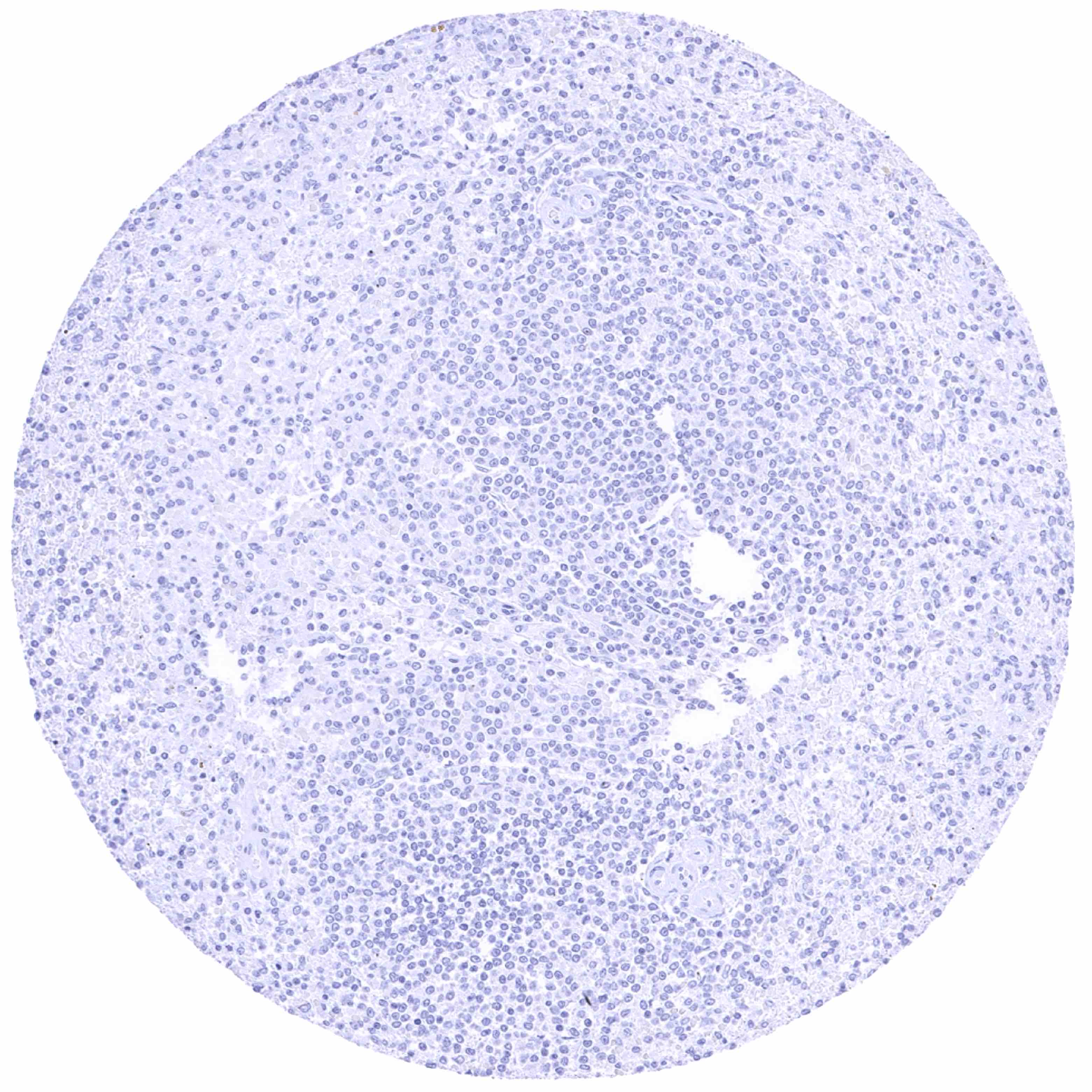
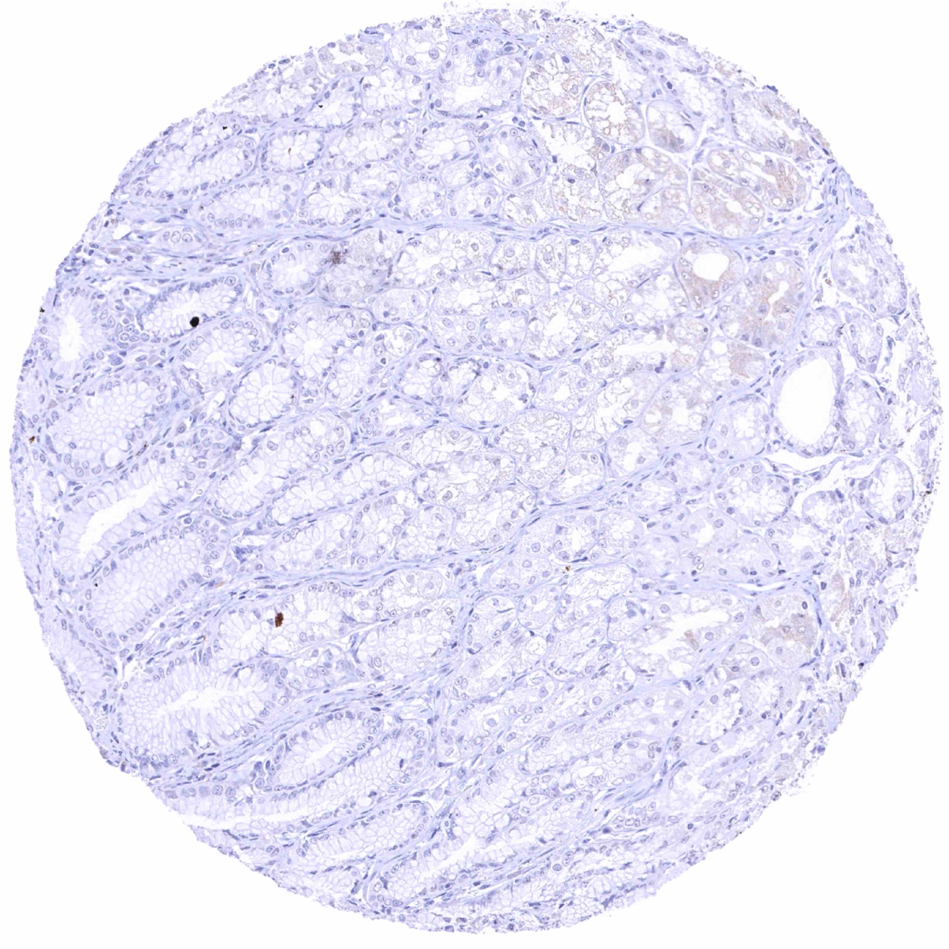
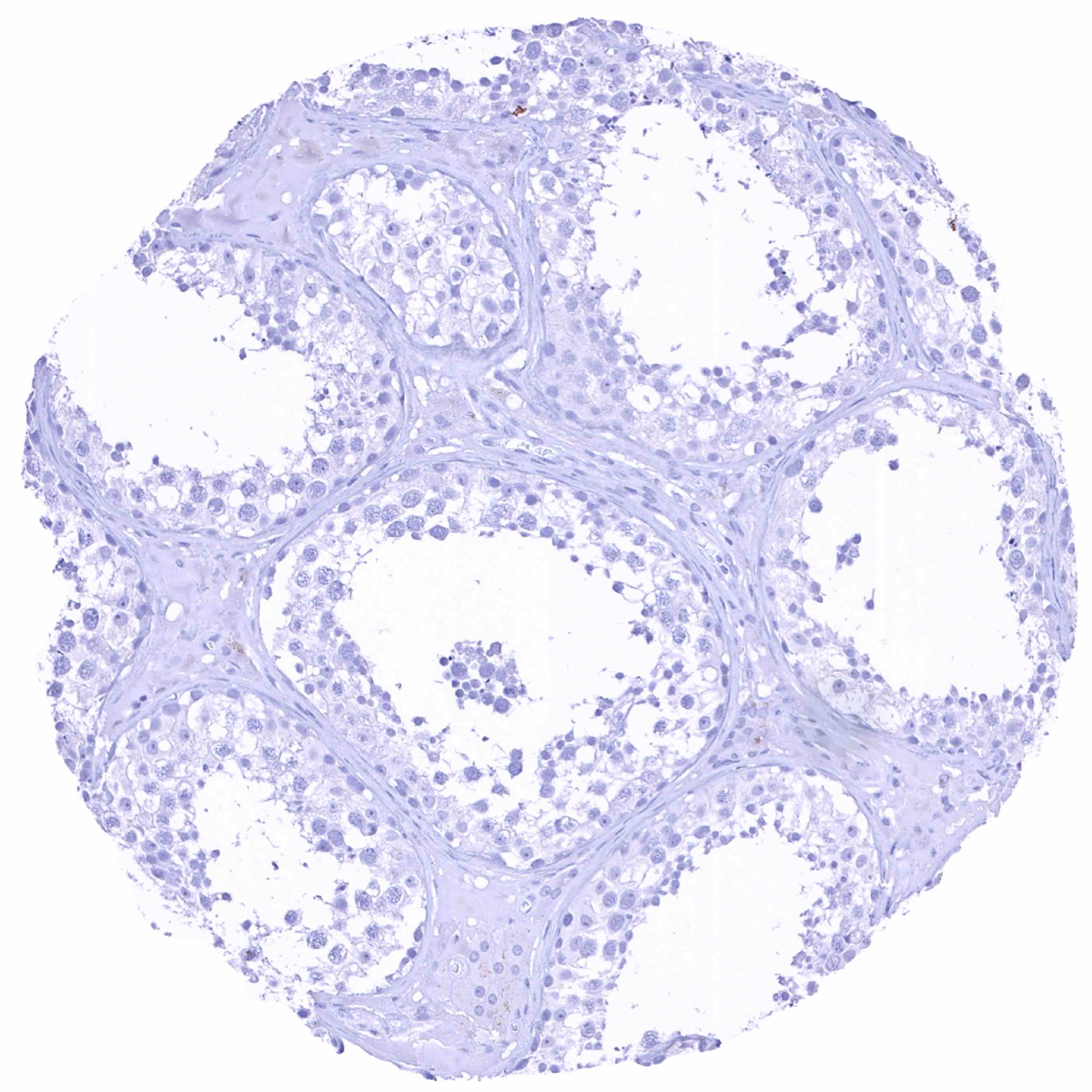
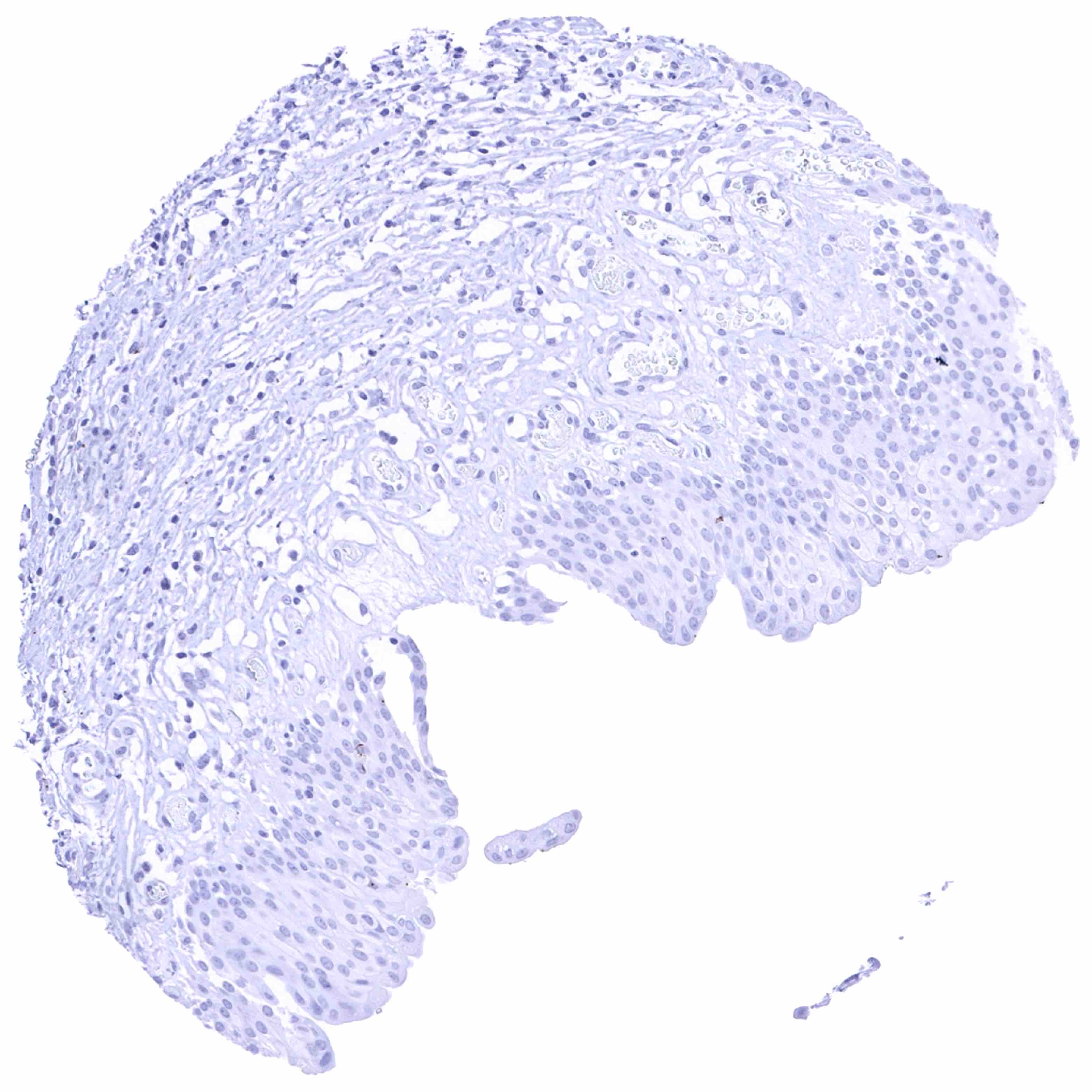
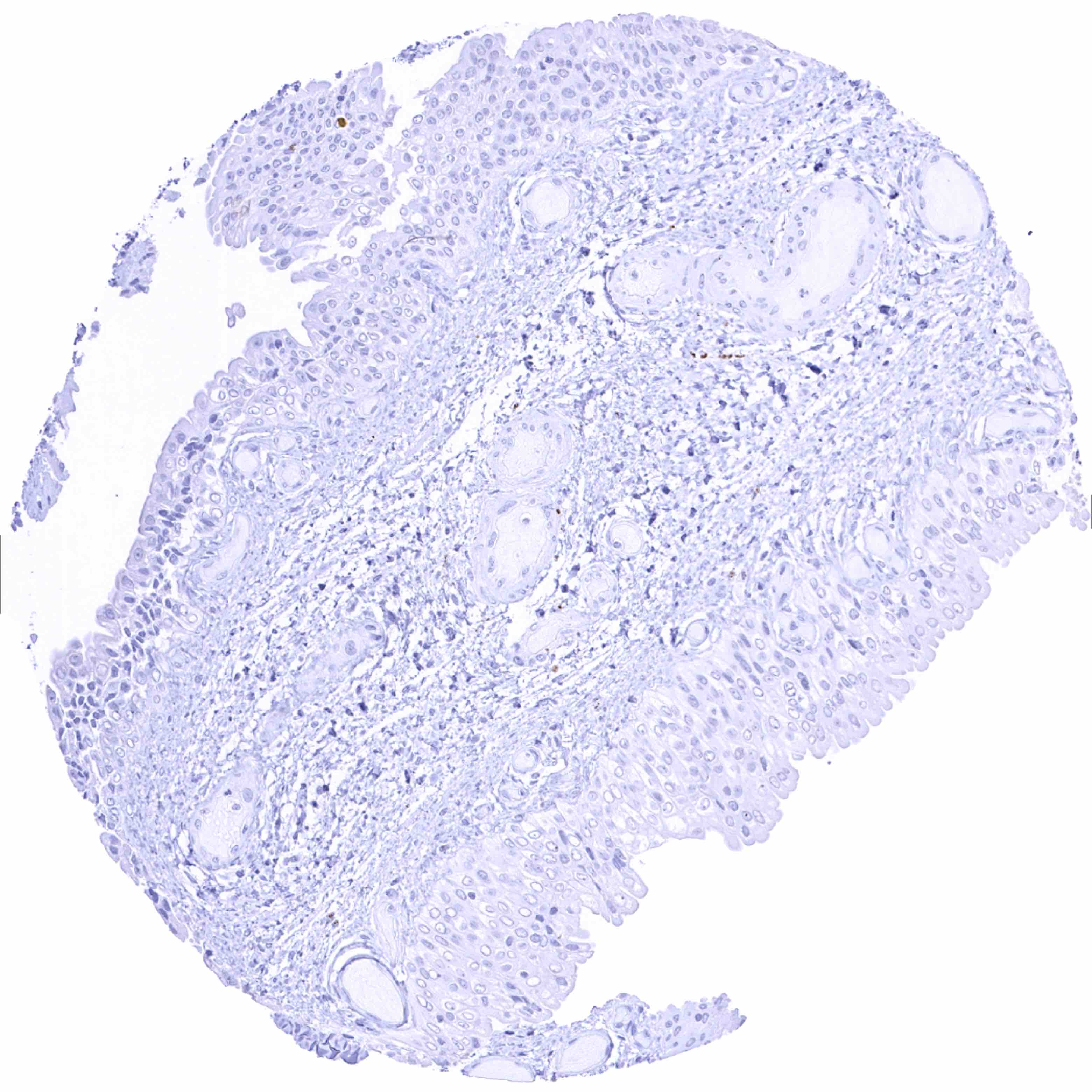
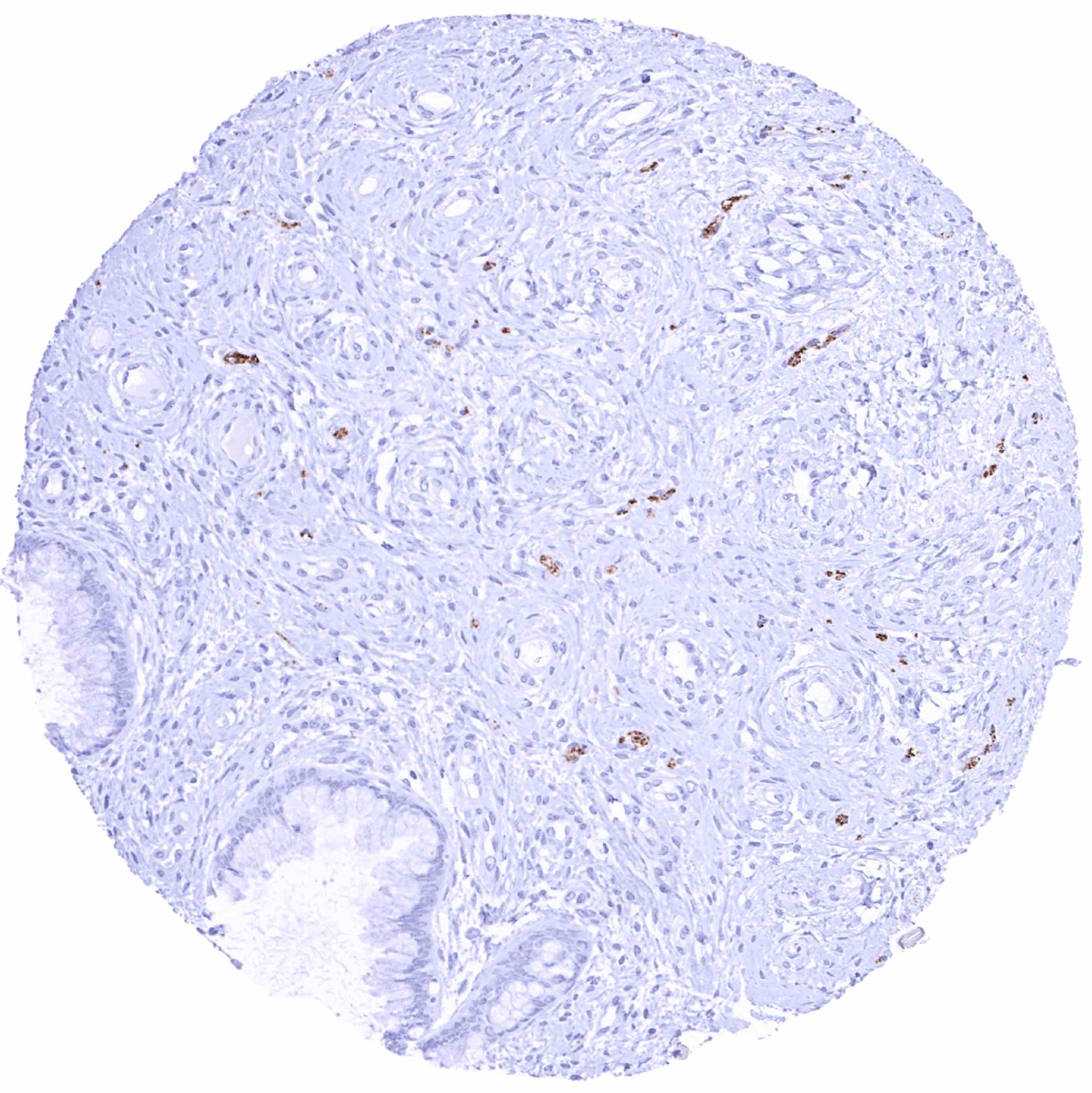
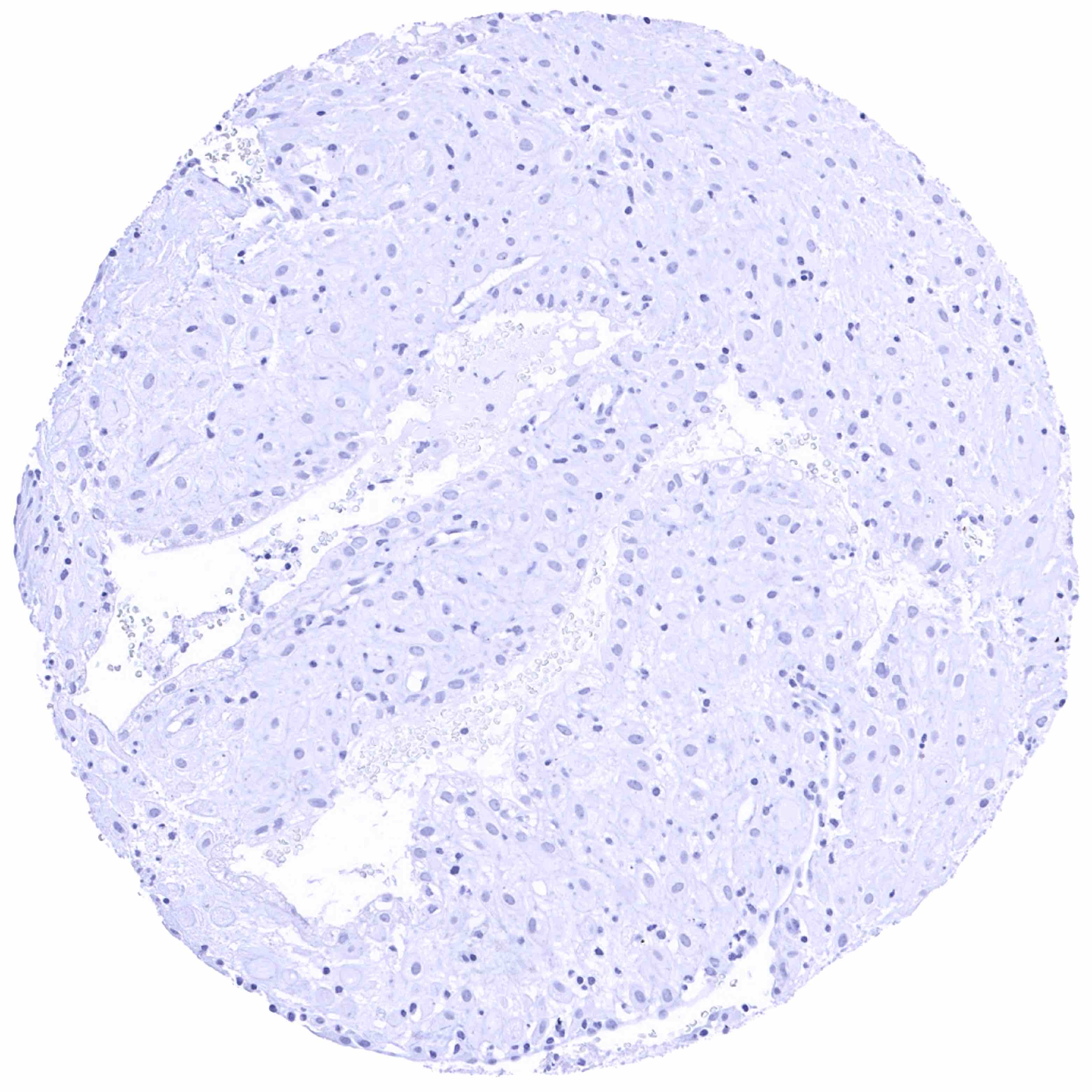
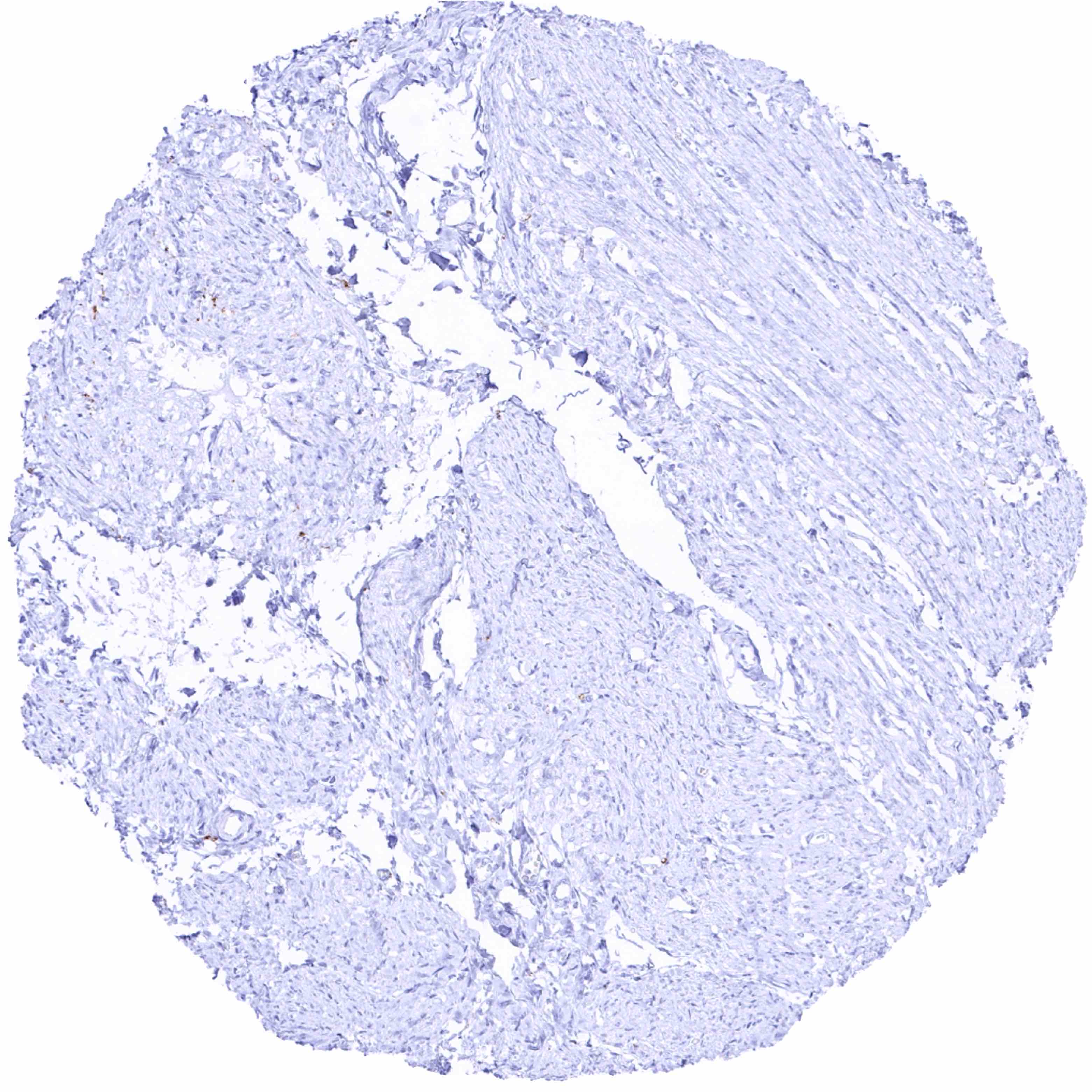
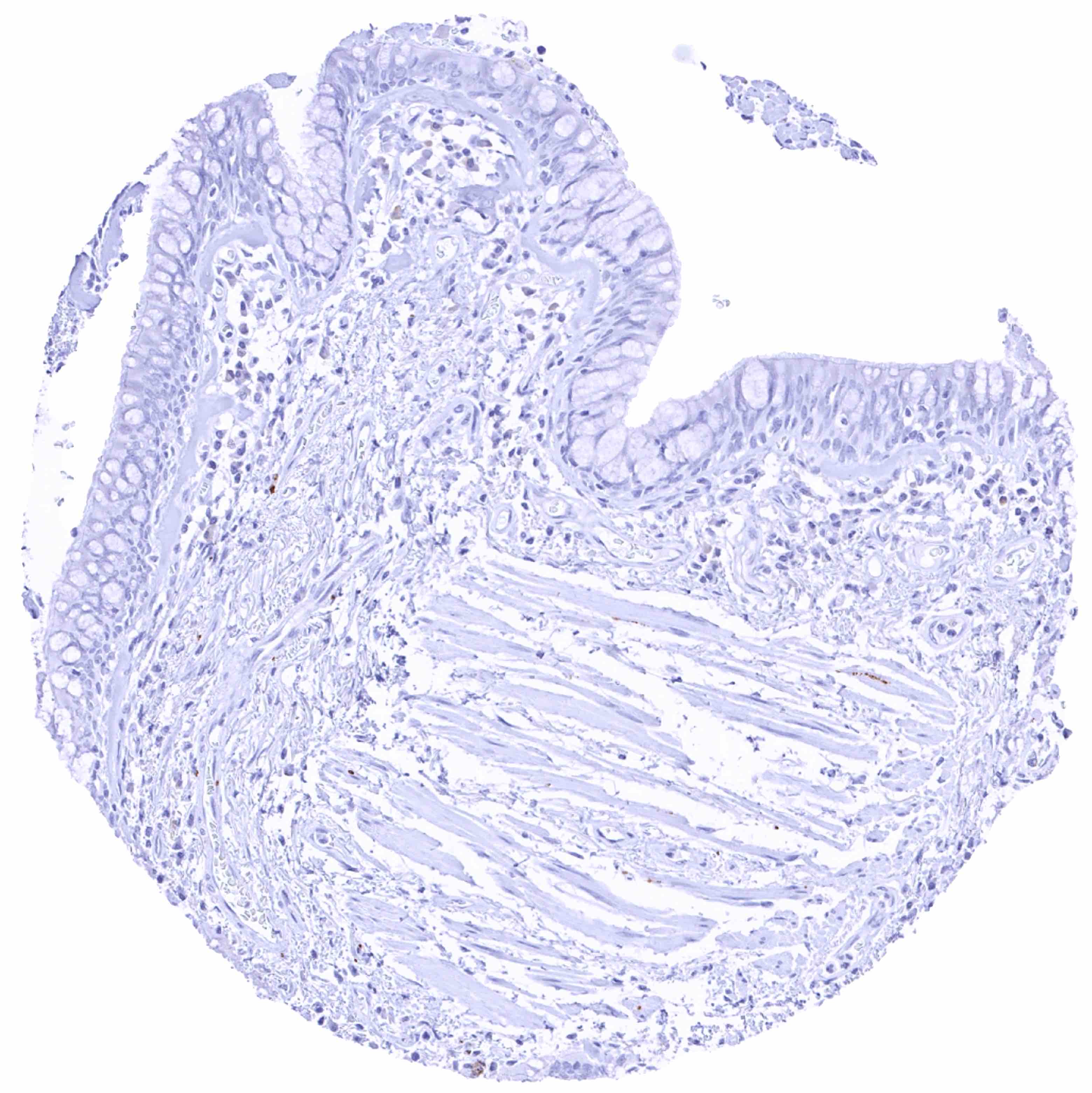
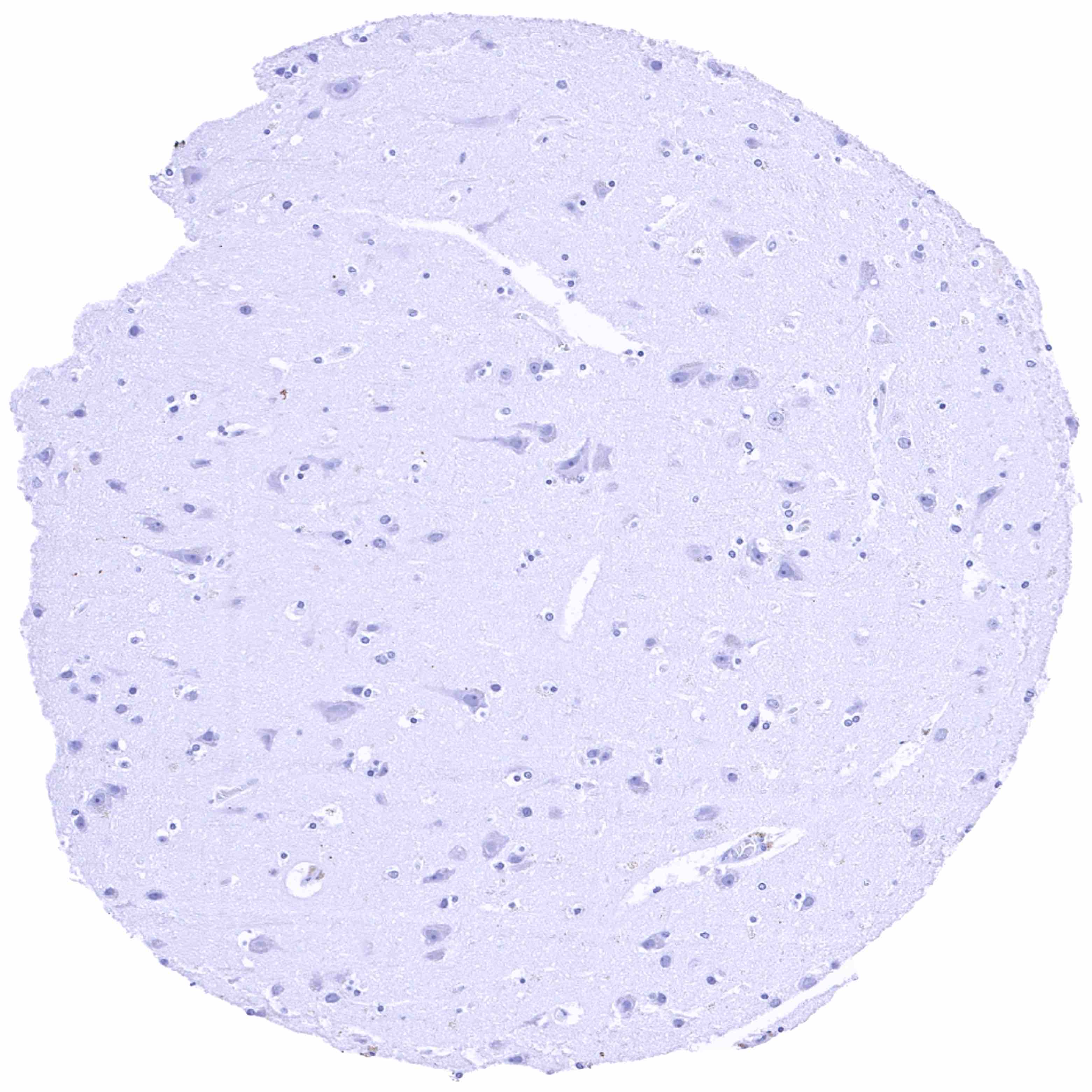
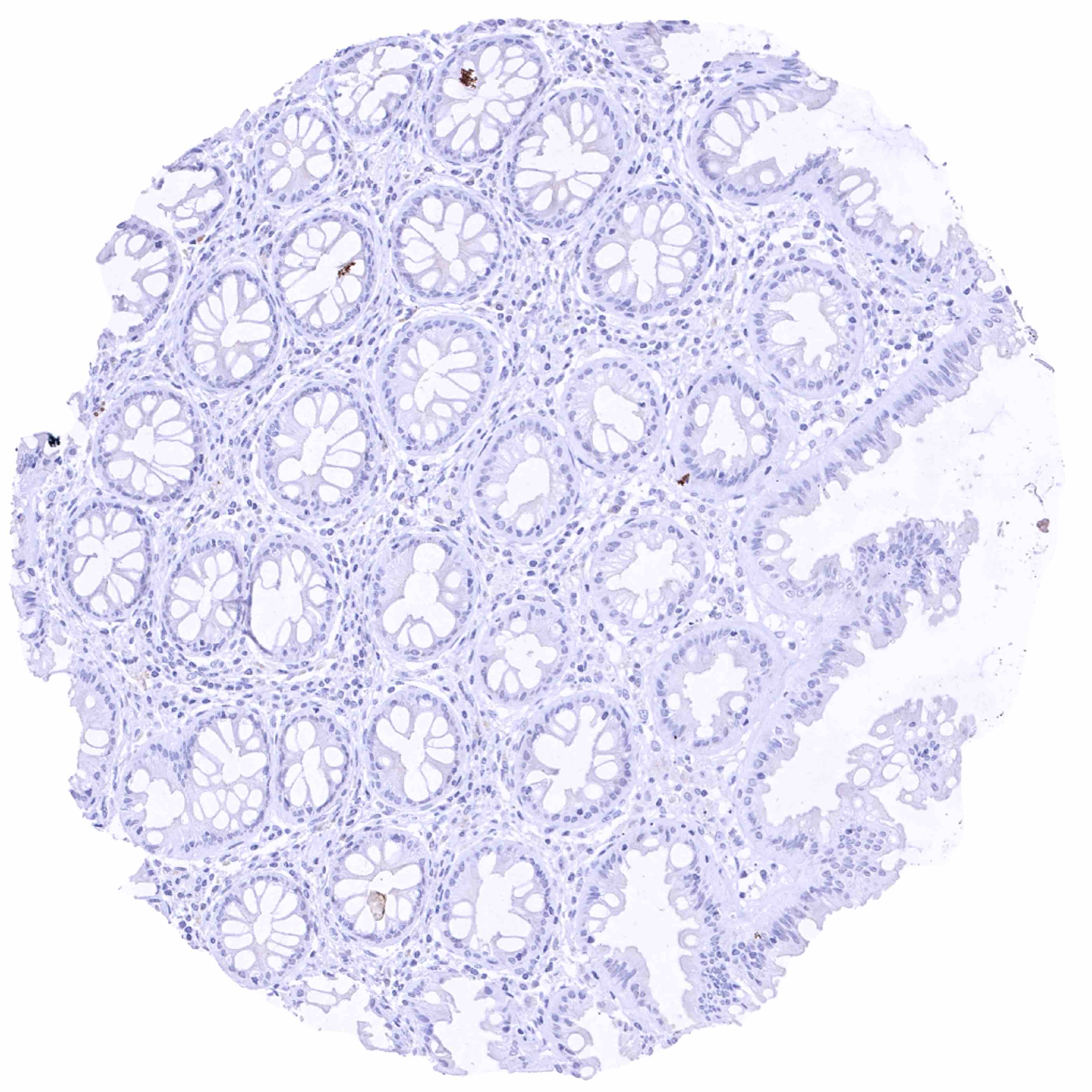
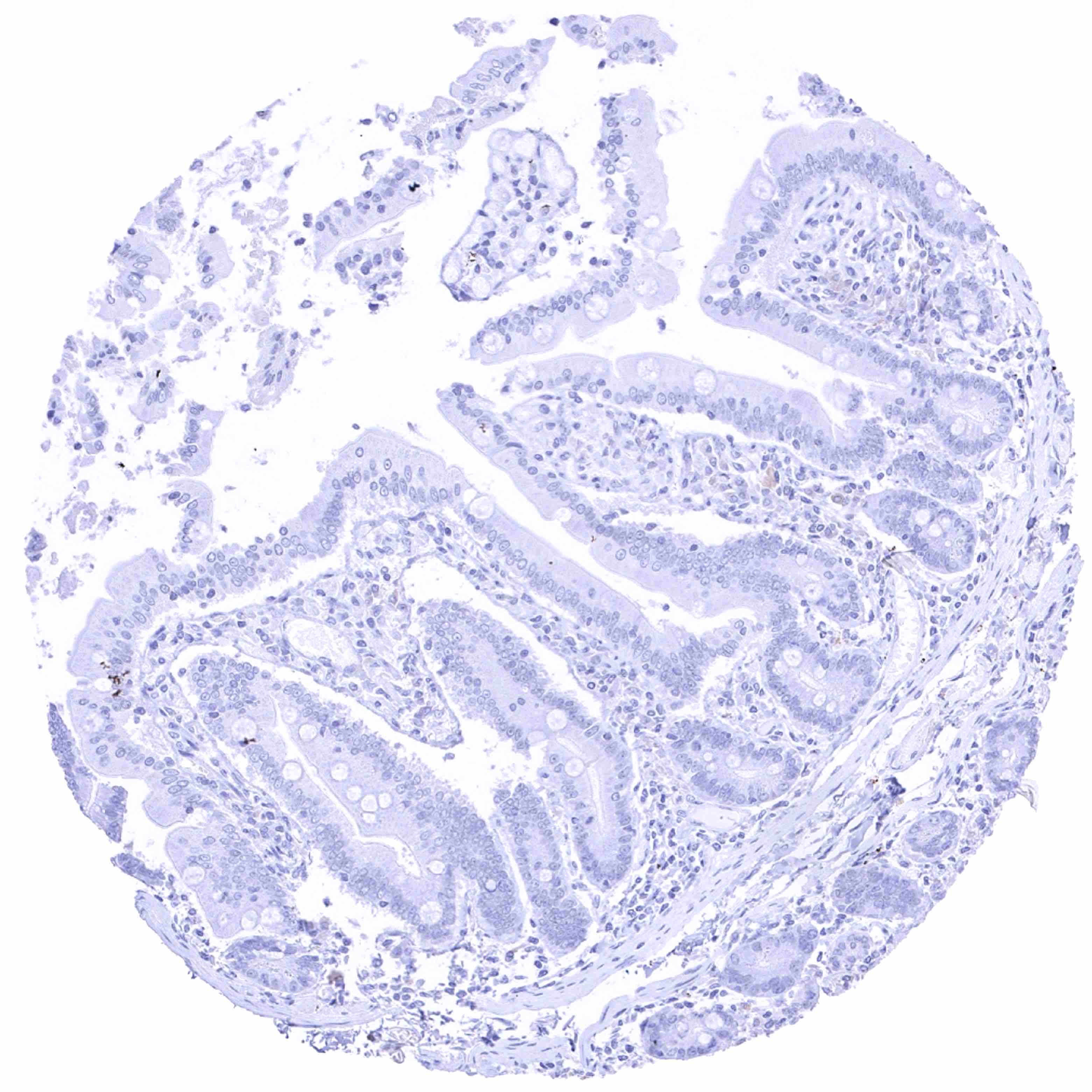
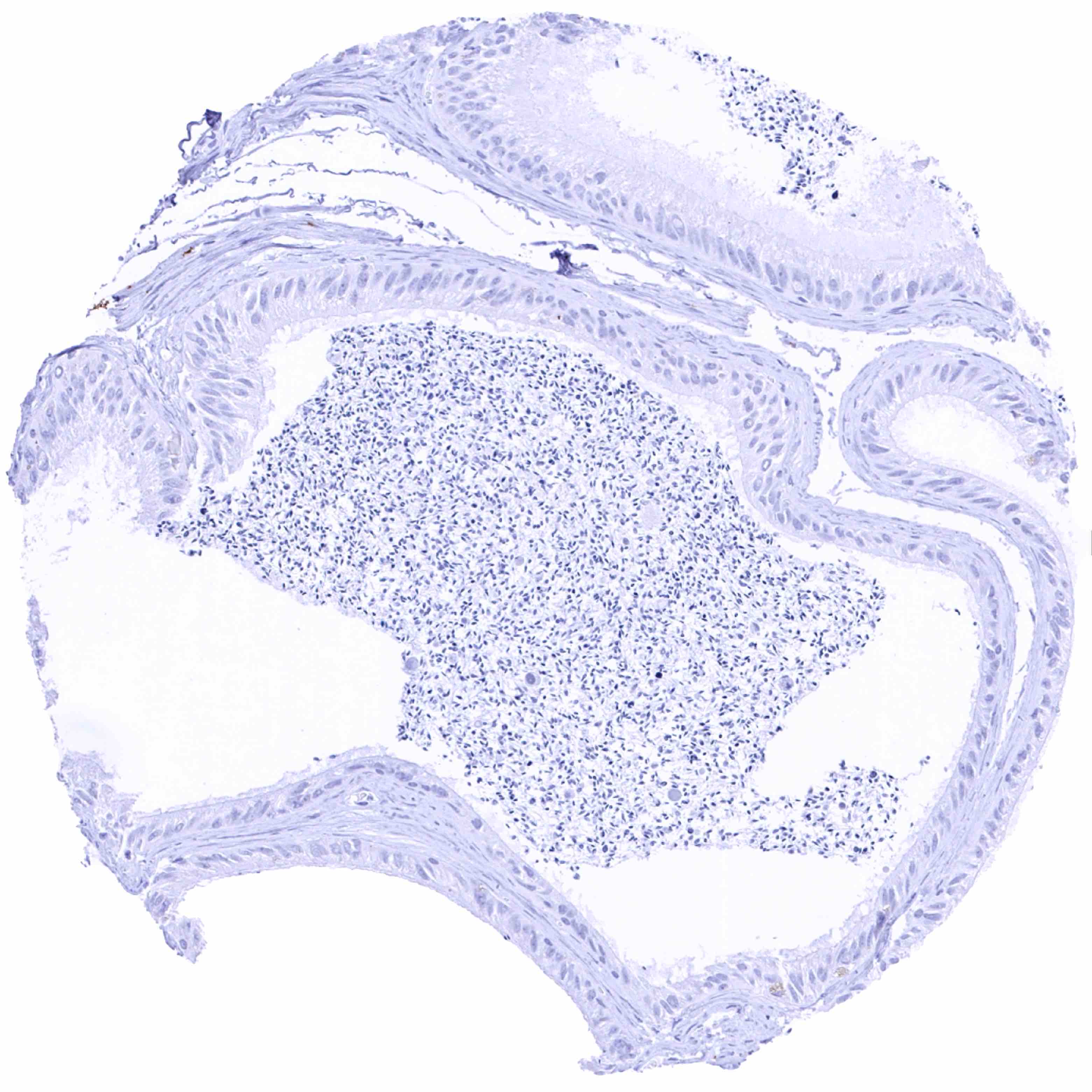
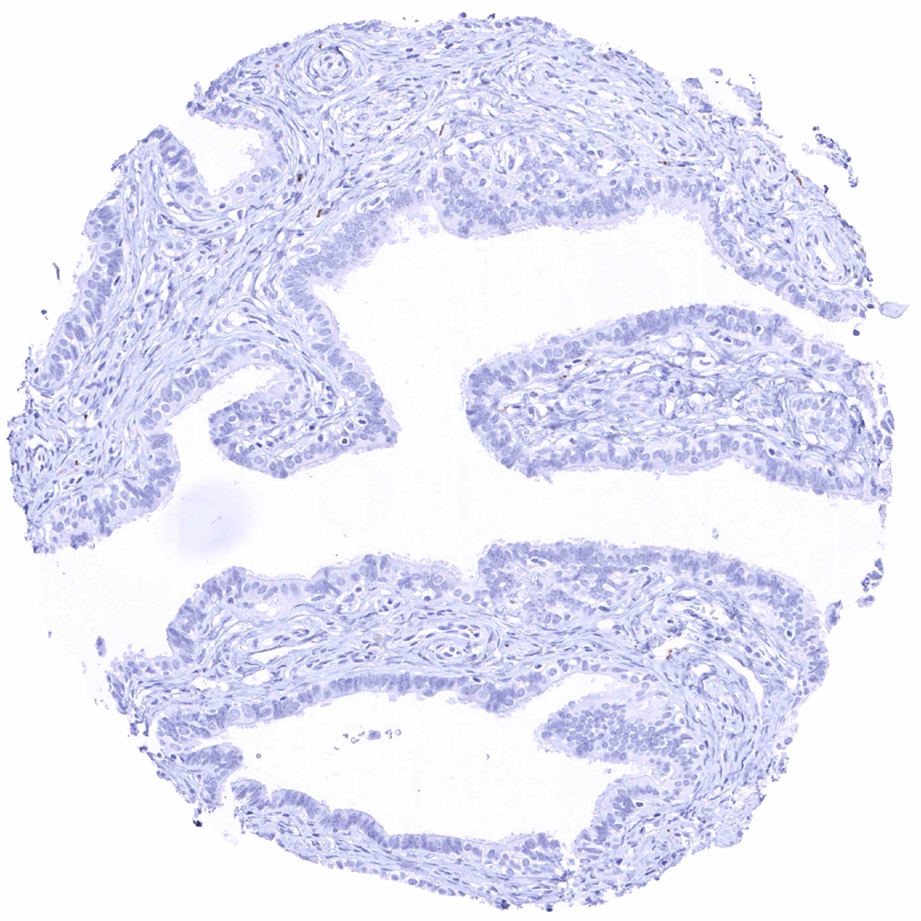
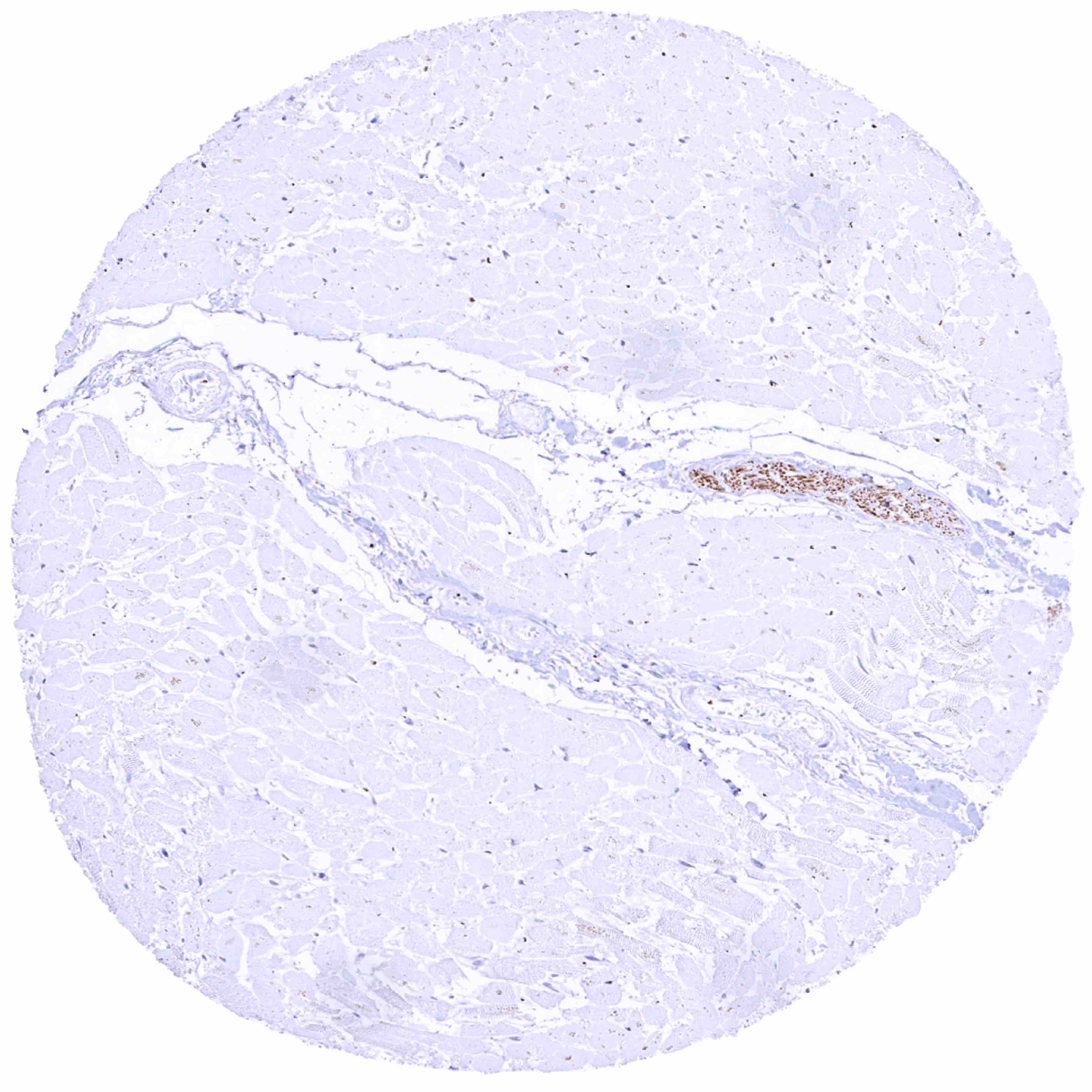
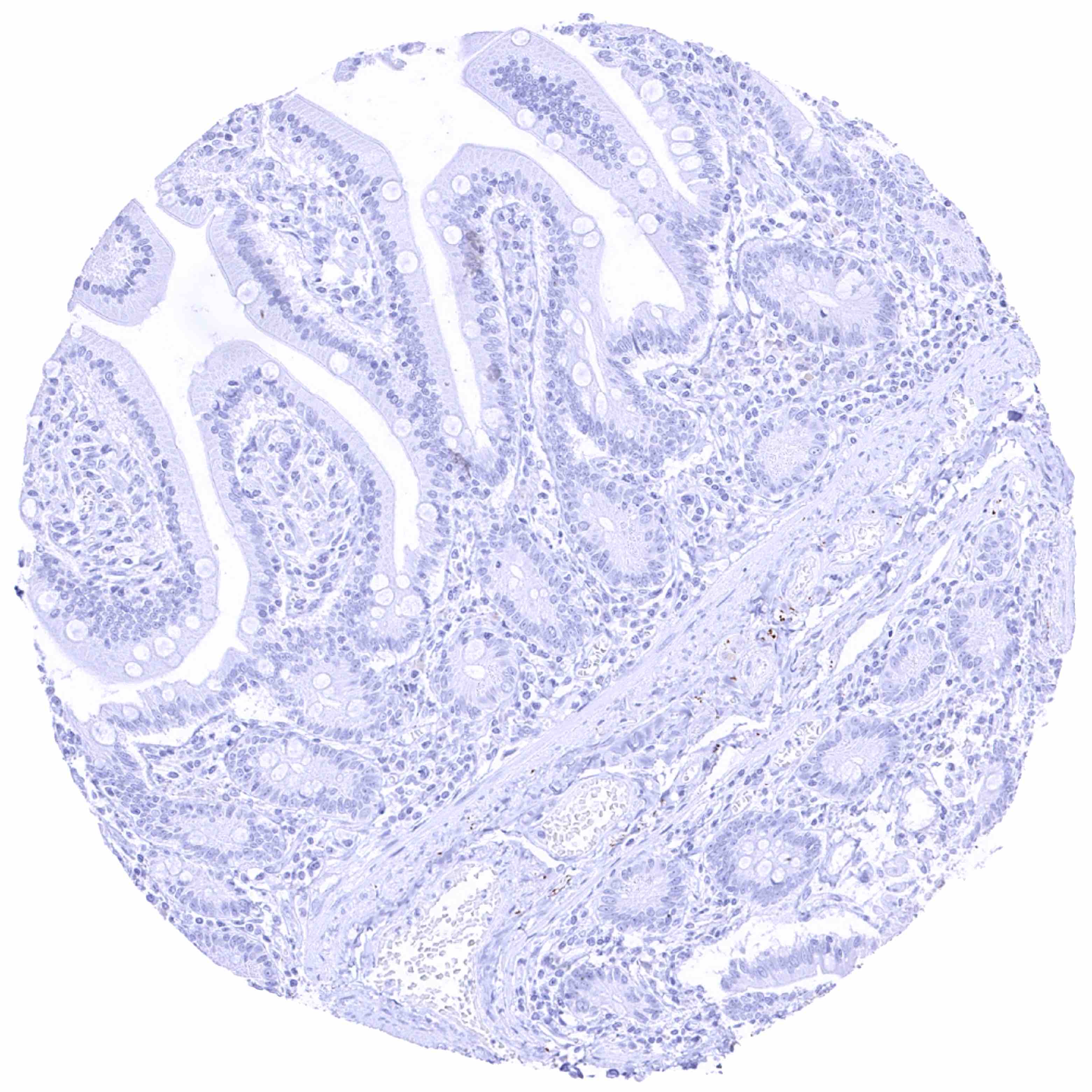
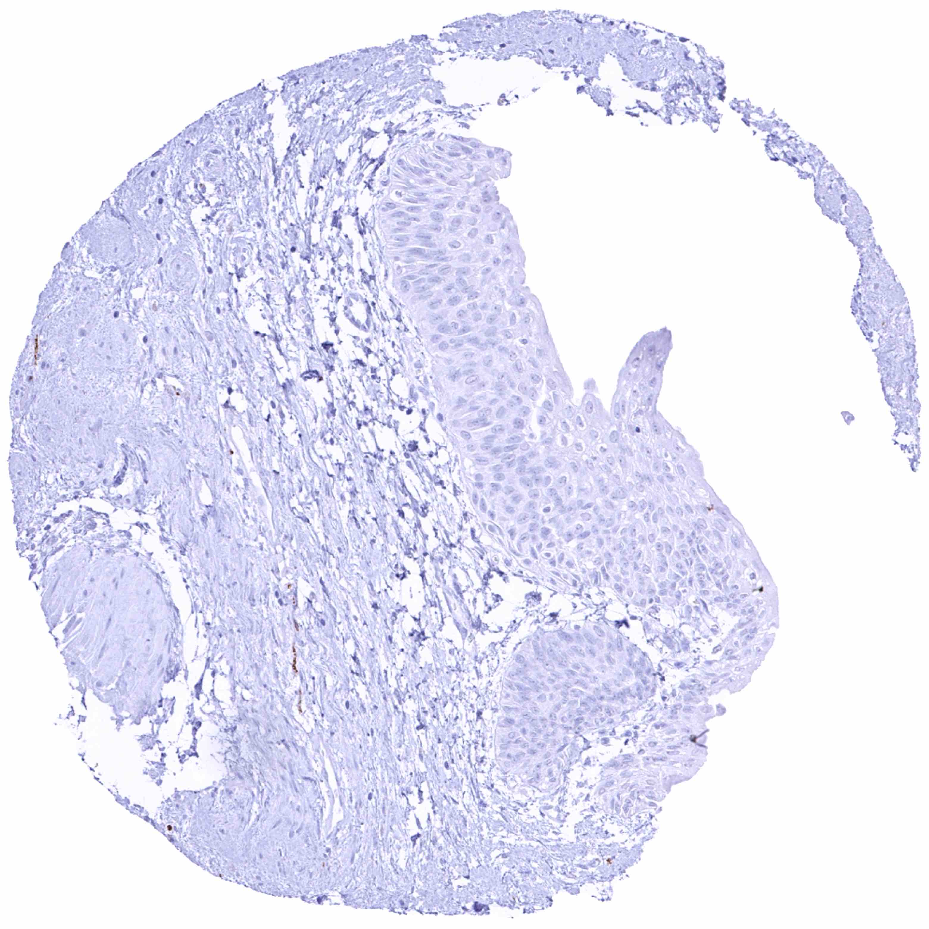
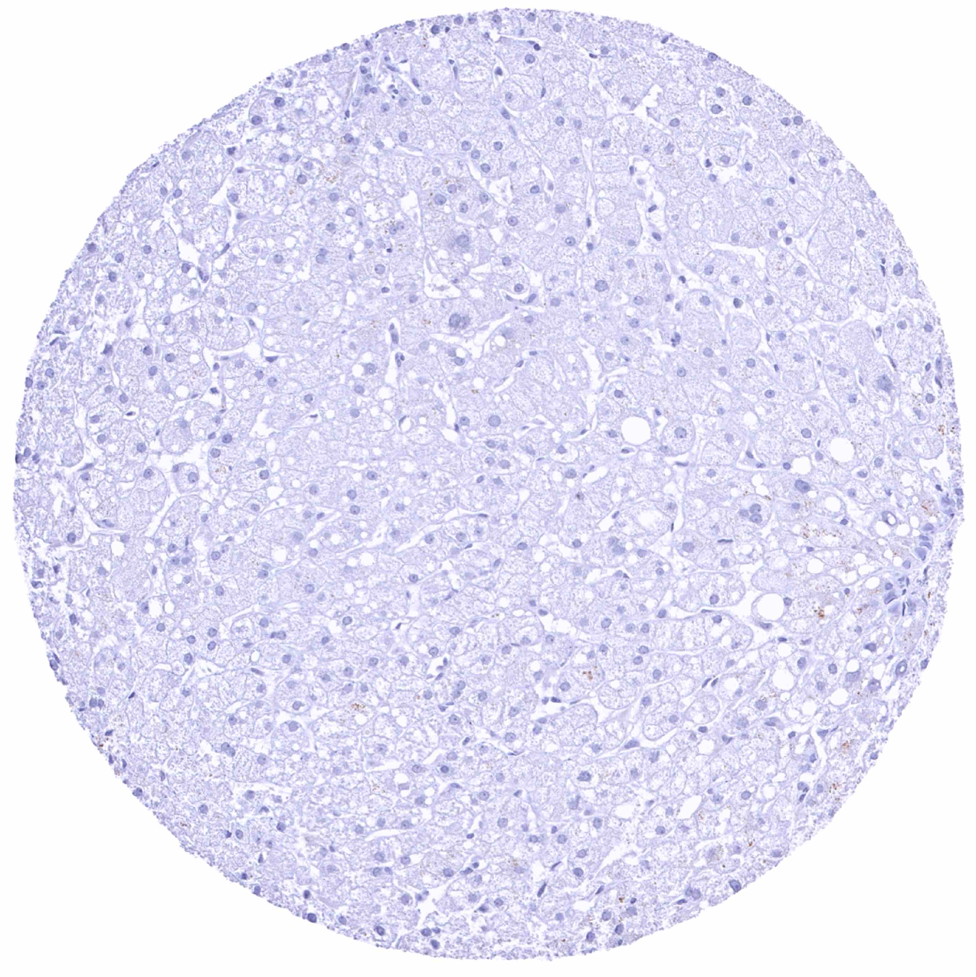
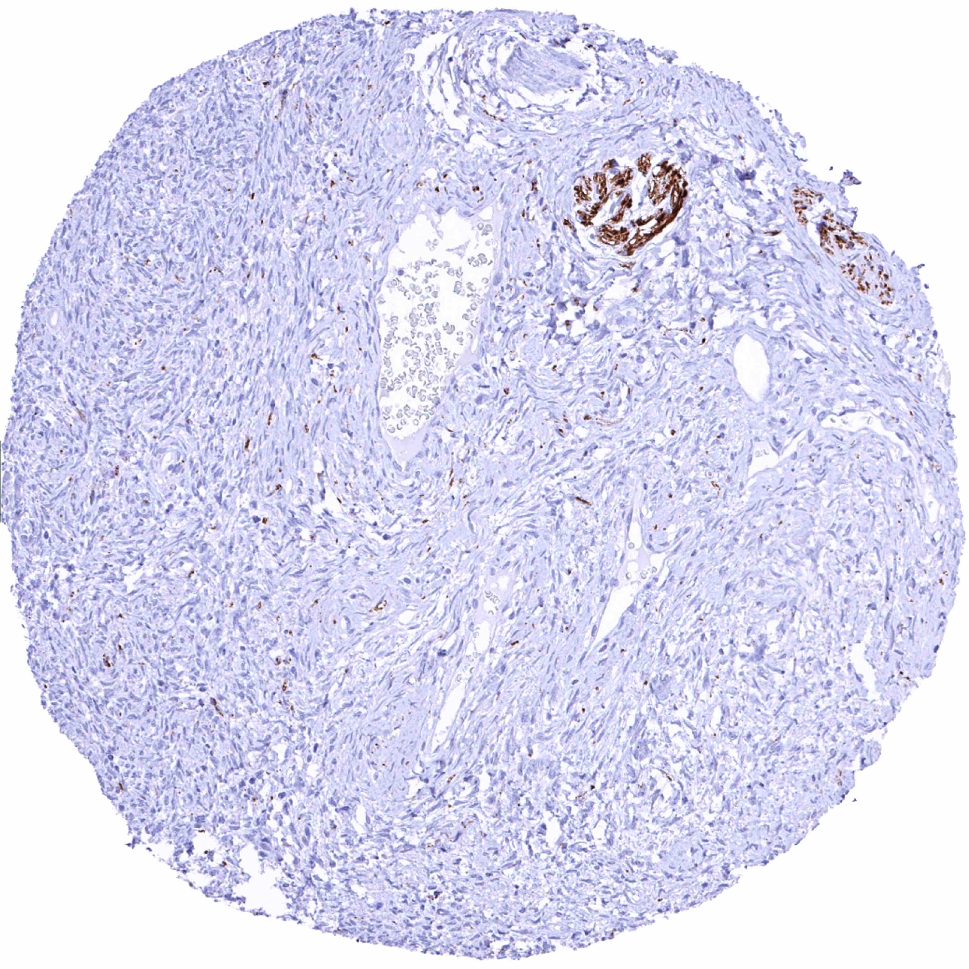
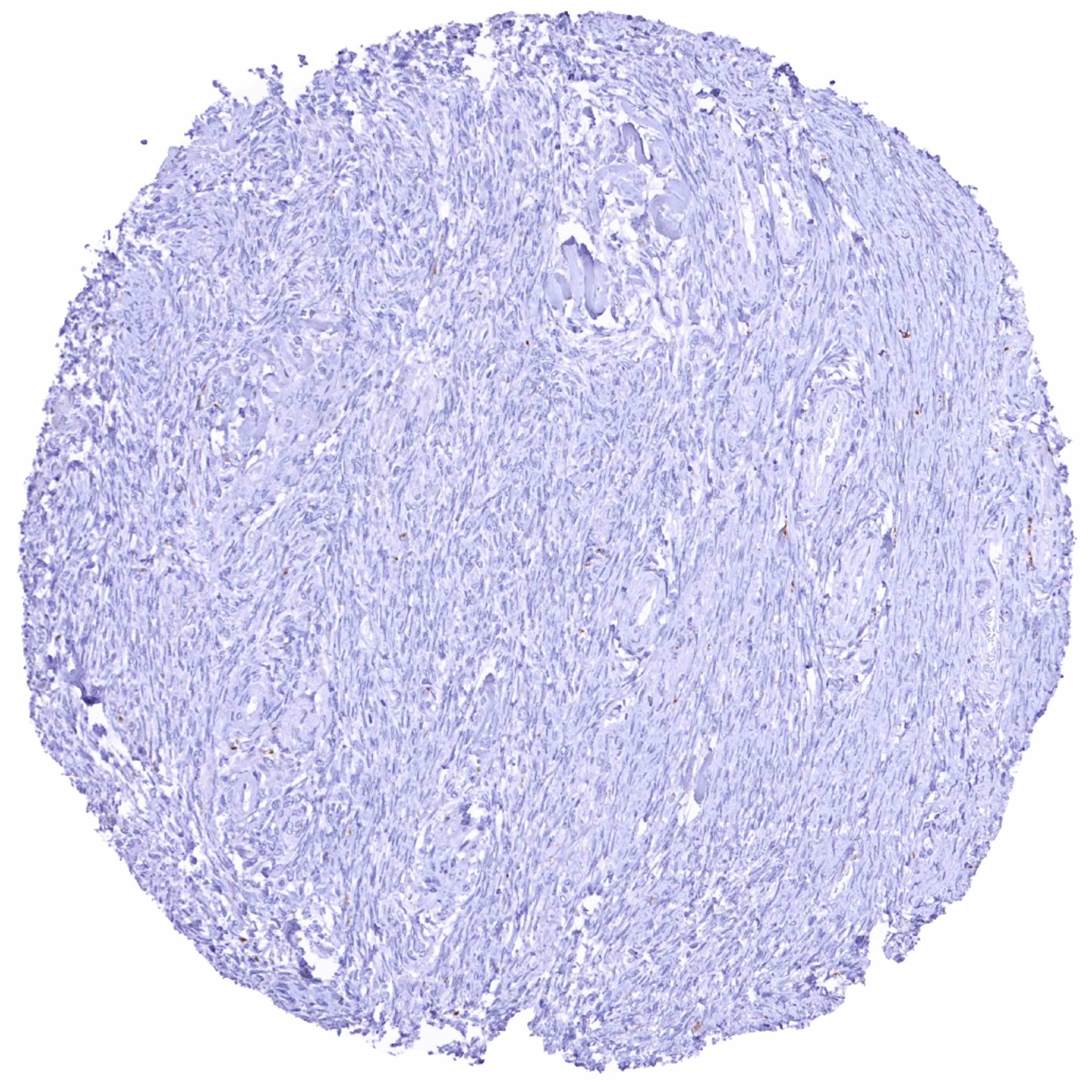
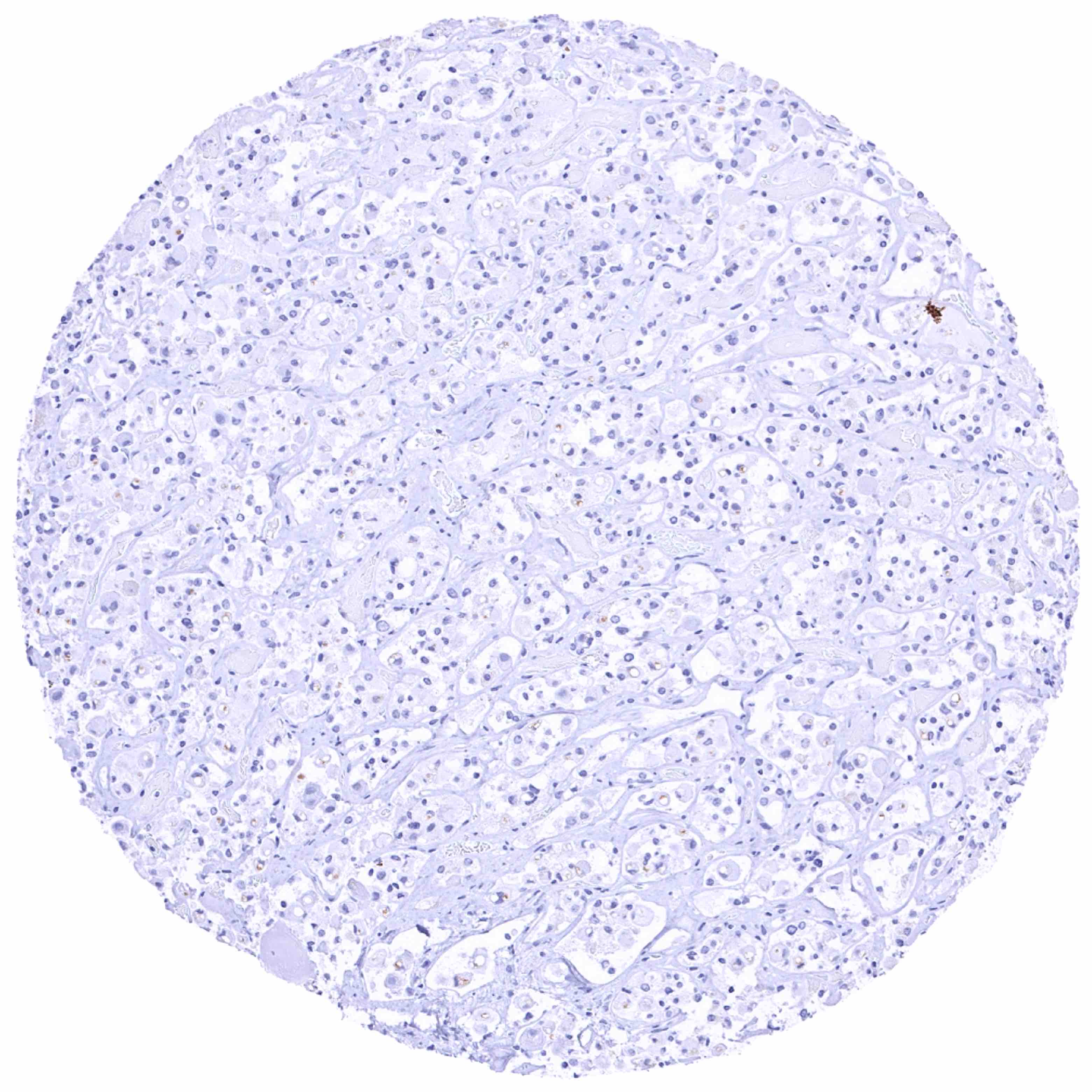
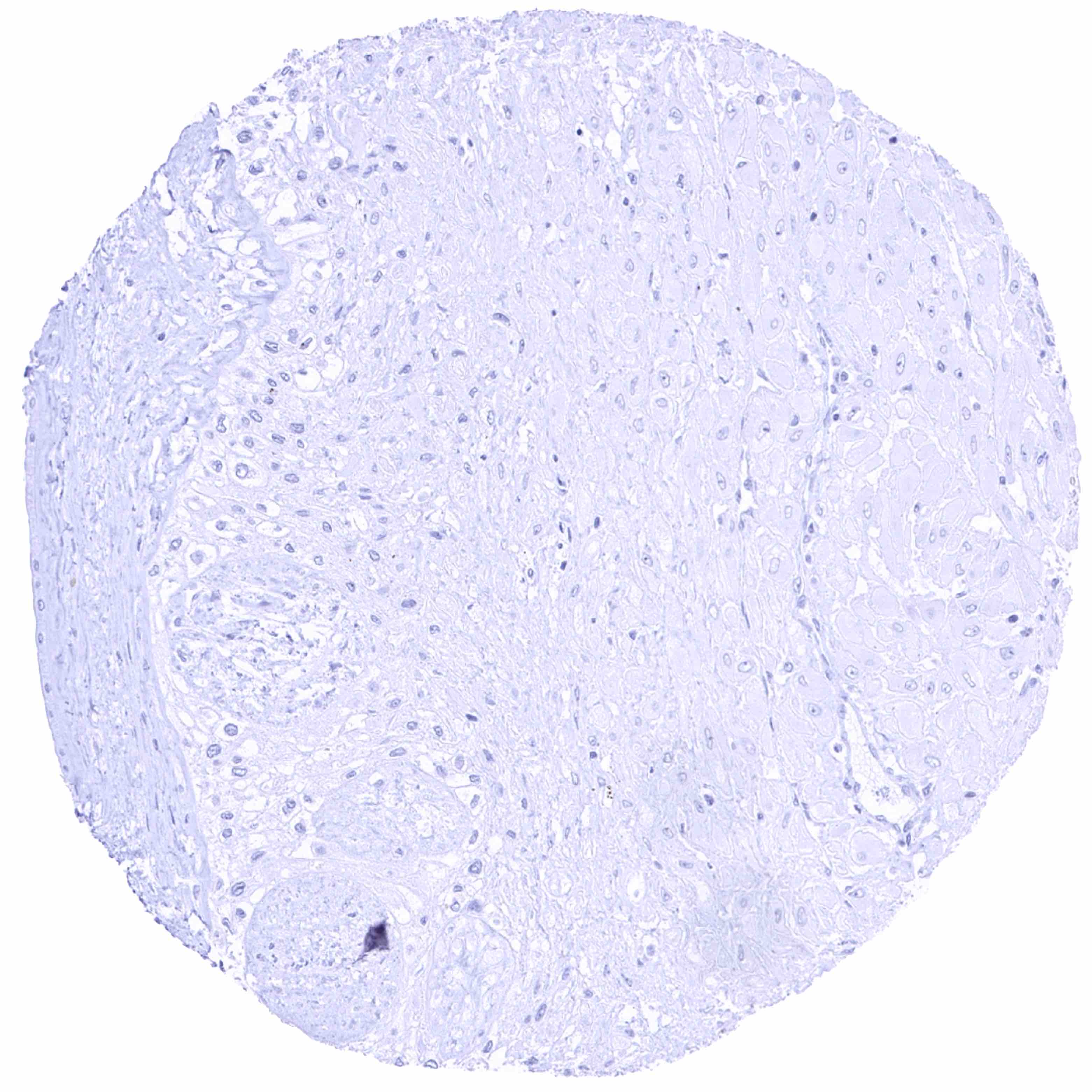
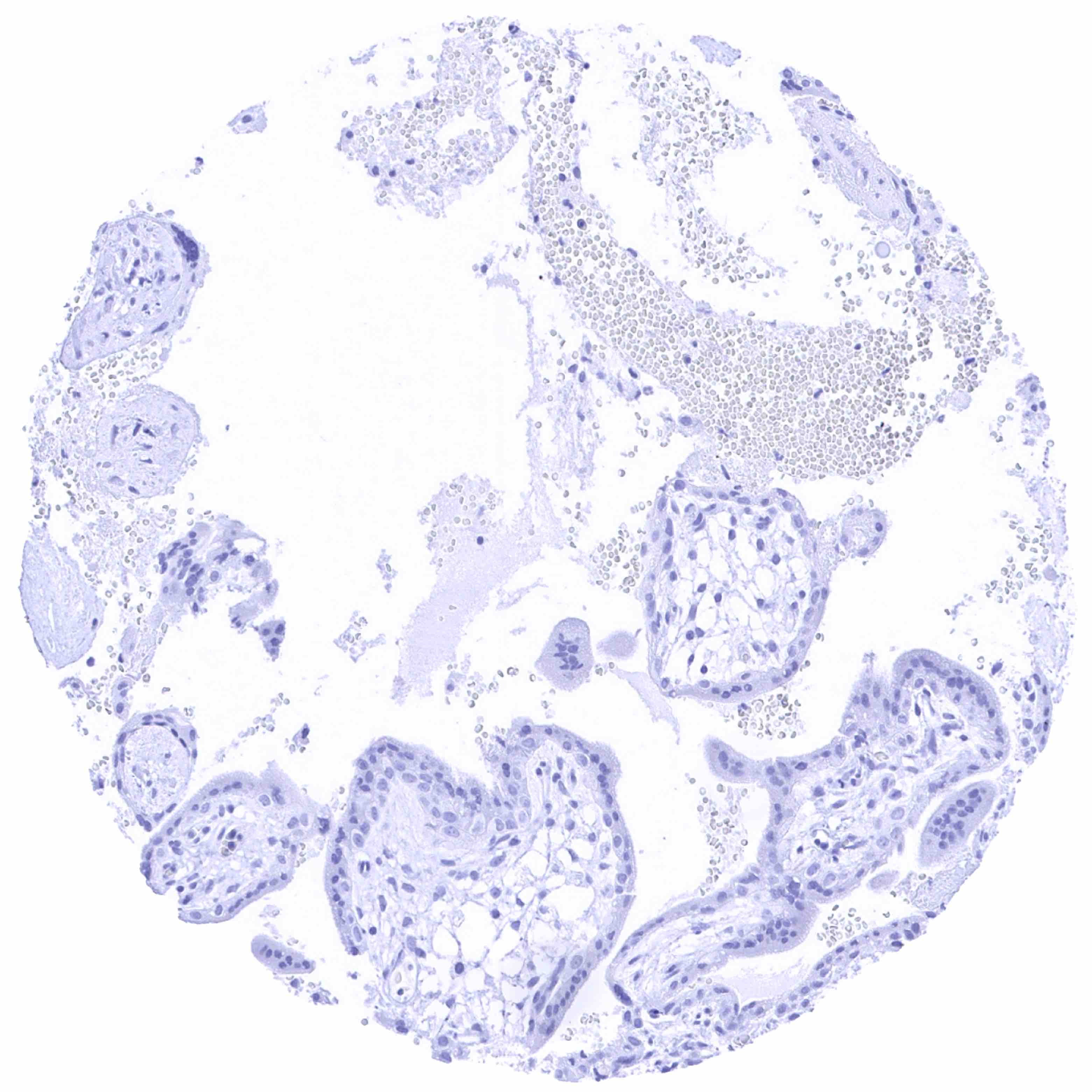
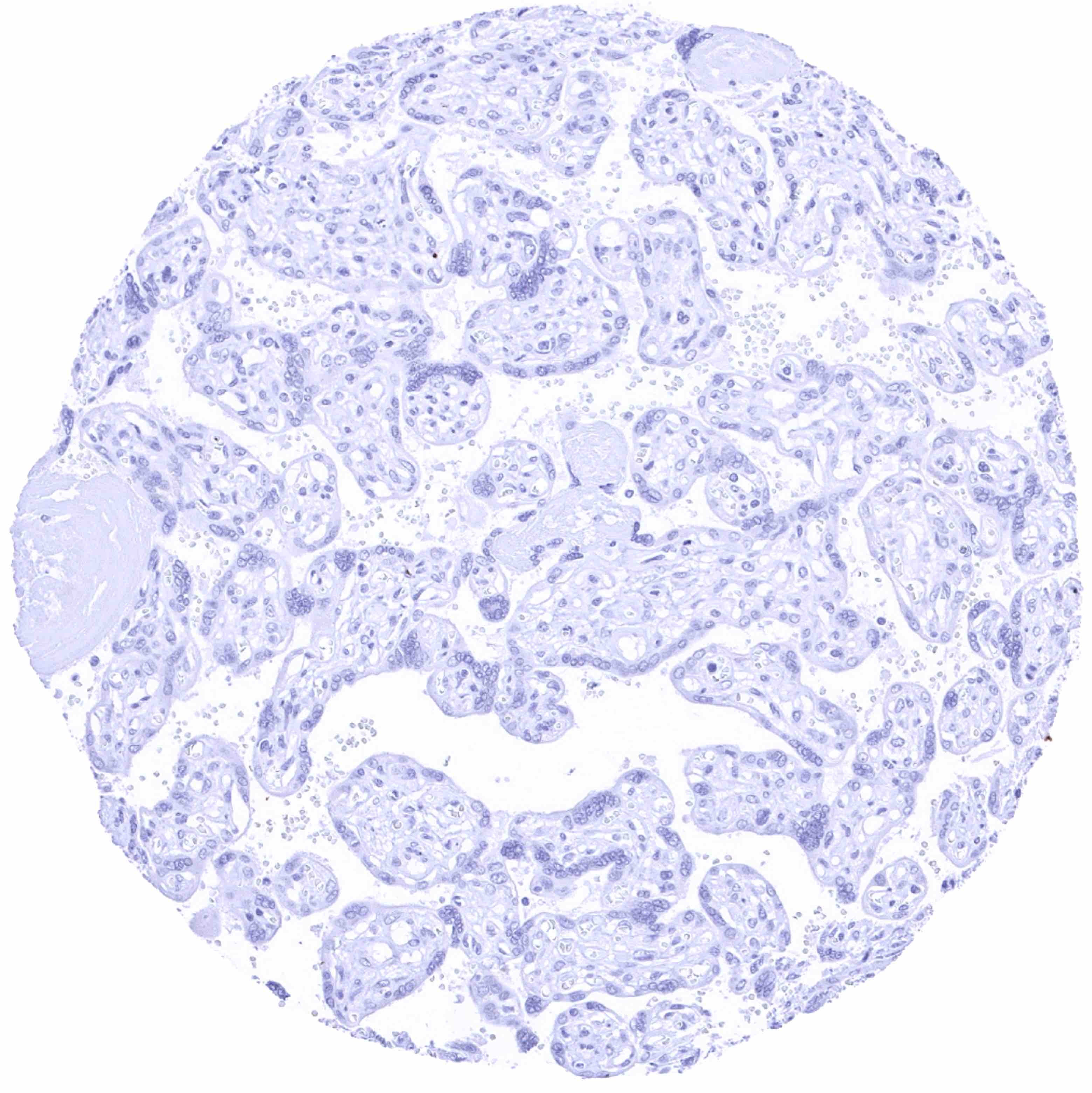
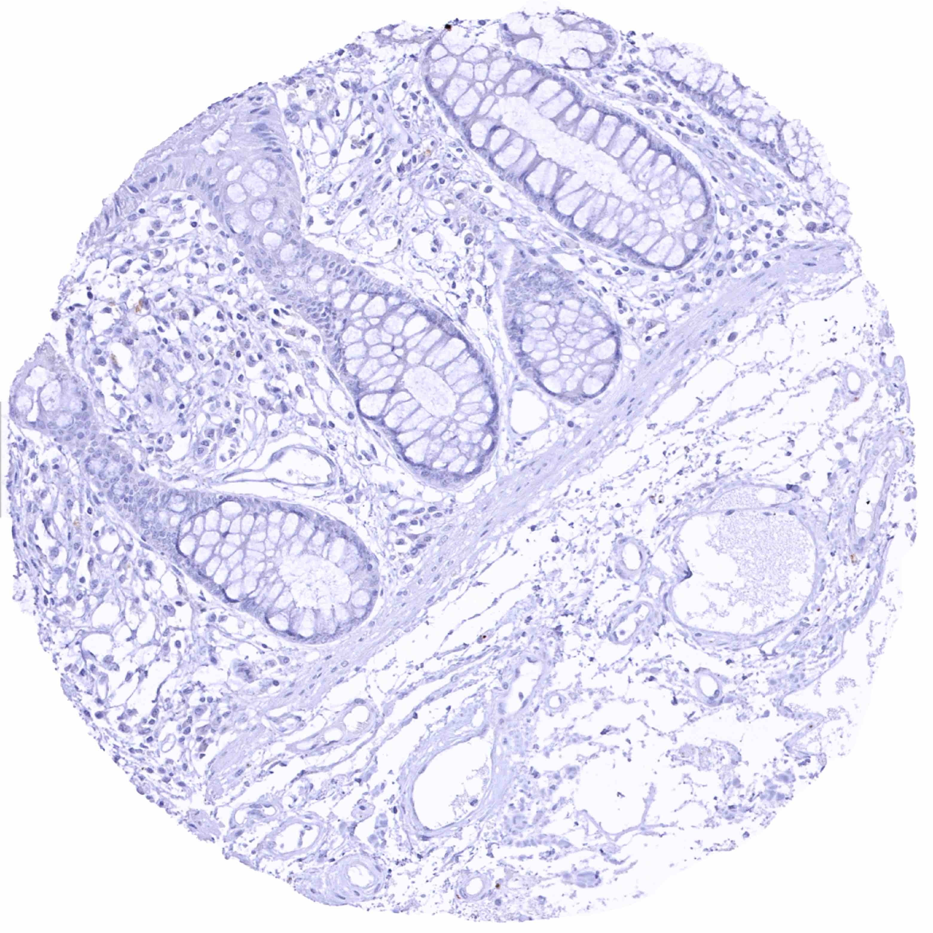
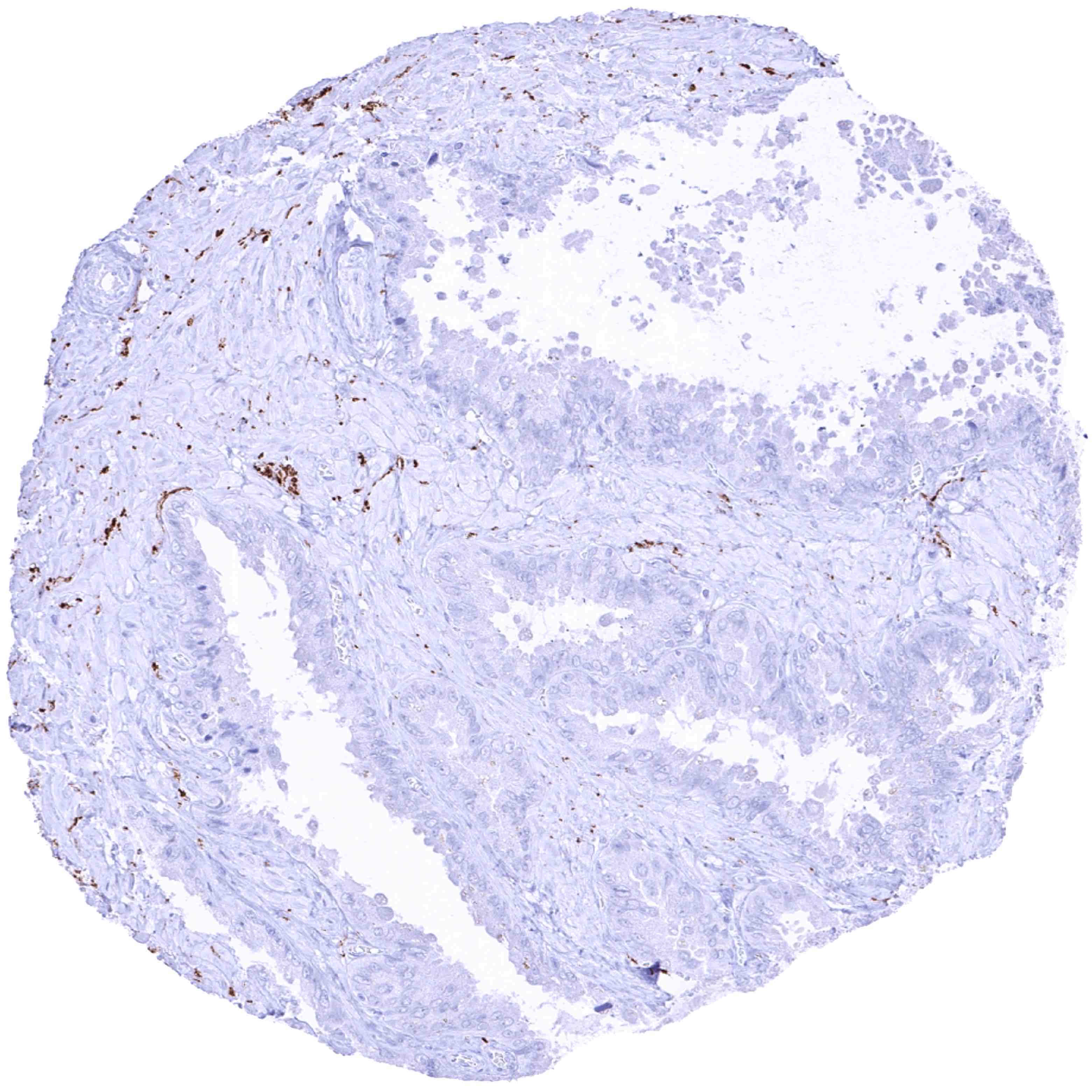
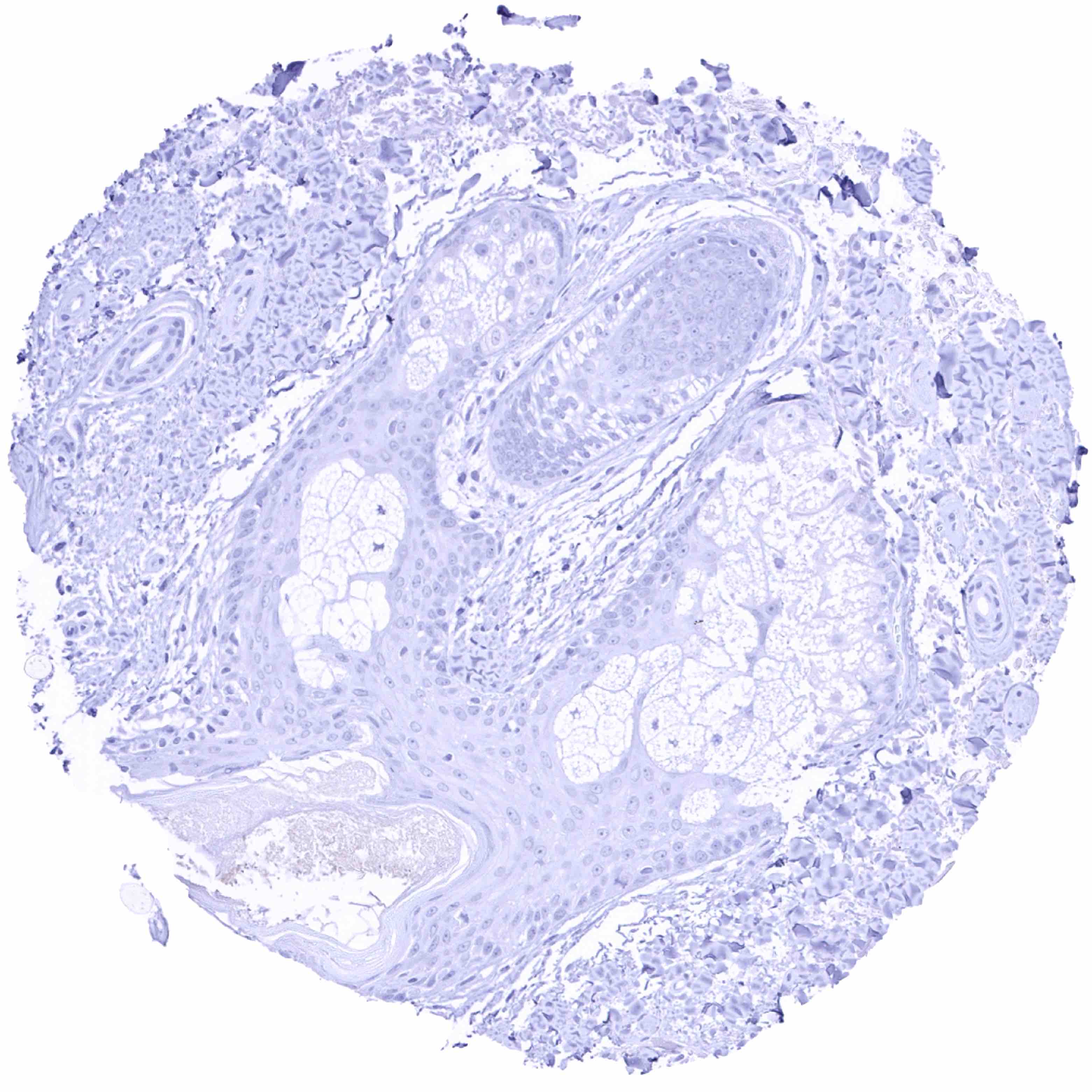
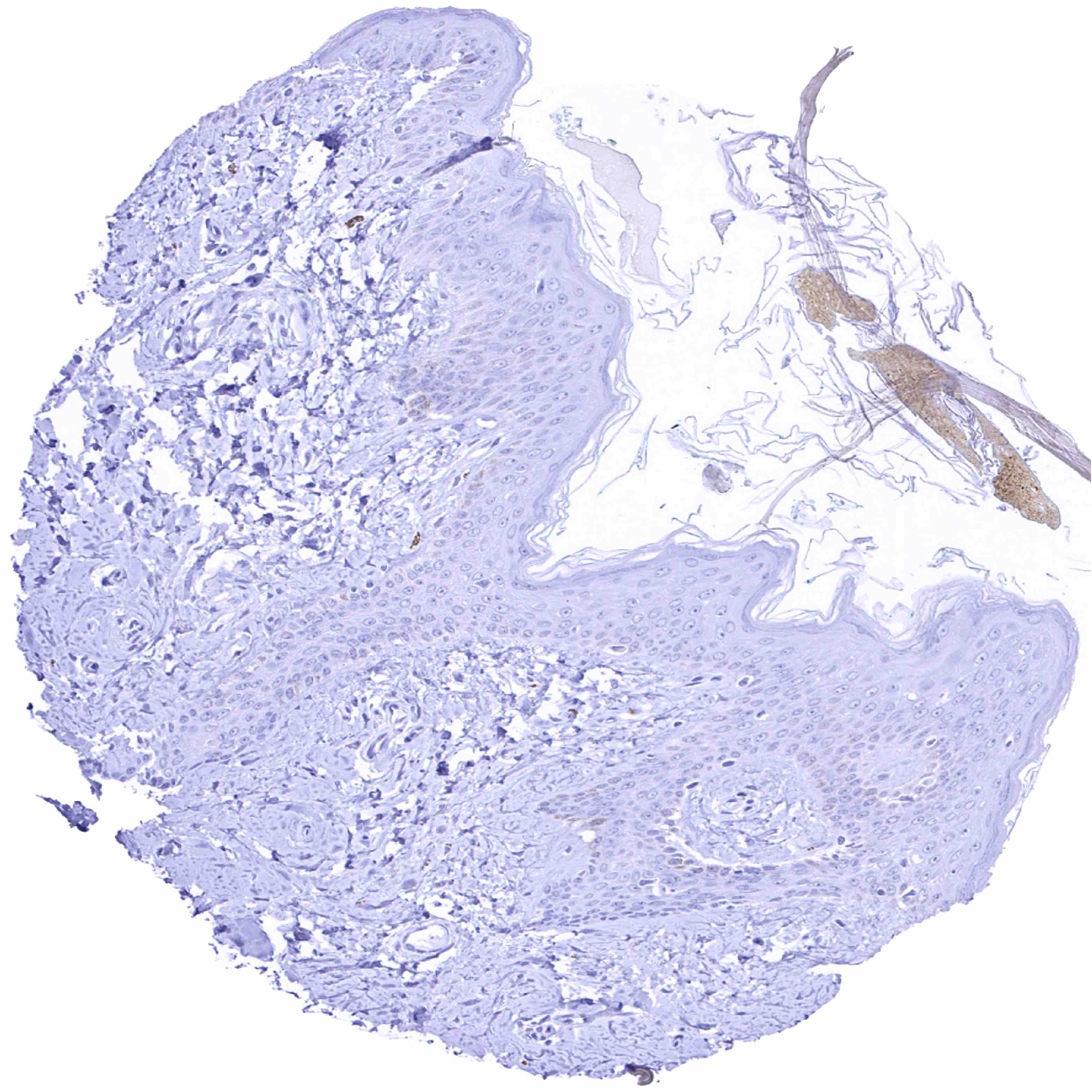
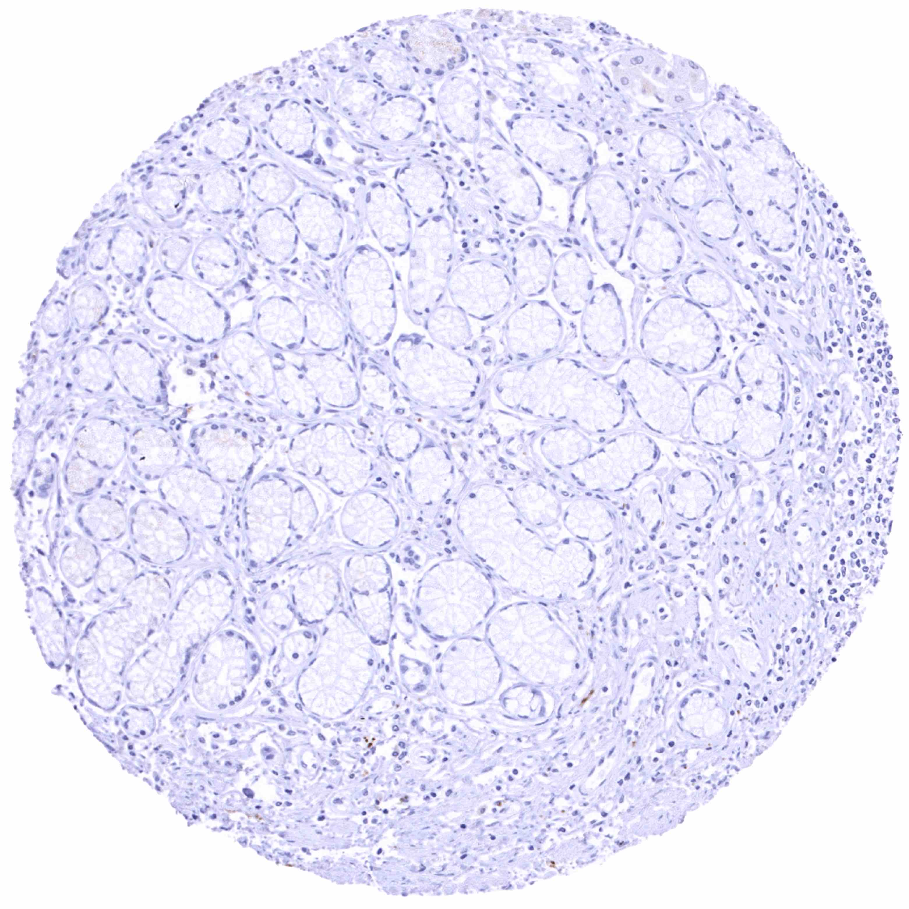
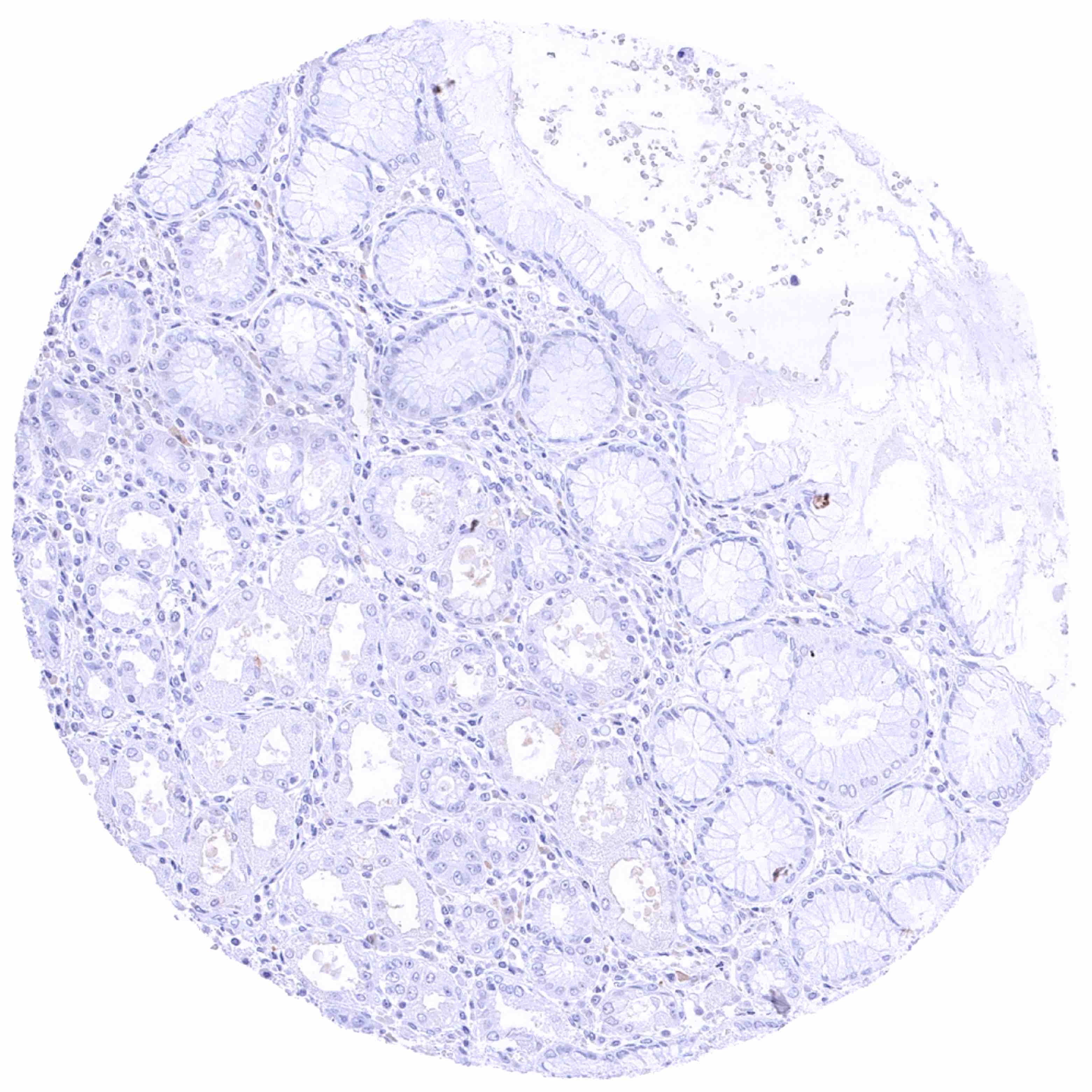
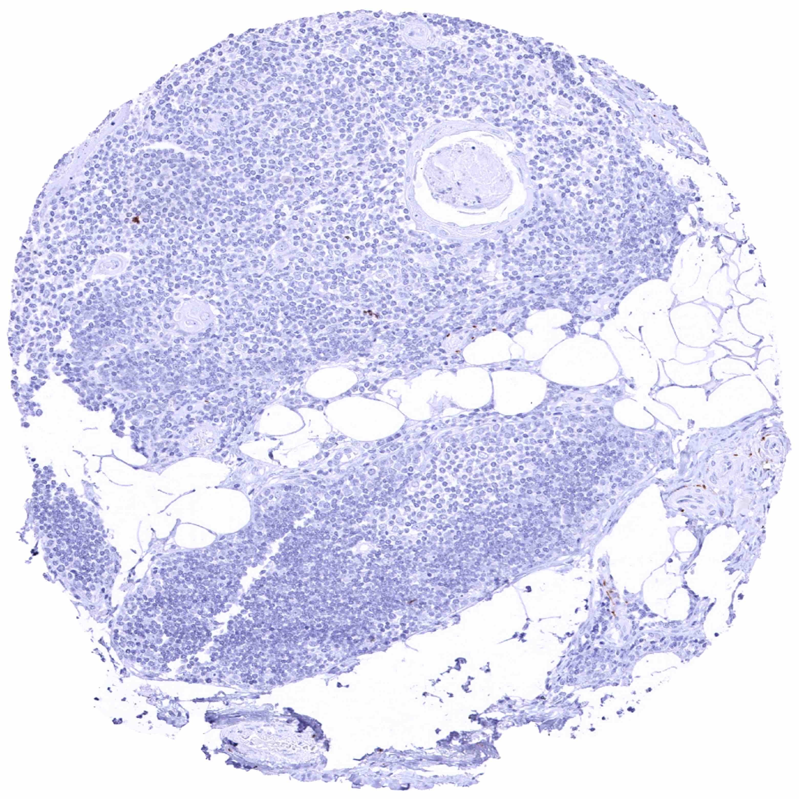
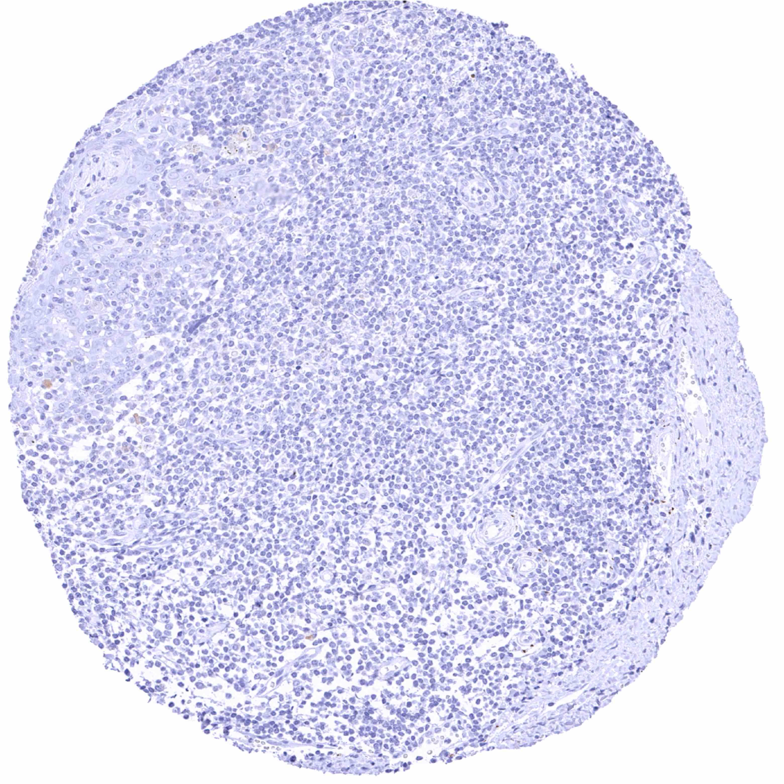
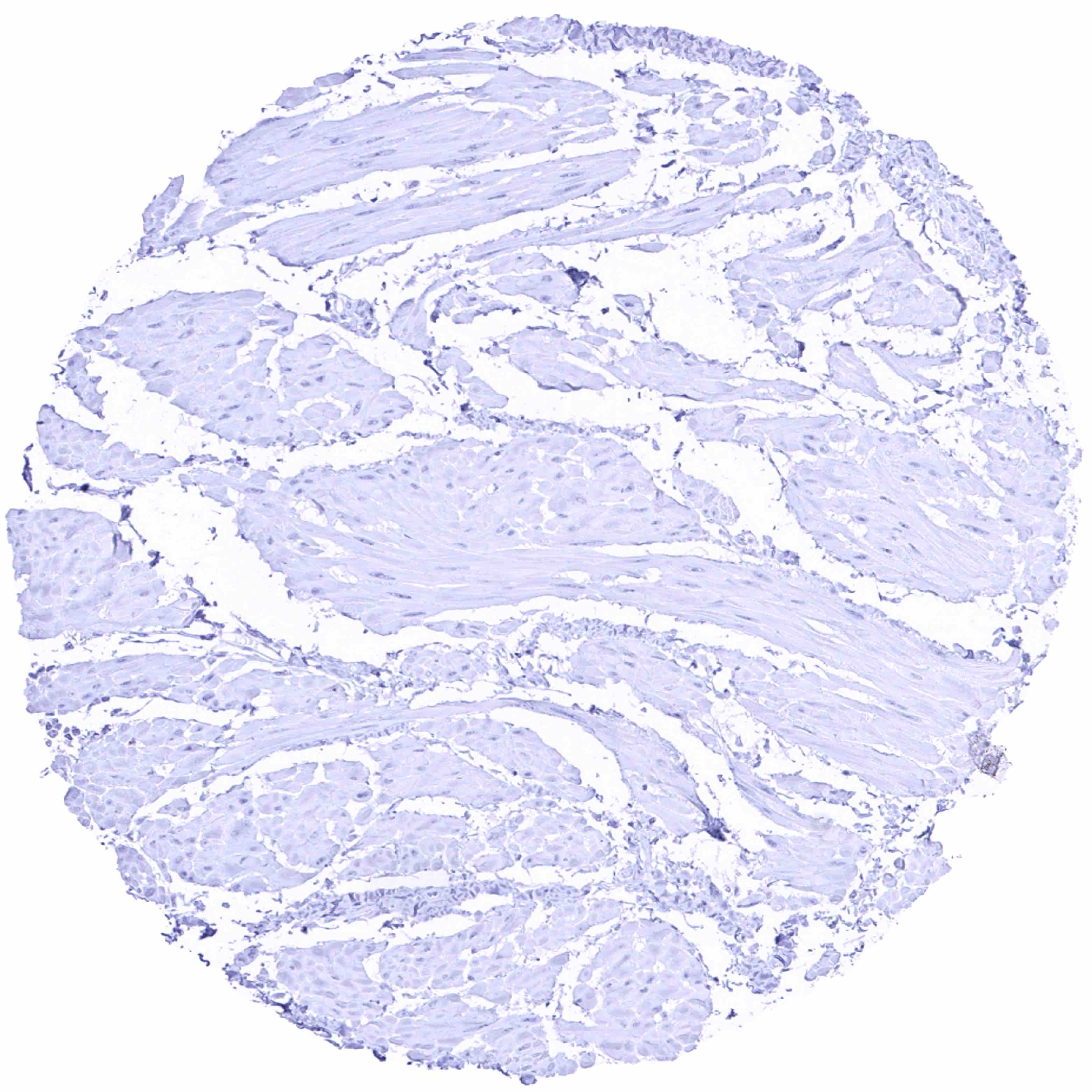
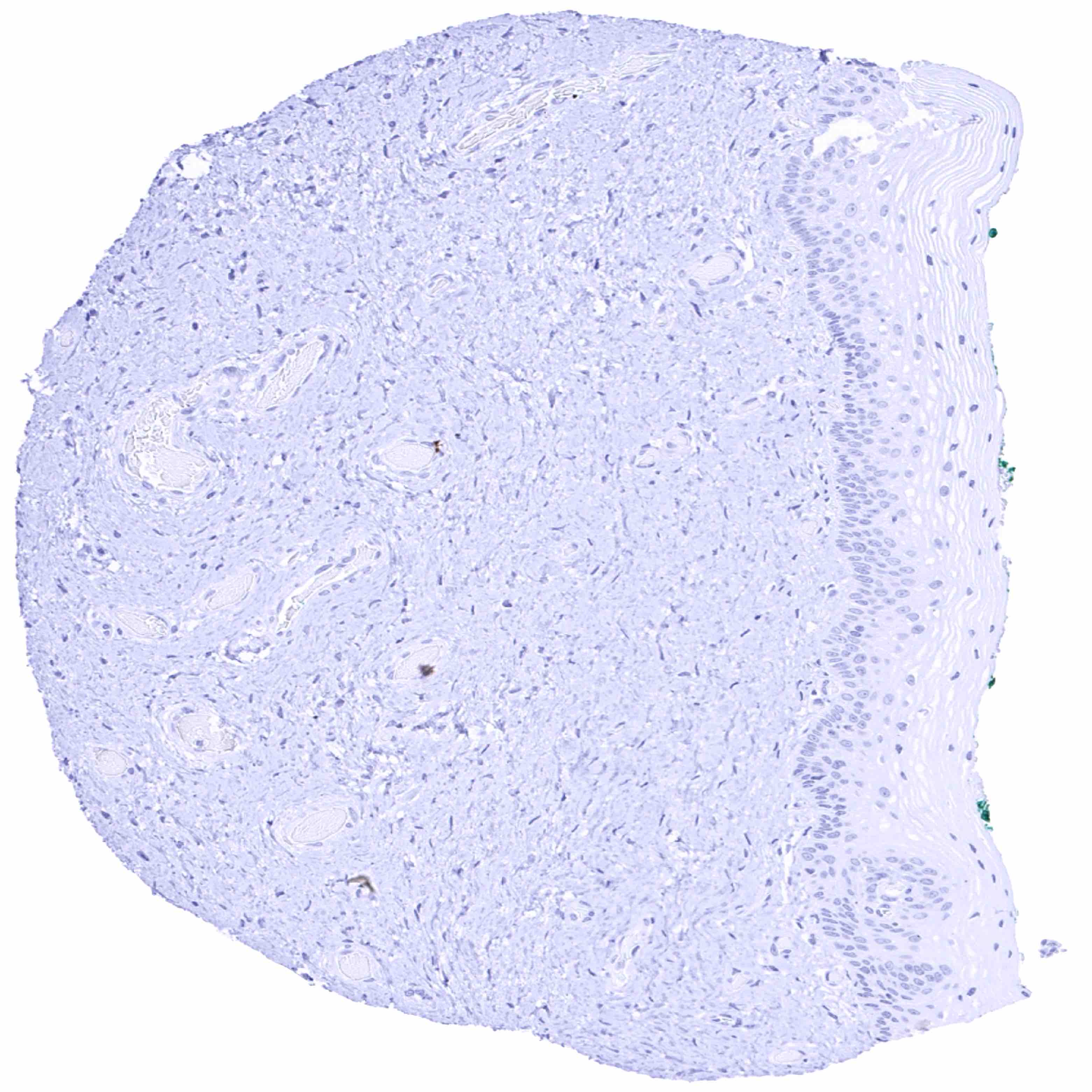
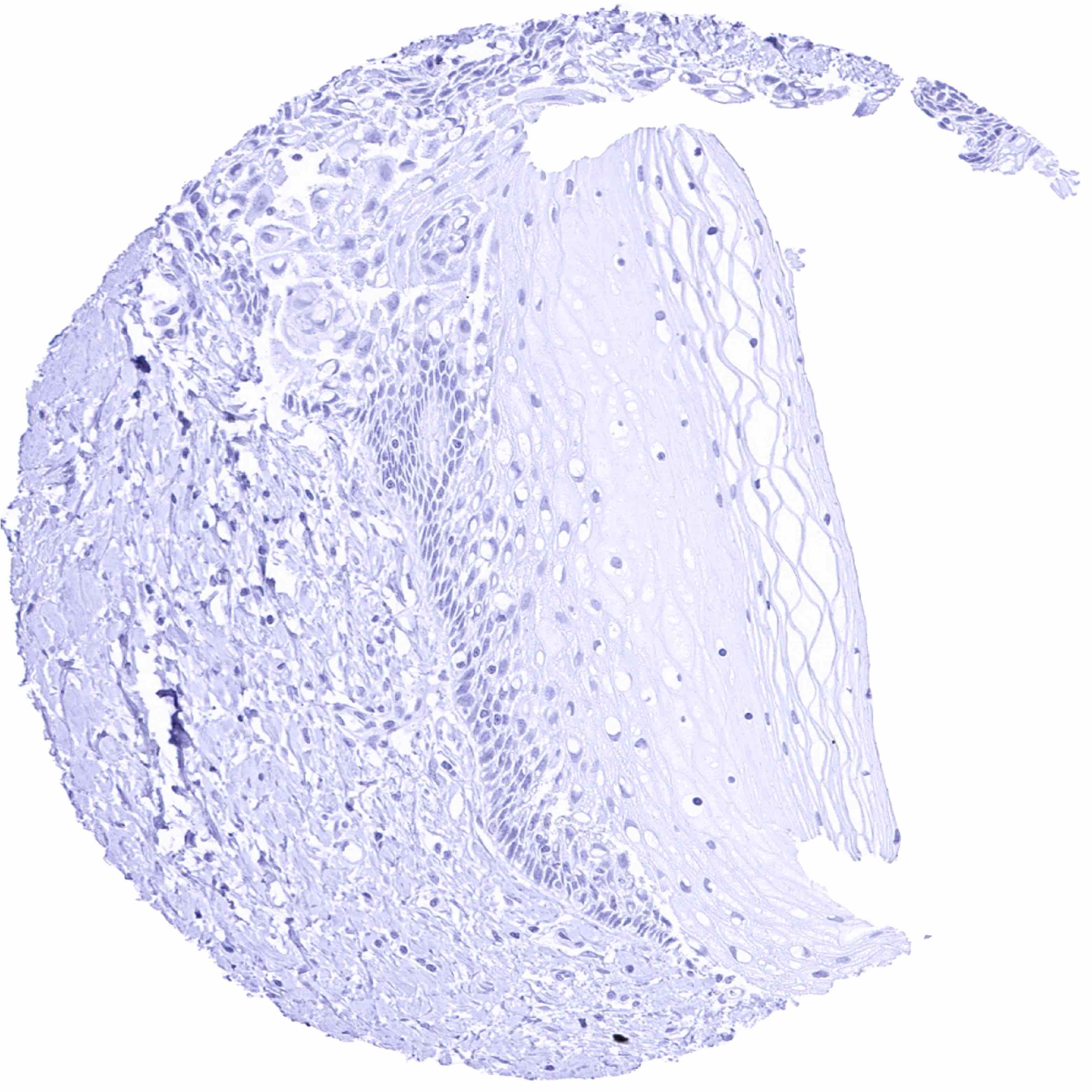
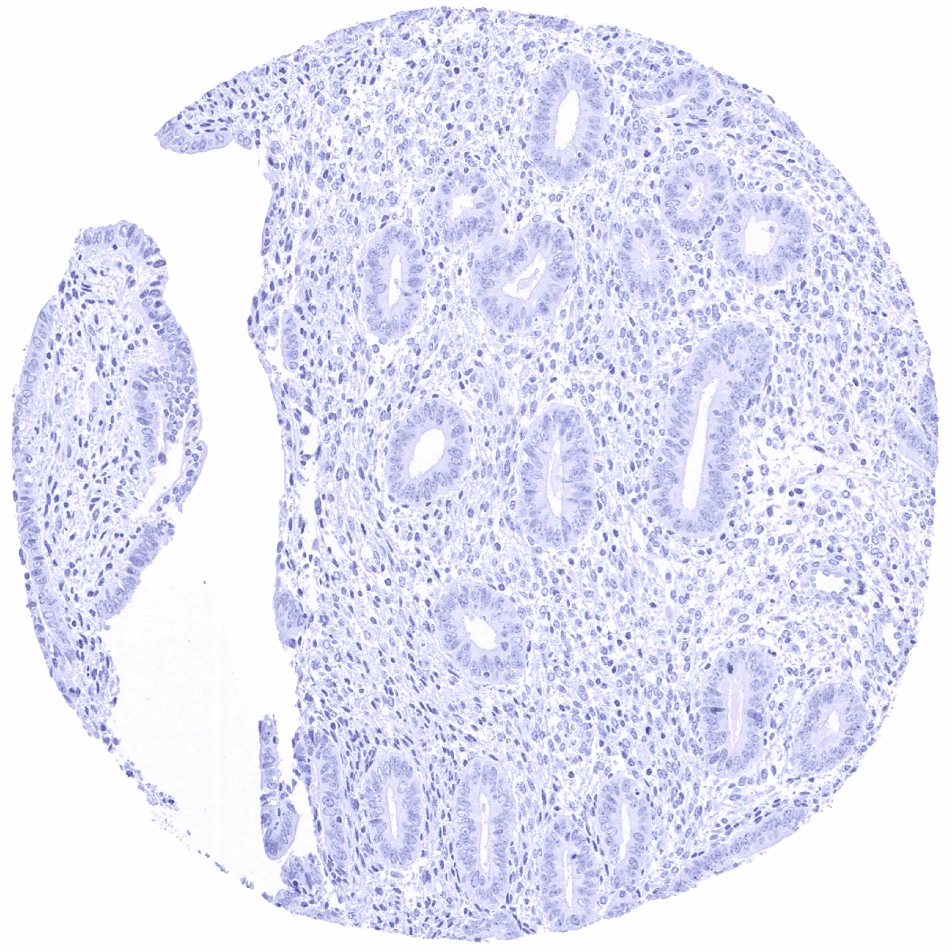
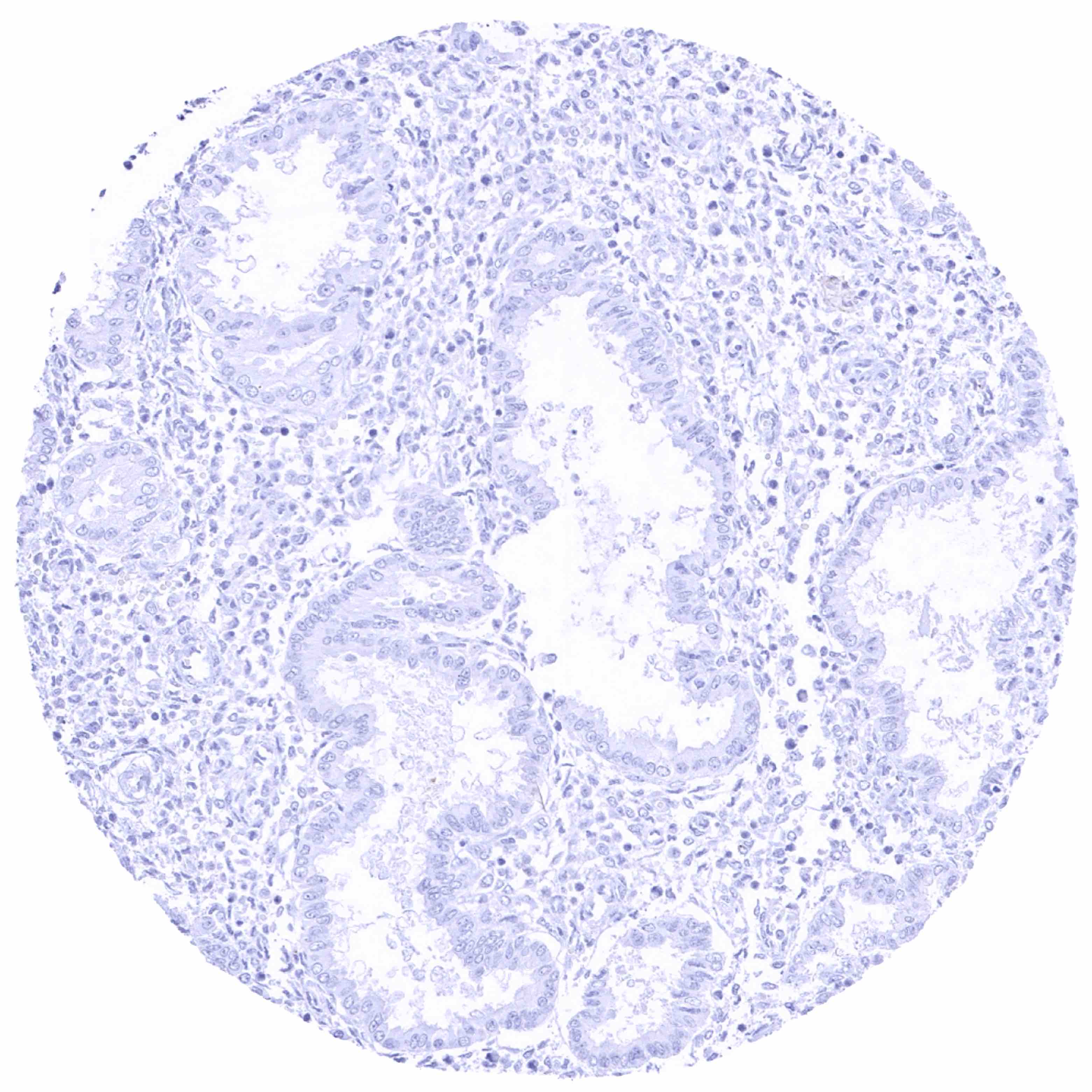
Tyrosine Hydroxylase antibody [HMV312] HistoMAXTM


Go to Cancer Tissue Gallery
  
 Go back to product page
  

Go to HistoMAX page


Adrenal gland – Distinct TH staining of at least a subset nerve fibers in the cortex

Adrenal gland – Strong membranous and cytoplasmic TH staining of medullary cells

Aorta, media

Appendix, mucosa


Appendix, muscular wall – Distinct TH staining of few nerve fibers

Bone marrow

Breast

Bronchus mucosa


Cerebellum (white matter)

Cerebellum, cortex (molecular layer, Purkinje cell layer, granule cell layer, white matter)

Cerebrum, grey matter

Cerebrum, white matter


Colon descendens, mucosa

Colon descendens, muscular wall

Duodenum, Brunner gland

Duodenum, mucosa


Epididymis (caput)

Epididymis (cauda)

Esophagus, squamous epithelium

Esophagus, squamous epithelium


Fallopian tube, mucosa

Fat

Gallbladder, epithelium

Heart muscle – Strong TH staining of fibers in a small nerve


Ileum, mucosa

Kidney, cortex

Kidney, medulla

Kidney, pelvis, muscular wall – Distinct TH staining of few nerve fibers


Liver

Lung

Lymph node

Ovary, corpus luteum


Ovary, stroma – A small nerve shows distinct TH positivity of nerve fibers. Individual nerve fibers also show TH staining

Ovary, stroma

Pancreas

Parathyroid gland – Distinct TH staining of few nerve fibers


Parotid gland – Distinct TH staining of fibers of a small nerve few as well as of small individual nerve fibers

Pituitary gland, anterior lobe

Pituitary gland, posterior lobe

Placenta (amnion and chorion)


Placenta (amnion) – Distinct cytoplasmic and membranous TH positivity of a small subset of amnion cells

Placenta (chorion)

Placenta, early

Placenta, mature


Prostate – Distinct TH staining of nerve fibers

Prostate – Strong TH staining of numerous nerve fibers

Rectum, mucosa

Seminal vesicle – Distinct TH staining of numerous nerve fibers


Sinus paranasales

Skeletal muscle

Skin, hairfollicel and sebaceous glands

Skin


Spleen

Stomach, antrum – A weak cytoplasmic background staining may occur in some glandular cells of the stomach

Stomach, antrum – A weak cytoplasmic background staining may occur in some glandular cells of the stomach

Stomach, corpus


Stomach, muscular wall

Testis

Thymus

Thyroid gland


Tonsil, surface epithelium

Tonsil

Urinary bladder, muscular wall

Urinary bladder, urothelium


Urinary bladder, urothelium

Uterus, ectocervix

Uterus, ectocervix

Uterus, endocervix – Distinct TH staining of some nerve fibers


Uterus, endometrium (pregnancy)

Uterus, endometrium (proliferation)

Uterus, endometrium (secretion)

Uterus, myometrium


| TOP |