GeneTex
  • Country / Location Selection

United States (US)

Normal Tissue Gallery- GTX04454

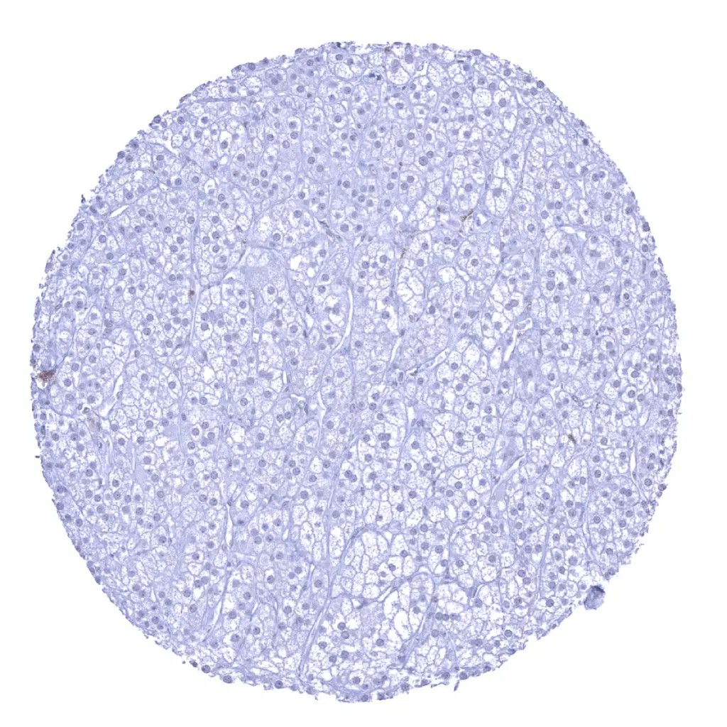
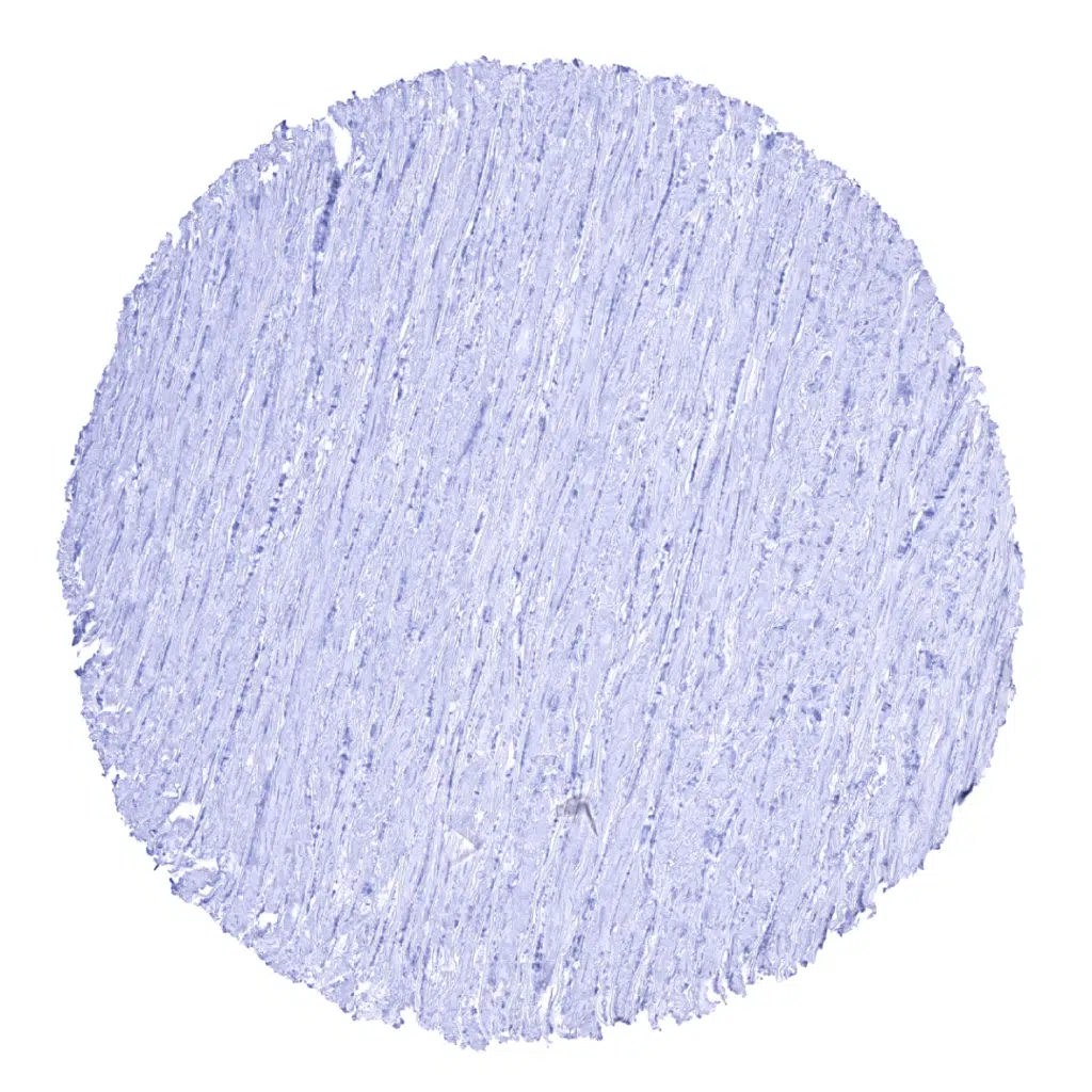
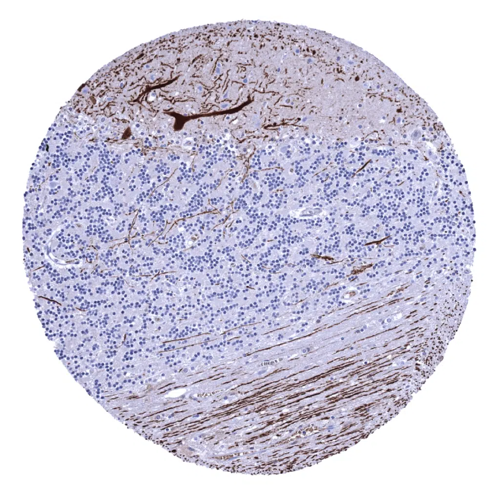
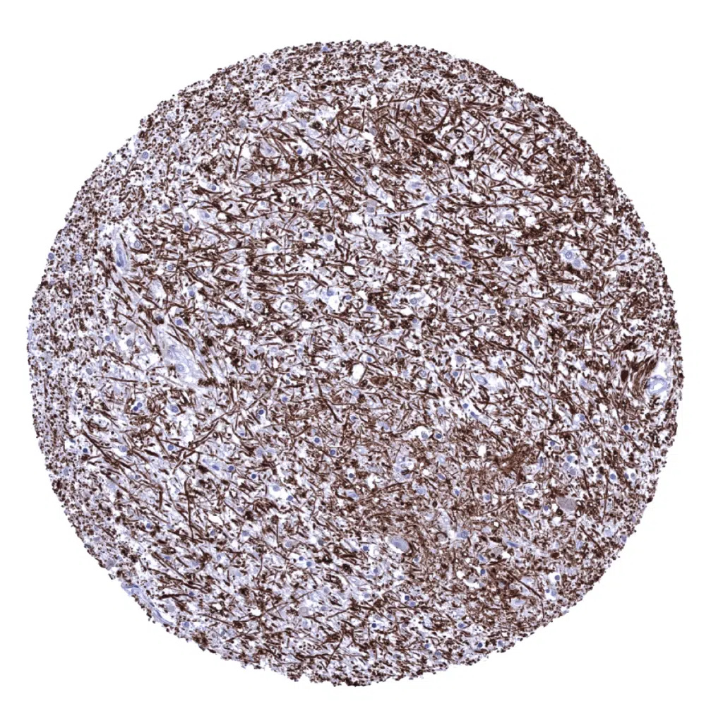
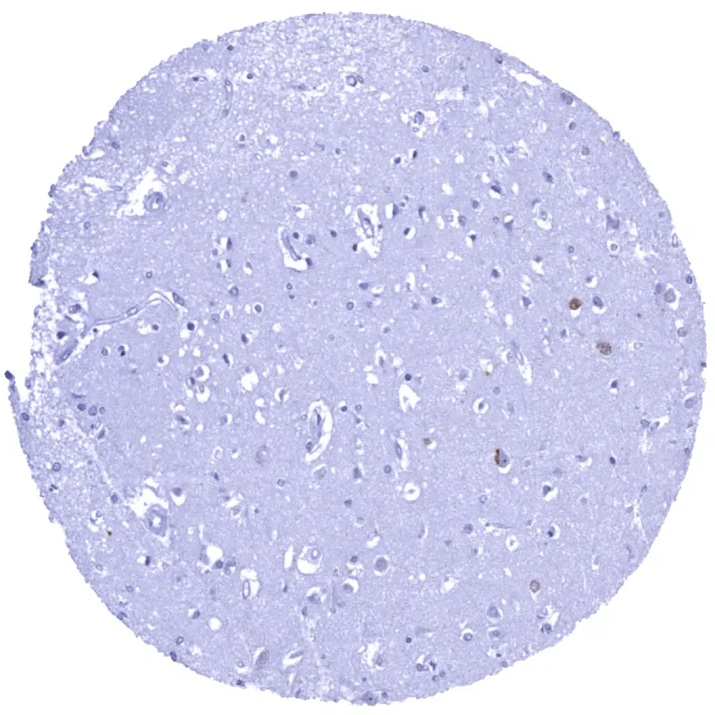
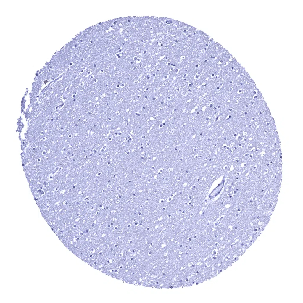
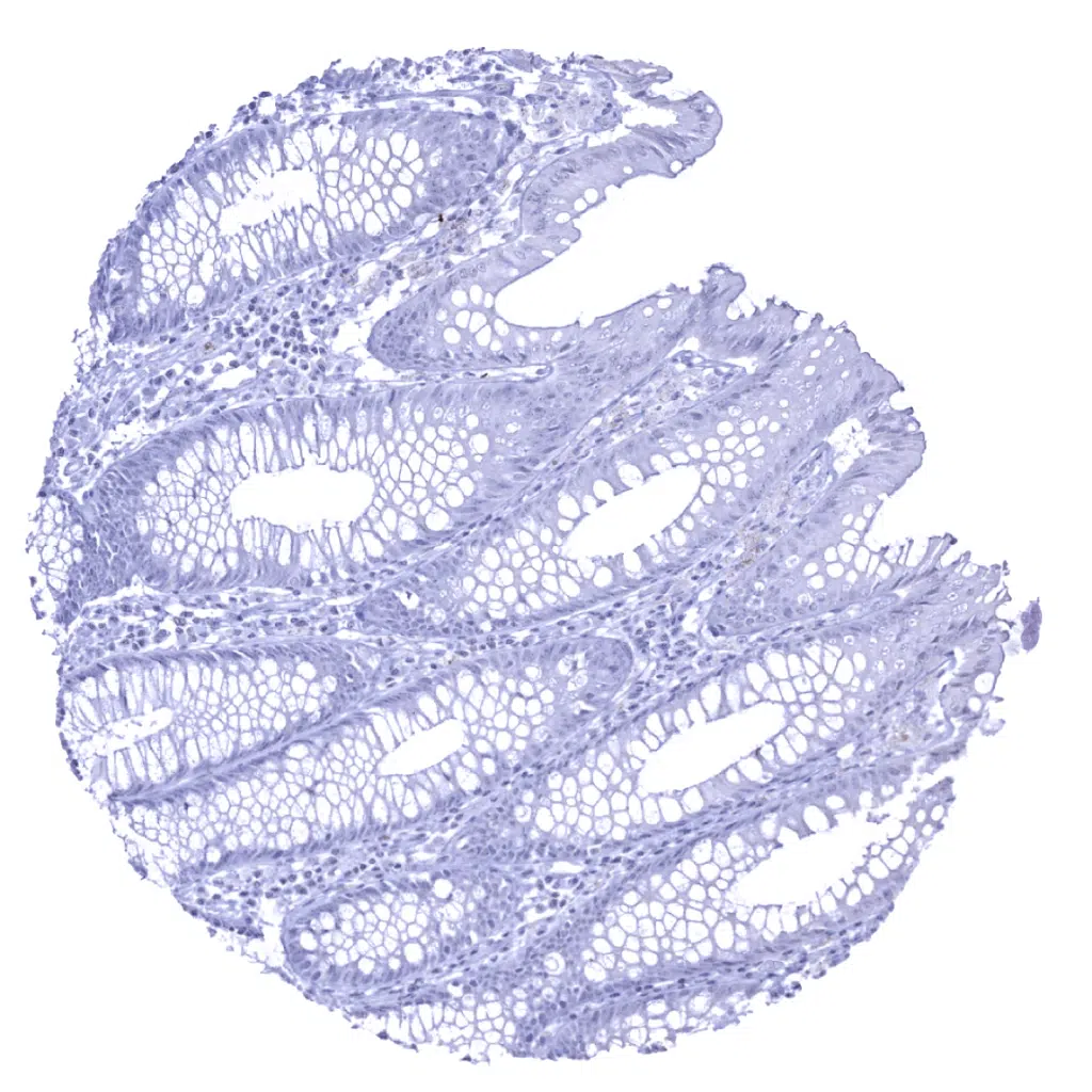
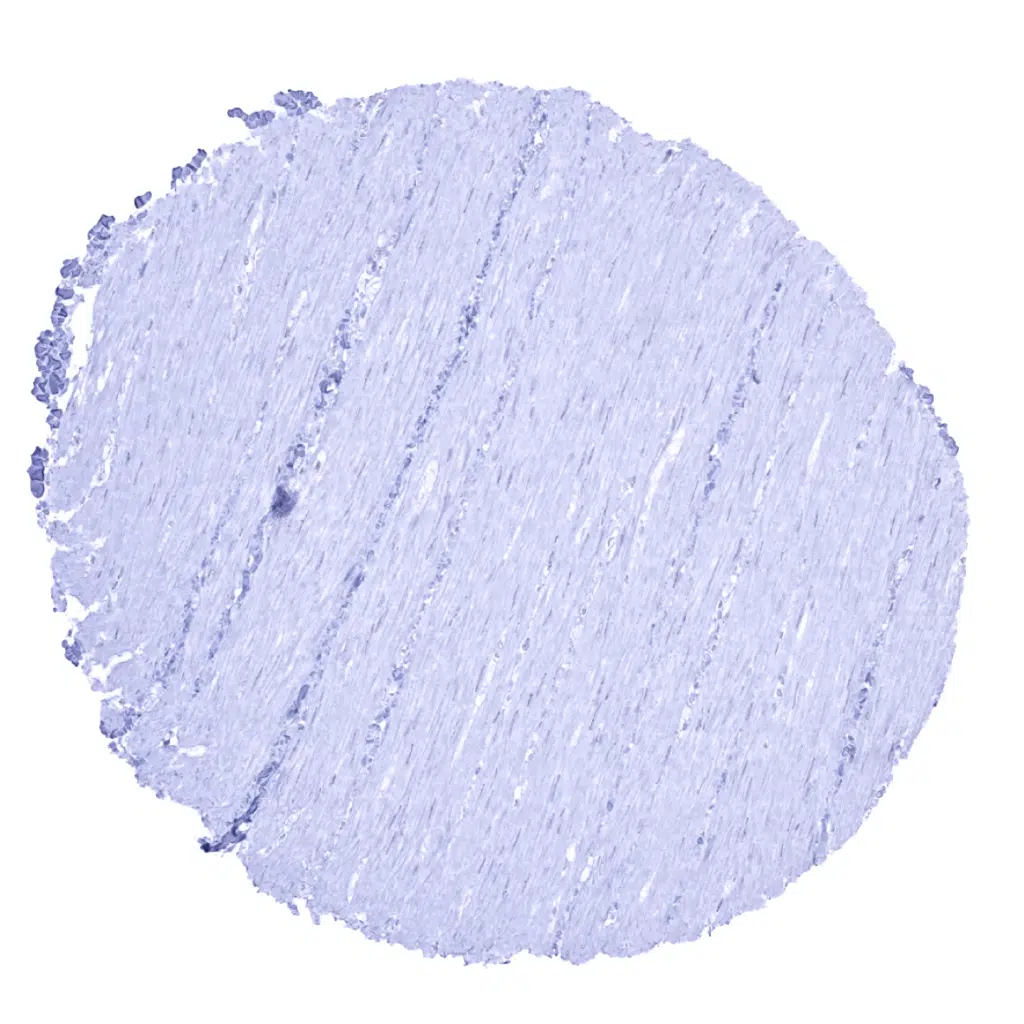
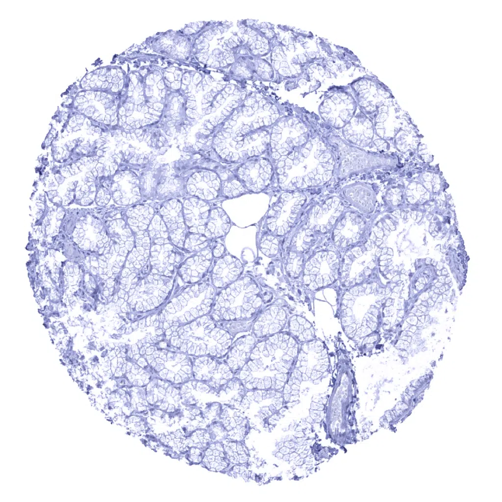
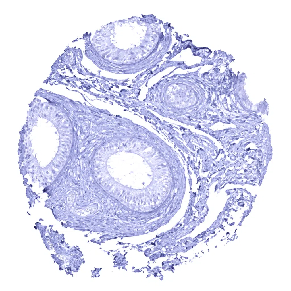
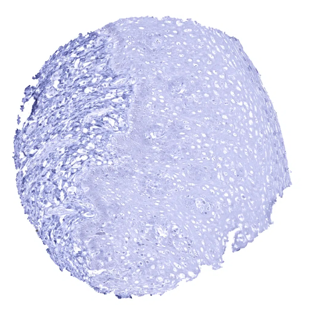
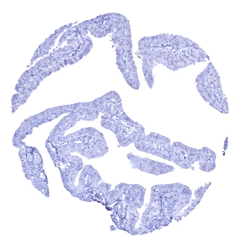
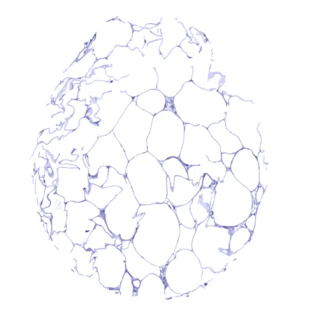
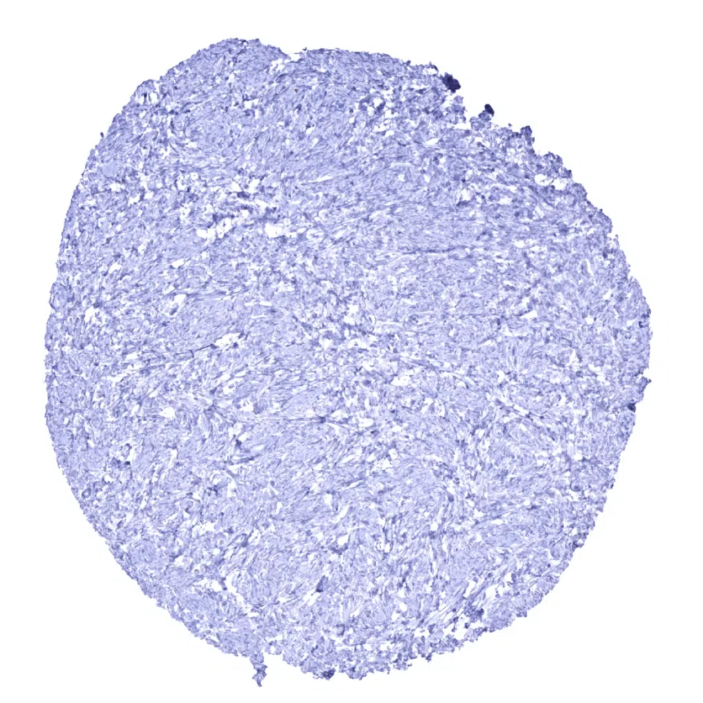
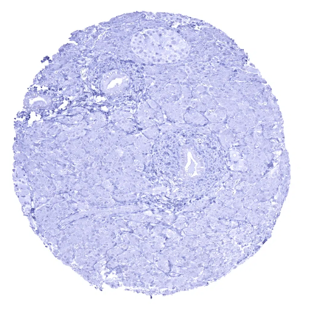
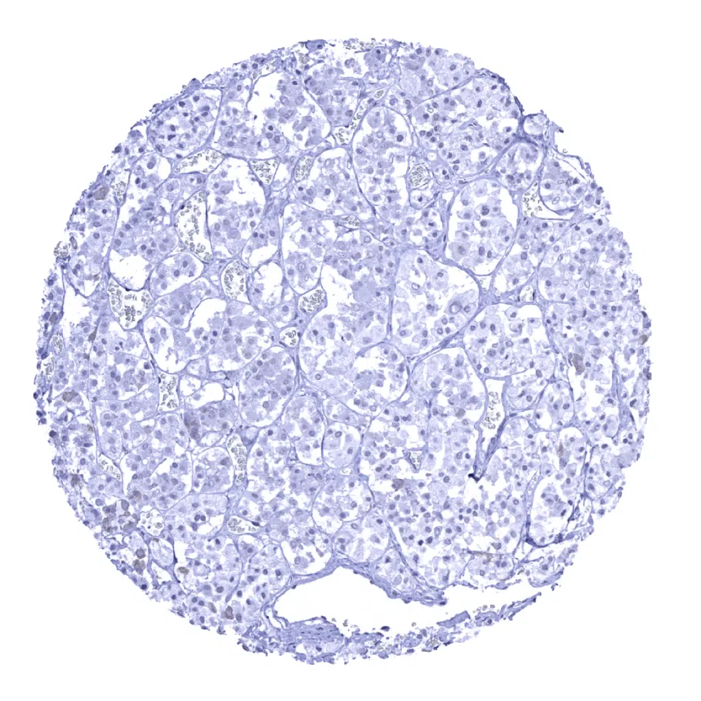
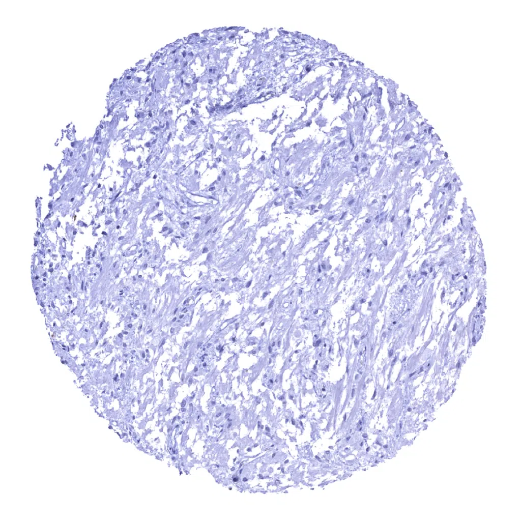
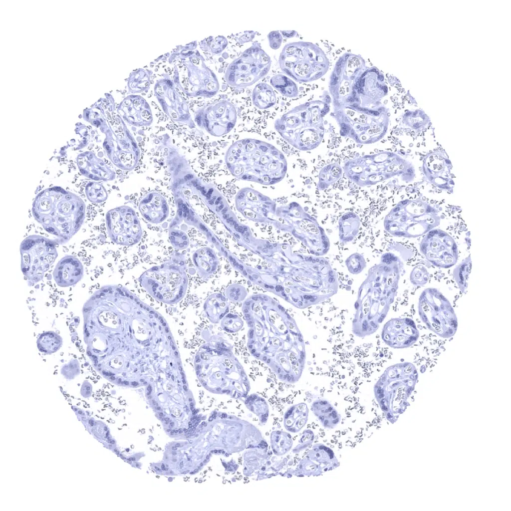
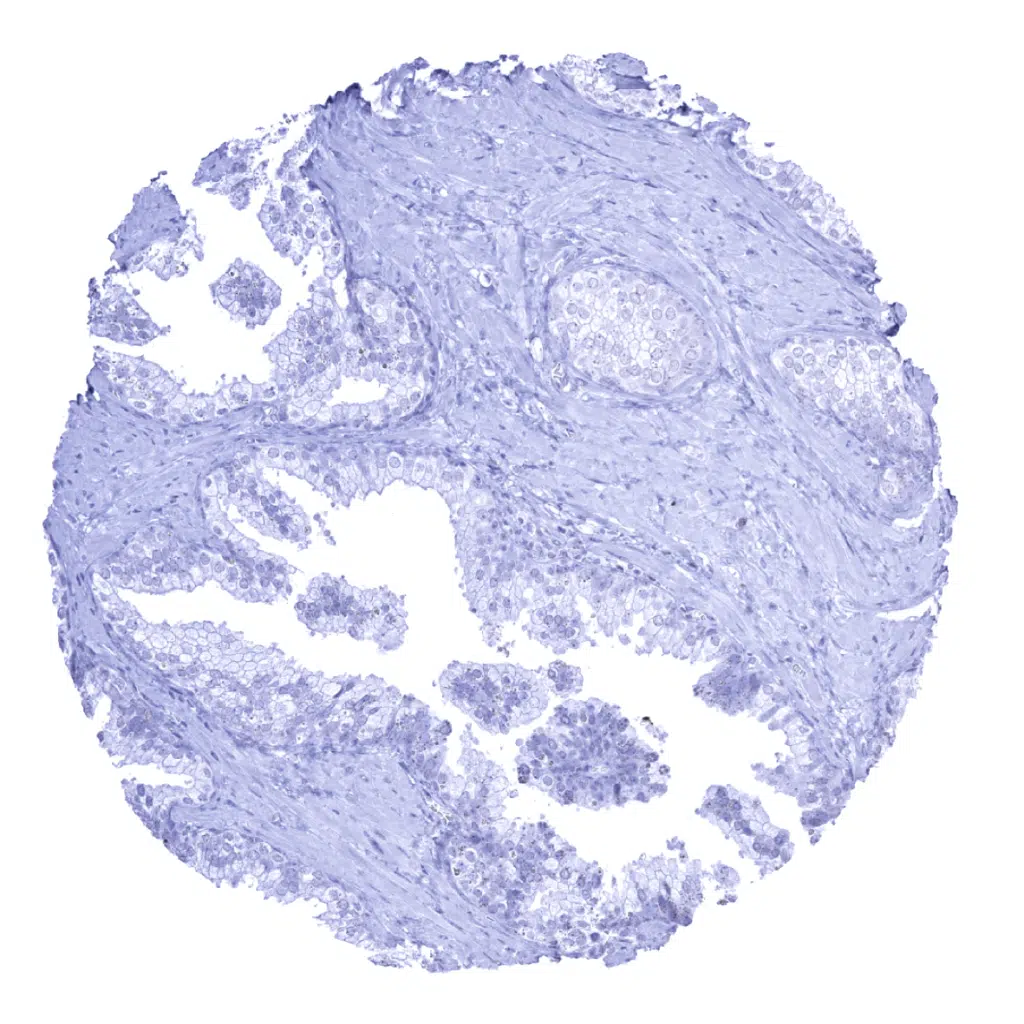
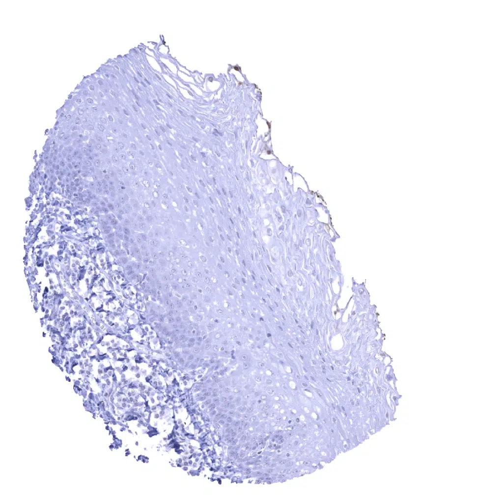
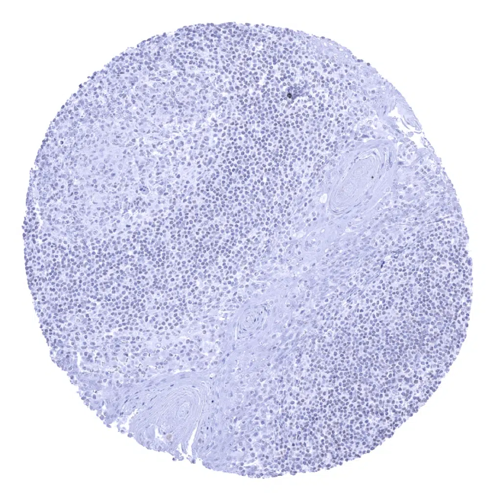
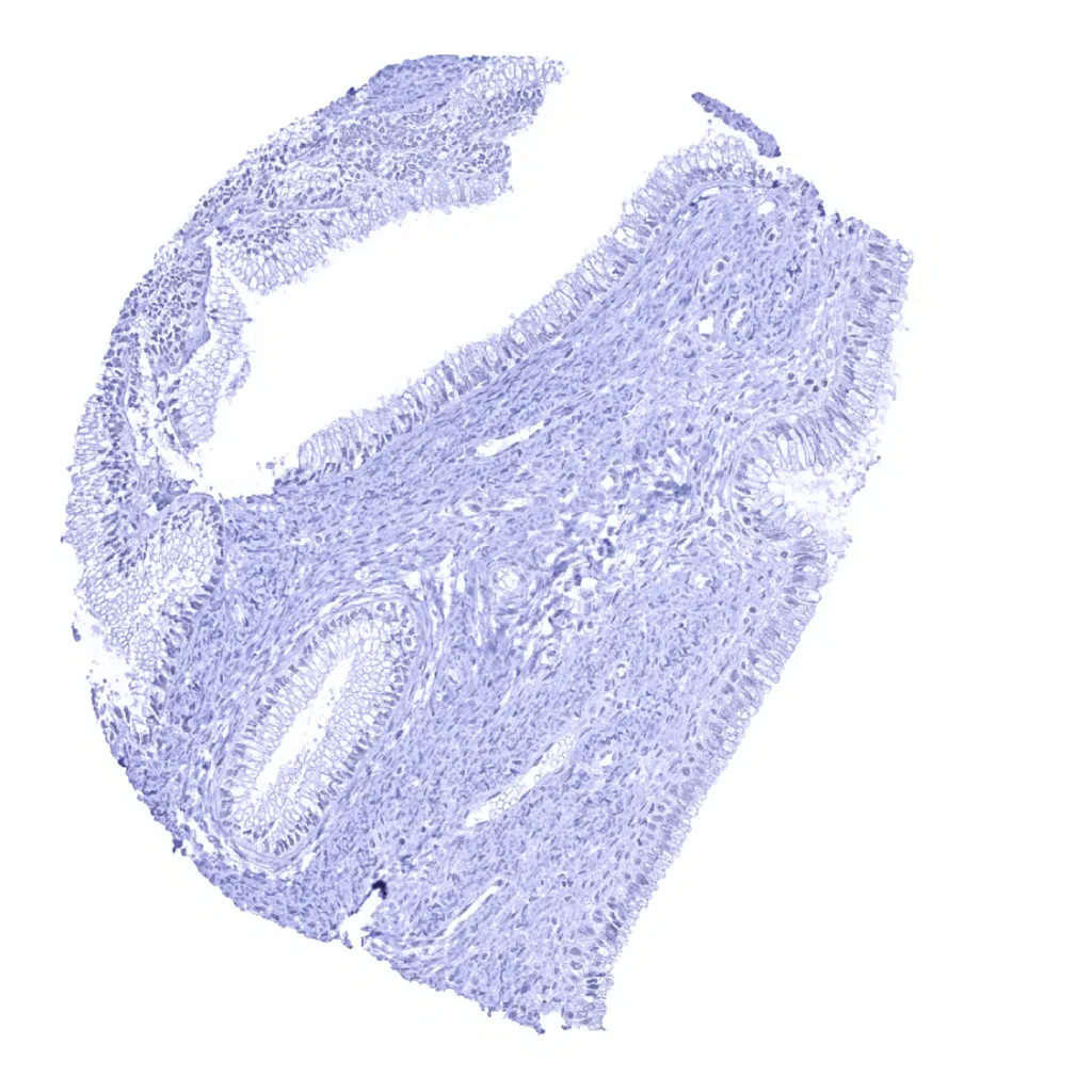
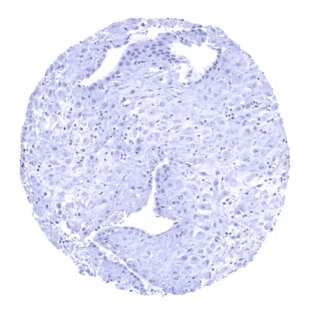
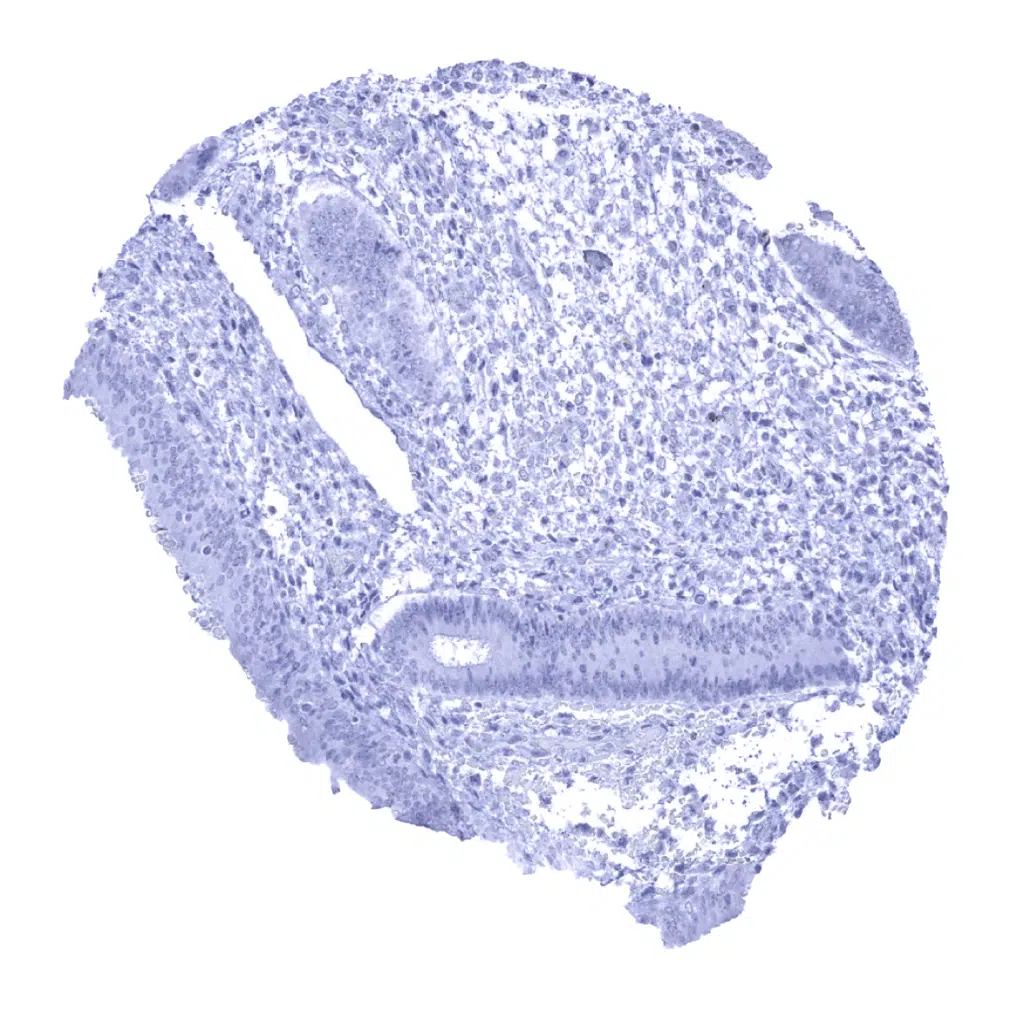
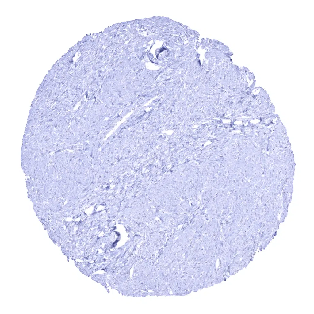
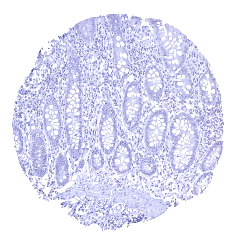
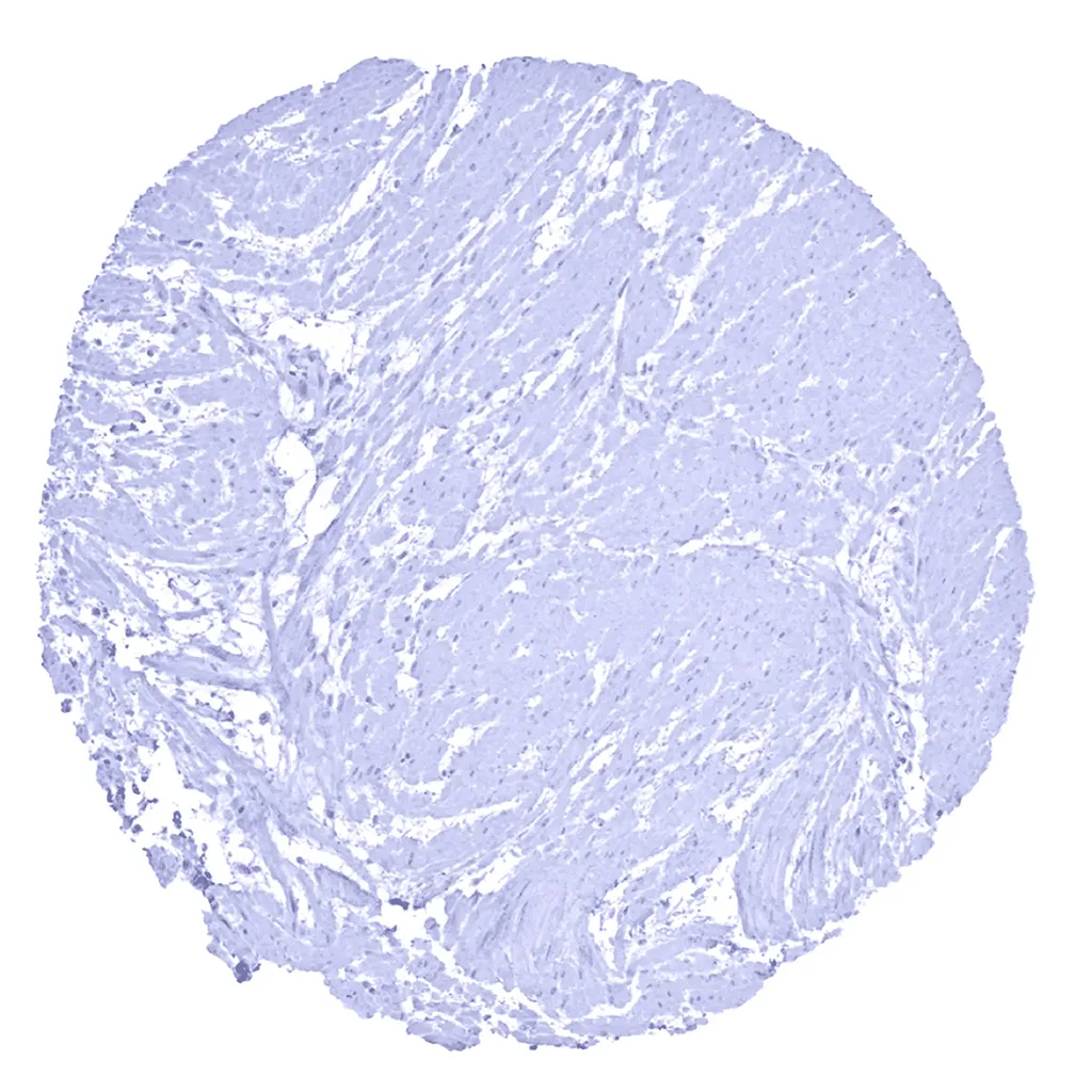
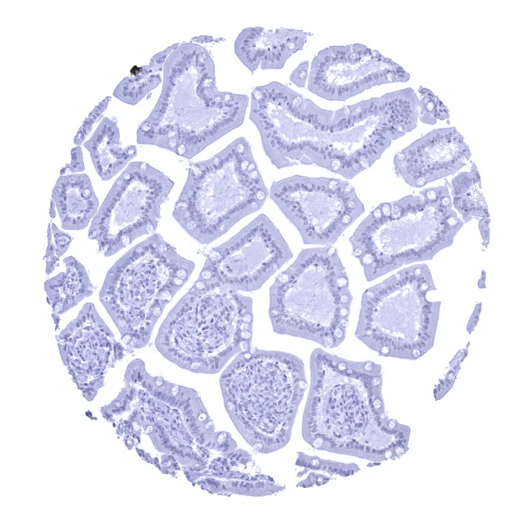
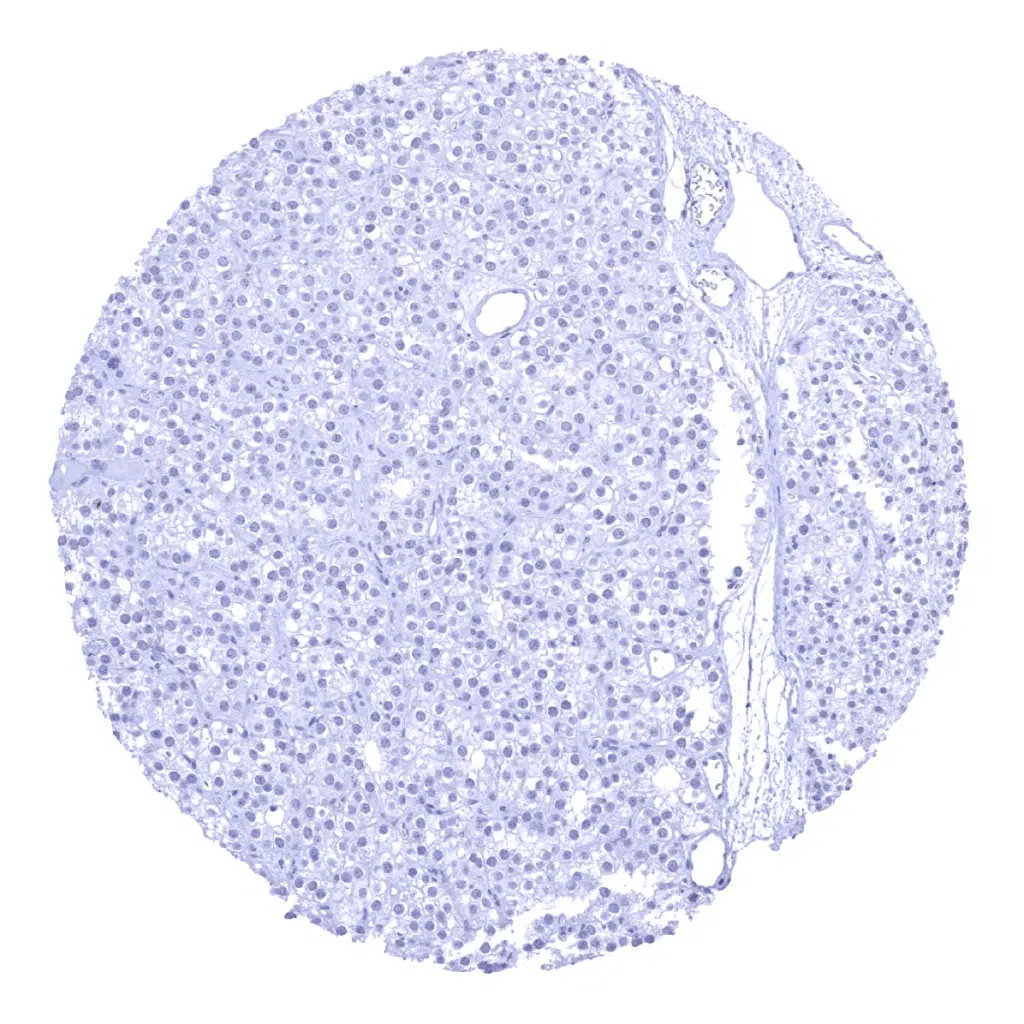
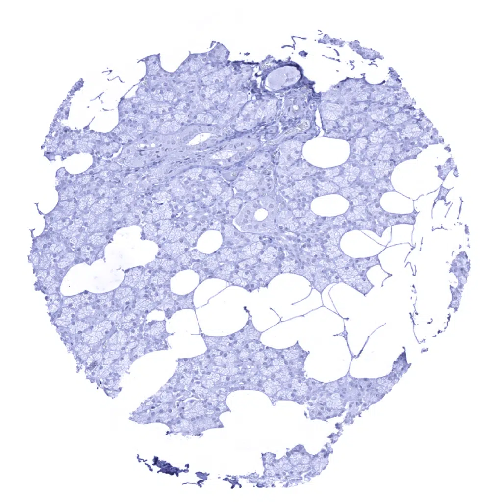
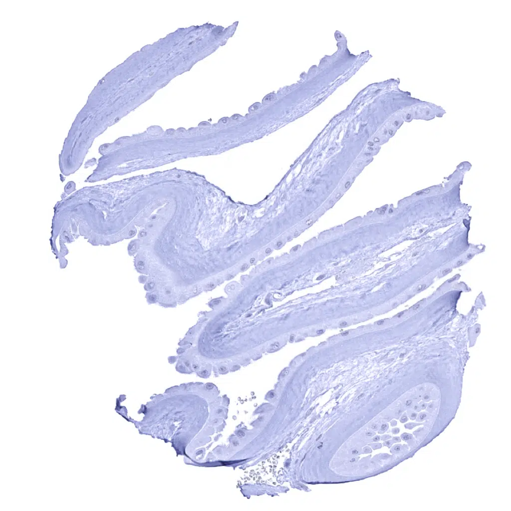
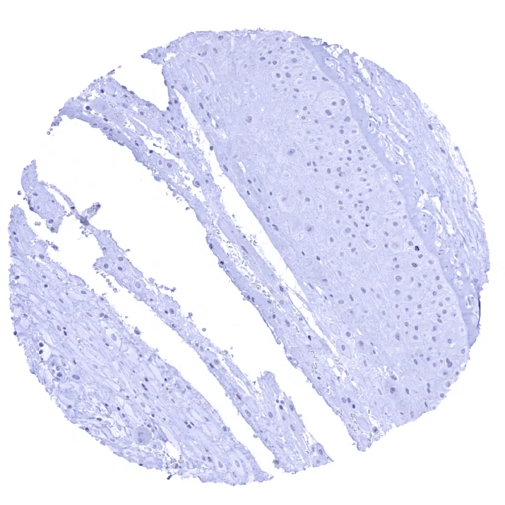
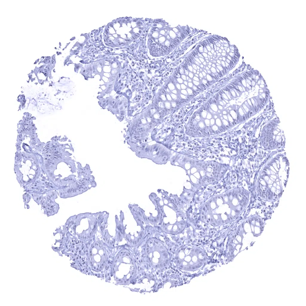
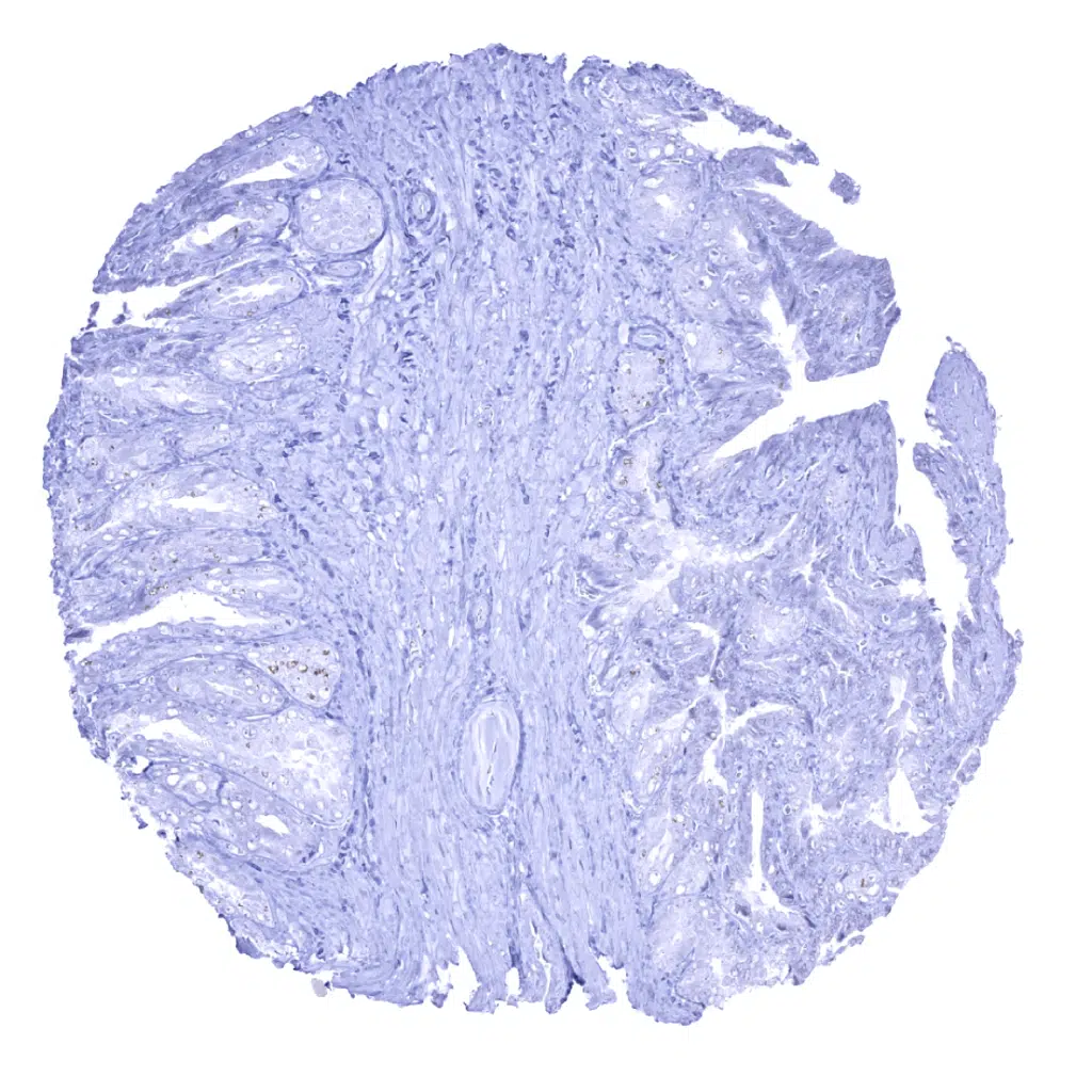
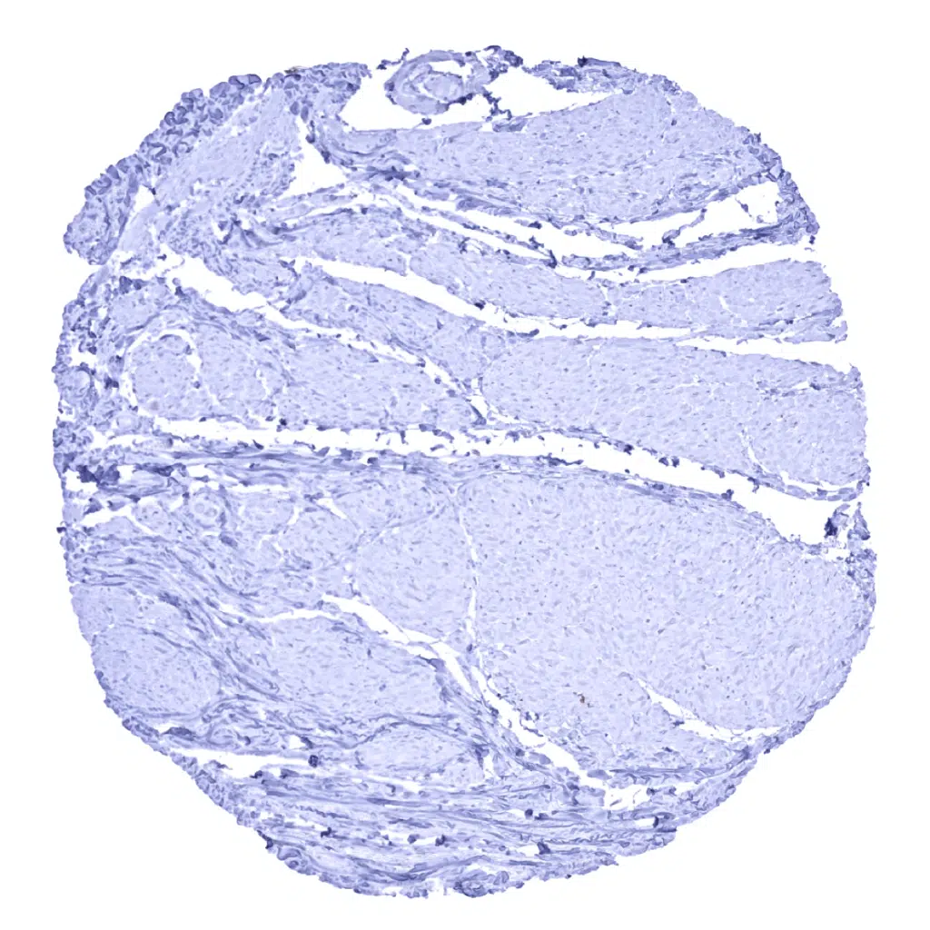
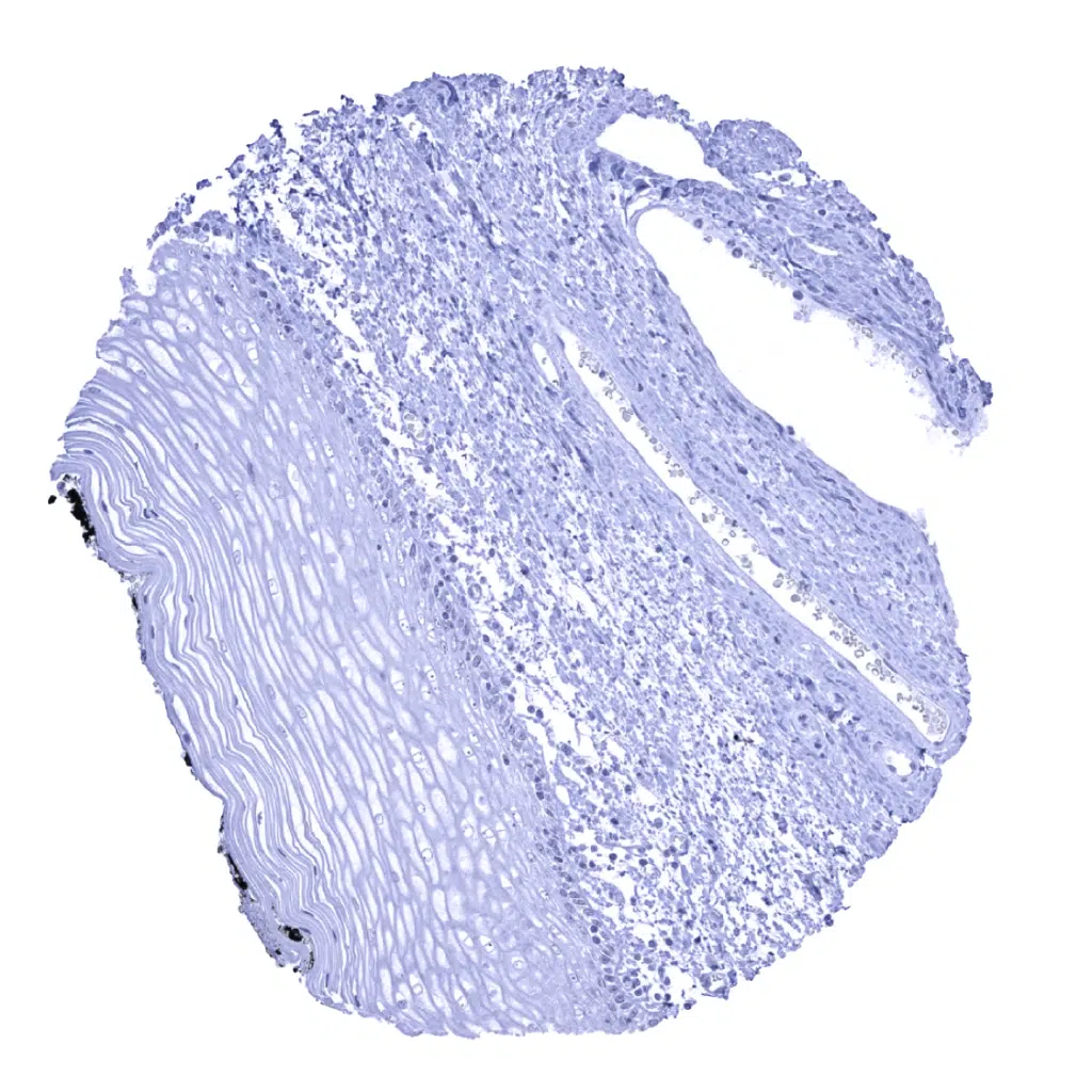
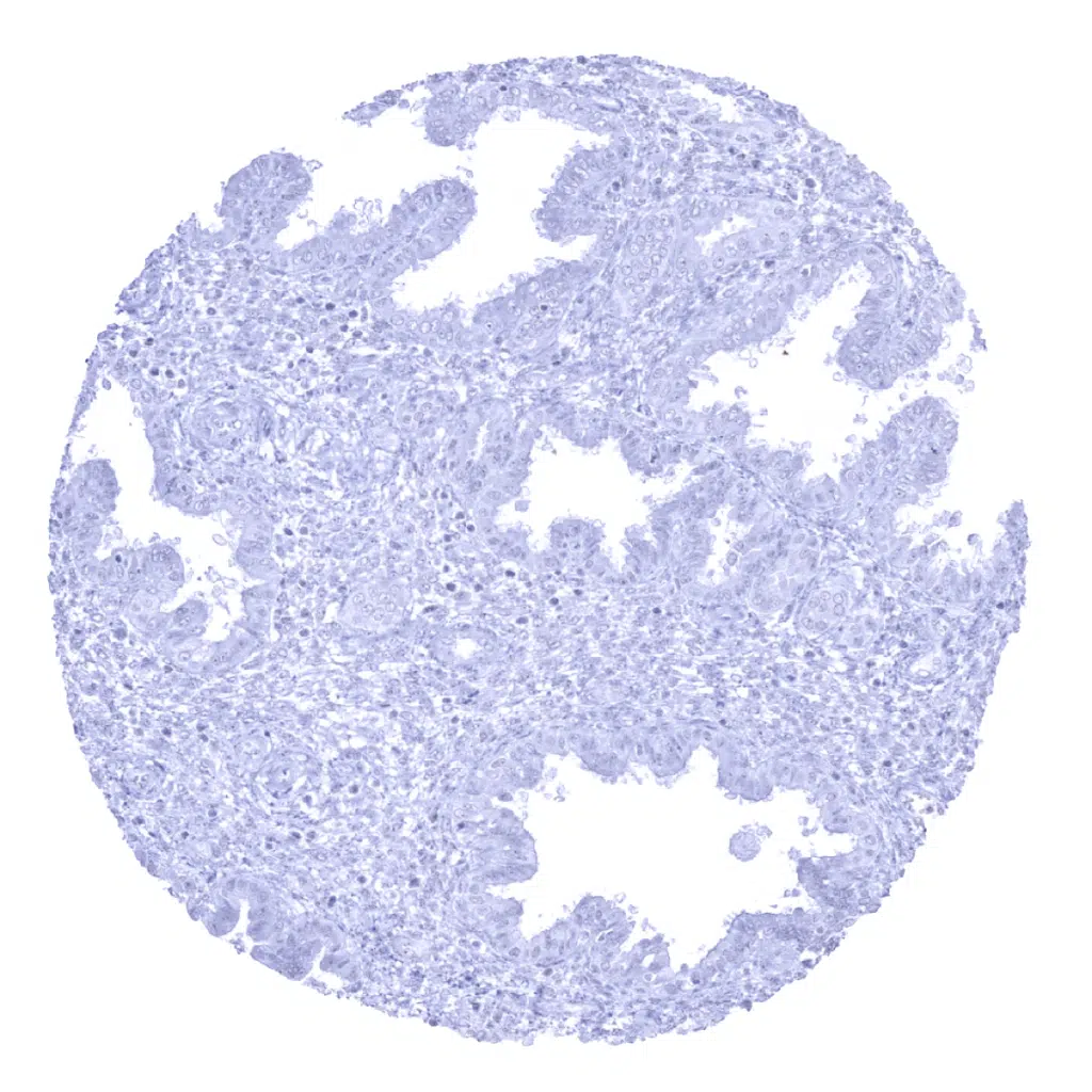
Calbindin antibody [MSVA-471M] HistoMAX


Go to Cancer Tissue Gallery Go back to product page Go to HistoMAX page


Adrenal gland
Anal canal, skin
Aorta, media
Appendix, mucosa
Appendix, muscular wall
Bone marrow
Breast
Bronchus, mucosa
Cerebellum (molecular layer, granule cell layer)
Cerebellum (molecular layer, Purkinje cell layer, granule cell layer, white matter) - Strong Calbindin staining of Purkinje cells and of associated axonal fibres in the cerebrum.
Cerebellum (white matter) - Strong Calbindin staining of axonal fibres in the cerebrum.
Cerebrum, grey matter - A weak to moderate cytoplasmic Calbindin immunostaining is seen in some neuronal cells.
Cerebrum, white matter
Colon descendens, mucosa
Colon descendens, muscular wall
Duodenum, Brunner gland
Duodenum, mucosa
Epididymis
Esophagus, squamous epithelium
Fallopian tube, mucosa
Fat
Gallbladder, epithelium
Ileum, mucosa
Kidney, cortex - Strong cytoplasmic Calbindin immunostaining occurs in a fraction of distal tubuli of the kidney.
Kidney, medulla - Collecting ducts of the kidney are Calbindin negative.
Kidney, pelvis, urothelium
Liver
Lung
Lymph node
Ovary, stroma
Pancreas
Parathyroid
Parotid gland
Pituitary gland, anterior lobe
Pituitary gland, posterior lobe
Placenta (amnion)
Placenta (chorion)
Placenta, early
Placenta, mature
Prostate
Rectum, mucosa
Seminal vesicle
Sinus paranasales
Skeletal muscle
Spleen
Stomach, antrum
Stomach, corpus
Testis
Thymus
Thyroid gland
Tonsil, surface epithelium
Tonsil
Urinary bladder, muscular wall
Uterus, ectocervix
Uterus, endocervix
Uterus, endometrium (pregnancy)
Uterus, endometrium (proliferation)
Uterus, endometrium (secretion)
Uterus, myometrium
 

| TOP |