GeneTex
  • Country / Location Selection

United States (US)

Normal Tissue Gallery- GTX04443

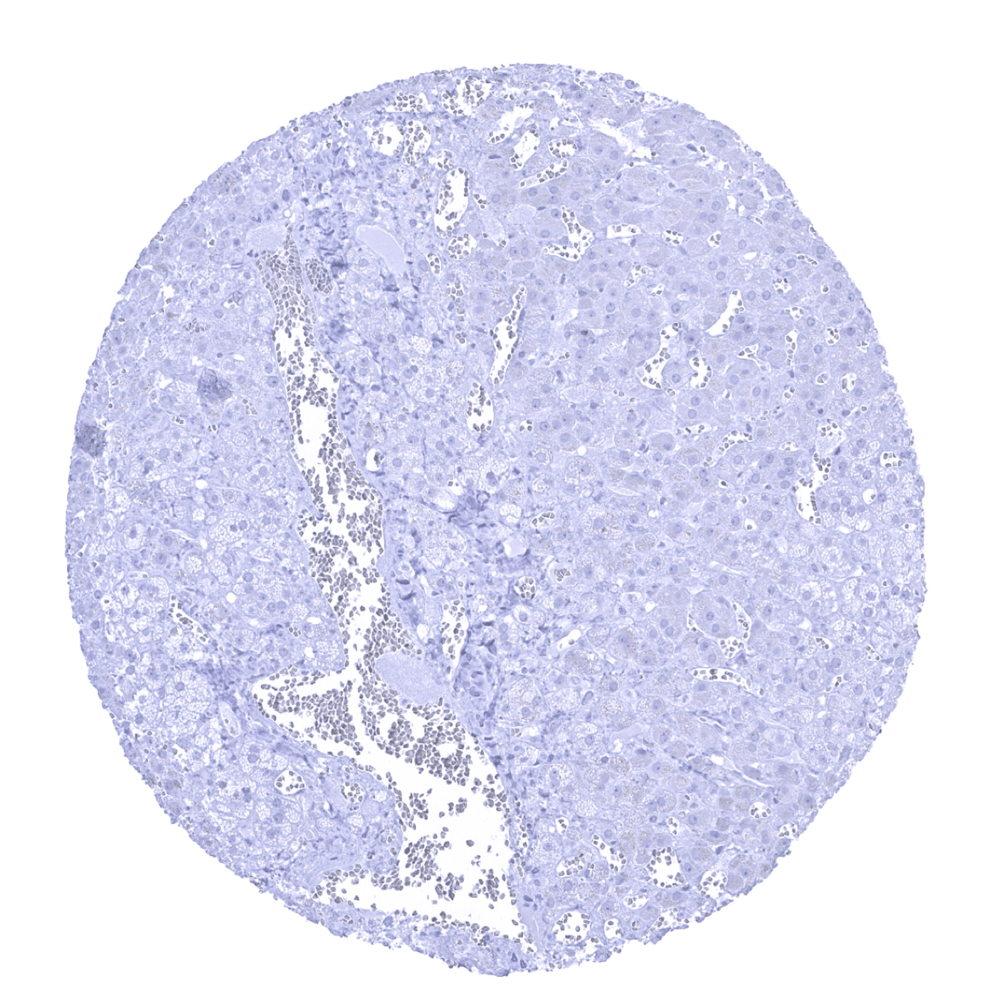
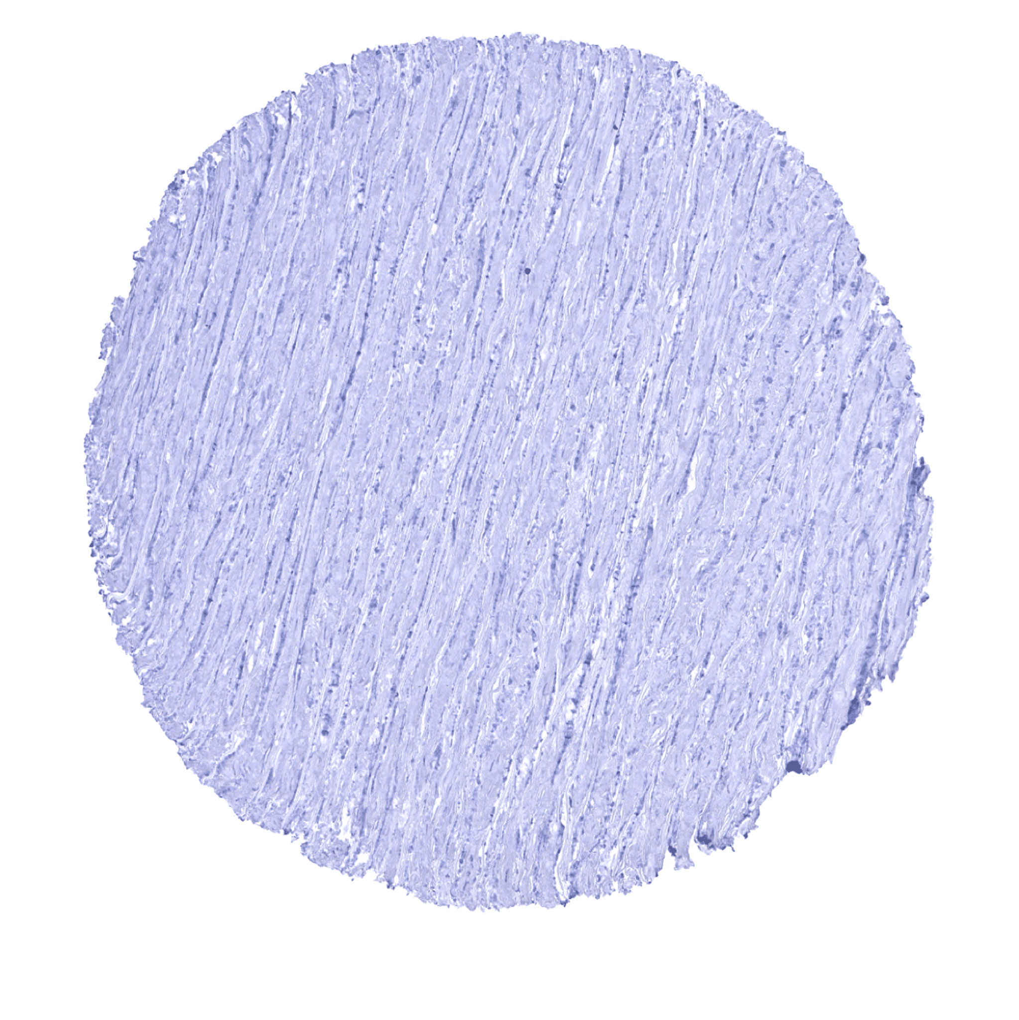
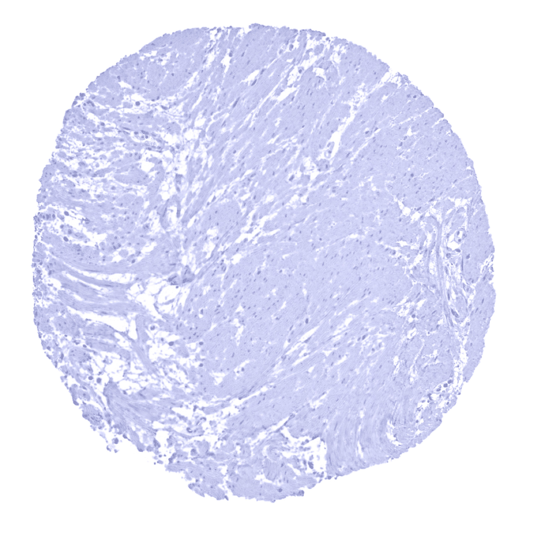
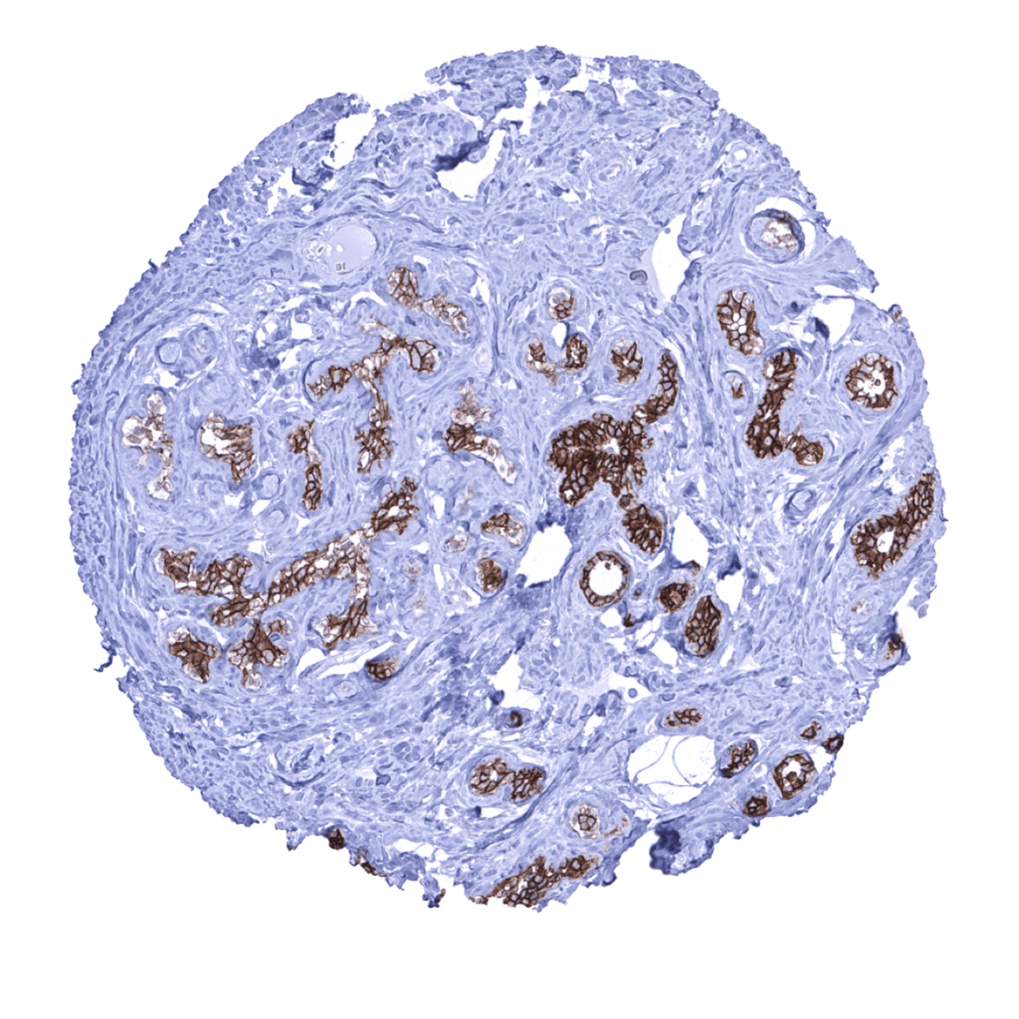
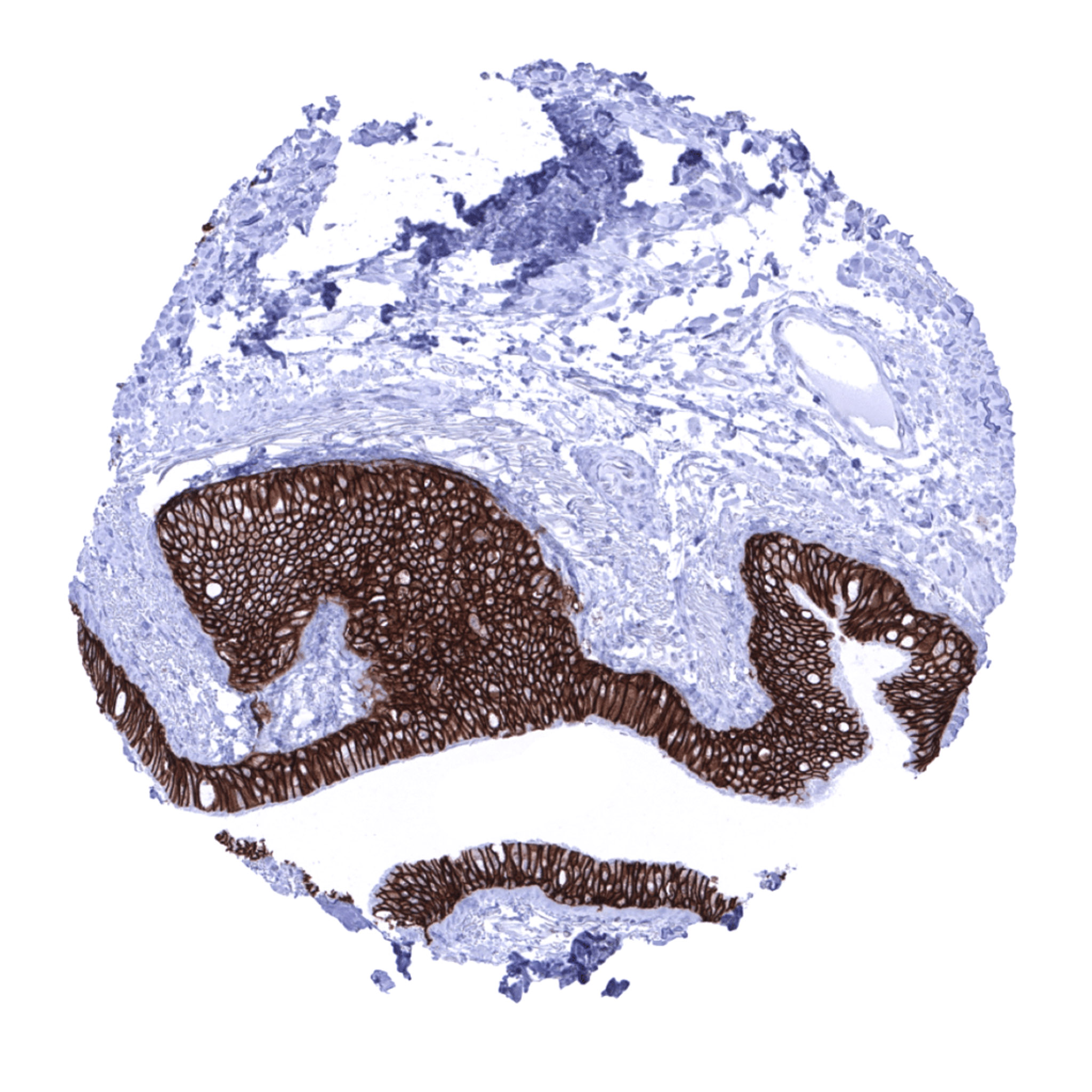
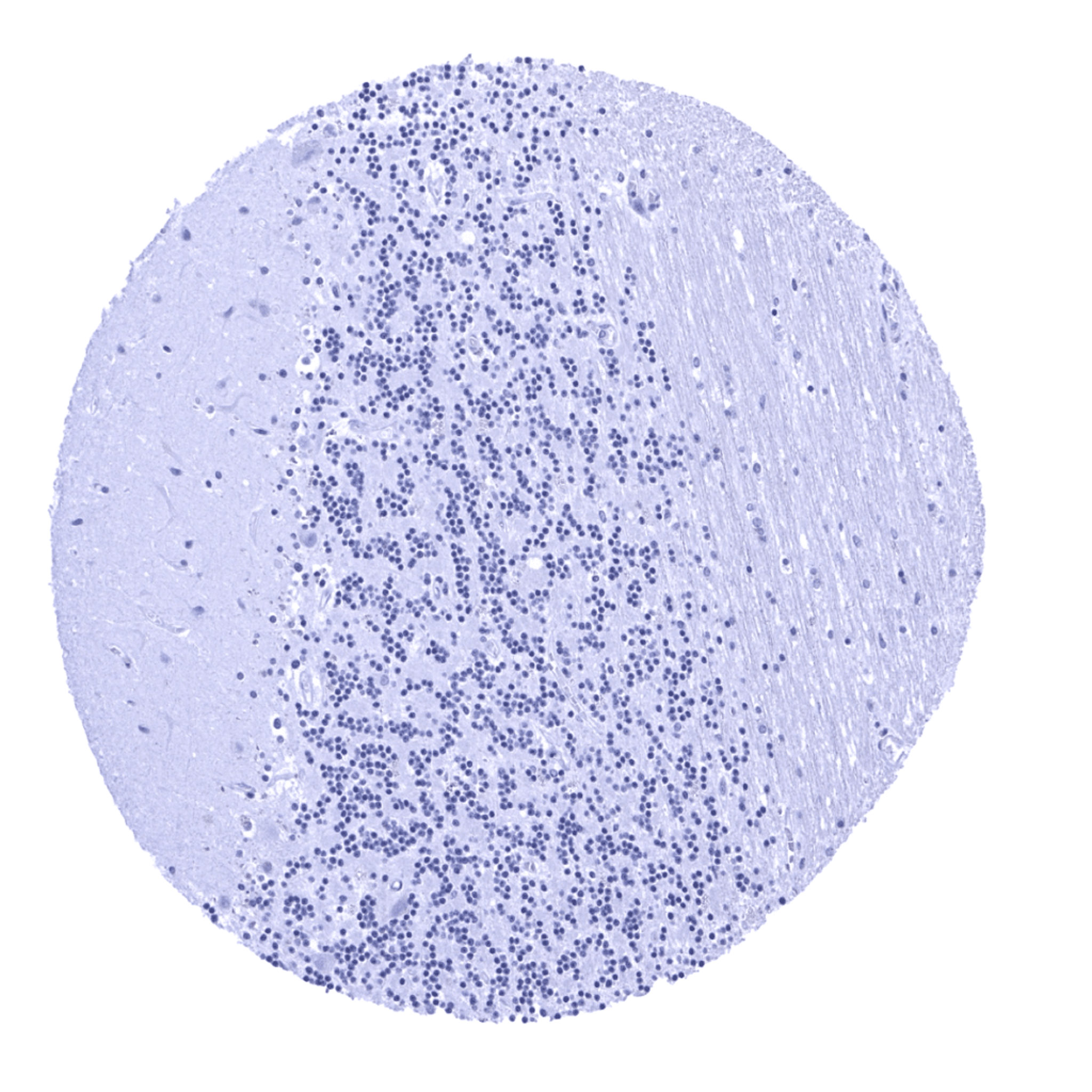
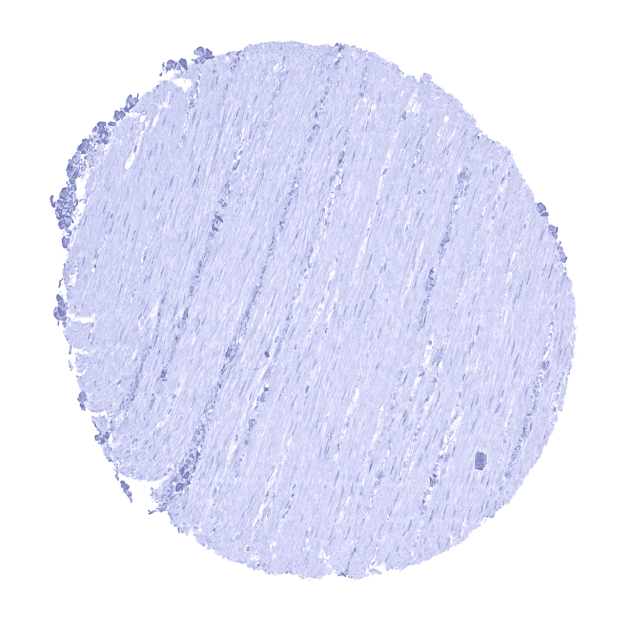
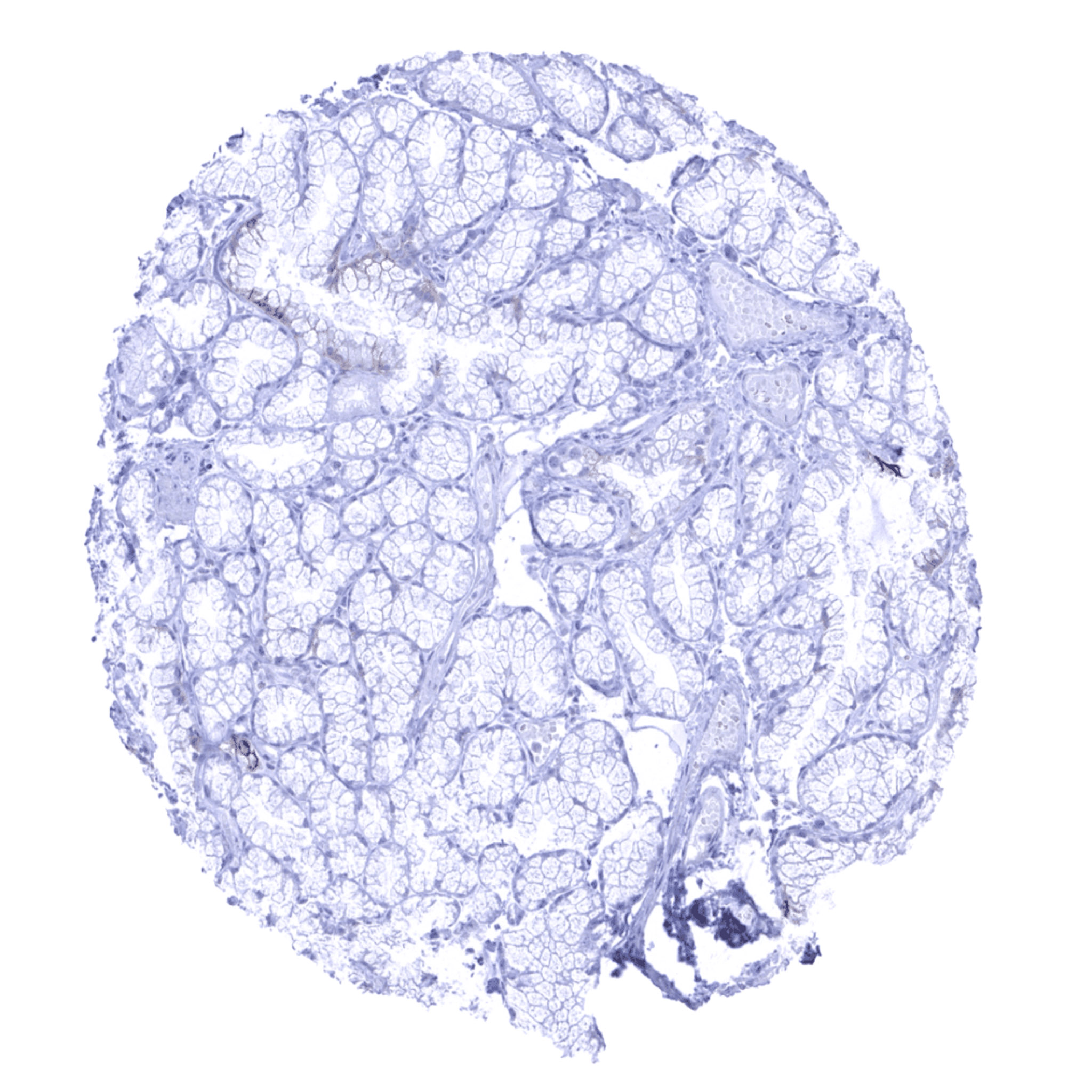
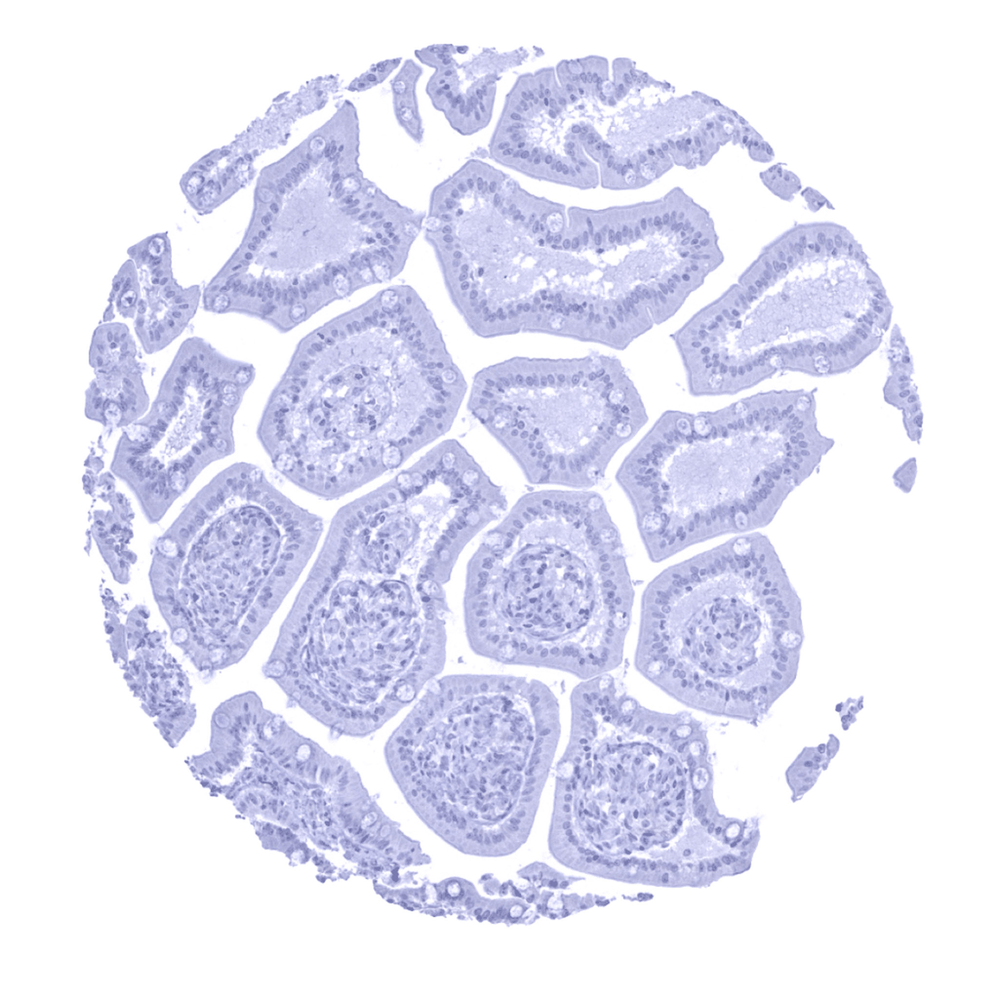
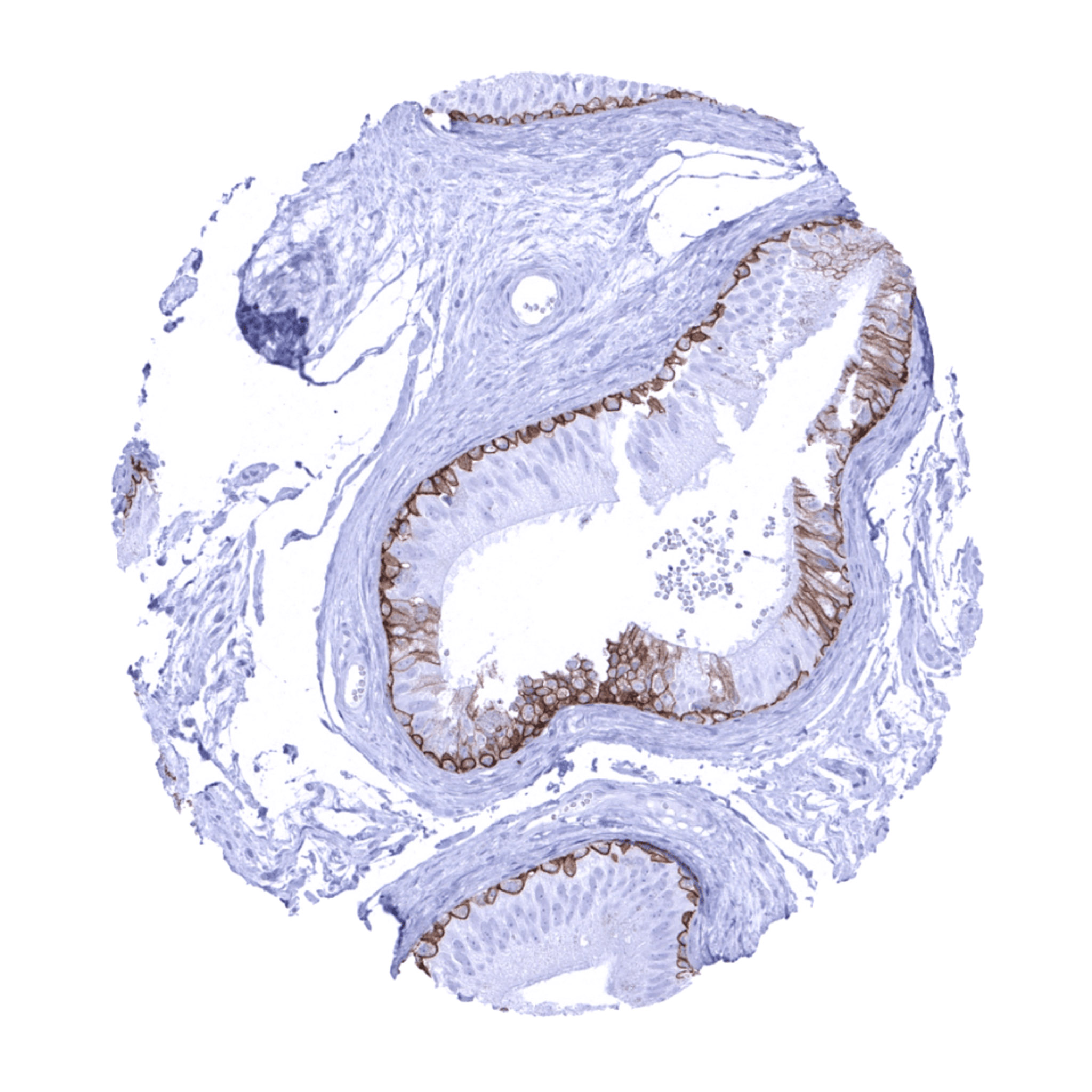
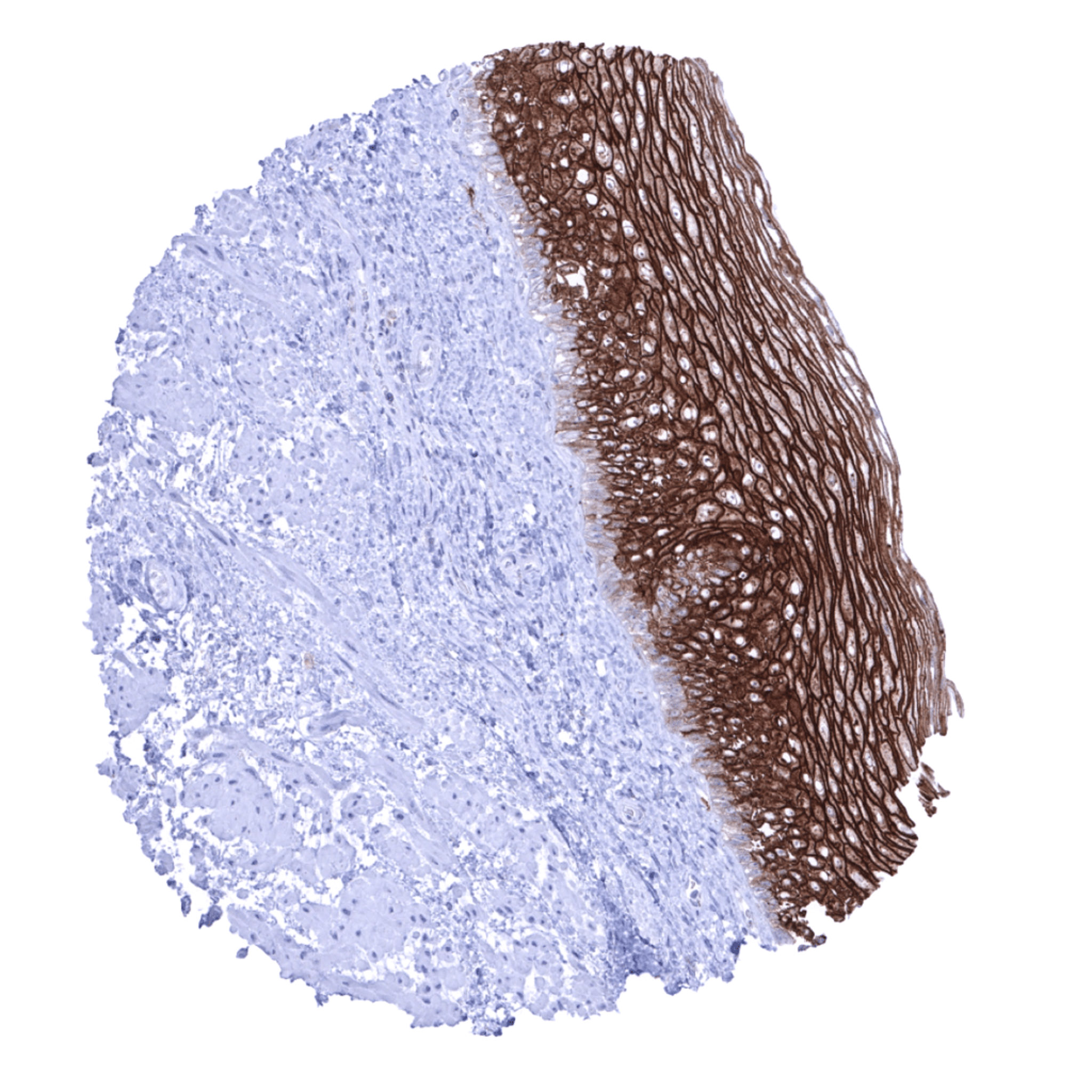
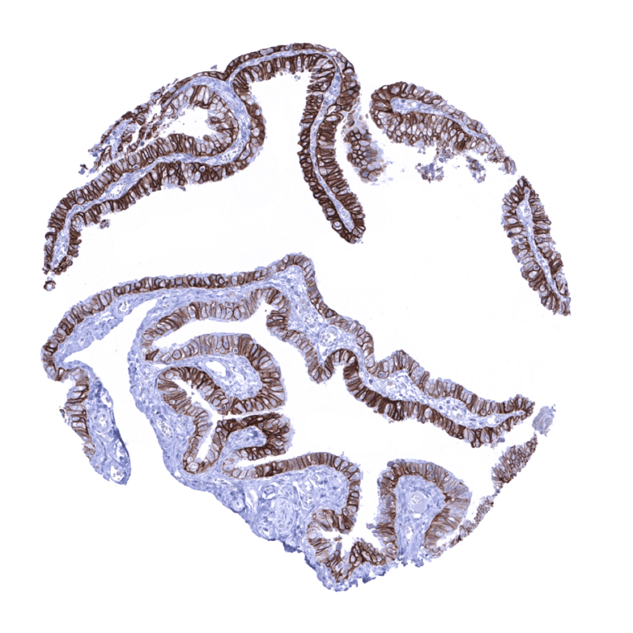
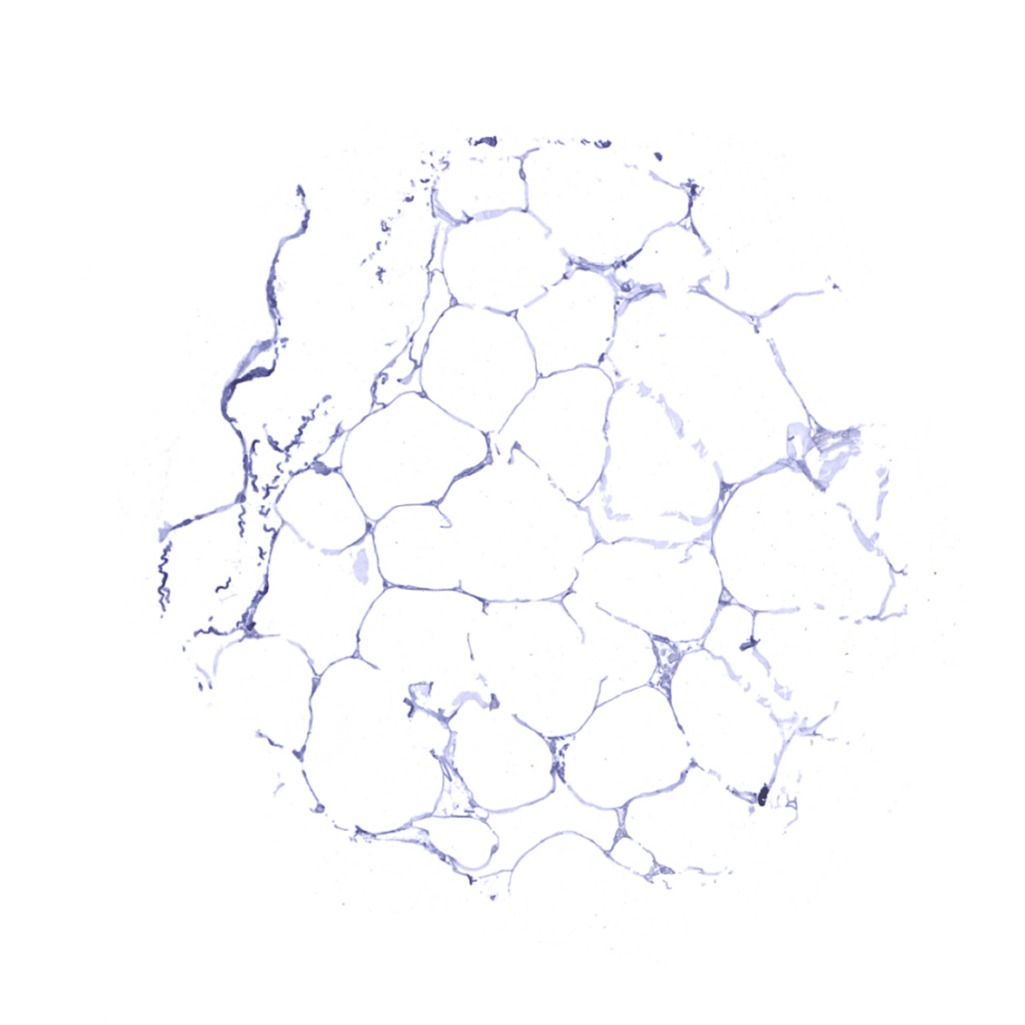
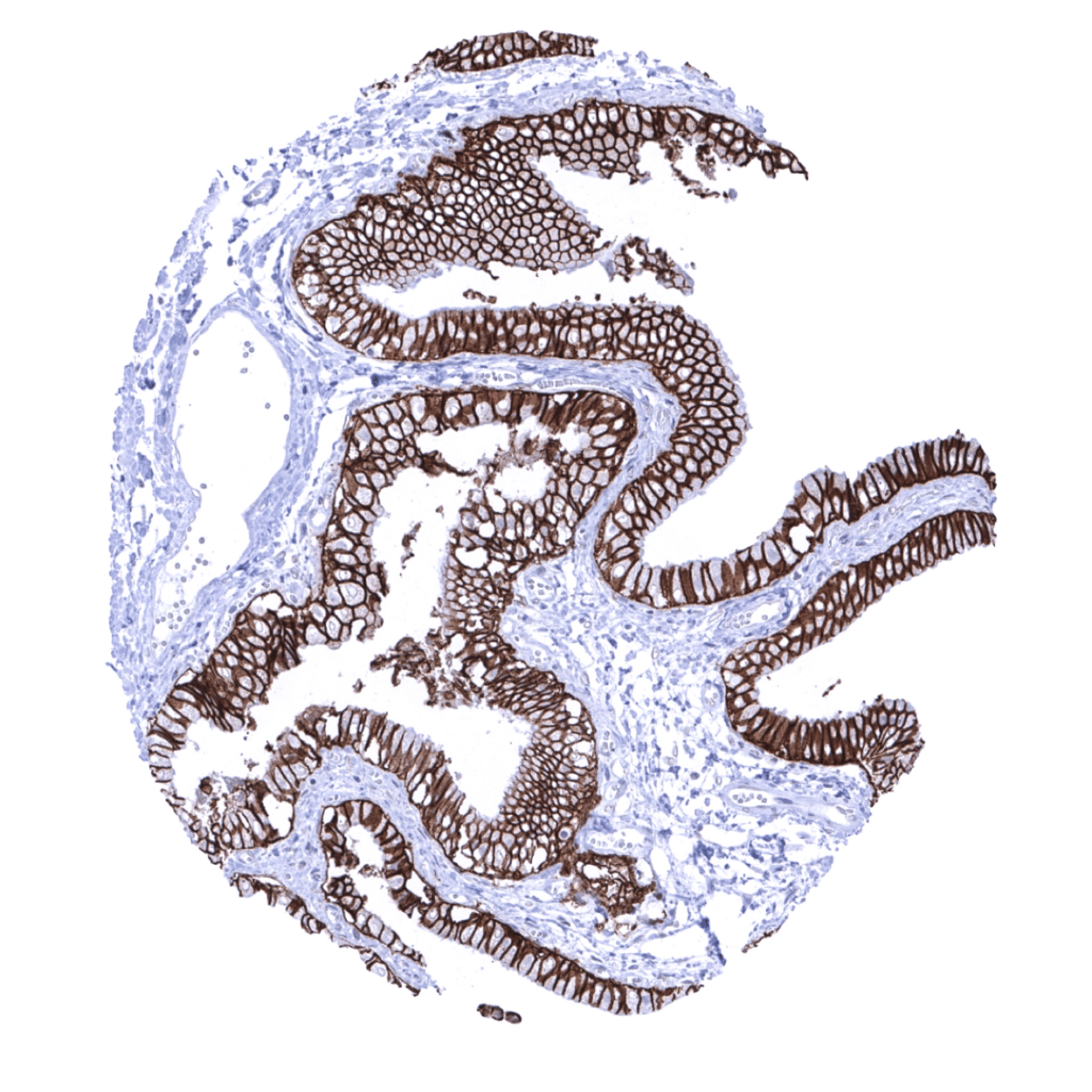
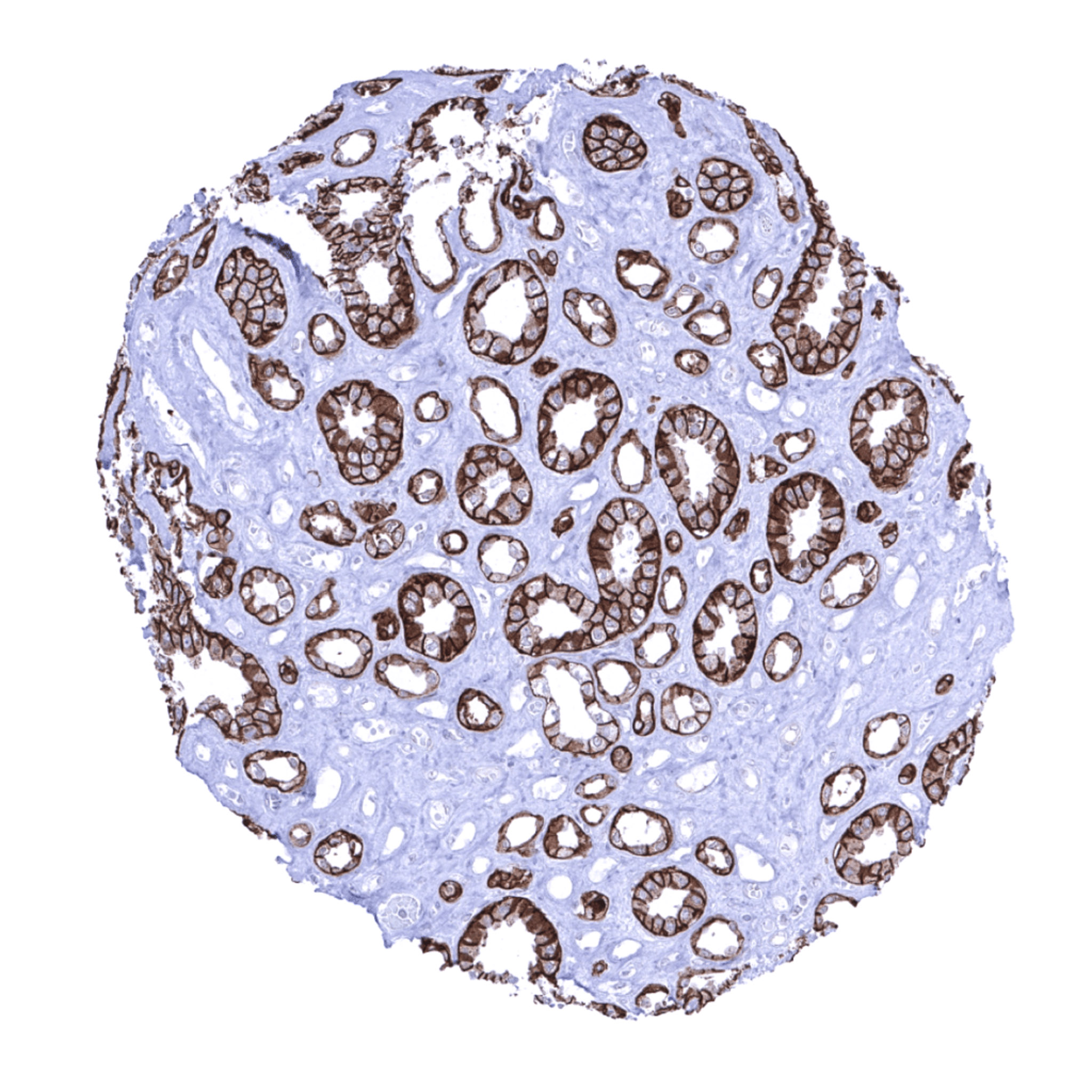
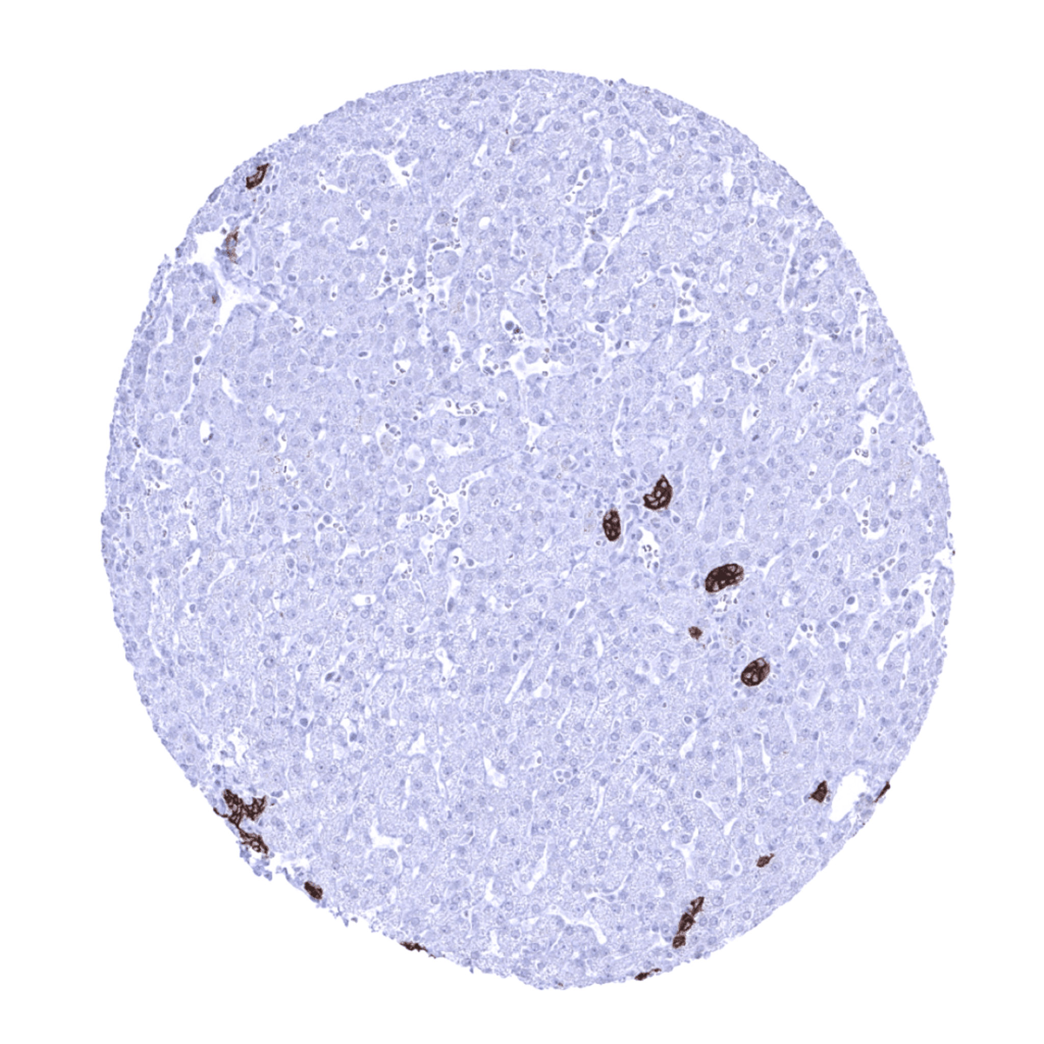
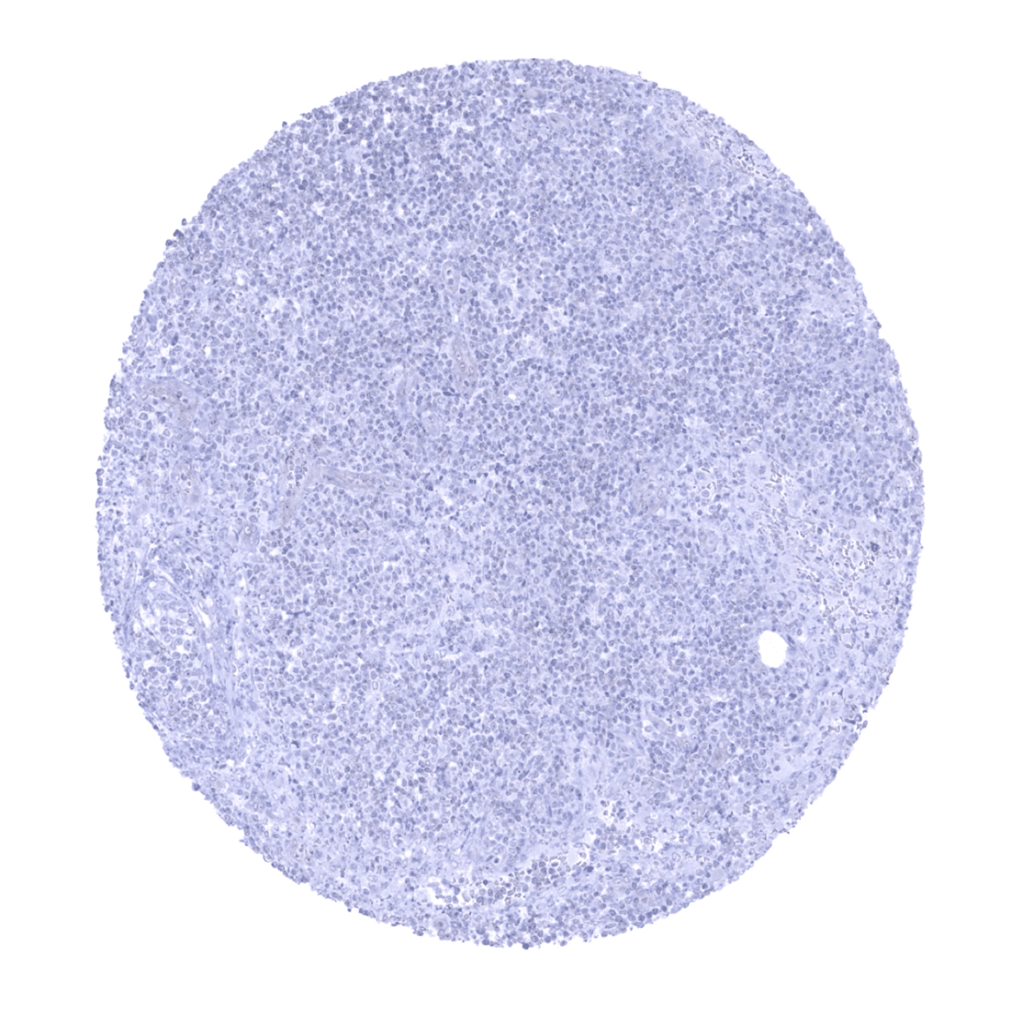
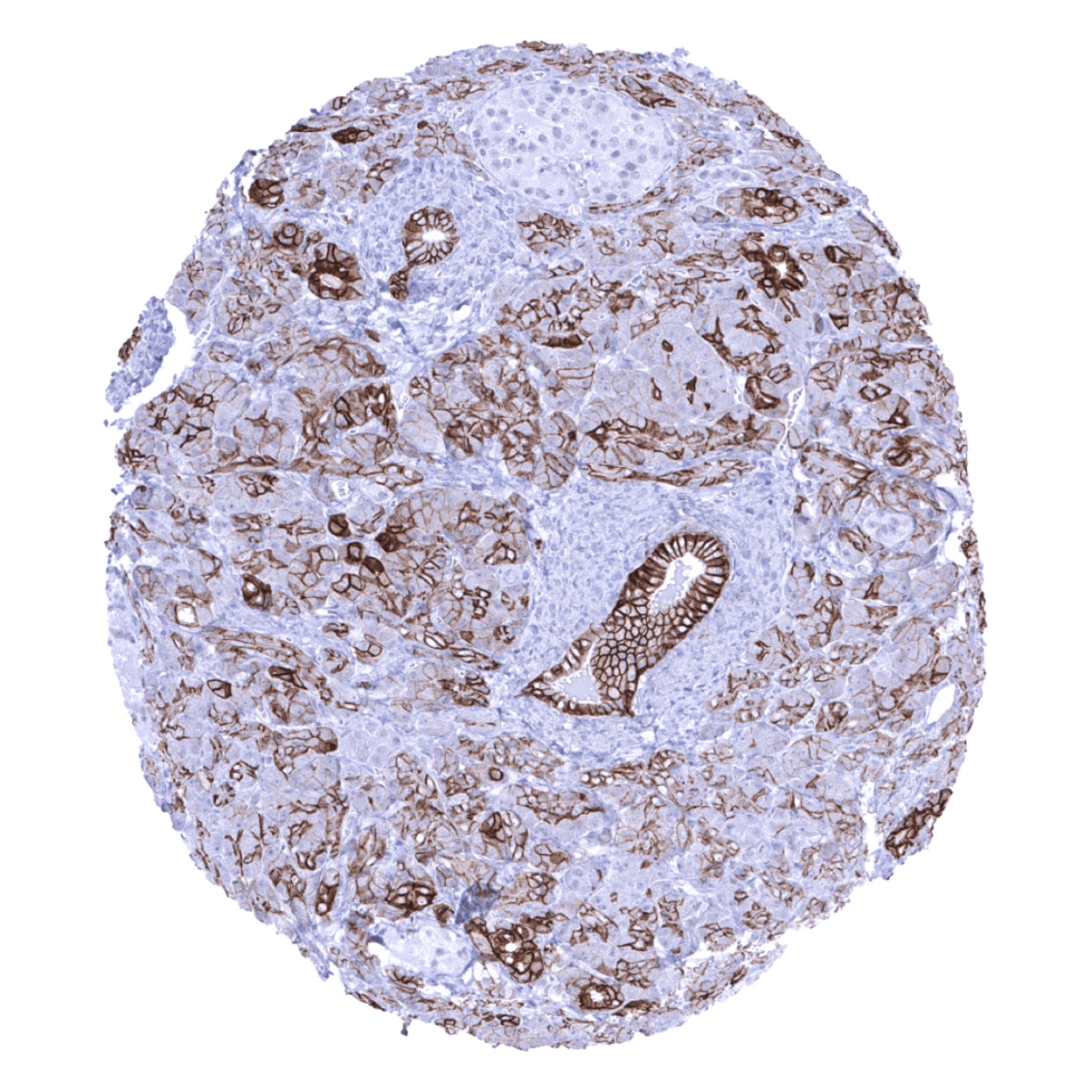
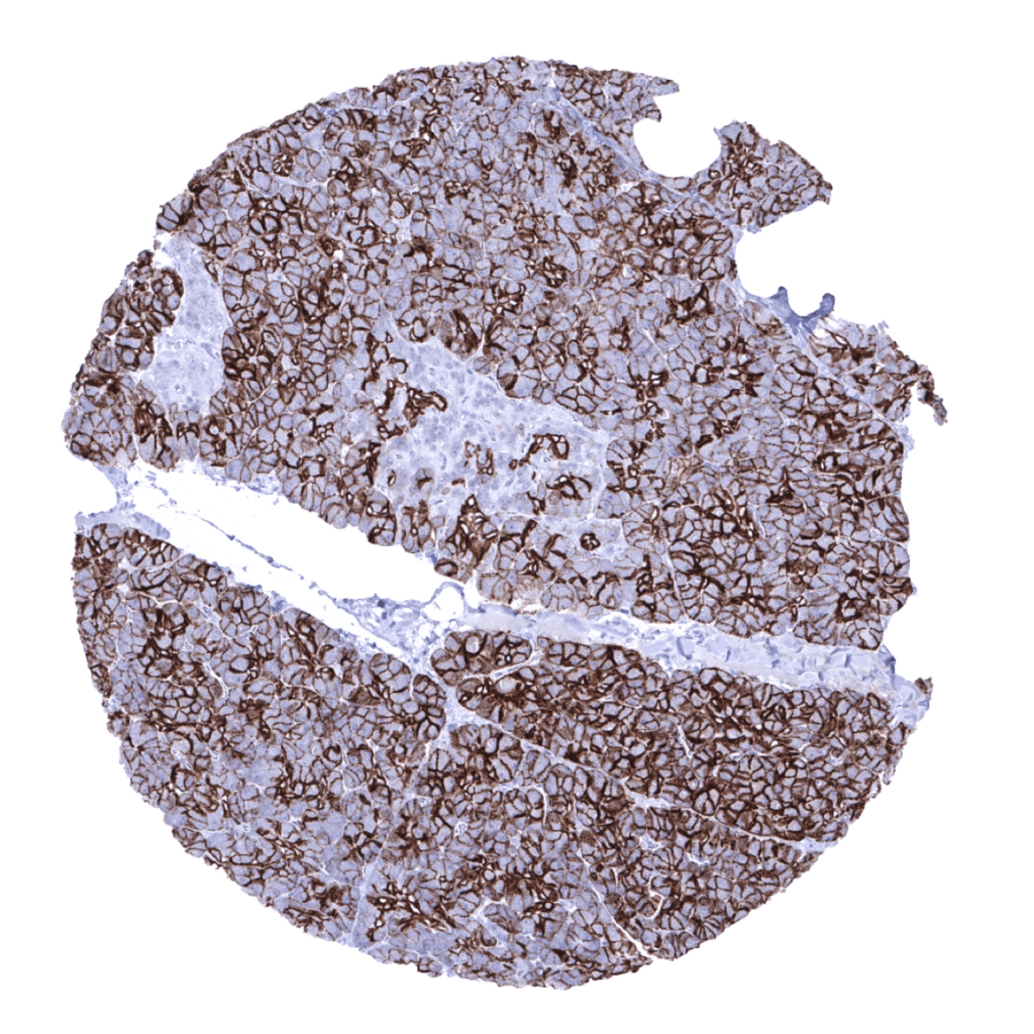
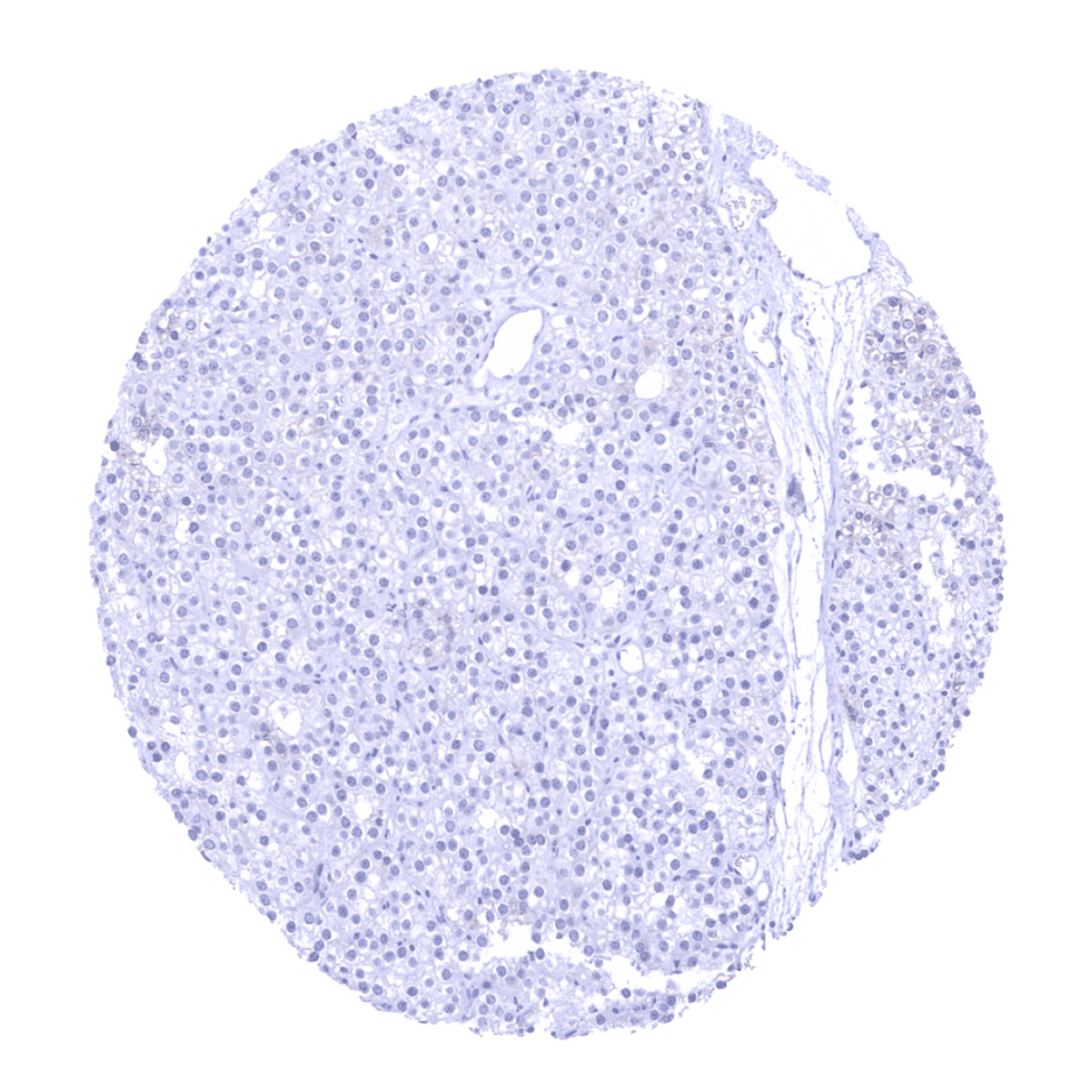
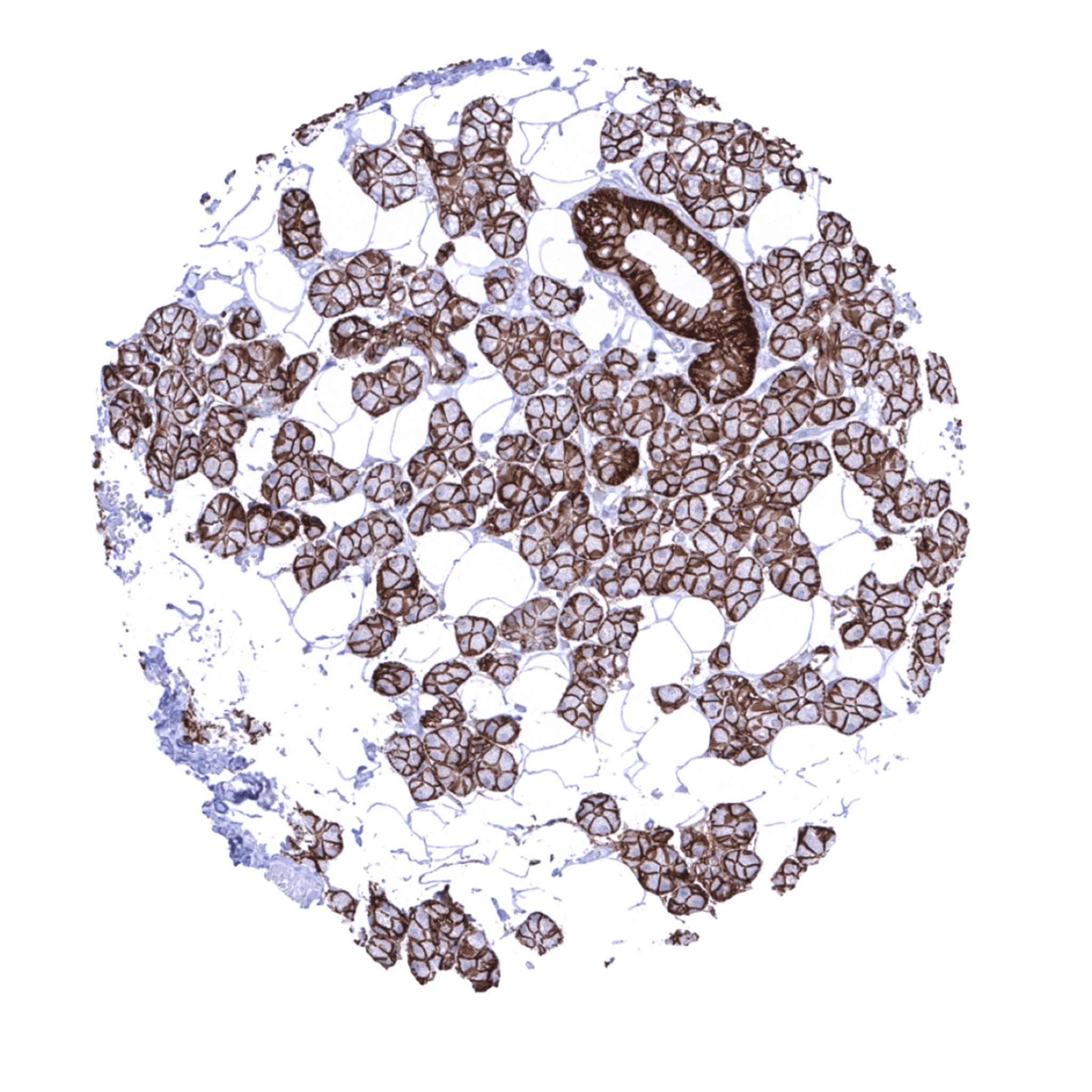
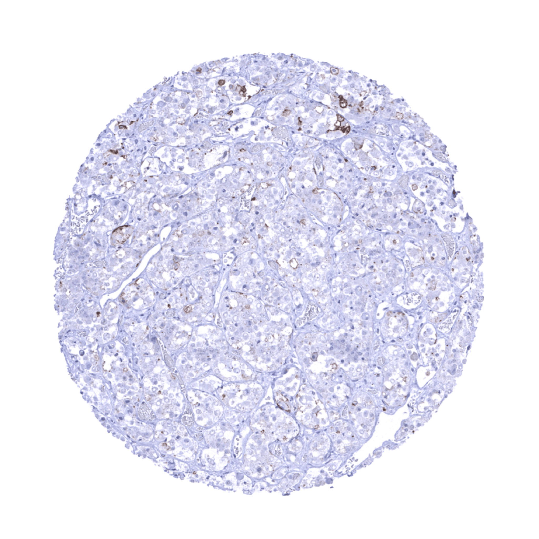
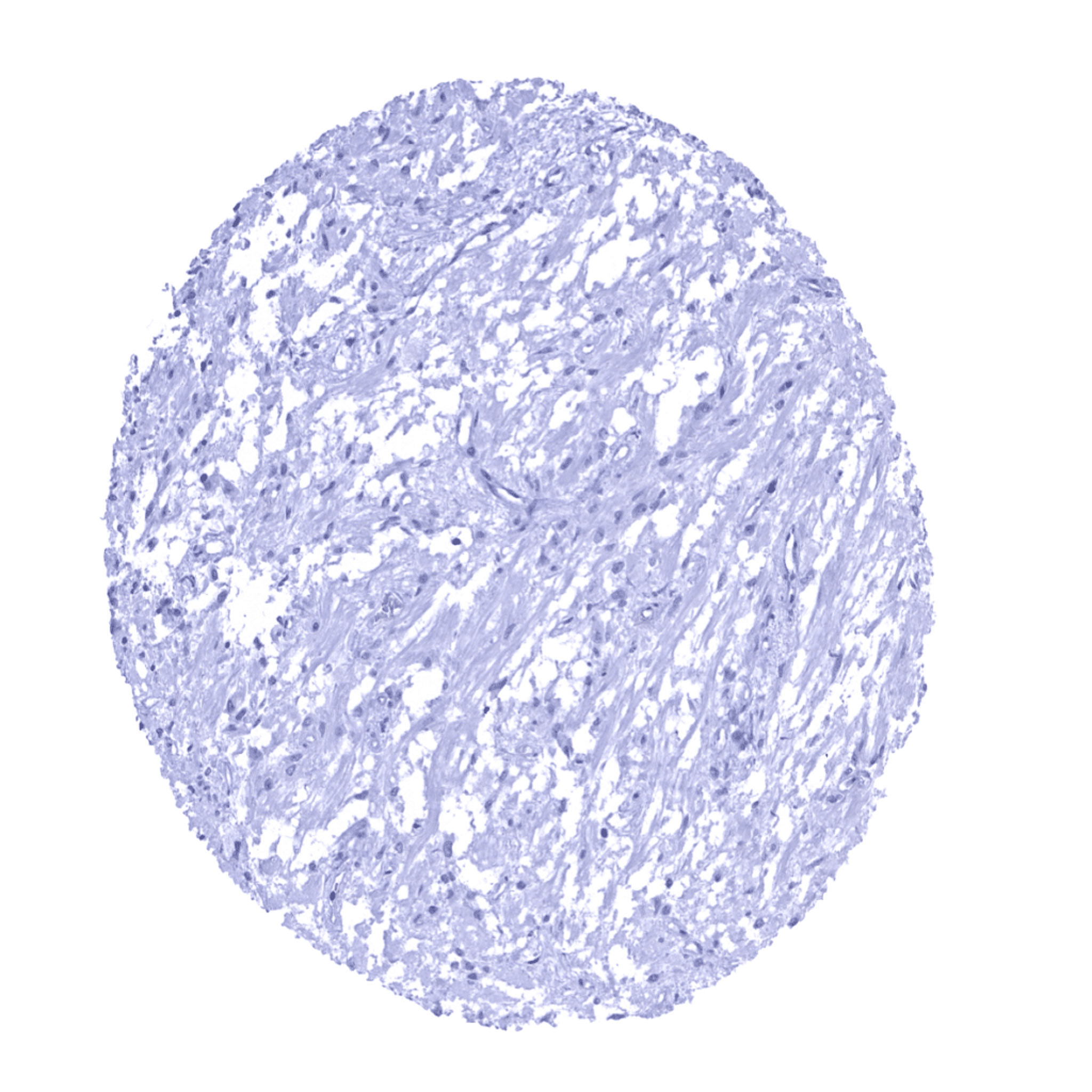
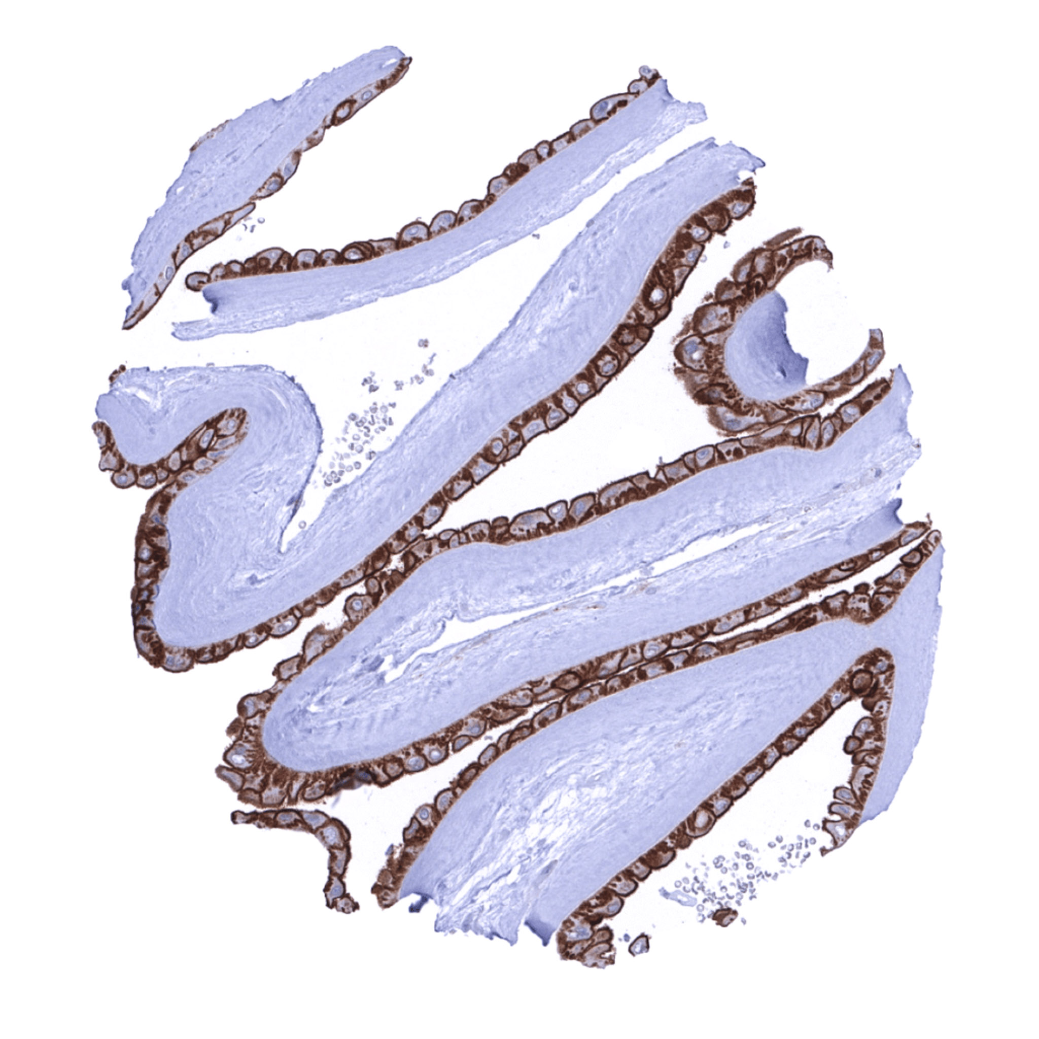
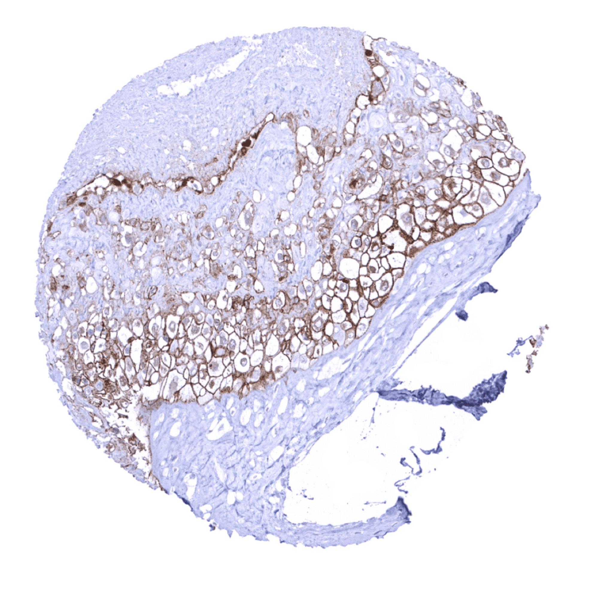
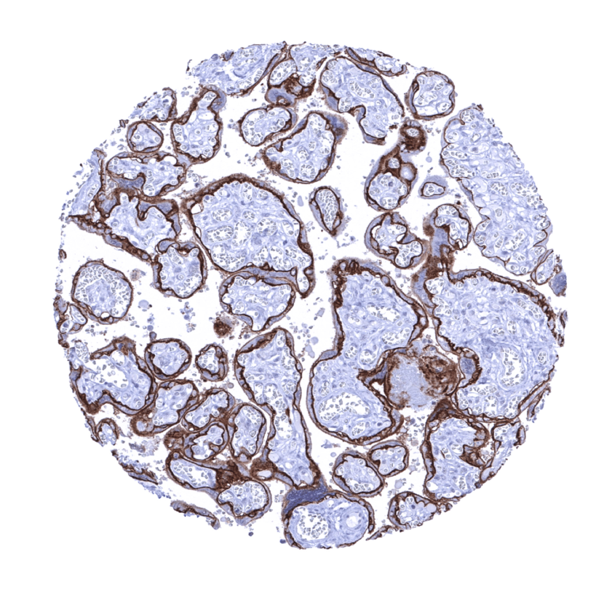
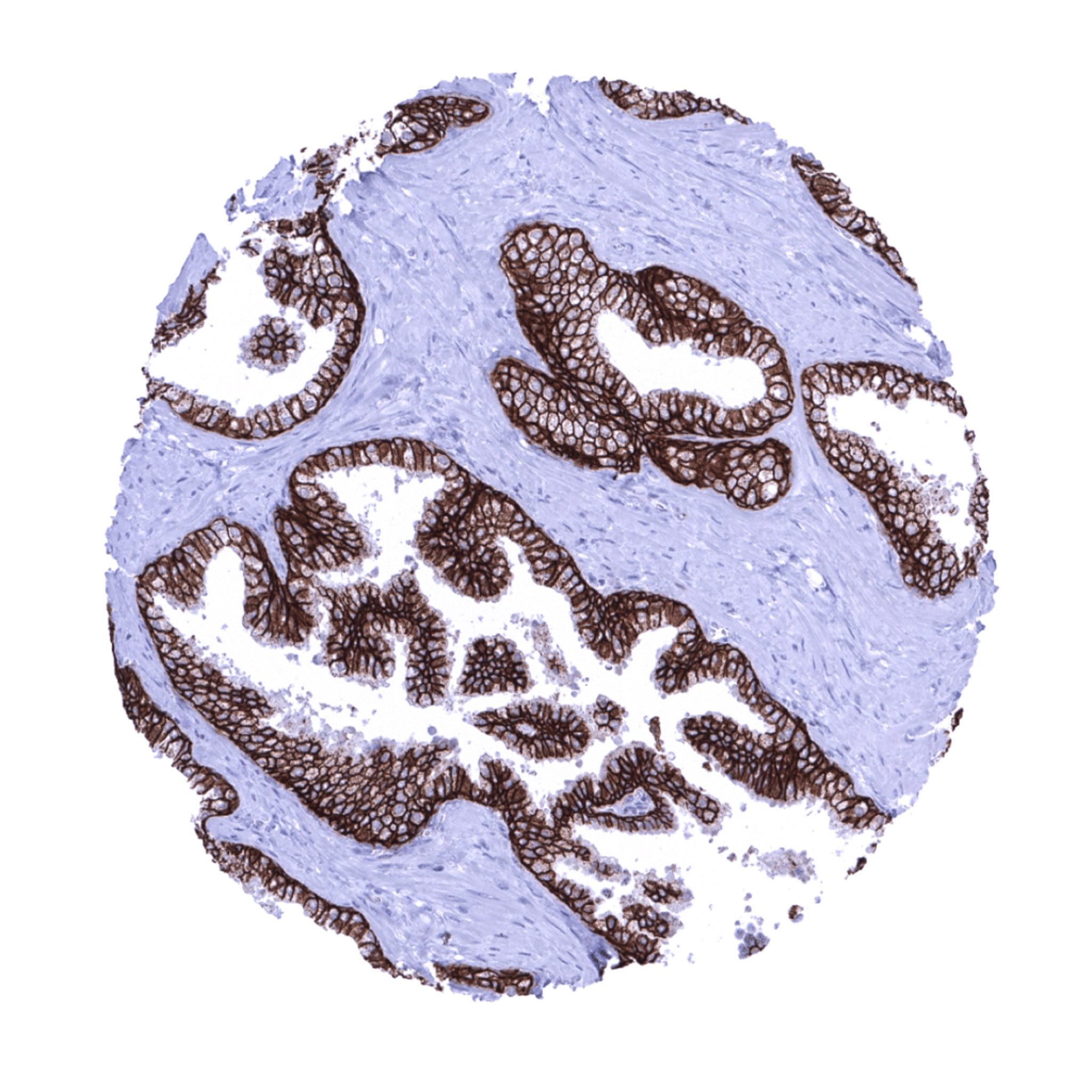
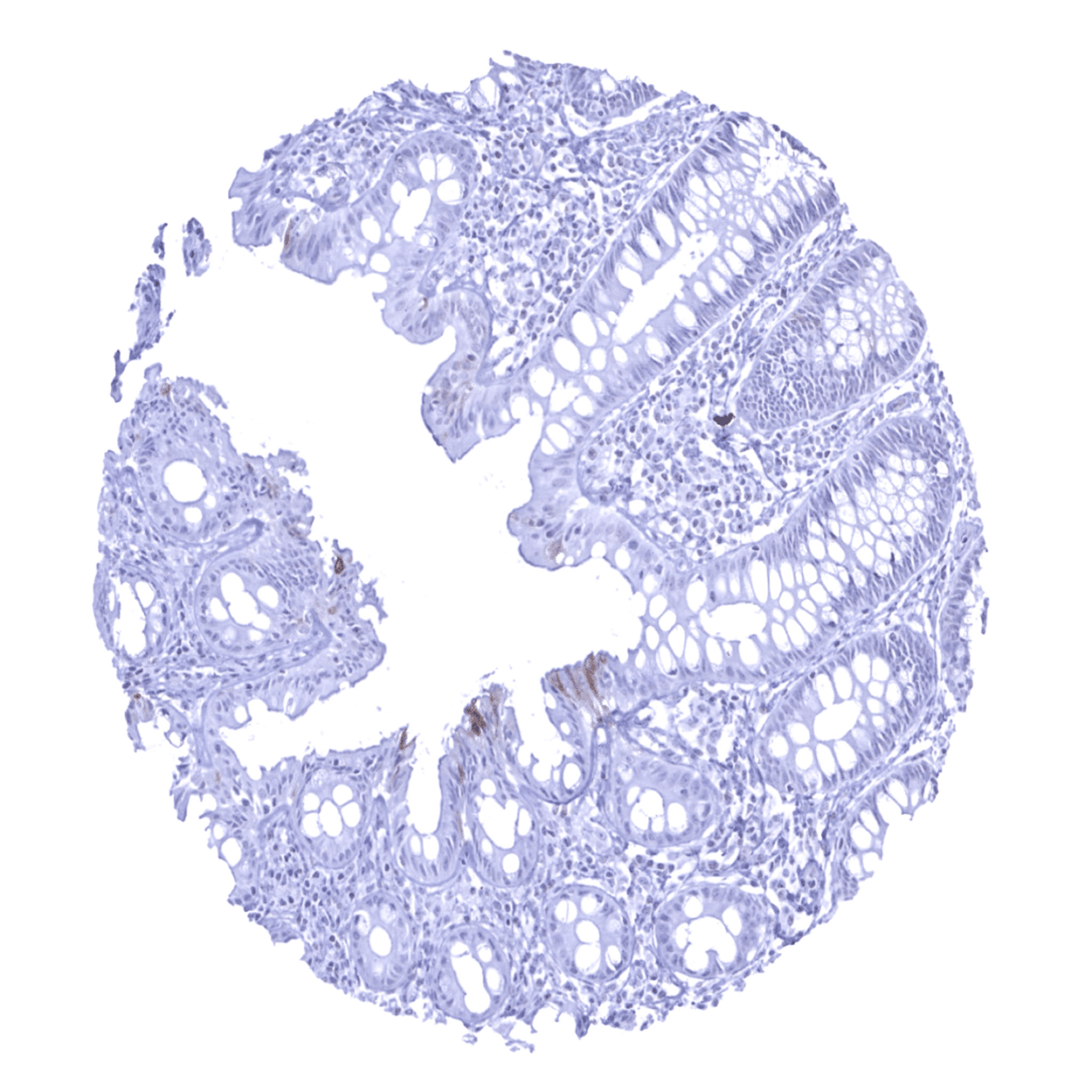
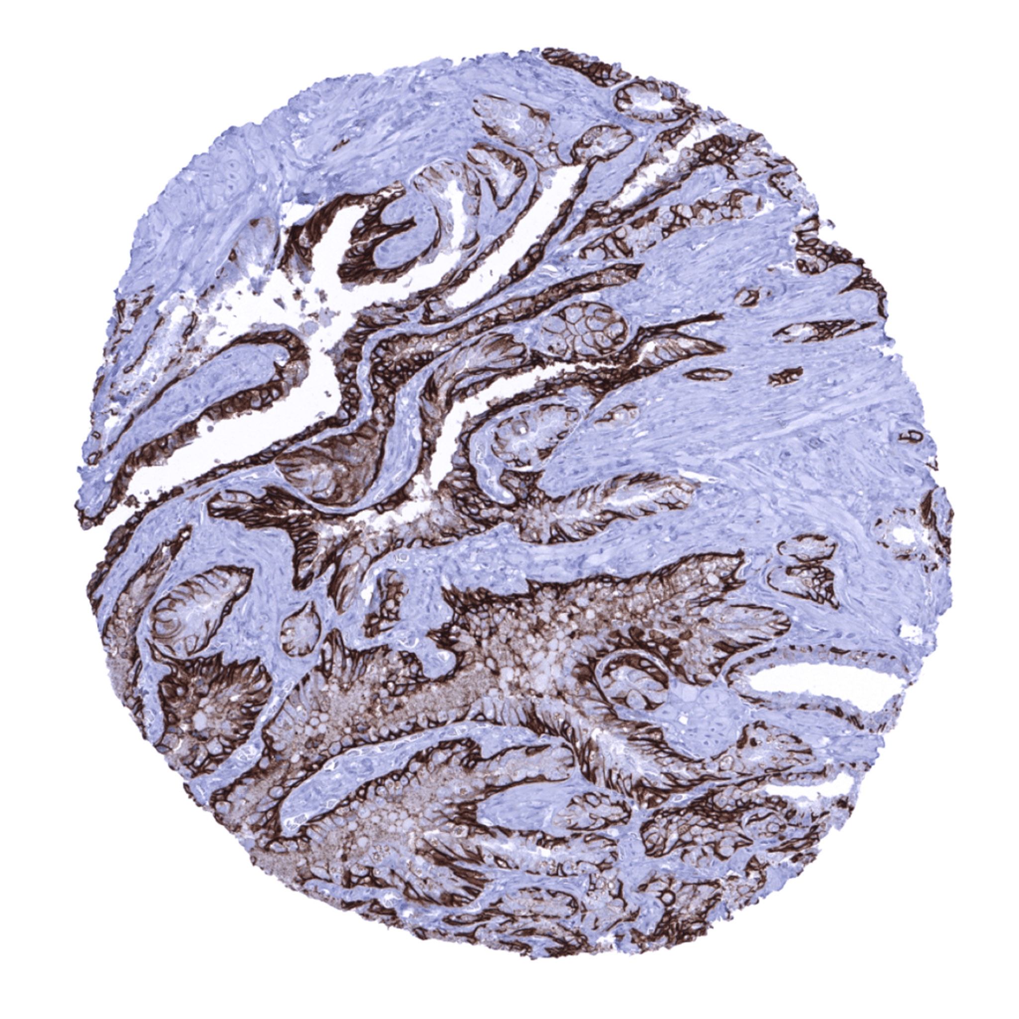
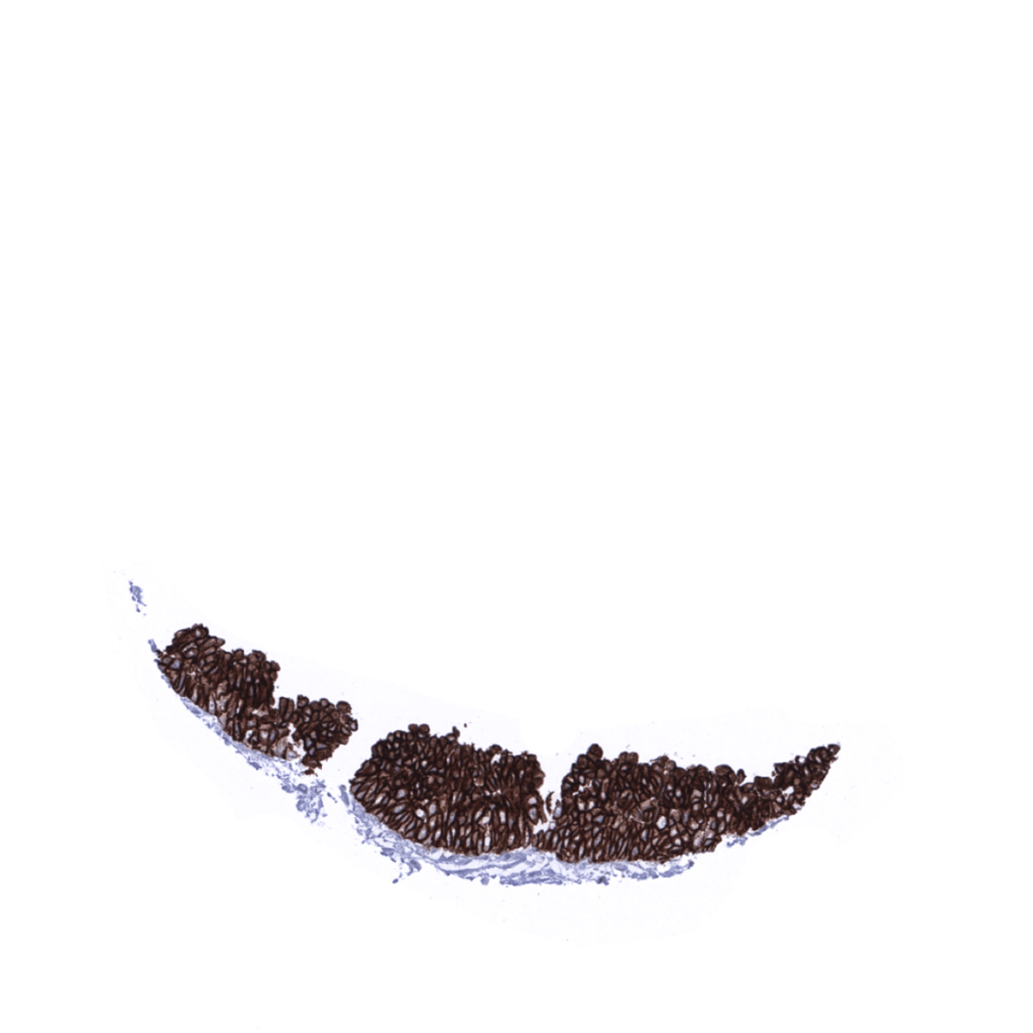
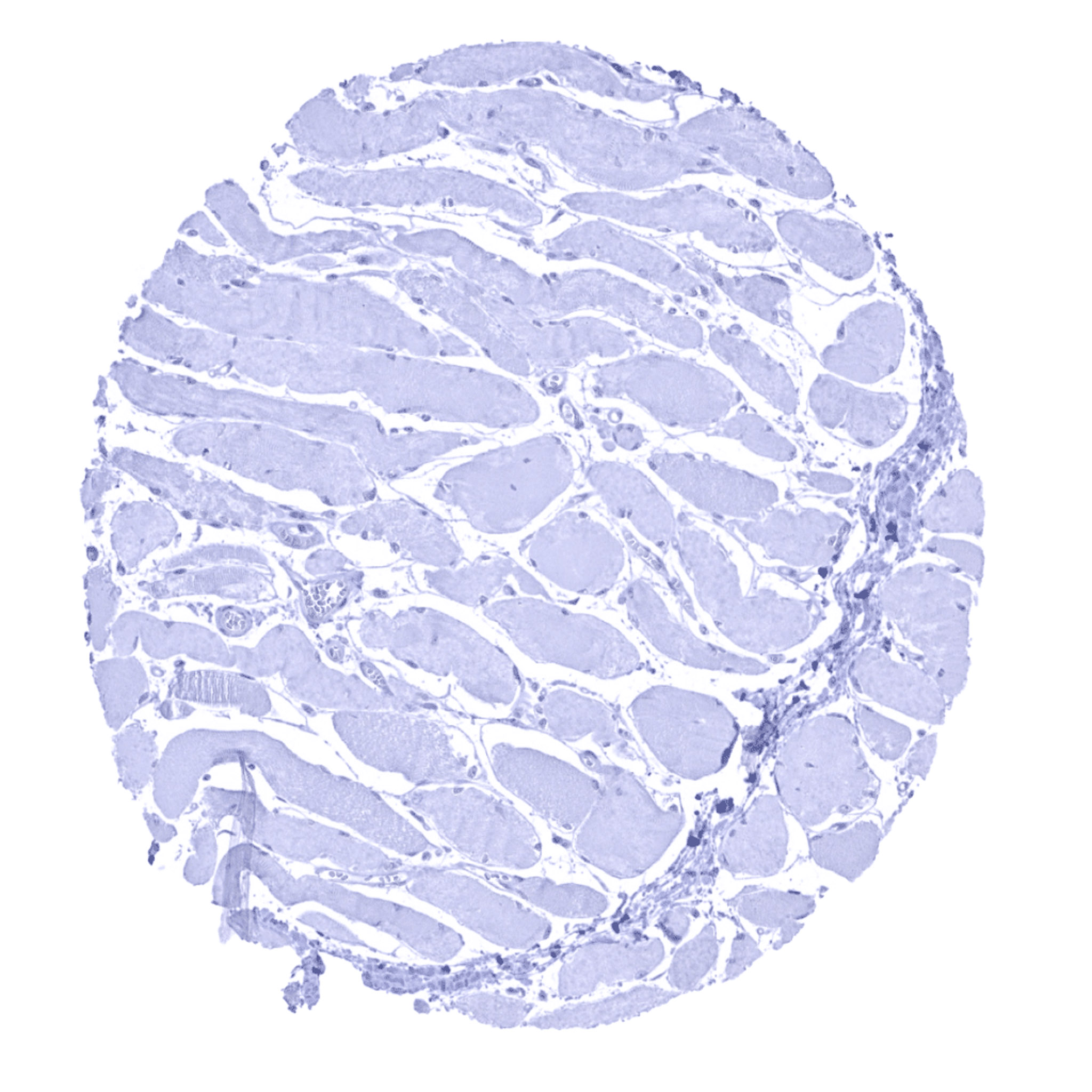
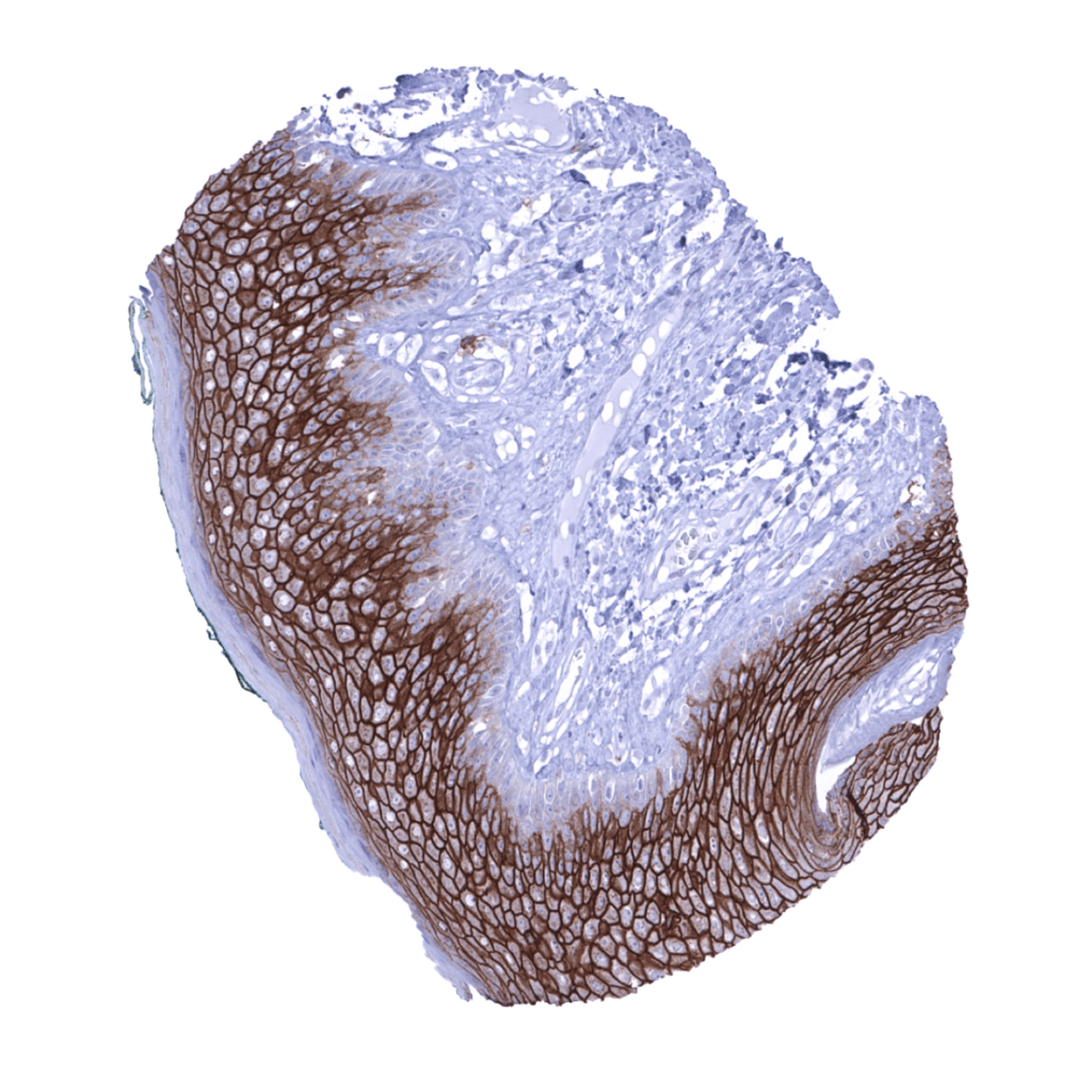
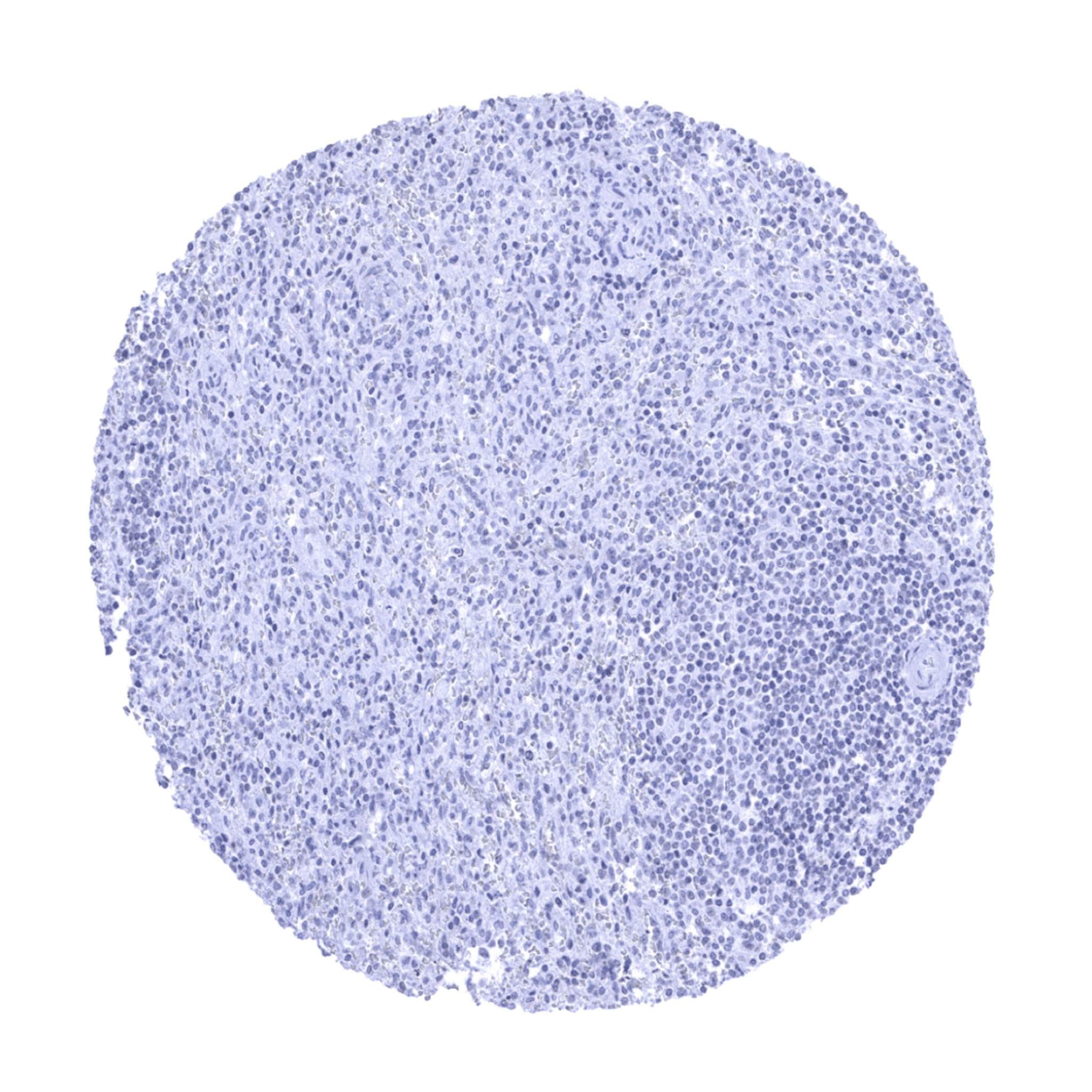
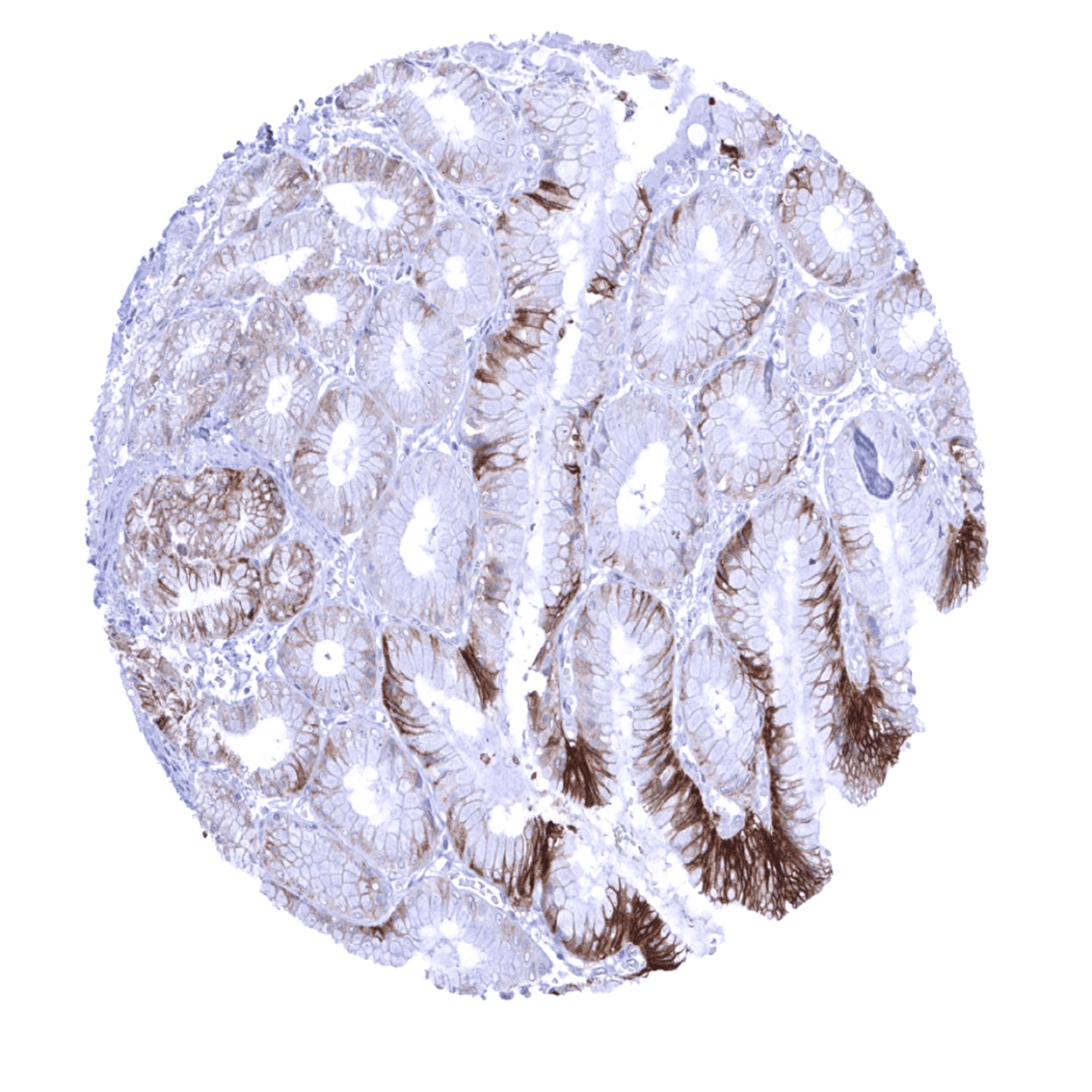
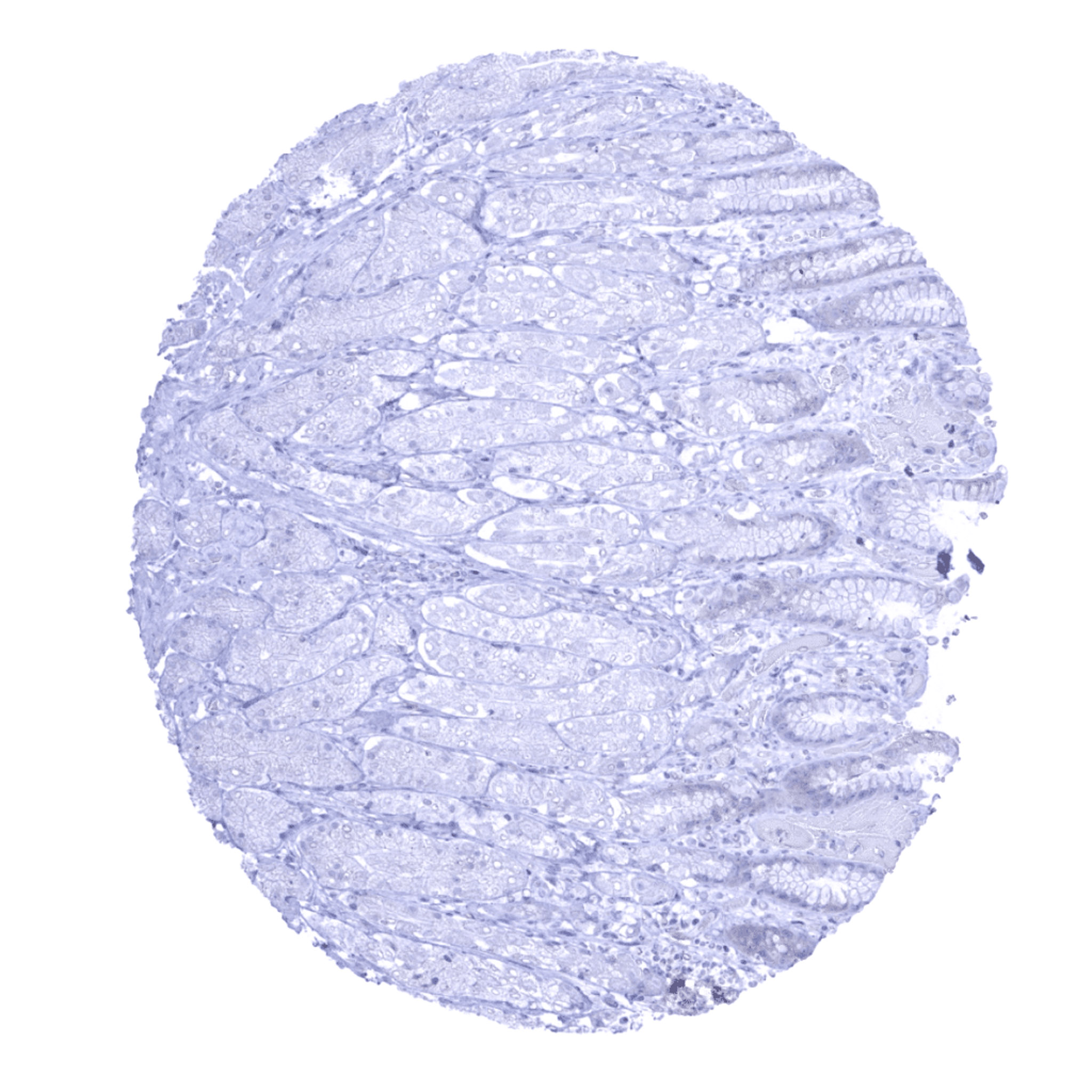
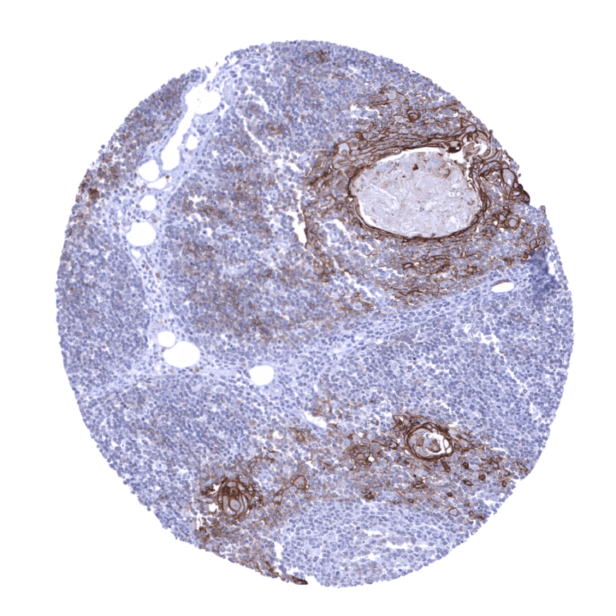
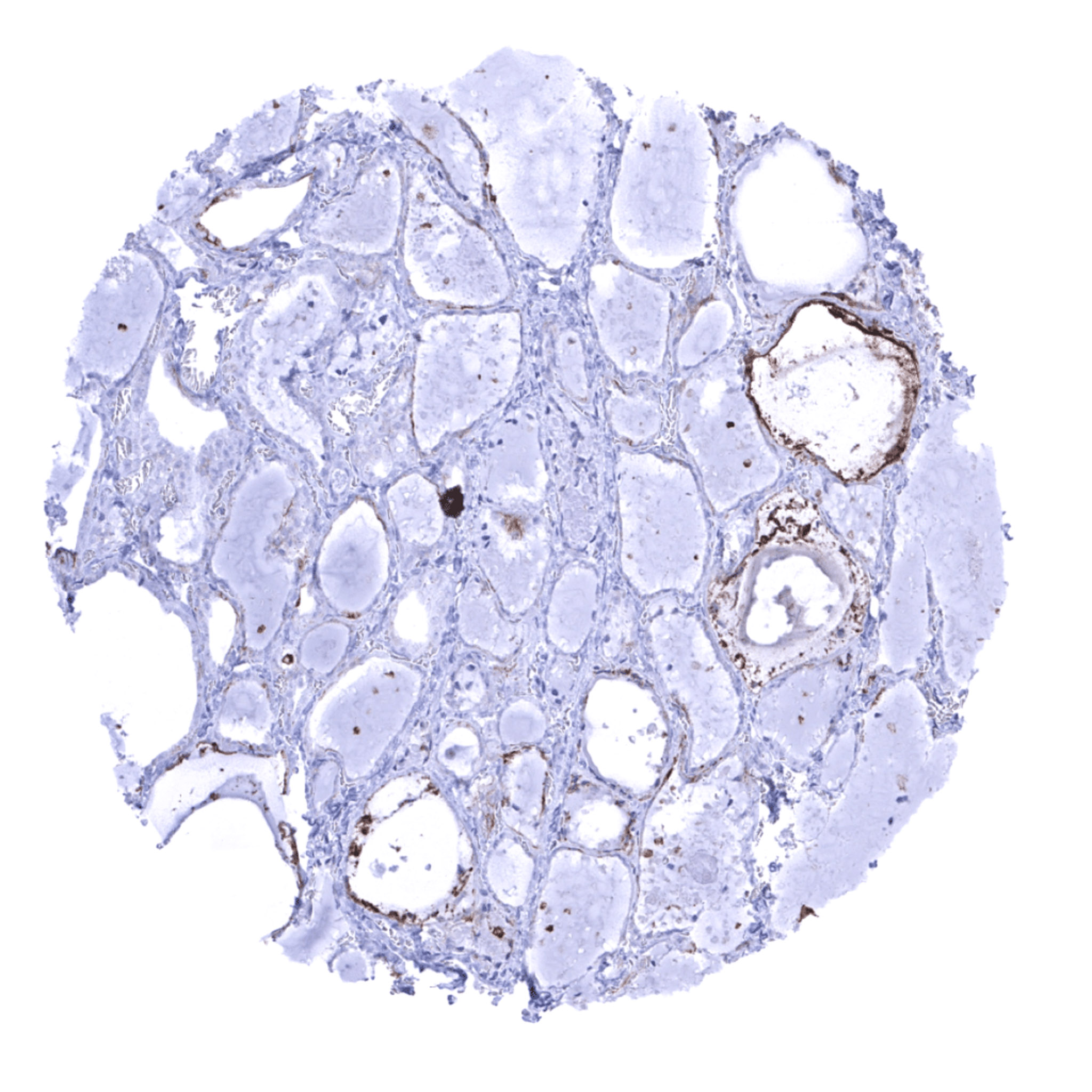
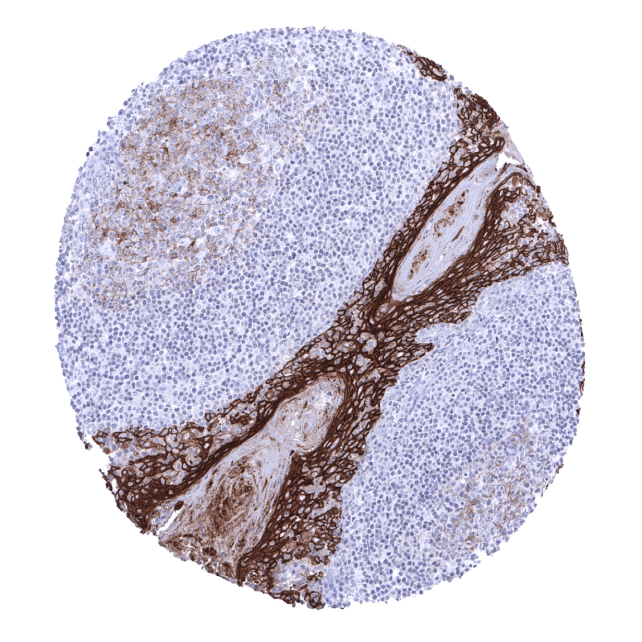
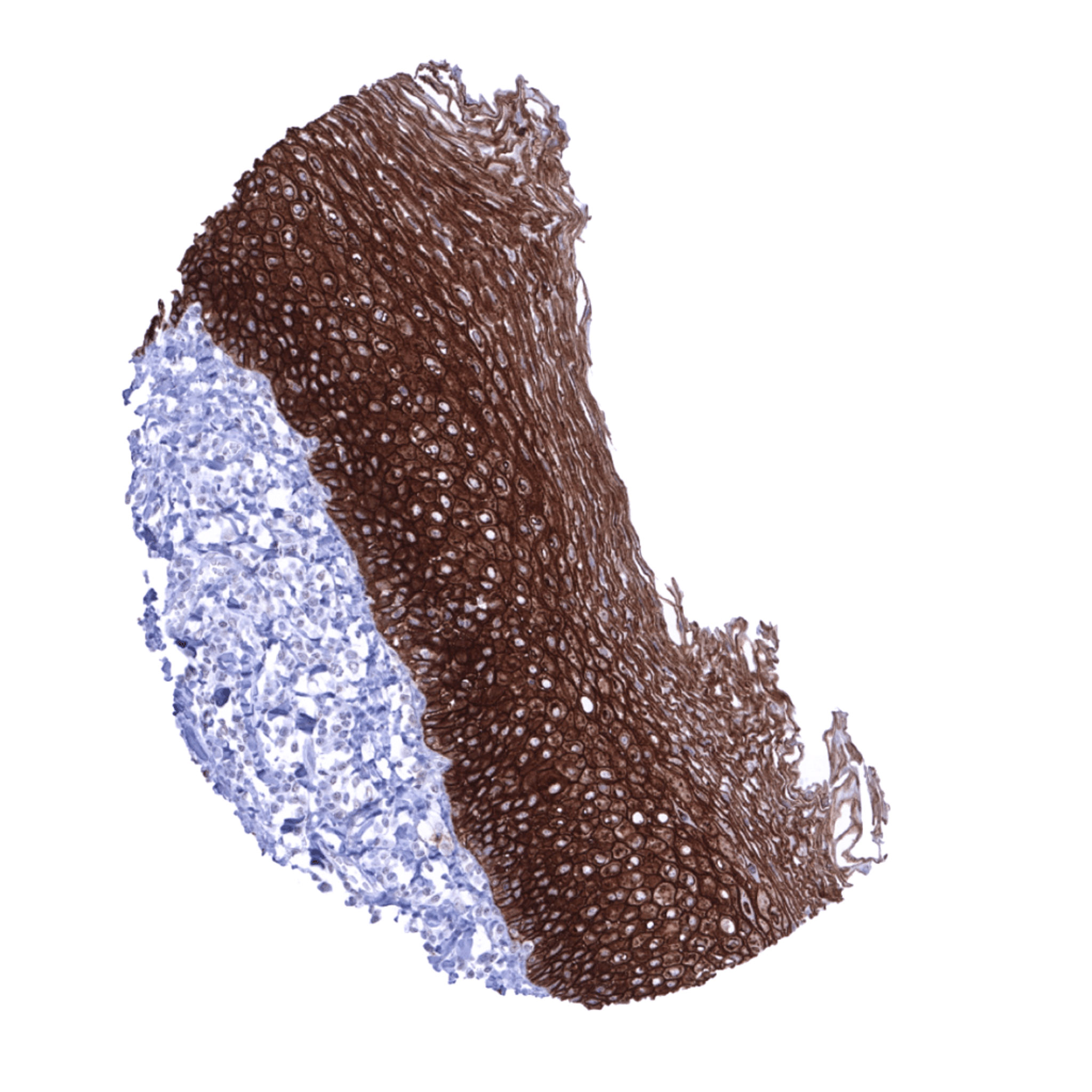
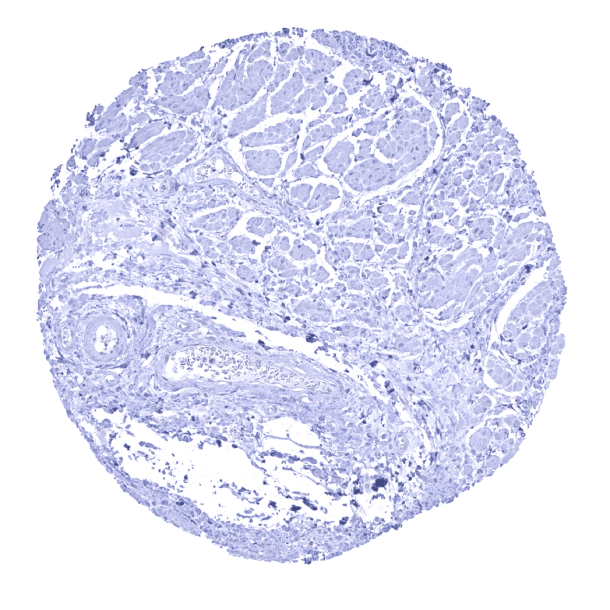
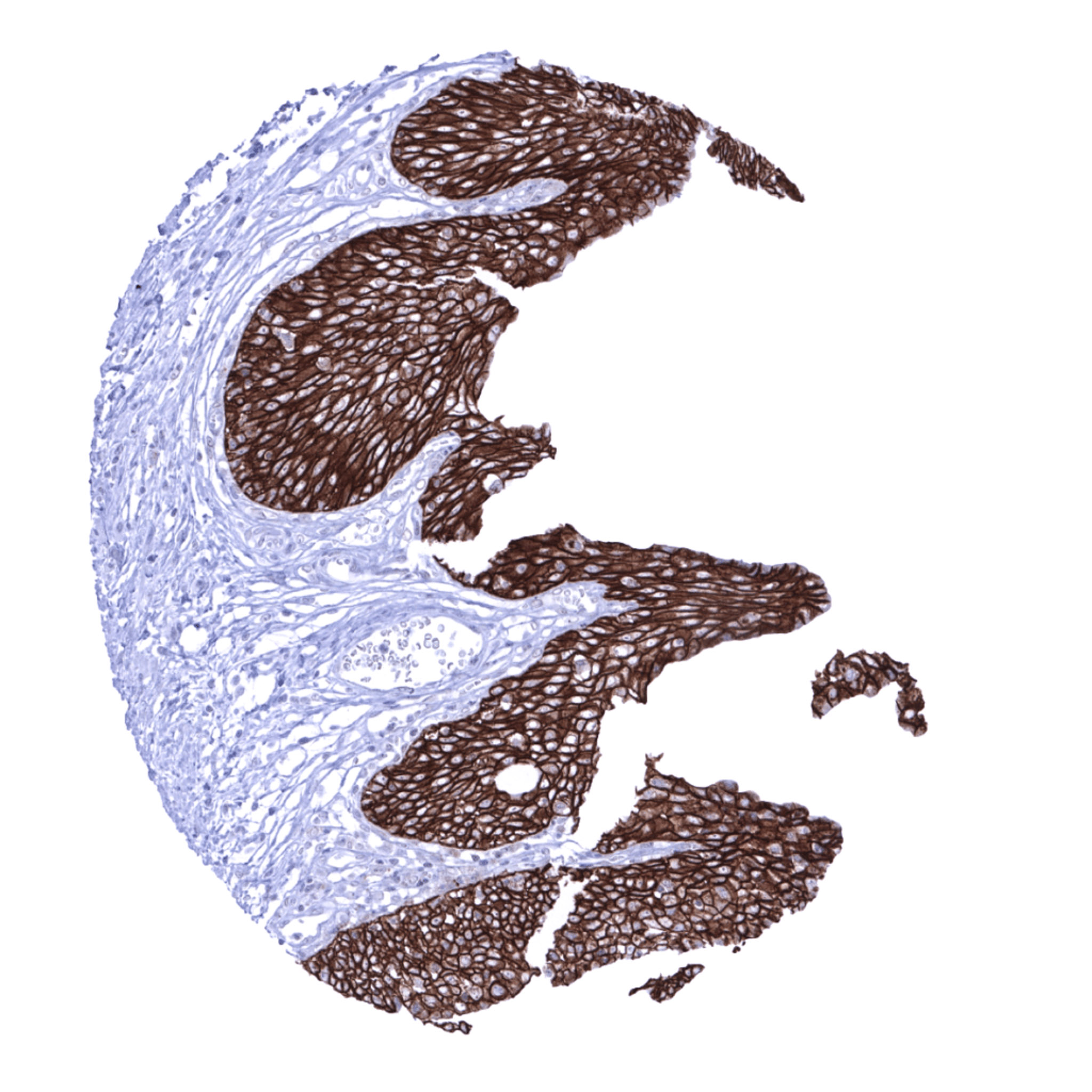
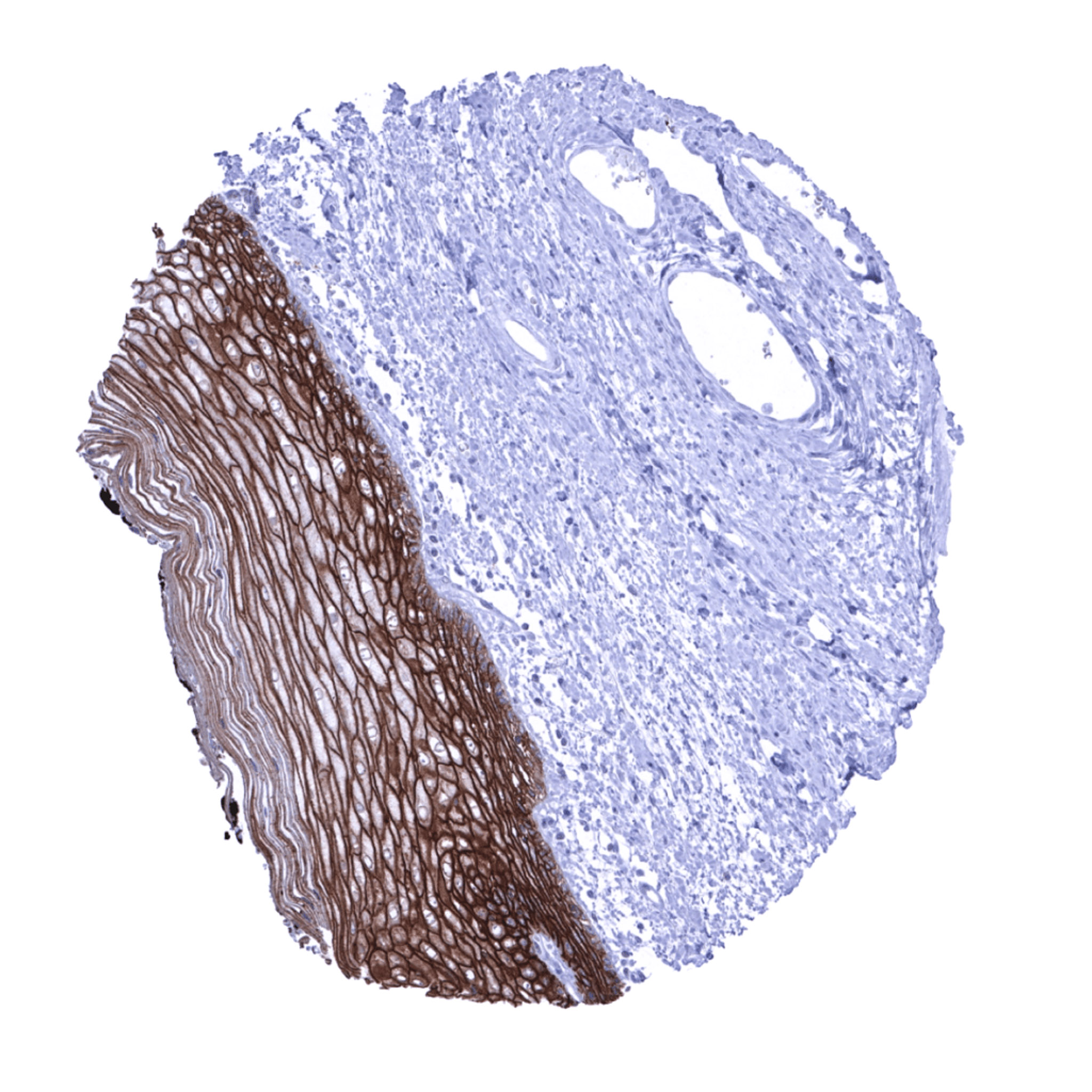
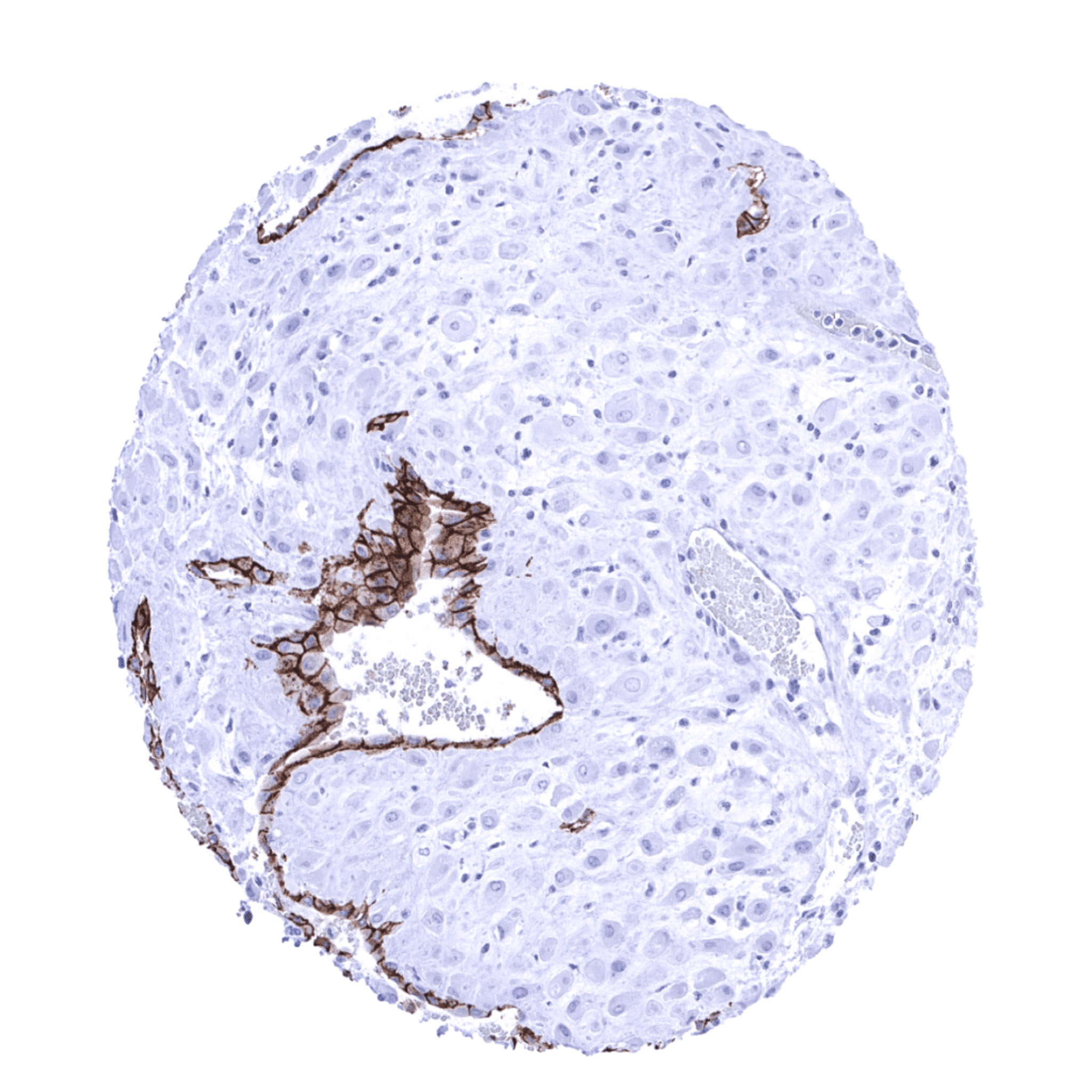
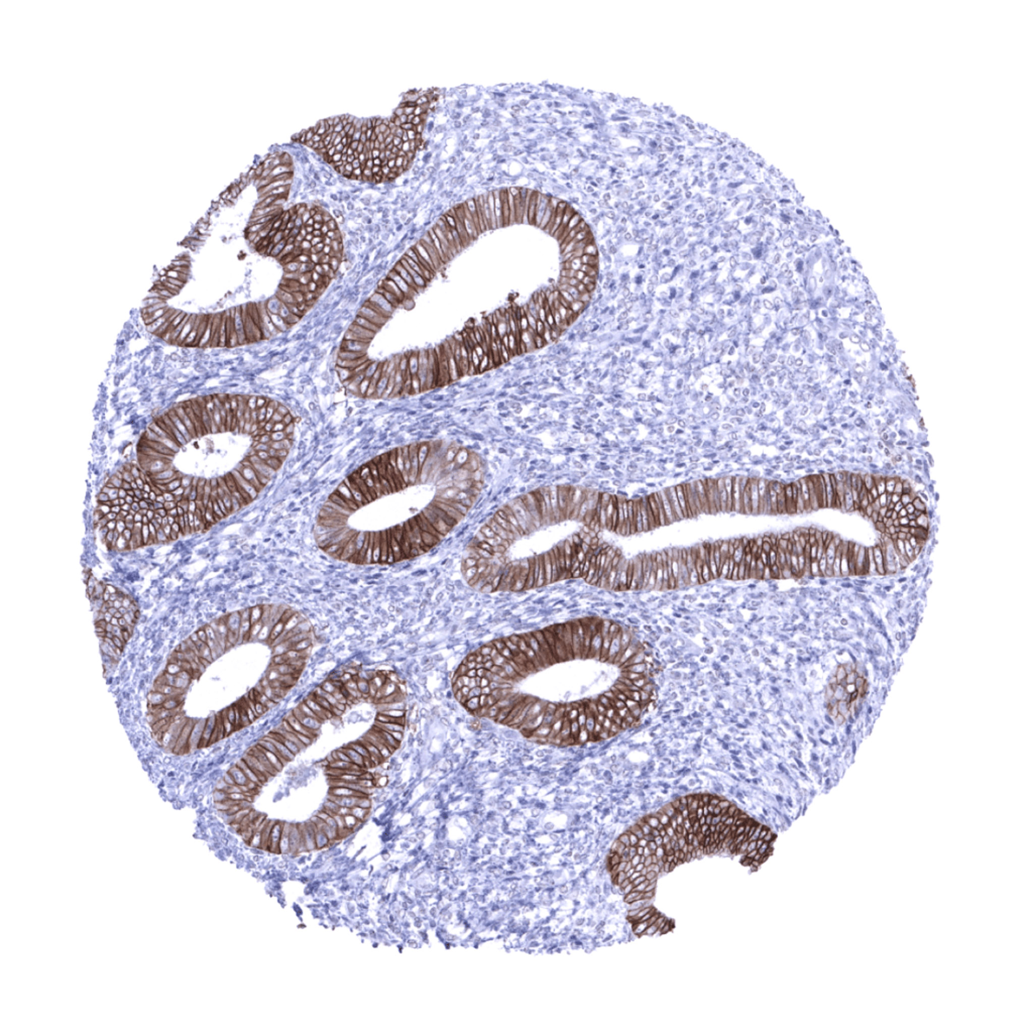
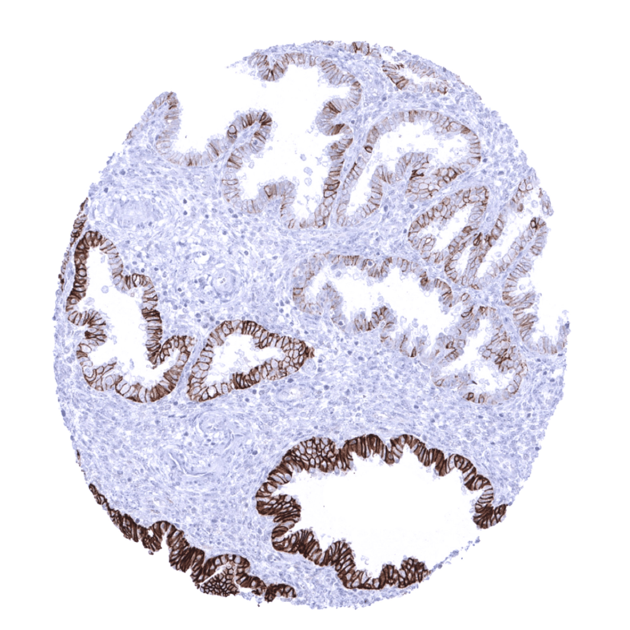
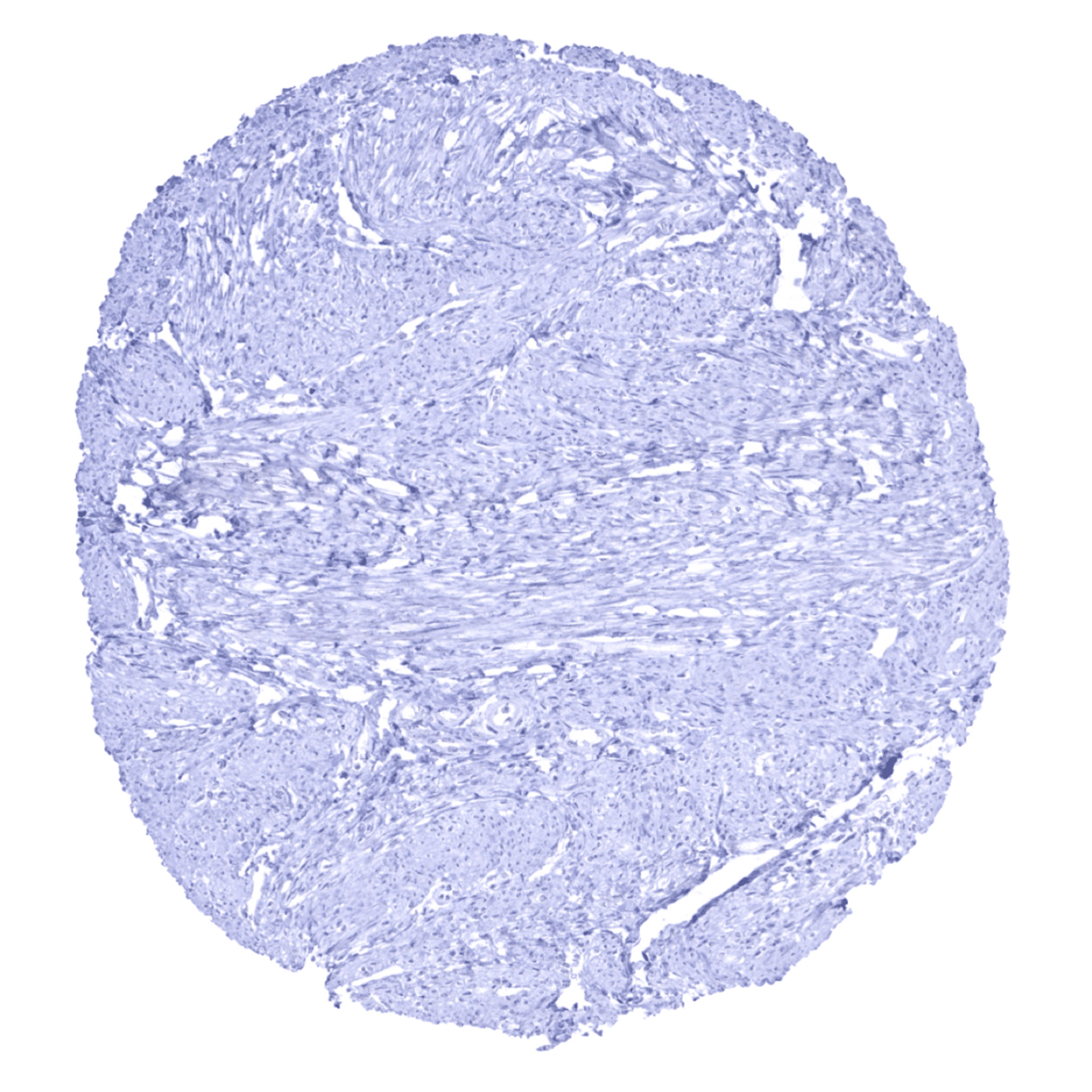
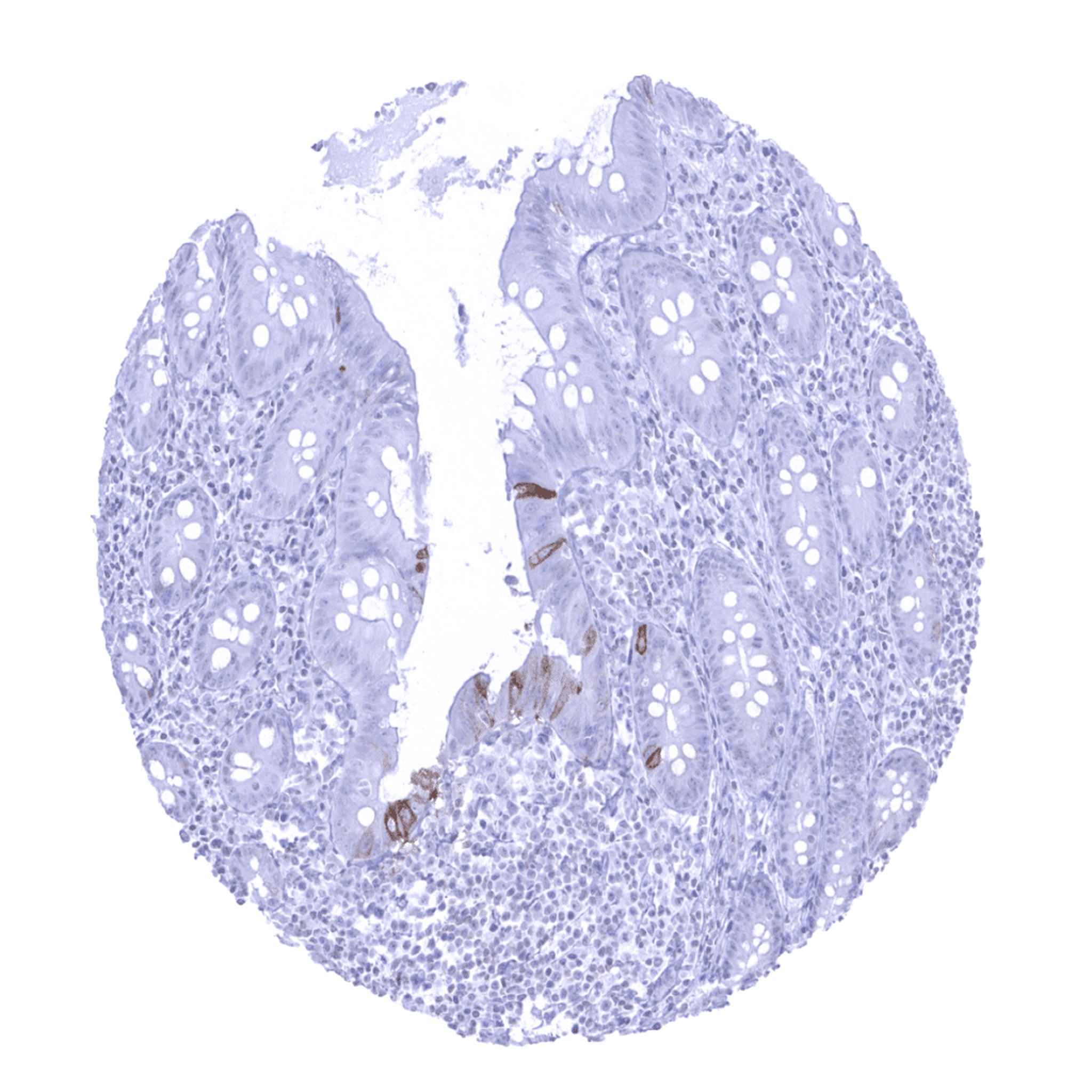
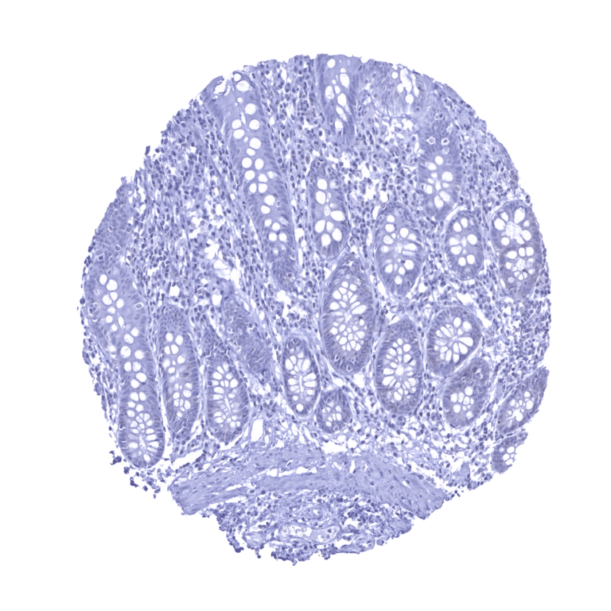
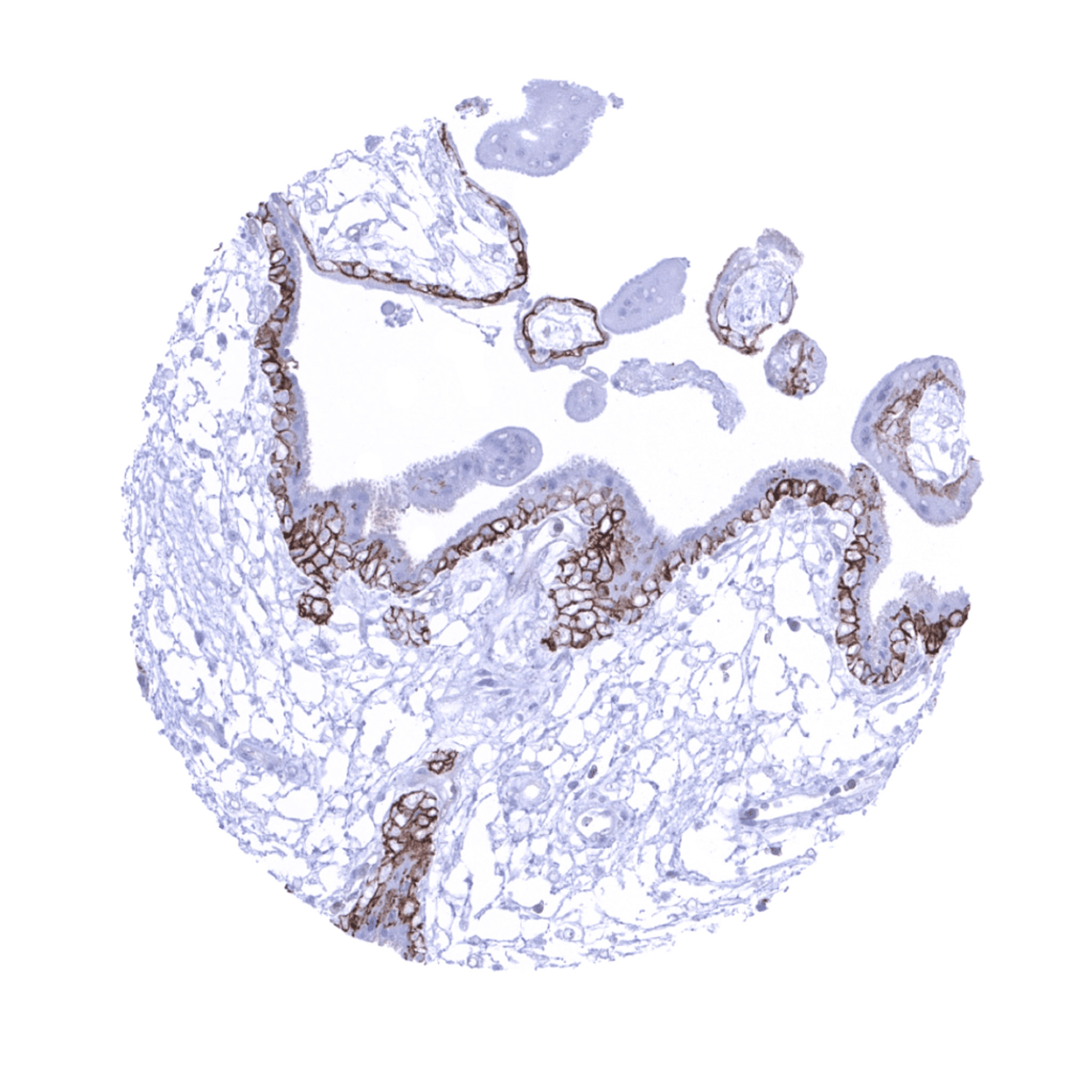
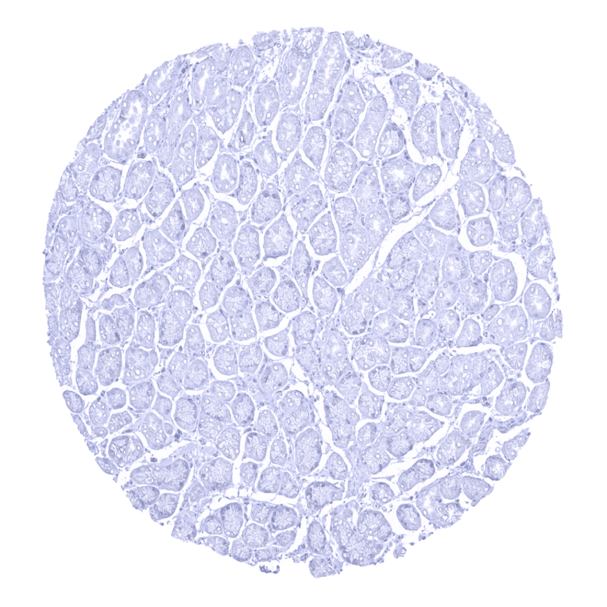
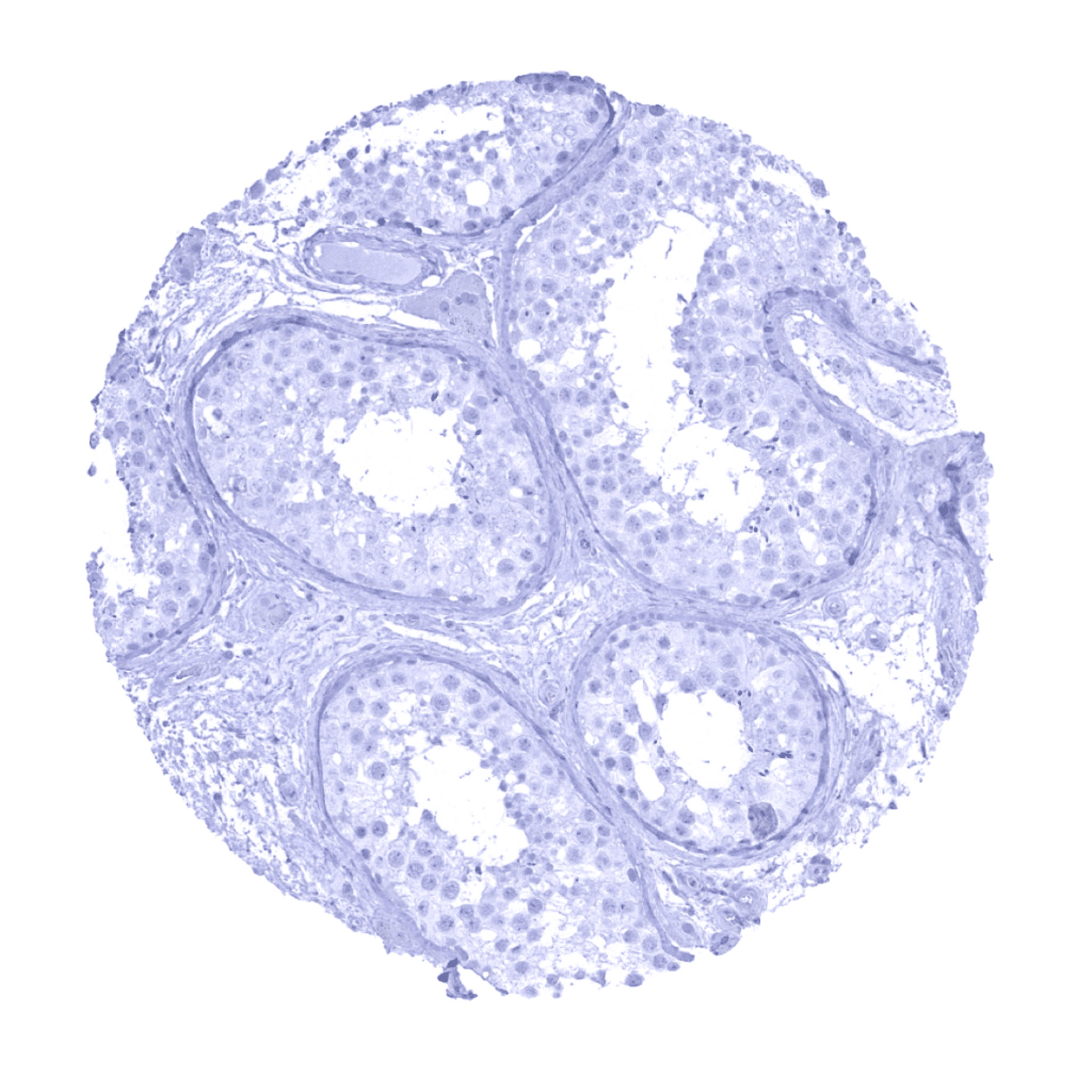
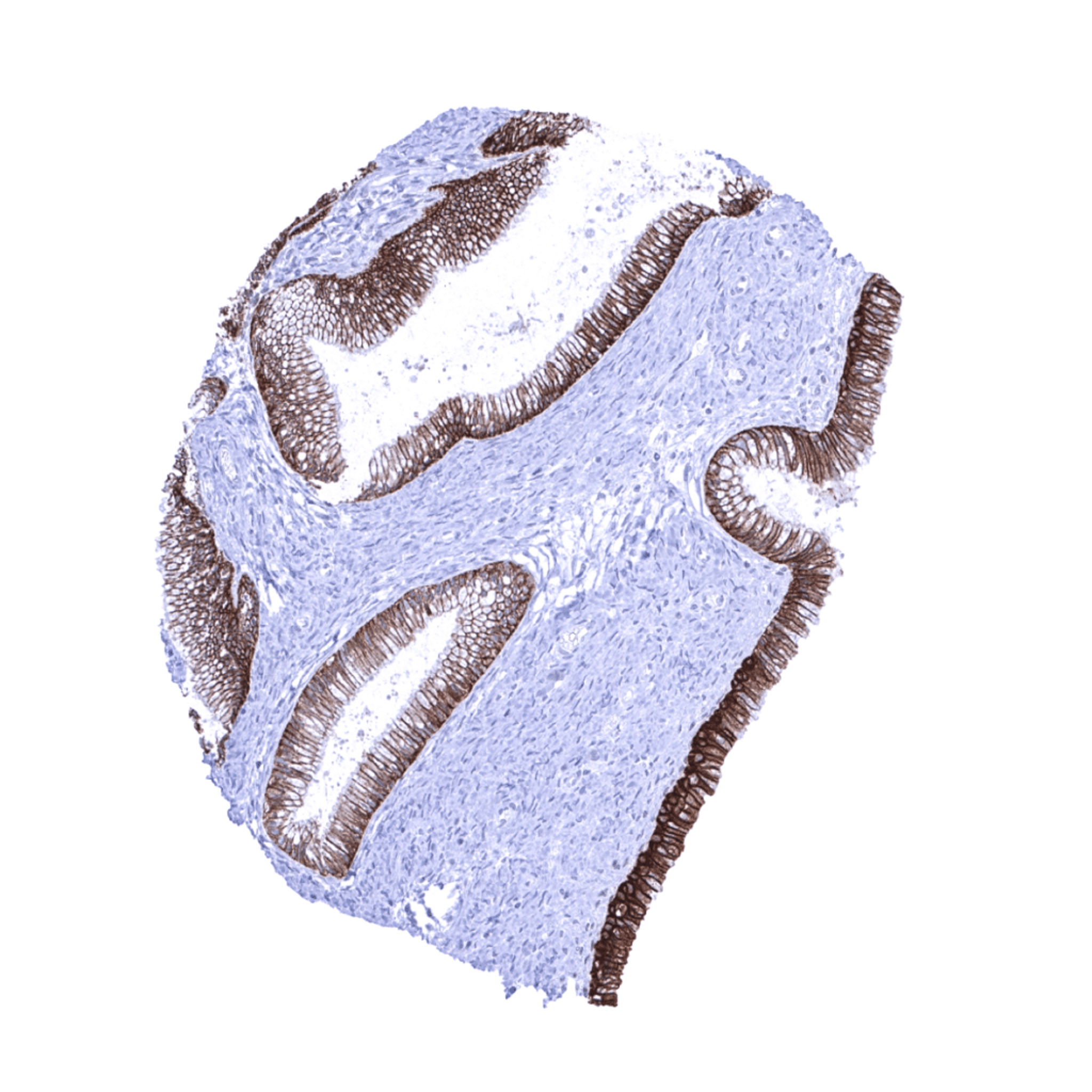
TACSTD2 antibody [MSVA-733R] HistoMAXTM


Go to Cancer Tissue Gallery Go back to product page Go to HistoMAX page


Adrenal gland
Aorta, media
Appendix, mucosa – In the entire GIT system, few scattered TACSTD2 positive epithelial cells can always occur.
Appendix, mucosa
Appendix, muscular wall
Bone marrow – A small subset of positive cells is seen in the bone marrow, perhaps reflecting granulocytes and their precursor cells.
Breast – A strong TACSTD2 immunostaining is seen in all epithelial cells of breast glands.
Bronchus, mucosa – A strong TACSTD2 positivity is seen in all cells of the respiratory epithelium.
Cerebellum (molecular layer, Purkinje cell layer, granule cell layer, white matter)
Cerebellum (molecular layer, Purkinje cell layer, granule cell layer)
Cerebrum, grey matter
Cerebrum, white matter
Colon descendens, muscular wall
Duodenum, Brunner gland
Duodenum, mucosa
Epididymis – TACSTD2 staining is most prominent in basal cells, but tall columnar cells also show a variable staining.
Esophagus, squamous epithelium – A strong, predominantly membranous TACSTD2 positivity occurs in the squamous epithelium but the basal cell layers are either negative or markedly less stained than the more superficial cell layers.
Fallopian tube, mucosa
Fat
Gallbladder, epithelium – A strong TACSTD2 staining is seen in all epithelial cells of the gallbladder epithelium.
Heart muscle
Ileum, mucosa – In the entire GIT system, few scattered TACSTD2 positive epithelial cells can always occur.
Ileum, mucosa
Kidney, cortex – A strong TACSTD2 immunostaining occurs in distal tubuli and collecting ducts while staining is weak to moderate in the parietal layer of the Bowman capsule.
Kidney, medulla – Strong TACSTD2 immunostaining in collecting ducts.
Liver – A strong TACSTD2 staining is seen in intrahepatic bile ducts.
Lung – A moderate TACSTD2 staining occurs in all pneumocytes of the lung.
Lymph node
Ovary, stroma
Pancreas – A strong positivity is seen in excretory and intercalated ducts while acinar cells show a variable staining intensity predominantly occurring at the apical membranes. Staining is largely absent in an islet of Langerhans.
Pancreas – A strong positivity is seen in excretory and intercalated ducts while acinar cells show a variable staining intensity predominantly occurring at the apical membranes.
Parathyroid gland
Parotid gland
Pituitary gland, anterior lobe – A small subset of epithelial cells of the adenohypophysis show a moderate TACSTD2 positivity.
Pituitary gland, posterior lobe
Placenta (amnion) – Strong TACSTD2 staining of amnion cells.
Placenta (chorion) – Moderate to strong TACSTD2 staining in chorion cells.
Placenta, early – TACSTD2 staining is strong in the cytotrophoblast of the placenta.
Placenta, mature – TACSTD2 staining is strongest in the cytotrophoblast of the placenta. An additional superficial membranous staining occurs in the syncytiotrophoblast of the mature placenta.
Prostate
Rectum, mucosa – In the entire GIT system, few scattered TACSTD2 positive epithelial cells can always occur.
Seminal vesicle
Sinus paranasales – A strong TACSTD2 positivity is seen in all cells of the respiratory epithelium.
Skeletal muscle
Skin – A strong, predominantly membranous TACSTD2 immunostaining is seen the squamous epithelium of the in skin but the basal cell layers are either negative or markedly less stained than the more superficial cell layers.
Spleen
Stomach, antrum – The superficial epithelial cell layers of the stomach can stain TACSTD2 positive.
Stomach, antrum – TACSTD2 positivity is not always seen in stomach samples
Stomach, corpus
Testis
Thymus – In the thymus, cells of corpuscles of Hassall’s show strong TACSTD2 staining while staining is weaker in other thymic epithelial cells.
Thyroid gland – A moderate to strong TACSTD2 staining occurs at the apical membranes of cells in a variable number of follicles.
Tonsil – In the tonsil, a strong TACSTD2 staining is seen in squamous epithelium of the crypts. In addition, a faint TACSTD2 staining occurs in a fraction of germinal centre cells.
Tonsil, surface epithelium – A strong, predominantly membranous TACSTD2 staining occurs in the squamous epithelium of tonsil surface where all cell layers are equally stained.
Urinary bladder, muscular wall
Urinary bladder, urothelium – A strong, predominantly membranous TACSTD2 staining occurs in the urothelium. All cell layers are equally stained.
Uterus, ectocervix – A strong, predominantly membranous TACSTD2 positivity is seen in the squamous epithelium of uterine cervix. All cell layers are equally stained
Uterus, endocervix – A strong TACSTD2 immunostaining is seen in all epithelial cells of the endocervix.
Uterus, endometrium (pregnancy) – Strong TACSTD2 immunostaining of endometrial glands while decidua cells are TACSTD2 negative.
Uterus, endometrium (proliferation) – Strong TACSTD2 immunostaining in endometrial glands.
Uterus, endometrium (secretion) – TACSTD2 immunostaining is variable in the endometrium and does not always involve all glands.
Uterus, myometrium
 

| TOP |