PRODUCT
Summary
HER2 antibody recognizes HER2 protein, also known as proto-oncogene Neu or ERBB2 (predicted molecular weight of 138 kDa). HER2 is a member of the human epidermal growth factor receptor (EGFR) family of receptor tyrosine kinases. It shares 50% sequence identity with EGFR protein, but involves different signaling pathways. HER2 can heterodimerize with EGFR protein or homodimerize when it is present in high concentrations.
APPLICATION
Application Note
*Optimal dilutions/concentrations should be determined by the researcher.
| Application |
Recommended Dilution |
| 1:500-1:20000 |
| 1:100-1:1000 |
Not tested in other applications.
Calculated MW
Positive Control
HCT116 , A549 , H1299 , HCT116 , MCF-7
Predict Reactivity
Mouse, Rat, Bovine, Cat, Dog, Pig(>80% identity)
PROPERTIES
Form
Liquid
Buffer
PBS, 1% BSA, 20% Glycerol
Preservative
0.01% Thimerosal
Storage
Store as concentrated solution. Centrifuge briefly prior to opening vial. For short-term storage (1-2 weeks), store at 4ºC. For long-term storage, aliquot and store at -20ºC or below. Avoid multiple freeze-thaw cycles.
Concentration
0.07 mg/ml (Please refer to the vial label for the specific concentration.)
Antigen Species
Human
Immunogen
Recombinant protein encompassing a sequence within the Intracellular domain of human Her2 / ErbB2. The exact sequence is proprietary.
Purification
Purified by antigen-affinity chromatography.
Conjugation
Unconjugated
RRID
AB_10618490
Note
For laboratory research use only. Not for any clinical, therapeutic, or diagnostic use in humans or animals. Not for animal or human consumption.
Purchasers shall not, and agree not to enable third parties to, analyze, copy, reverse engineer or otherwise attempt to determine the structure or sequence of the product.
TARGET
Synonyms
erb-b2 receptor tyrosine kinase 2 , CD340 , HER-2 , HER-2/neu , HER2 , MLN 19 , NEU , NGL , TKR1
Cellular Localization
Membrane; Single-pass type I membrane protein
Background
This gene encodes a member of the epidermal growth factor (EGF) receptor family of receptor tyrosine kinases. This protein has no ligand binding domain of its own and therefore cannot bind growth factors. However, it does bind tightly to other ligand-bound EGF receptor family members to form a heterodimer, stabilizing ligand binding and enhancing kinase-mediated activation of downstream signalling pathways, such as those involving mitogen-activated protein kinase and phosphatidylinositol-3 kinase. Allelic variations at amino acid positions 654 and 655 of isoform a (positions 624 and 625 of isoform b) have been reported, with the most common allele, Ile654/Ile655, shown here. Amplification and/or overexpression of this gene has been reported in numerous cancers, including breast and ovarian tumors. Alternative splicing results in several additional transcript variants, some encoding different isoforms and others that have not been fully characterized. [provided by RefSeq]
Database
Research Area
DATA IMAGES

|
GTX100509 IHC-P Image
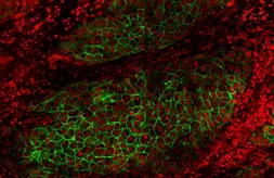
Her2 / ErbB2 antibody detects Her2 / ErbB2 protein at cell membrane by immunohistochemical analysis.Sample: Paraffin-embedded human breast carcinoma.Green: Her2 / ErbB2 stained by Her2 / ErbB2 antibody (GTX100509) diluted at 1:50.
|

|
GTX100509 WB Image
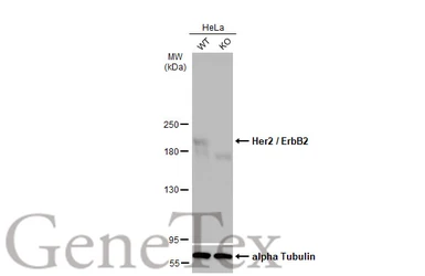
Wild-type (WT) and Her2 / ErbB2 knockout (KO) HeLa cell extracts (30 μg) were separated by 5% SDS-PAGE, and the membrane was blotted with Her2 / ErbB2 antibody (GTX100509) diluted at 1:5000. The HRP-conjugated anti-rabbit IgG antibody (GTX213110-01) was used to detect the primary antibody, and the signal was developed with Trident femto Western HRP Substrate.
|

|
GTX100509 IHC-P Image
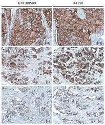
Her2 / ErbB2 antibody [C2C3], C-term detects Her2 / ErbB2 protein at cell membrane in human breast carcinoma by immunohistochemical analysis.
Sample: Strong positive (++), low positive (+) and negative tissue slides cores assessed using Quantitative Digital Pathology.
Her2 / ErbB2 antibody [C2C3], C-term (GTX100509) diluted at 1:500, and competitor's antibody (CST#4290) diluted at 1:100.
Antigen Retrieval: Trilogy™ (EDTA based, pH 8.0) buffer, 15min
*The competitor is not affiliated with GeneTex and does not endorse this product.
|

|
GTX100509 WB Image
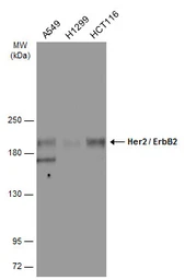
Various whole cell extracts (30 μg) were separated by 5% SDS-PAGE, and the membrane was blotted with Her2 / ErbB2 antibody [C2C3], C-term (GTX100509) diluted at 1:2000. The HRP-conjugated anti-rabbit IgG antibody (GTX213110-01) was used to detect the primary antibody.
|

|
GTX100509 WB Image
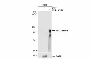
Non-transfected (–) and transfected (+) 293T whole cell extracts were separated by 5% SDS-PAGE, and the membrane was blotted with Her2 / ErbB2 antibody (GTX100509) diluted at 1:7000. The HRP-conjugated anti-rabbit IgG antibody (GTX213110-01) was used to detect the primary antibody.
|

|
GTX100509 WB Image
Various whole cell extracts (30 μg) were separated by 5% SDS-PAGE, and the membrane was blotted with Her2 / ErbB2 antibody [C2C3], C-term (GTX100509) diluted at 1:2000. The HRP-conjugated anti-rabbit IgG antibody (GTX213110-01) was used to detect the primary antibody.
|

|
GTX100509 IHC-P Image
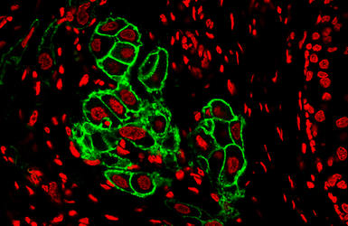
Her2 / ErbB2 antibody detects Her2 / ErbB2 protein at cell membrane by immunohistochemical analysis.
Sample: Human Breast Cancer.
Green: Her2 / ErbB2 stained by Dylight488-conjugated Her2 / ErbB2 antibody (GTX100509) diluted at 1:500.
|

|
GTX100509 WB Image
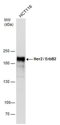
Whole cell extract (30 μg) was separated by 5% SDS-PAGE, and the membrane was blotted with Her2 / ErbB2 antibody [C2C3], C-term (GTX100509) diluted at 1:10000.
|
REFERENCE
REVIEW
Her2 / ErbB2 antibody
Cat. No. GTX100509
|
Rating
|
|
( Average 5 based on 2 users reviews)
|
Application
|
IHC (Paraffin sections)(IHC-P)
|
|
( Average 5 based on 2 users reviews)
|
Date :
Anonymous submitted on 18-Oct-2021
Application Tested :
IHC-P
Sample Species :
Hu
Sample :
human breast cancer
Primary Antibody :
1:50, 4°C, 16Hr
Date :
Anonymous submitted on 16-Jul-2021
Application Tested :
IHC-P
Sample Species :
Hu
Sample :
Breast cancer tissue
Primary Antibody :
1:500, 4°C, 16Hr


