APPLICATION
Application Note
*Optimal dilutions/concentrations should be determined by the researcher.
| Application |
Recommended Dilution |
| Assay dependent |
| Assay dependent |
Not tested in other applications.
Calculated MW
Predict Reactivity
Pig(>80% identity)
PROPERTIES
Form
Liquid
Buffer
PBS, 1% BSA
Preservative
0.025% Sodium azide
Storage
Store as concentrated solution. Centrifuge briefly prior to opening vial. For short-term storage (1-2 weeks), store at 4ºC. For long-term storage, aliquot and store at -20ºC or below. Avoid multiple freeze-thaw cycles.
Concentration
0.8 mg/ml (Please refer to the vial label for the specific concentration.)
Antigen Species
Human
Immunogen
Peptide RQELRKLKRRFLEEHEC, corresponding to amino acid residues 53-69 (Extracellular, near the P1 loop) of human KCNK1 (Accession : O00180).
Purification
Purified by antigen-affinity chromatography
Conjugation
Unconjugated
Note
For laboratory research use only. Not for any clinical, therapeutic, or diagnostic use in humans or animals. Not for animal or human consumption.
Purchasers shall not, and agree not to enable third parties to, analyze, copy, reverse engineer or otherwise attempt to determine the structure or sequence of the product.
TARGET
Synonyms
potassium two pore domain channel subfamily K member 1 , DPK , HOHO , K2P1 , K2p1.1 , KCNO1 , TWIK-1 , TWIK1
Cellular Localization
Cell membrane,Cell junction, synapse,Cytoplasmic vesicle,Cell projection, dendrite,Apical cell membrane
Background
This gene encodes one of the members of the superfamily of potassium channel proteins containing two pore-forming P domains. The product of this gene has not been shown to be a functional channel, however, it may require other non-pore-forming proteins for activity. [provided by RefSeq, Jul 2008]
Database
Research Area
DATA IMAGES

|
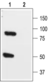
GTX16685 WB Image
WB analysis of rat brain lysate using GTX16685 KCNK1 antibody preincubated with or without immunogen peptide.
Dilution : 1:200
|

|
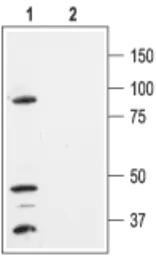
GTX16685 WB Image
WB analysis of HEK-293-KCNK1 transfected cell lysates using GTX16685 KCNK1 antibody preincubated with or without immunogen peptide.
Dilution : 1:300
|

|
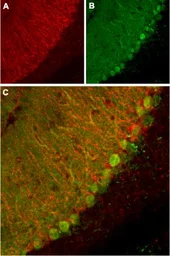
GTX16685 IHC-Fr Image
IHC-Fr analysis of mouse cerebellum tissue using GTX16685 KCNK1 antibody.
Panel A : KCNK1 channel appears in glial processes (red).
Panel B : Staining of Purkinje nerve cells with mouse anti-calbindin D28K (a calcium binding protein, green).
Panel C : Merge of KCNK1 channel and calbindin D28K demonstrates the separate localization of these proteins.
|
REFERENCE
There are currently no references for KCNK1 antibody (GTX16685). Be the first to share your publications with this product.
REVIEW
There are currently no reviews for KCNK1 antibody (GTX16685). Be the first to share your experience with this product.


