APPLICATION
Application Note
*Optimal dilutions/concentrations should be determined by the researcher.
| Application |
Recommended Dilution |
| 1:500 - 1:1000 |
| 1:50 - 1:100 |
Not tested in other applications.
Calculated MW
PROPERTIES
Form
Liquid
Buffer
PBS, 50% Glycerol
Preservative
0.02% Sodium azide
Storage
Store as concentrated solution. Centrifuge briefly prior to opening vial. For short-term storage (1-2 weeks), store at 4ºC. For long-term storage, aliquot and store at -20ºC or below. Avoid multiple freeze-thaw cycles.
Concentration
Batch dependent (Please refer to the vial label for the specific concentration.)
Antigen Species
Human
Immunogen
Recombinant fusion protein containing a sequence corresponding to amino acids 1-300 of human PKN1 (NP_002732.3).
Purification
Purified by affinity chromatography
Conjugation
Unconjugated
Note
For laboratory research use only. Not for any clinical, therapeutic, or diagnostic use in humans or animals. Not for animal or human consumption.
Purchasers shall not, and agree not to enable third parties to, analyze, copy, reverse engineer or otherwise attempt to determine the structure or sequence of the product.
TARGET
Synonyms
protein kinase N1 , DBK , PAK-1 , PAK1 , PKN , PKN-ALPHA , PRK1 , PRKCL1
Cellular Localization
Cytoplasm,Nucleus,Endosome,Cell membrane
Background
The protein encoded by this gene belongs to the protein kinase C superfamily. This kinase is activated by Rho family of small G proteins and may mediate the Rho-dependent signaling pathway. This kinase can be activated by phospholipids and by limited proteolysis. The 3-phosphoinositide dependent protein kinase-1 (PDPK1/PDK1) is reported to phosphorylate this kinase, which may mediate insulin signals to the actin cytoskeleton. The proteolytic activation of this kinase by caspase-3 or related proteases during apoptosis suggests its role in signal transduction related to apoptosis. Alternatively spliced transcript variants encoding distinct isoforms have been observed. [provided by RefSeq, Jul 2008]
Database
Research Area
DATA IMAGES

|
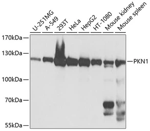
GTX64829 WB Image
WB analysis of various sample lysates using GTX64829 PKN1 antibody.
Dilution : 1:1000
Loading : 25μg per lane
|

|
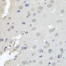
GTX64829 IHC-P Image
IHC-P analysis of mouse brain tissue using GTX64829 PKN1 antibody.
Dilution : 1:100
|

|
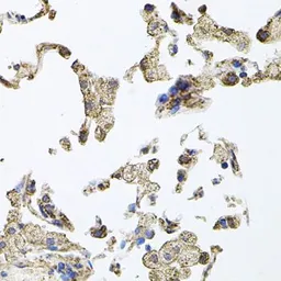
GTX64829 IHC-P Image
IHC-P analysis of human lung tissue using GTX64829 PKN1 antibody.
Dilution : 1:100
|

|
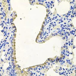
GTX64829 IHC-P Image
IHC-P analysis of mouse lung tissue using GTX64829 PKN1 antibody.
Dilution : 1:100
|
REFERENCE
There are currently no references for PKN1 antibody (GTX64829). Be the first to share your publications with this product.
REVIEW
There are currently no reviews for PKN1 antibody (GTX64829). Be the first to share your experience with this product.


