GeneTex
  • Country / Location Selection

United States (US)

RIF1 antibody

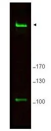
Anti-RIF1 antibody used in Western Blot (WB). GTX48737

Cat. No. GTX48737

Host

Rabbit

Clonality

Polyclonal

Isotype

IgG

Application

WB, ELISA

Reactivity

Human, Mouse
Package
50 μg ($399)

APPLICATION

Application Note

*Optimal dilutions/concentrations should be determined by the researcher.
Application Recommended Dilution
WB 1:500-1:3000
ELISA 1:5000-1:40000
Not tested in other applications.

Calculated MW

266 kDa. ( Note )

PROPERTIES

Form

Liquid

Buffer

20mM Potassium Phosphate, 150mM NaCl

Preservative

0.01% Sodium azide

Storage

Store as concentrated solution. Centrifuge briefly prior to opening vial. For short-term storage (1-2 weeks), store at 4ºC. For long-term storage, aliquot and store at -20ºC or below. Avoid multiple freeze-thaw cycles.

Concentration

1 mg/ml (Please refer to the vial label for the specific concentration.)

Antigen Species

Mouse

Immunogen

Synthetic peptide corresponding to C-Terminal region near amino acids 2375-2419 of Mouse Rif1.

Purification

Purified by antigen-affinity chromatography.
From serum

Conjugation

Unconjugated

RRID

AB_11162229

Note

For laboratory research use only. Not for any clinical, therapeutic, or diagnostic use in humans or animals. Not for animal or human consumption.

Purchasers shall not, and agree not to enable third parties to, analyze, copy, reverse engineer or otherwise attempt to determine the structure or sequence of the product.

TARGET

Synonyms

replication timing regulatory factor 1 , 5730435J01Rik , 6530403D07Rik , AU016181 , AW549474 , D2Ertd145e

Cellular Localization

Nucleus

Background

Rif1 (also known as telomere-associated protein RIF1 and Rap1-interacting factor 1) is a mouse ortholog of the yeast Rif1 family of telomere-associated proteins identified on the basis of its high expression in primordial germ cells and embryo-derived pluripotent stem cell lines. Mouse Rif1 is a protein involved in negative regulation of telomere length. It is also localized to ATM dependent DNA damage foci and may act at the chromatin level to allow access to some factors.

Database

Research Area

DATA IMAGES

Anti-RIF1 antibody used in Western Blot (WB). GTX48737

GTX48737 WB Image

Western blot using GeneTex's Affinity Purified anti-Rif1 antibody shows detection of a band ~265 kDa corresponding to mouse Rif1 (arrowhead). Specific reactivity with this band is blocked when the antibody is pre-incubated with the immunizing peptide (data not shown). Approximately 25 μg of MEF whole cell lysate was separated by SDS-PAGE and transferred onto nitrocellulose. After blocking the membrane was probed with the primary antibody diluted to 1.0 ug/ml for 2 h at room temperature followed by washes and reaction with a 1:10,000 dilution of IRDye800 conjugated goat anti-Rabbit IgG [H&L] MX for 45 min at room temperature. IRDye800 fluorescence image was captured using the OdysseyR Infrared Imaging System developed by LI-COR. IRDye is a trademark of LI-COR, Inc. Other detection systems will yield similar results.

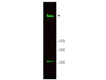
Anti-RIF1 antibody used in Western Blot (WB). GTX48737

GTX48737 WB Image

WB analysis of MEF whole cell lysate using GTX48737 RIF1 antibody.
Loading : 25 μg
Dilution : 1 μg/mL

REFERENCE

REVIEW

There are currently no reviews for RIF1 antibody (GTX48737). Be the first to share your experience with this product.
SDS
Sodium Azide.pdf
Package List Price ($)
$ 399