GeneTex
  • Country / Location Selection

United States (US)
Cancer Tissue Gallery- GTX04457

HLA-DRB1 antibody [MSVA-478R] HistoMAX


Go to Normal Tissue Gallery Go back to product page Go to HistoMAX page


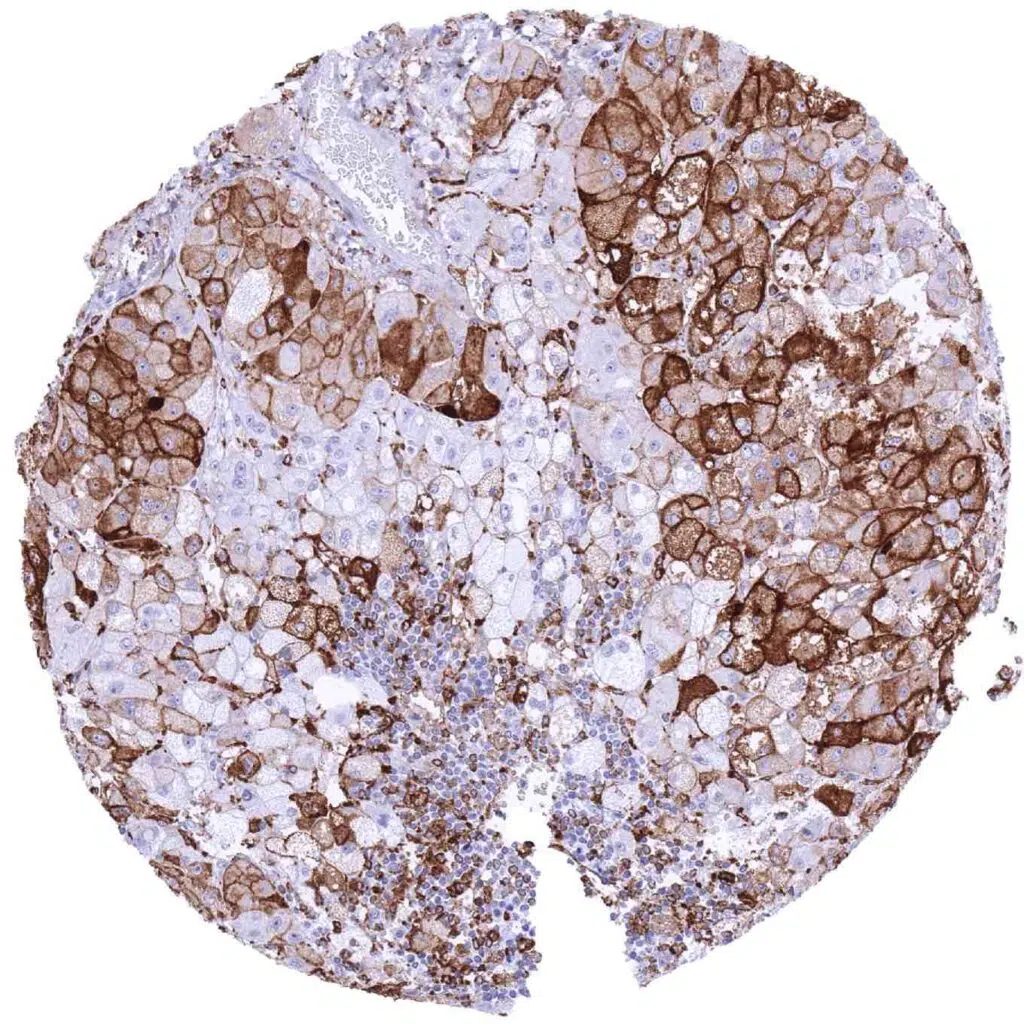
Adrenal gland – Adrenocortical adenoma with variable HLA-DRB1 immunostaining of tumor cells and of a subset of inflammatory cells (HLA-DRB1 immunohistochemistry)
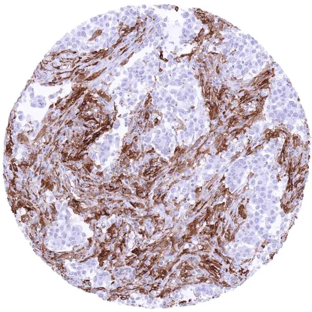
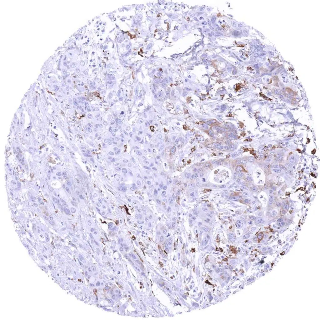
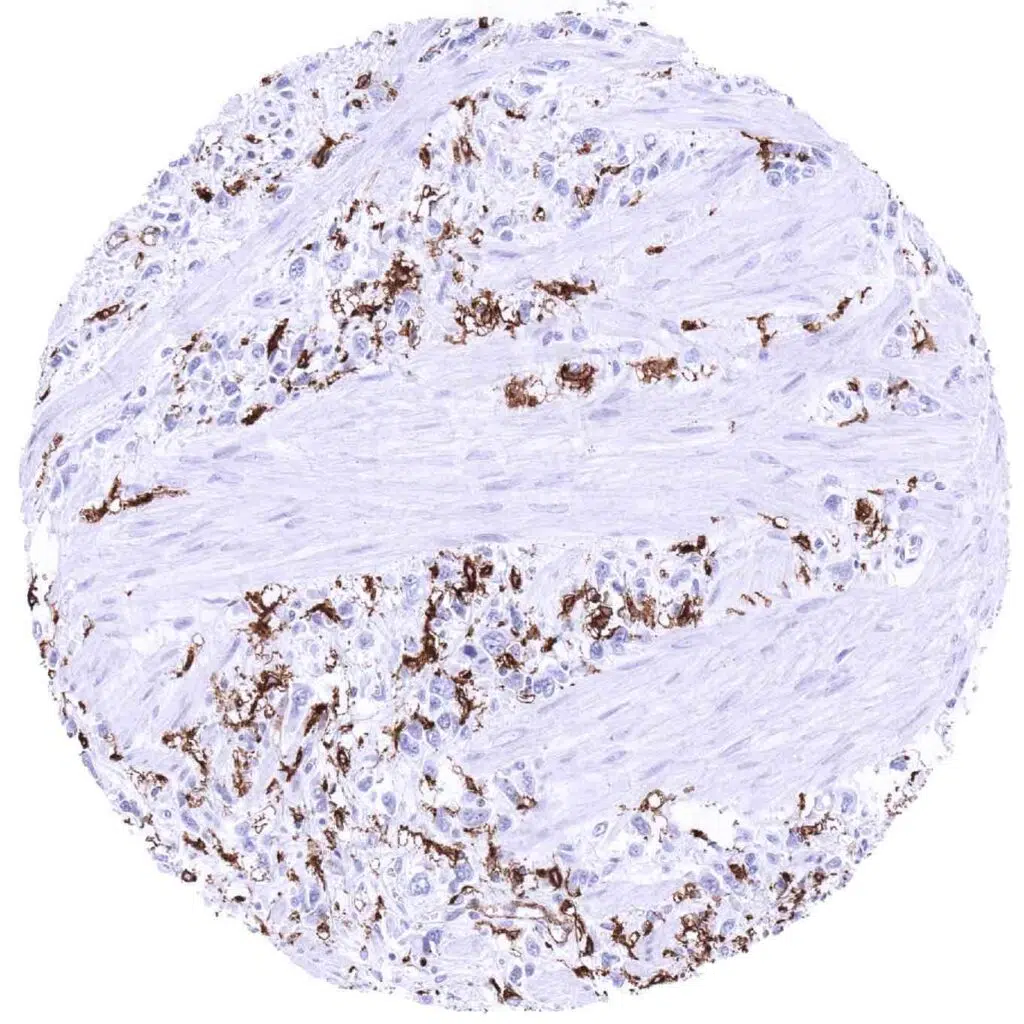
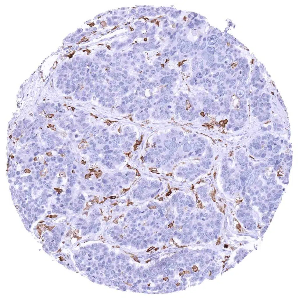
Breast – HLA-DRB1 negative invasive breast cancer of no special type (NST including numerous positive inflammatory cells (HLA-DRB1 immunohistochemistry)
Breast – HLA-DRB1 negative invasive lobular breast cancer accompanied by numerous positive inflammatory cells (HLA-DRB1 immunohistochemistry)
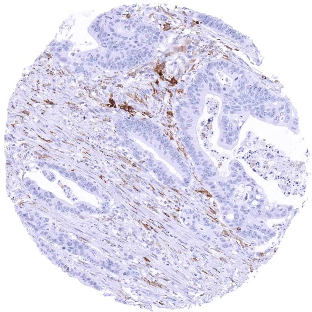
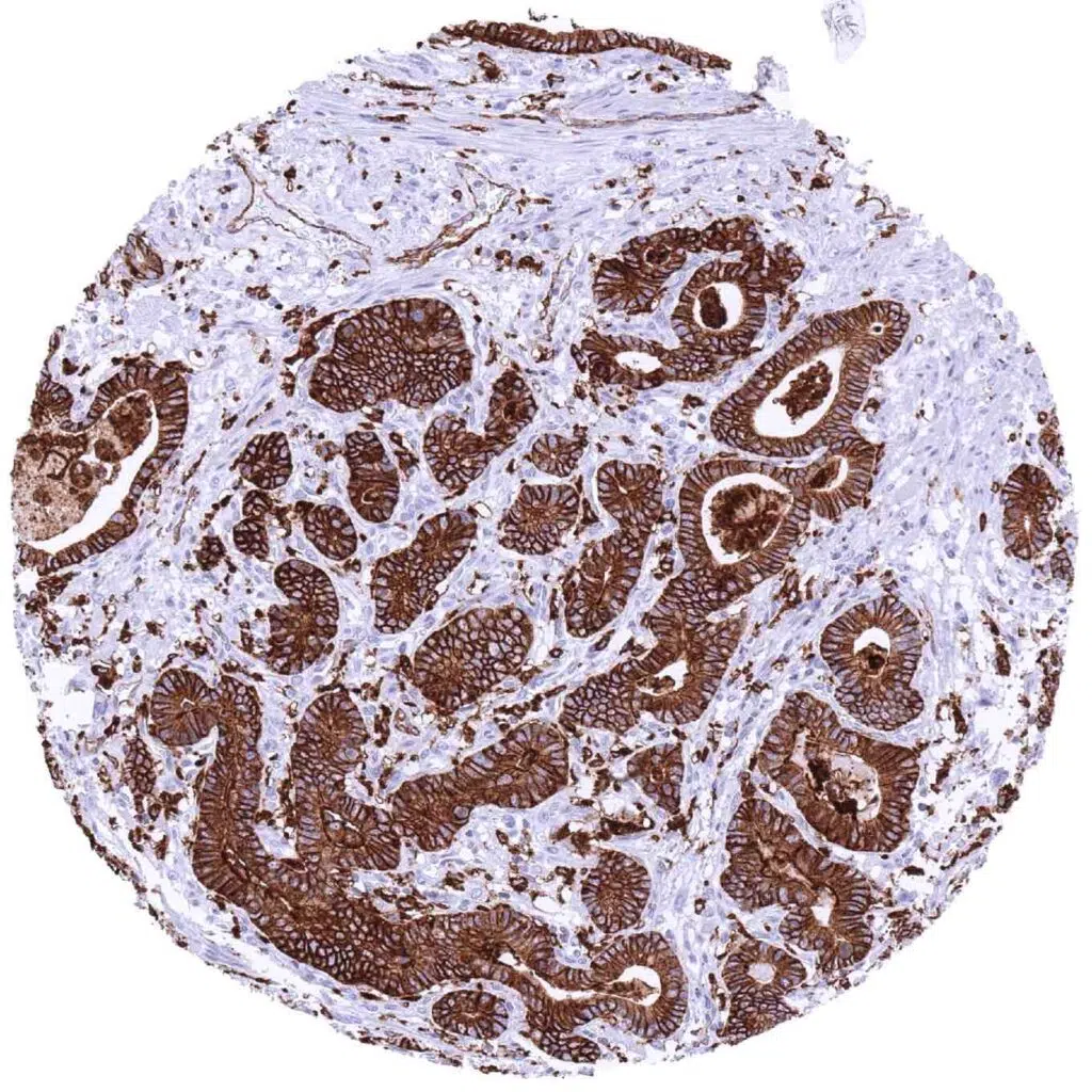
Colon – HLA-DRB1 negative colorectal adenocarcinoma including several positive inflammatory cells (HLA-DRB1 immunohistochemistry)
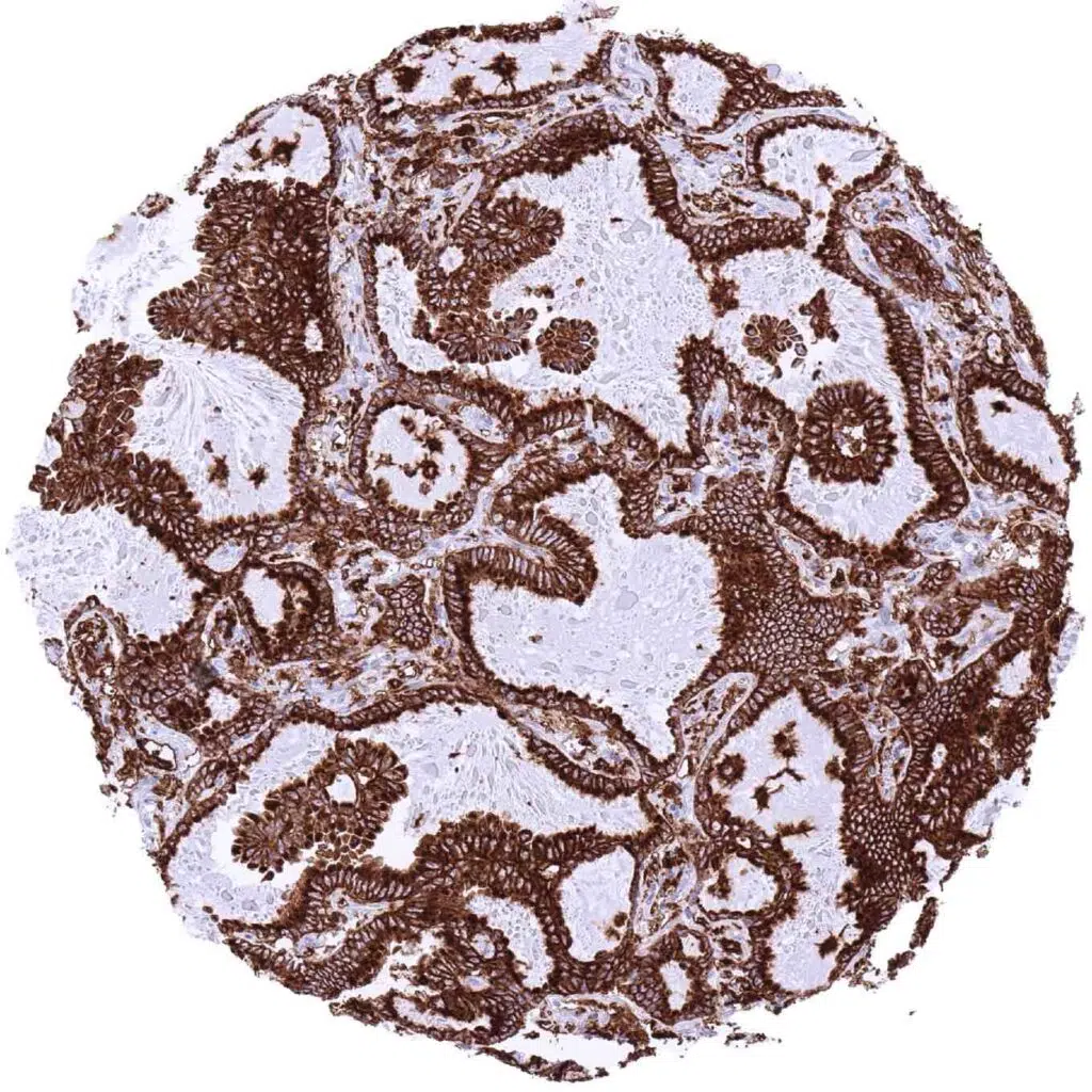
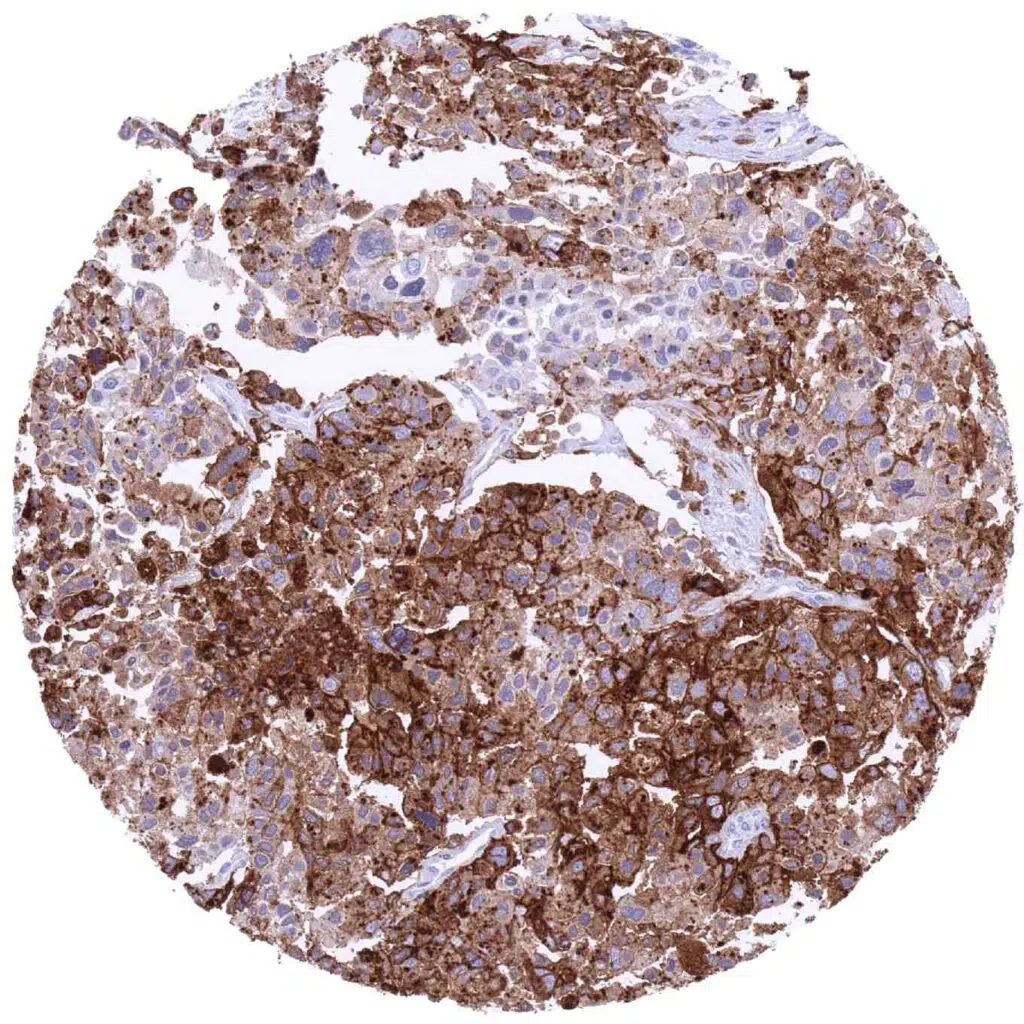
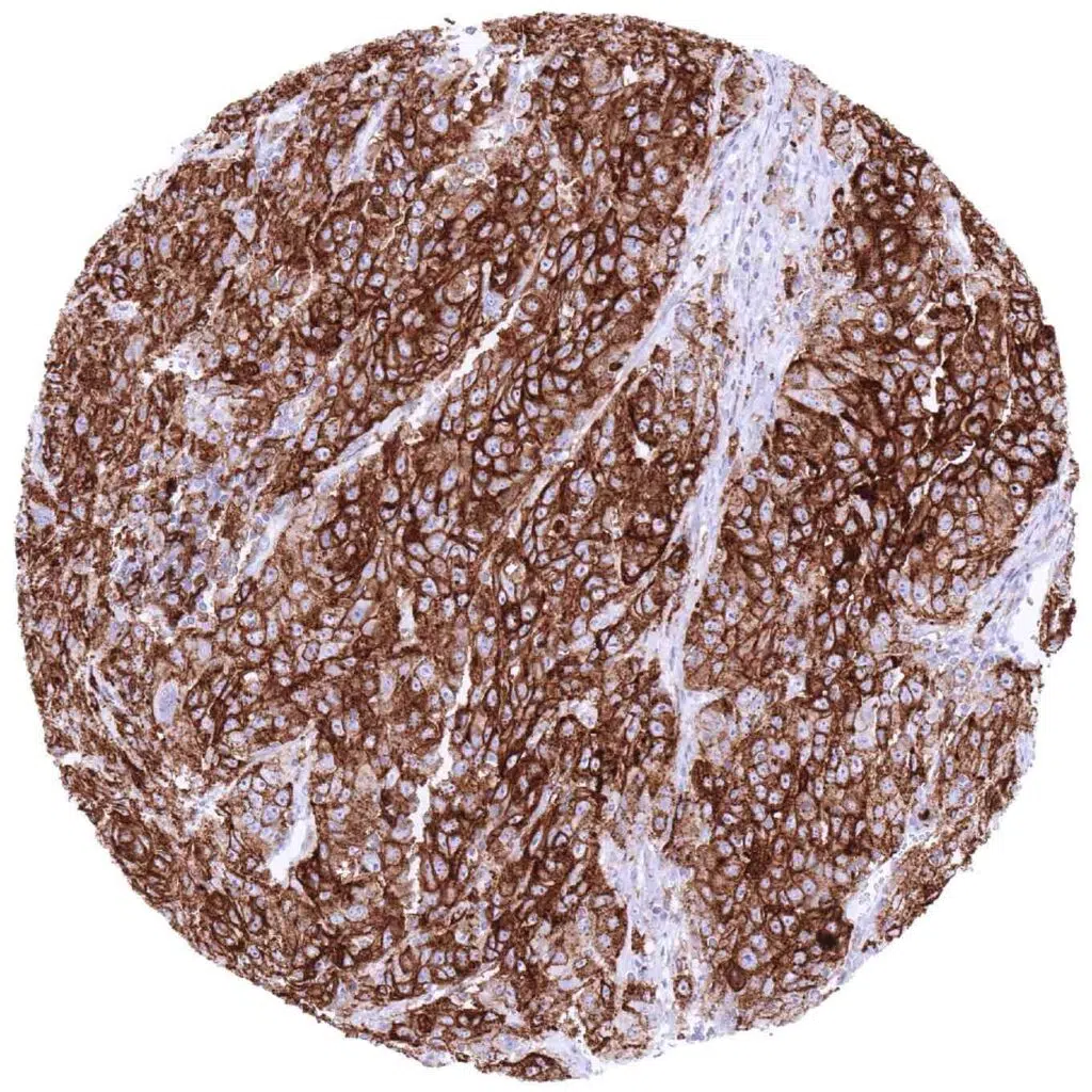
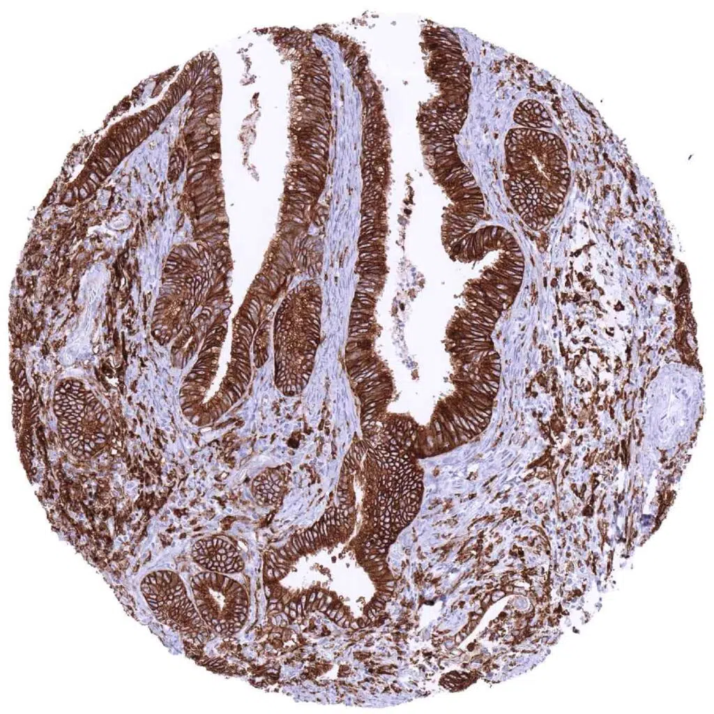
Esophagus – Adenocarcinoma with strong HLA-DRB1 immunostaining of 100_ of tumor cells (HLA-DRB1 immunohistochemistry)
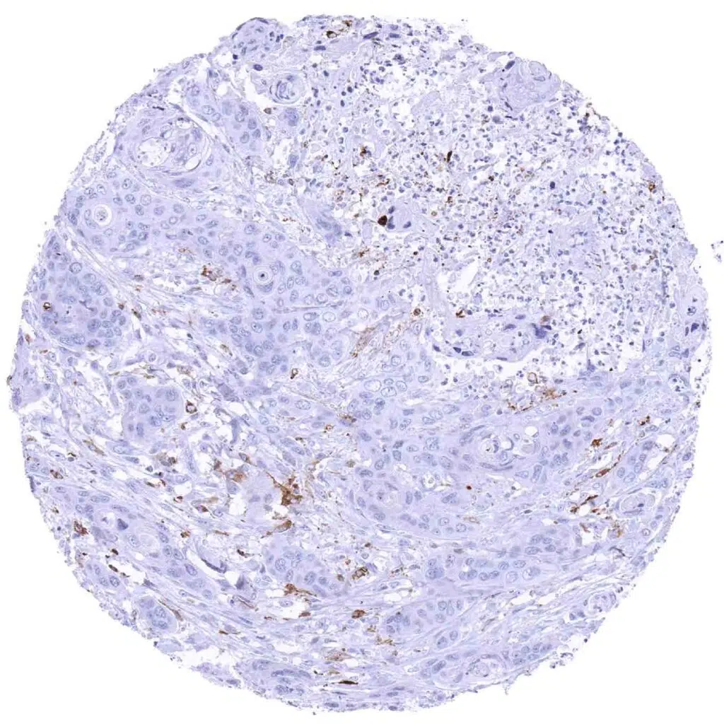
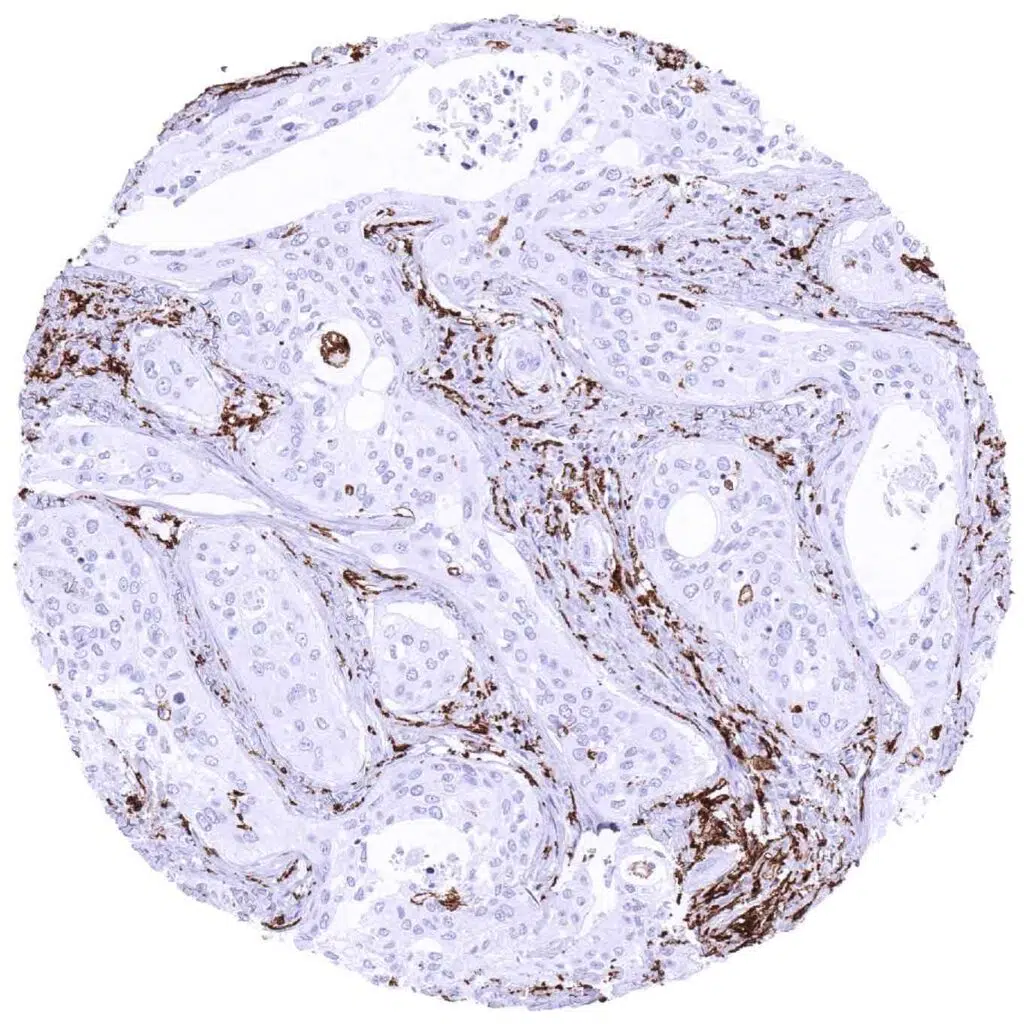
Esophagus – HLA-DRB1 negative squamous cell carcinoma containing few positive inflammatory cells (HLA-DRB1 immunohistochemistry)
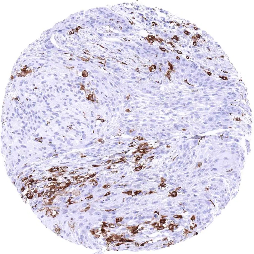
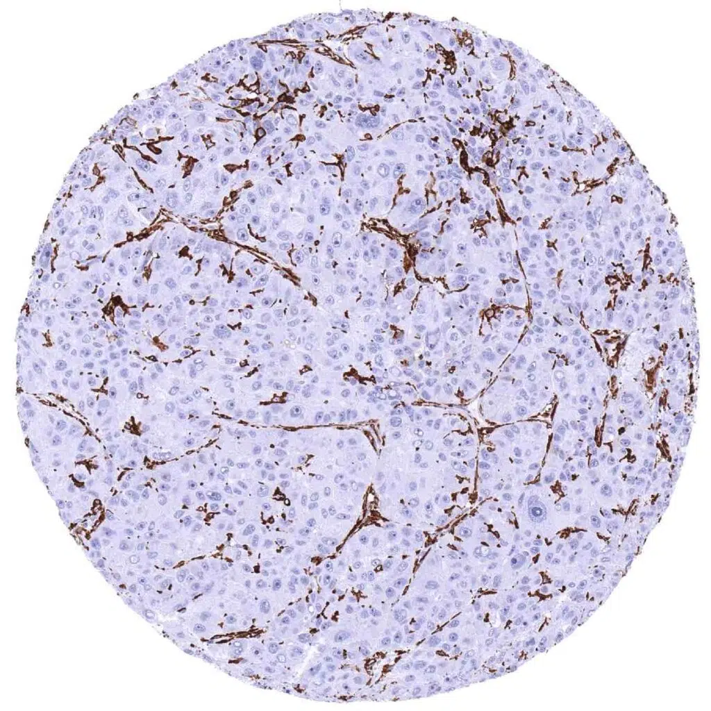
Larynx – HLA-DRB1 negative squamous cell carcinoma showing a strong staining of inflammatory cells (HLA-DRB1 immunohistochemistry)
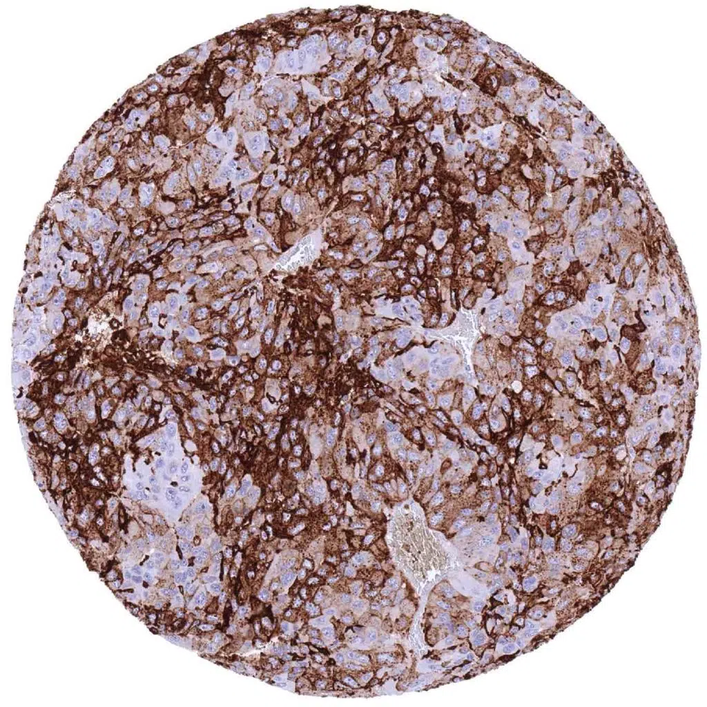
Liver – Hepatocellular carcinoma with strong HLA-DRB1 immunostaining of a large fraction of tumor cells (HLA-DRB1 immunohistochemistry)
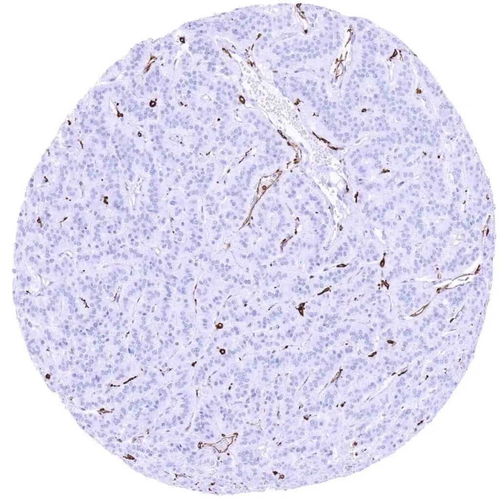
Liver – HLA-DRB1 negative hepatocellular carcinoma showing a strong staining of small vessels and inflammatory cells (HLA-DRB1 immunohistochemistry)
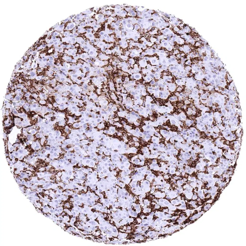
Lung – HLA-DRB1 negative malignant mesothelioma (epitheloid). Strong immunostaining of endothelial and inflammatory cells (HLA-DRB1 immunohistochemistry)
Lung – Strong HLA-DRB1 immunostaining of all tumor cells of an adenocarcinoma (HLA-DRB1 immunohistochemistry)
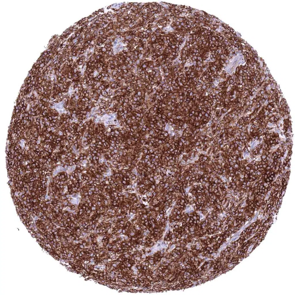
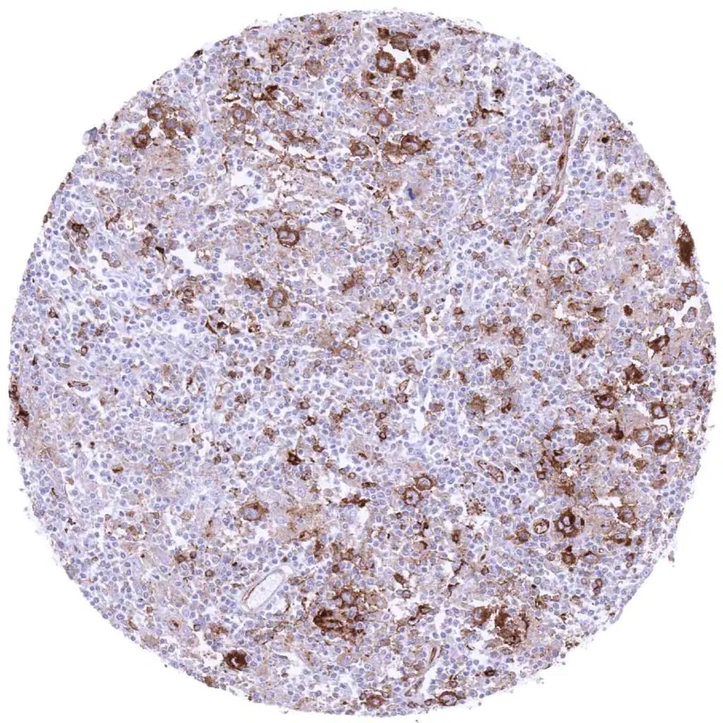
Lymph node – Diffuse large B-cell lymphoma exhibiting a strong HLA-DRB1 immunostaining of all tumor cells (HLA-DRB1 immunohistochemistry)
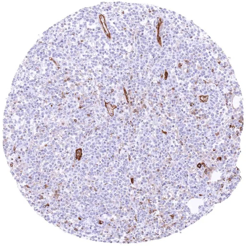
Lymph node – HLA-DRB1 negative diffuse large B-cell lymphoma. Some blood vessels are HLA-DRB1 positive (HLA-DRB1 immunohistochemistry)
Lymph node – Hodgkin’s lymphoma containing HLA-DRB1 positive Hodgkin’s and Reed-Sternberg cells (HLA-DRB1 immunohistochemistry).
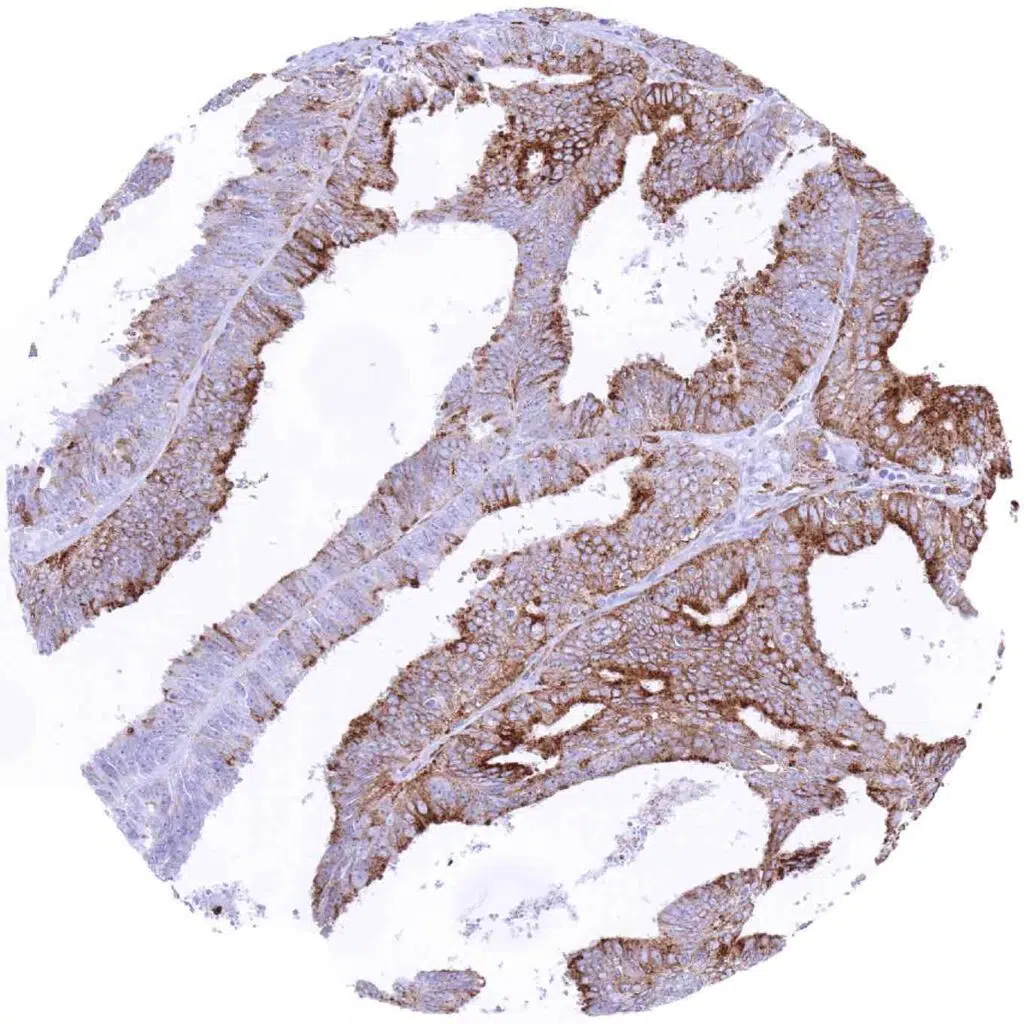
Ovary – Endometroid carcinoma with HLA-DRB1 immunostaining of variable intensity in tumor cells (HLA-DRB1 immunohistochemistry)
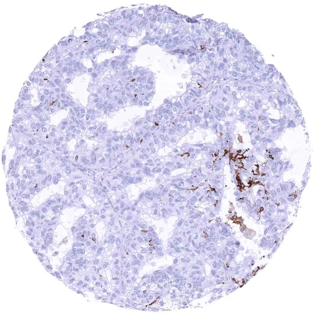
Ovary – HLA-DRB1 negative clear cell carcinoma containing few positive inflammatory cells (HLA-DRB1 immunohistochemistry)
Ovary – HLA-DRB1 negative clear cell carcinoma with distinct HLA-DRB1 staining of small vessels (HLA-DRB1 immunohistochemistry)
Ovary – Serous high-grade carcinoma showing a strong HLA-DRB1 immunostaining of most tumor cells (HLA-DRB1 immunohistochemistry)
Pancreas – Ductal adenocarcinoma with weak to moderate HLA-DRB1 immunostaining of about 40_ of tumor cells. Inflammatory cells are also positive (HLA-DRB1 immunohistochemistry)
Pancreas – HLA-DRB1 negative neuroendocrine tumor showing a distinct positivity of associated small vessels and inflammatory cells (HLA-DRB1 immunohistochemistry)
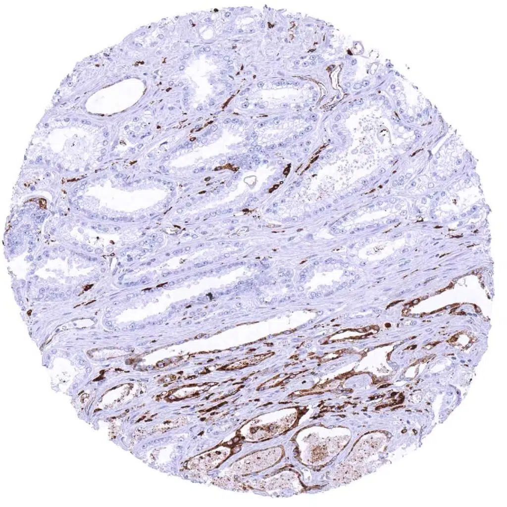
Prostate – HLA-DRB1 negative adenocarcinoma (Gleason 3+3=6) showing strong staining in adjacent atrophic glands (HLA-DRB1 immunohistochemistry)
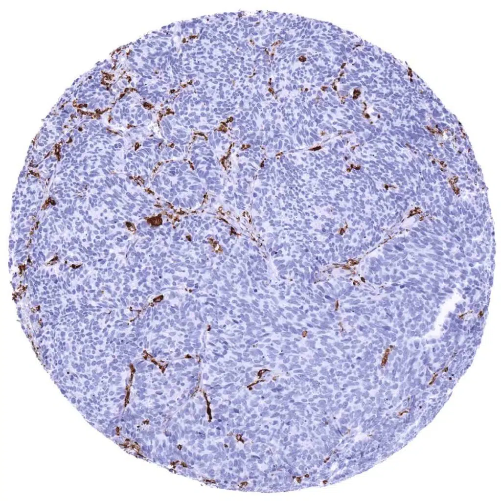
Prostate – HLA-DRB1 negative small cell carcinoma containing few positive endothelial and inflammatory cells (HLA-DRB1 immunohistochemistry)
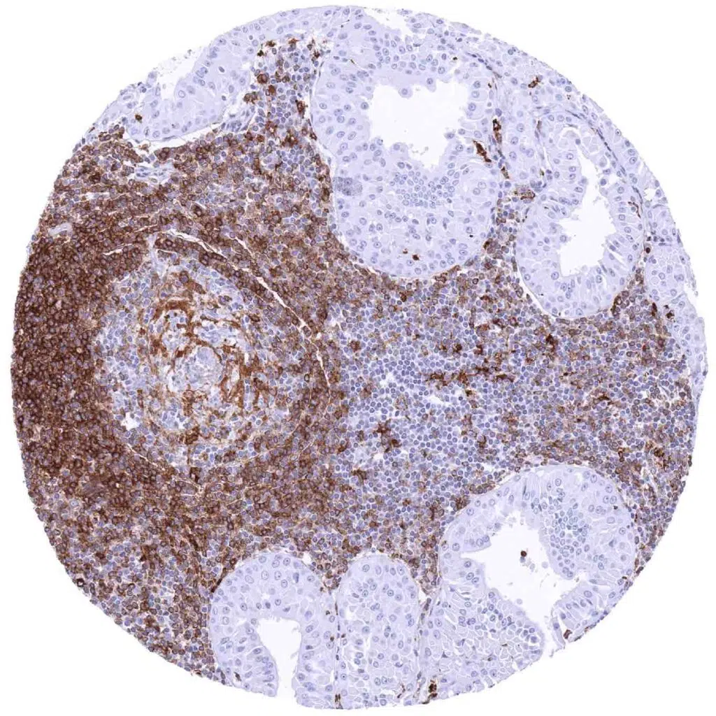
Salivary gland – HLA-DRB1 negative Warthin tumor with strong staining of associated inflammatory cells (HLA-DRB1 immunohistochemistry)
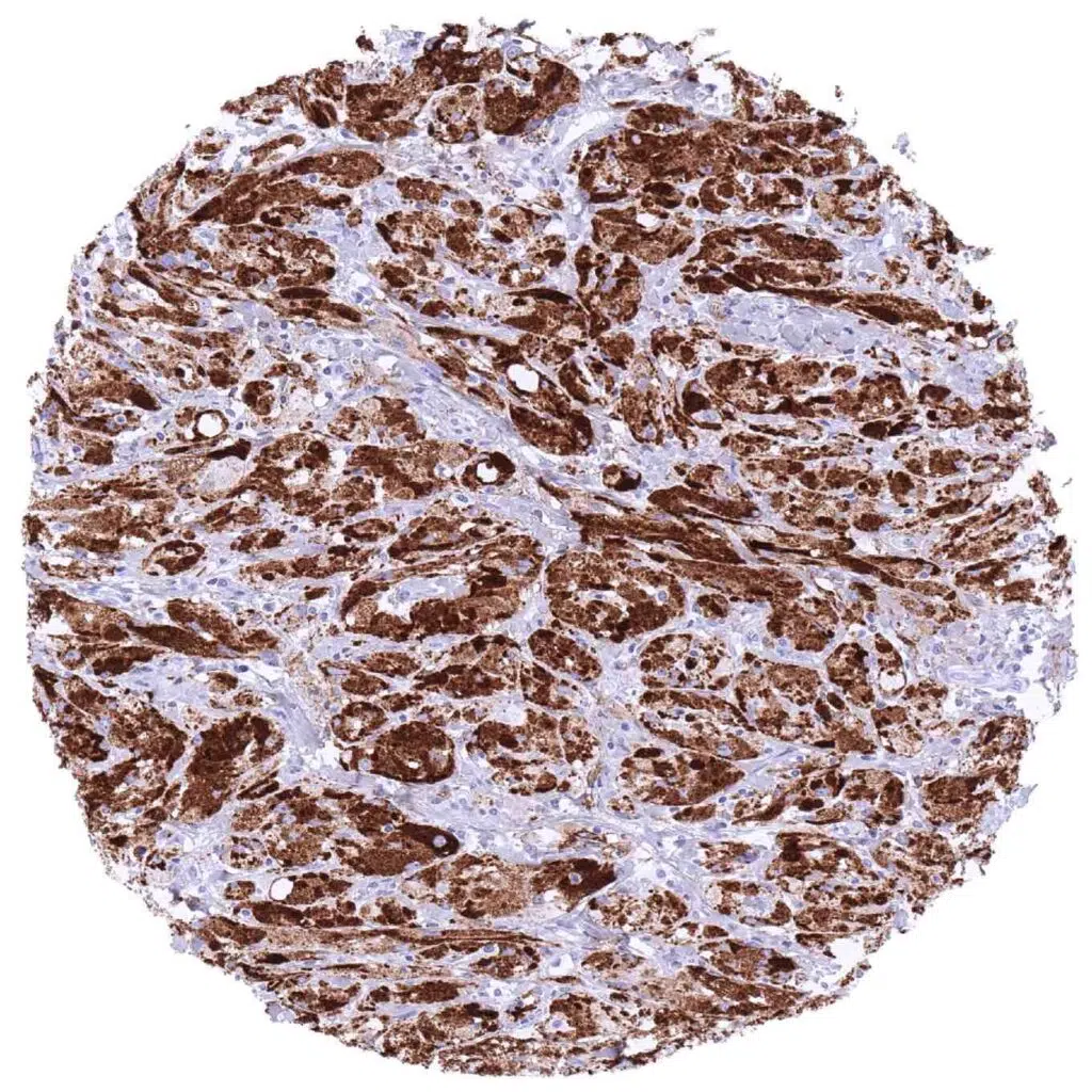
Soft tissue – Granular cell tumor exhibiting a strong HLA-DRB1 immunostaining of all tumor cells (HLA-DRB1 immunohistochemistry)
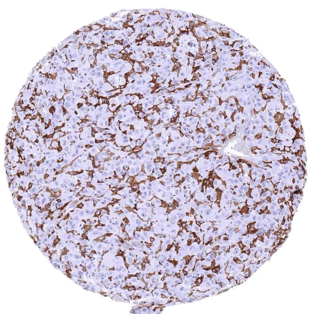
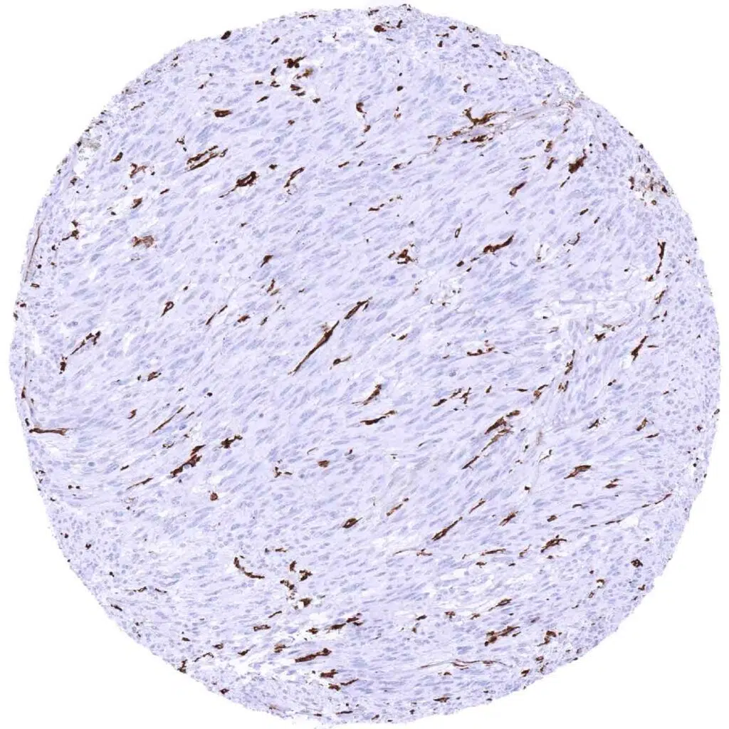
Soft tissue – HLA-DRB1 negative liposarcoma. Strong positivity of endothelial and inflammatory cells (HLA-DRB1 immunohistochemistry)
Stomach – Gastric adenocarcinoma with strong HLA-DRB1 immunostaining of all tumor cells (HLA-DRB1 immunohistochemistry)
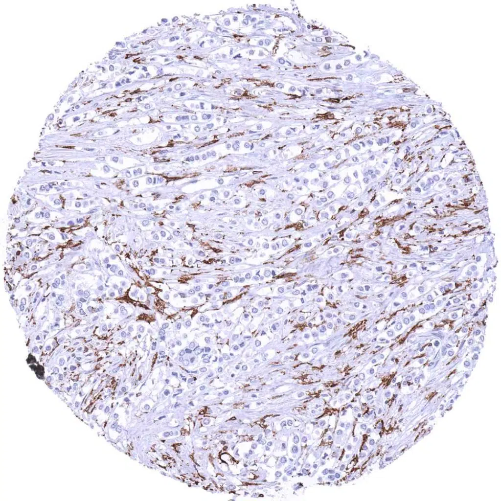
Stomach – HLA-DRB1 negative gastric adenocarcinoma (diffuse type). Strong positivity of associated inflammatory cells (HLA-DRB1 immunohistochemistry)
Stomach – HLA-DRB1 negative gastric adenocarcinoma. Associated inflammatory cells show strong staining (HLA-DRB1 immunohistochemistry)
Stomach – HLA-DRB1 negative gastrointestinal stromal tumor (GIST). Strong staining of small vessels and inflammatory cells (HLA-DRB1 immunohistochemistry)
Testis – HLA-DRB1 negative seminoma showing HLA-DRB1 staining of inflammatory cells and of small vessels (HLA-DRB1 immunohistochemistry)
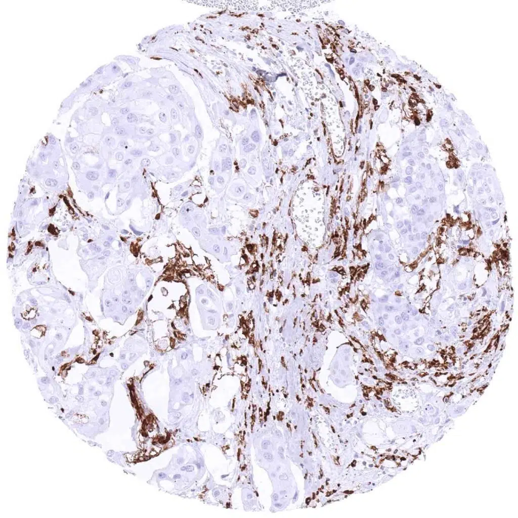
Urinary bladder – HLA-DRB1 negative muscle-invasive urothelial carcinoma with strong HLA-DRB1 immunostaining of associated inflammatory cells (HLA-DRB1 immunohistochemistry)
Urinary bladder – Muscle-invasive urothelial carcinoma with strong HLA-DRB1 immunostaining of tumor cells (HLA-DRB1 immunohistochemistry)
Uterus, cervix – Adenocarcinoma with strong HLA-DRB1 immunostaining of tumor cells and of associated inflammatory cells (HLA-DRB1 immunohistochemistry)
Uterus, cervix – HLA-DRB1 negative squamous cell carcinoma including positive inflammatory cells (HLA-DRB1 immunohistochemistry)
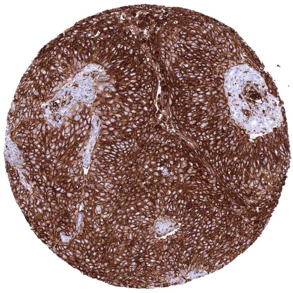
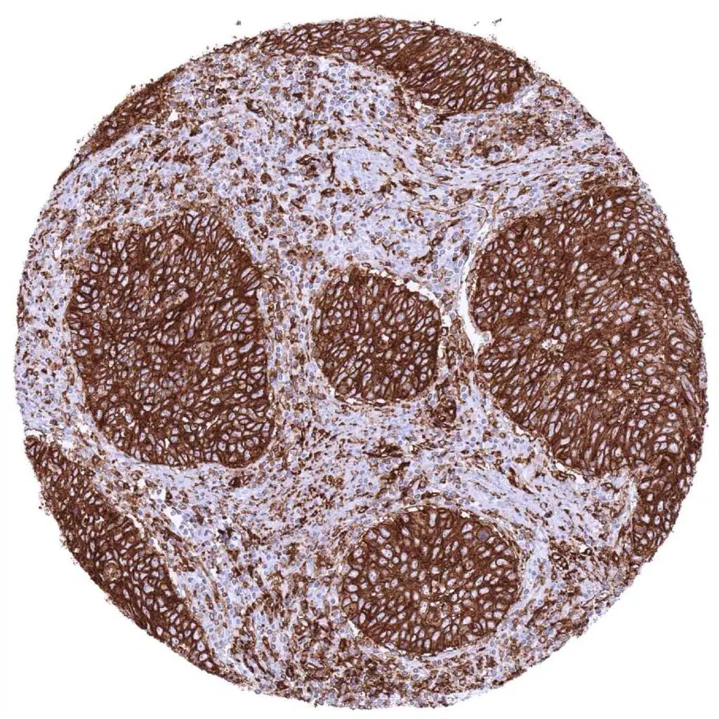
Uterus, cervix – Squamous cell carcinoma with intense HLA-DRB1 immunostaining of tumor cells (HLA-DRB1 immunohistochemistry)
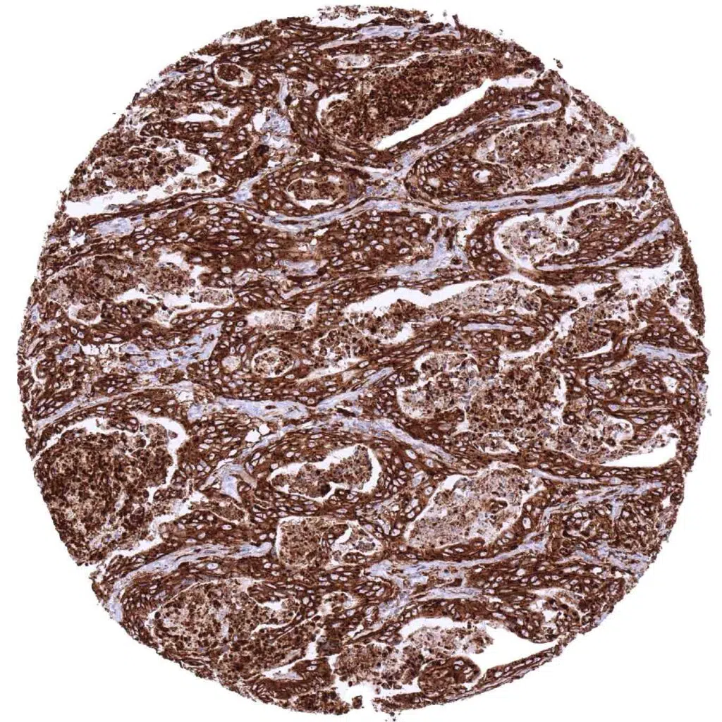
Uterus, cervix – Squamous cell carcinoma with strong HLA-DRB1 immunostaining of tumor cells and of intermingled inflammatory cells (HLA-DRB1 immunohistochemistry)

| TOP |