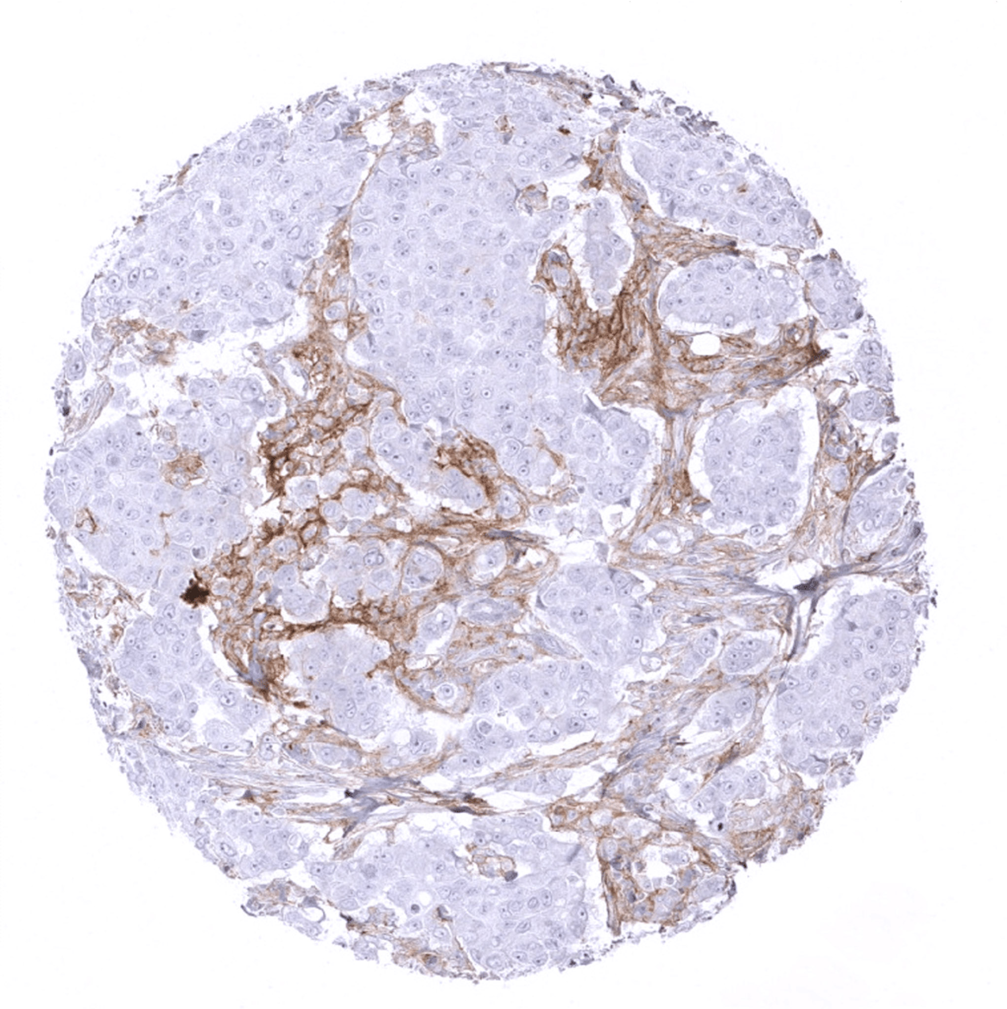
Breast – Breast cancer NST with PD-L1 negative tumor cells but intense PD-L1 staining in tumor associated inflammatory cells.
Breast – Breast cancer NST with PD-L1 negative tumor cells. Tumor associated inflammatory cells show strongest PD-L1 staining if they are immediately adjacent to tumor cells.
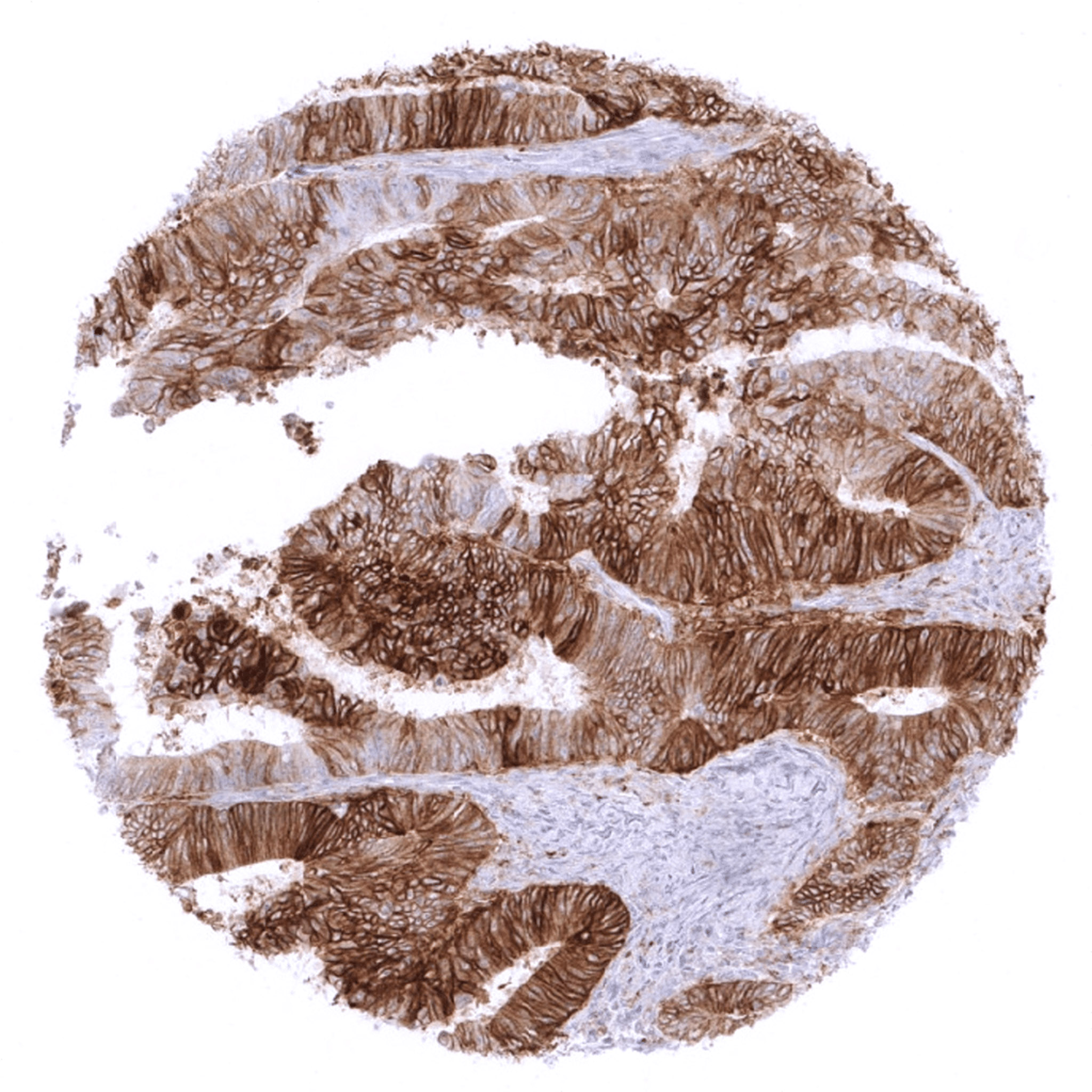
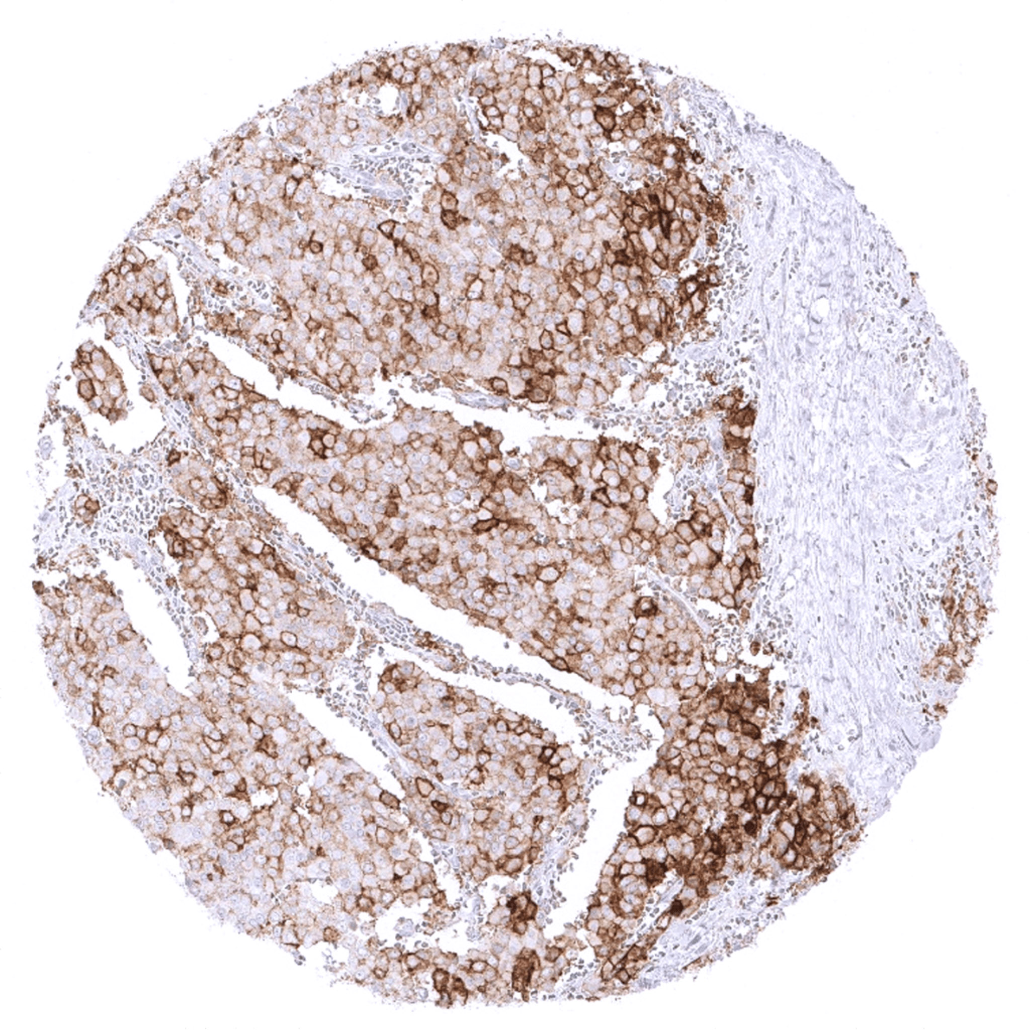
Colon – Colorectal adenocarcinoma with moderate to strong membranous PD-L1 staining in the vast majority of its tumor cells.
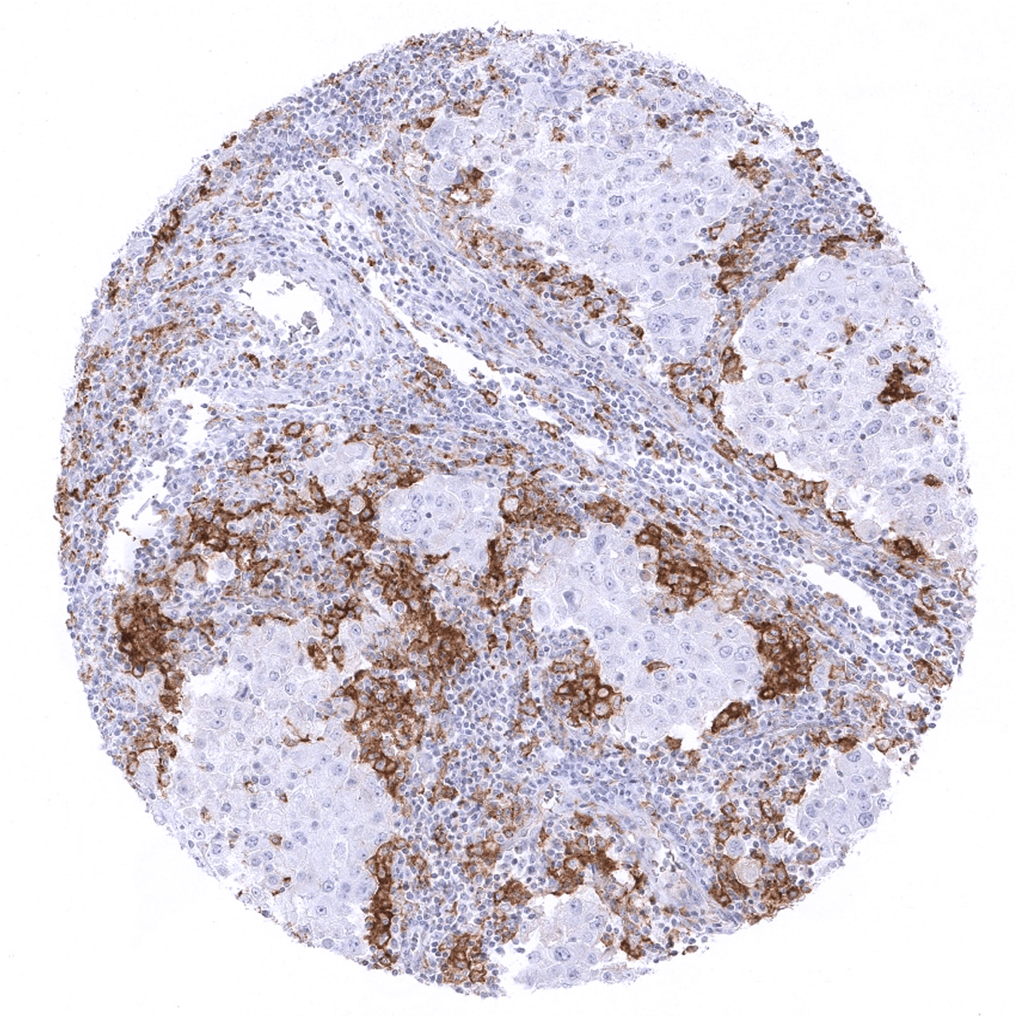
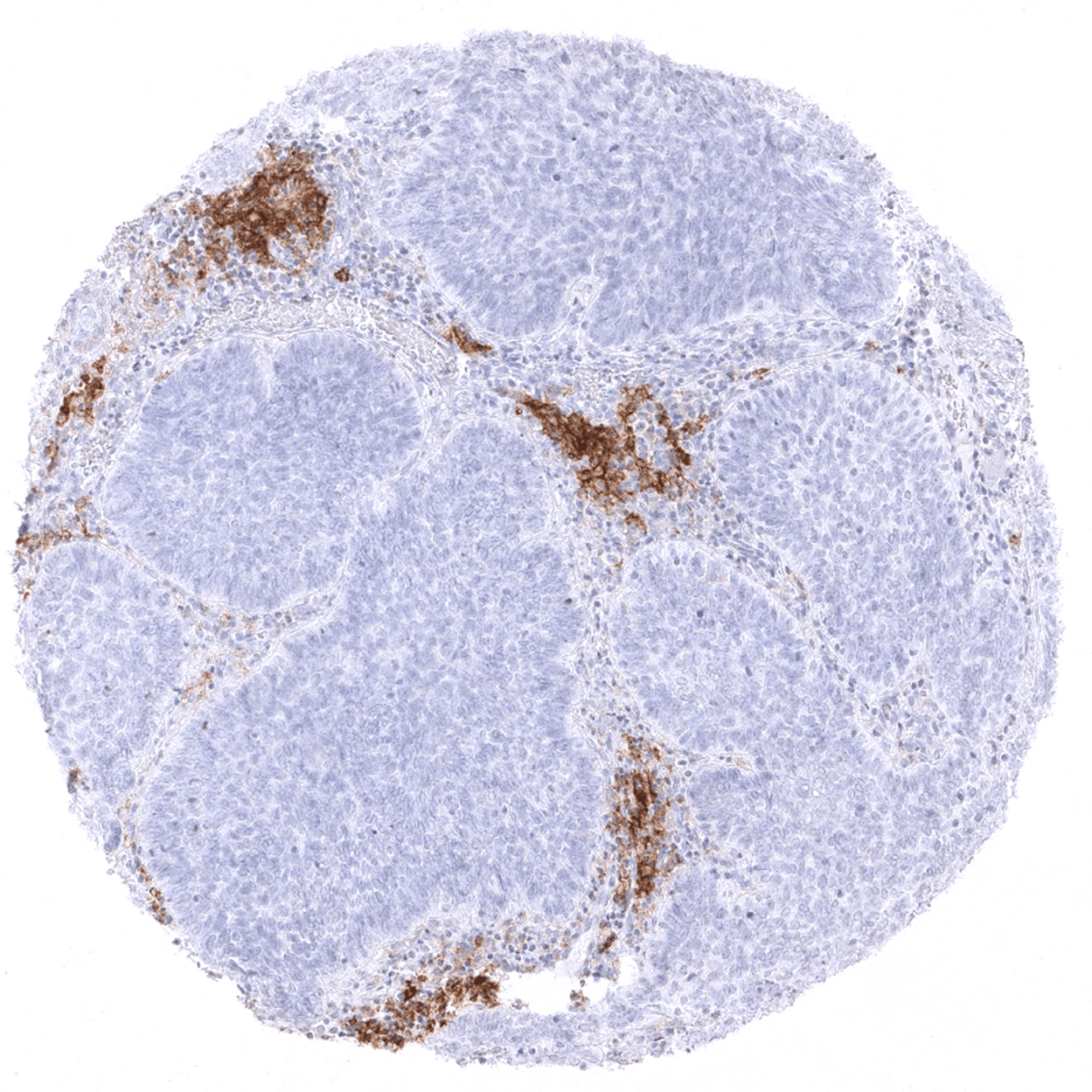
Colon – Colorectal adenocarcinoma with PD-L1 negative tumor cells surrounded by large numbers of tumor associated inflammatory cells with intense PD-L1 staining.
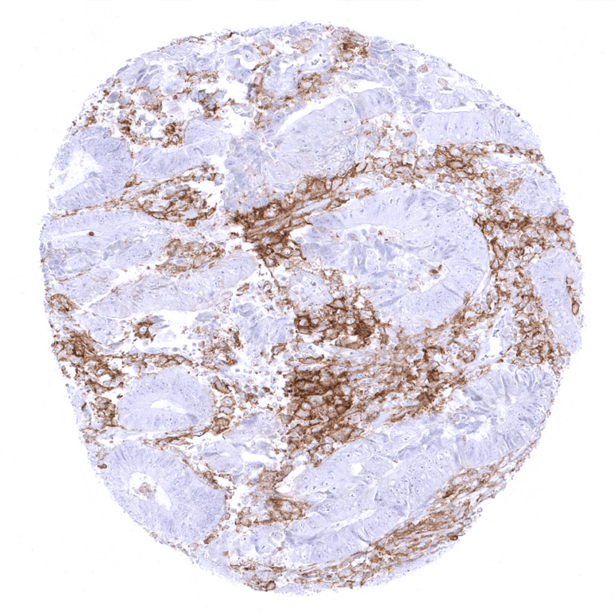
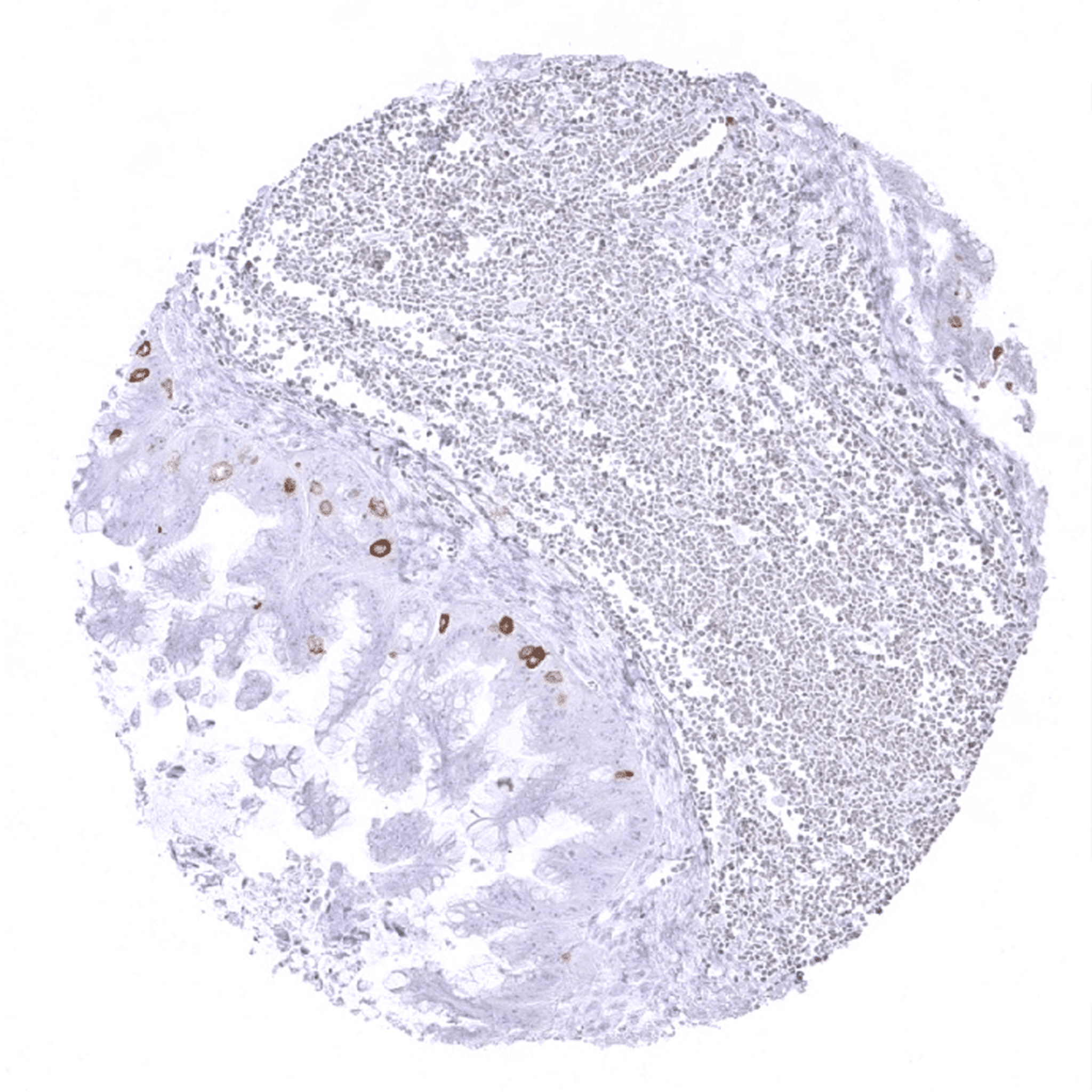
Colon – Colorectal adenocarcinoma with PD-L1 staining limited to intraglandular macrophages.
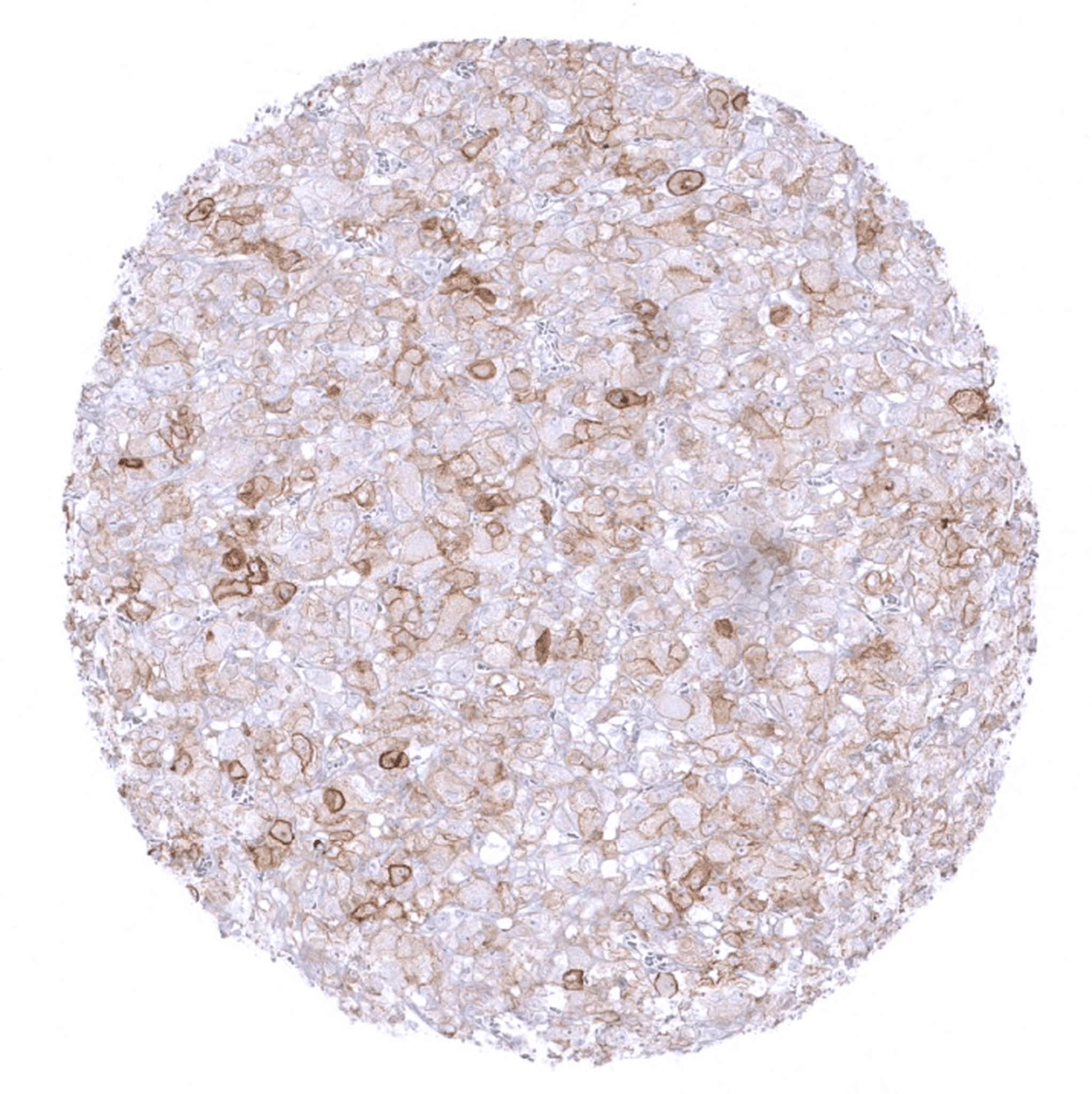
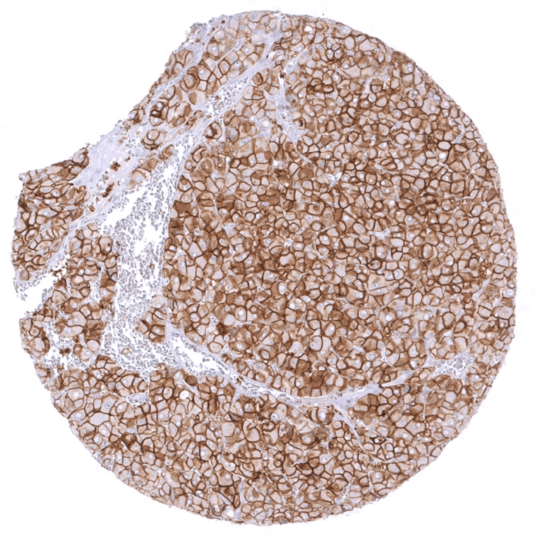
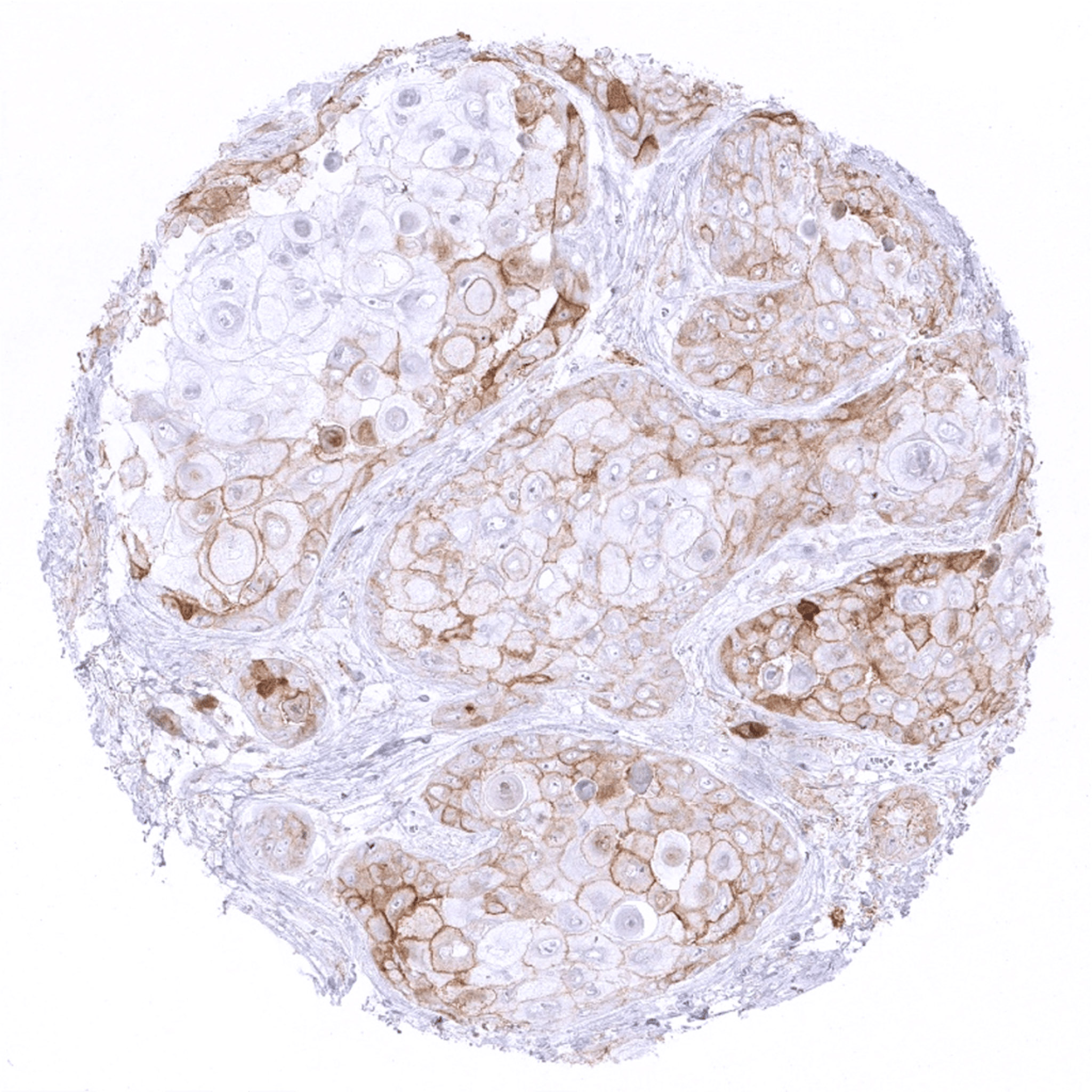
Kidney – Weak to moderate membranous PD-L1 staining in a clear cell renal cell carcinoma.
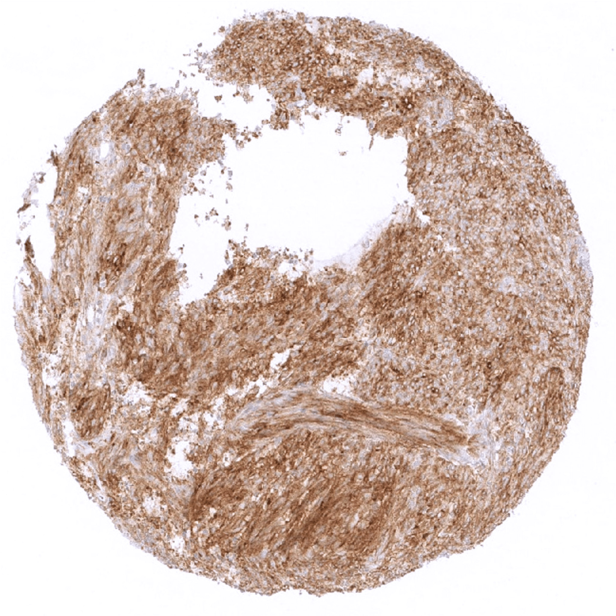
Lymph node – Strong PD-L1 immunostaining in all tumor cells of an anaplastic large cell lymphoma.
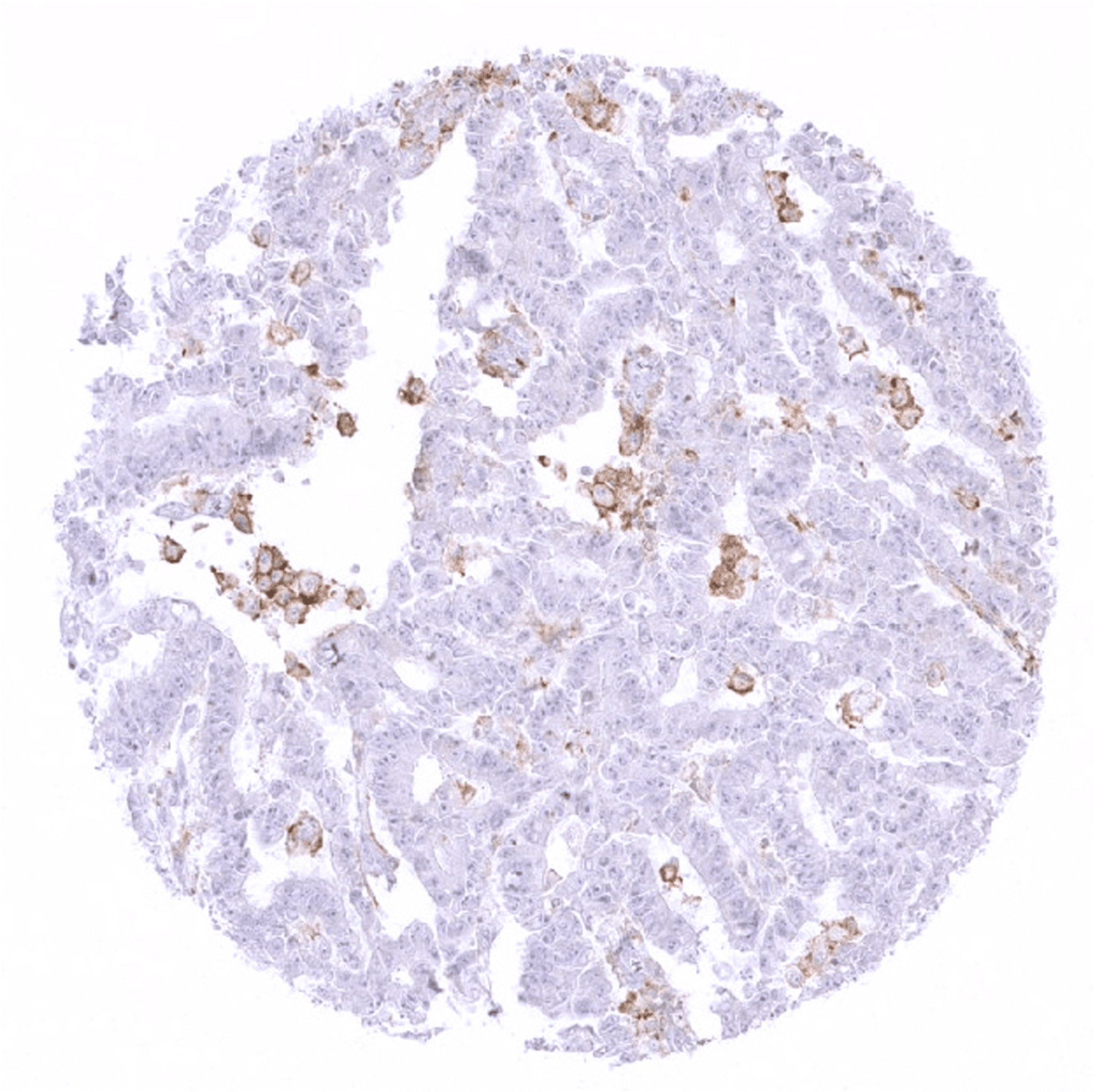
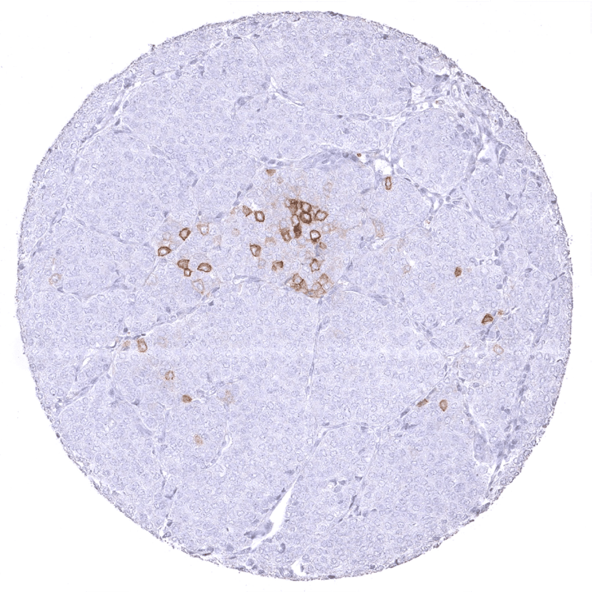
Ovary – Few scattered PD-L1 positive cells in a mucinous ovarian cancer. Most likely, these cells are tumor associated macrophages.
Prostate – Few scattered cells (tumor cells or tumor associated macrophages) with membranous PD-L1 staining in a recurrent Gleason 5+5 adenocarcinoma of the prostate.
Prostate – Moderate to strong membranous PD-L1 staining in virtually all tumor cells of a Gleason 5+5 prostate cancer.
Skin – Strong focal PD-L1 staining in tumor associated inflammatory cells in a basal cell carcinoma of the skin. PD-L1 staining is not seen in tumor cells.
Stomach – Strong PD-L1 positivity in all tumor cells of a gastrointestinal stromal tumor (GIST).
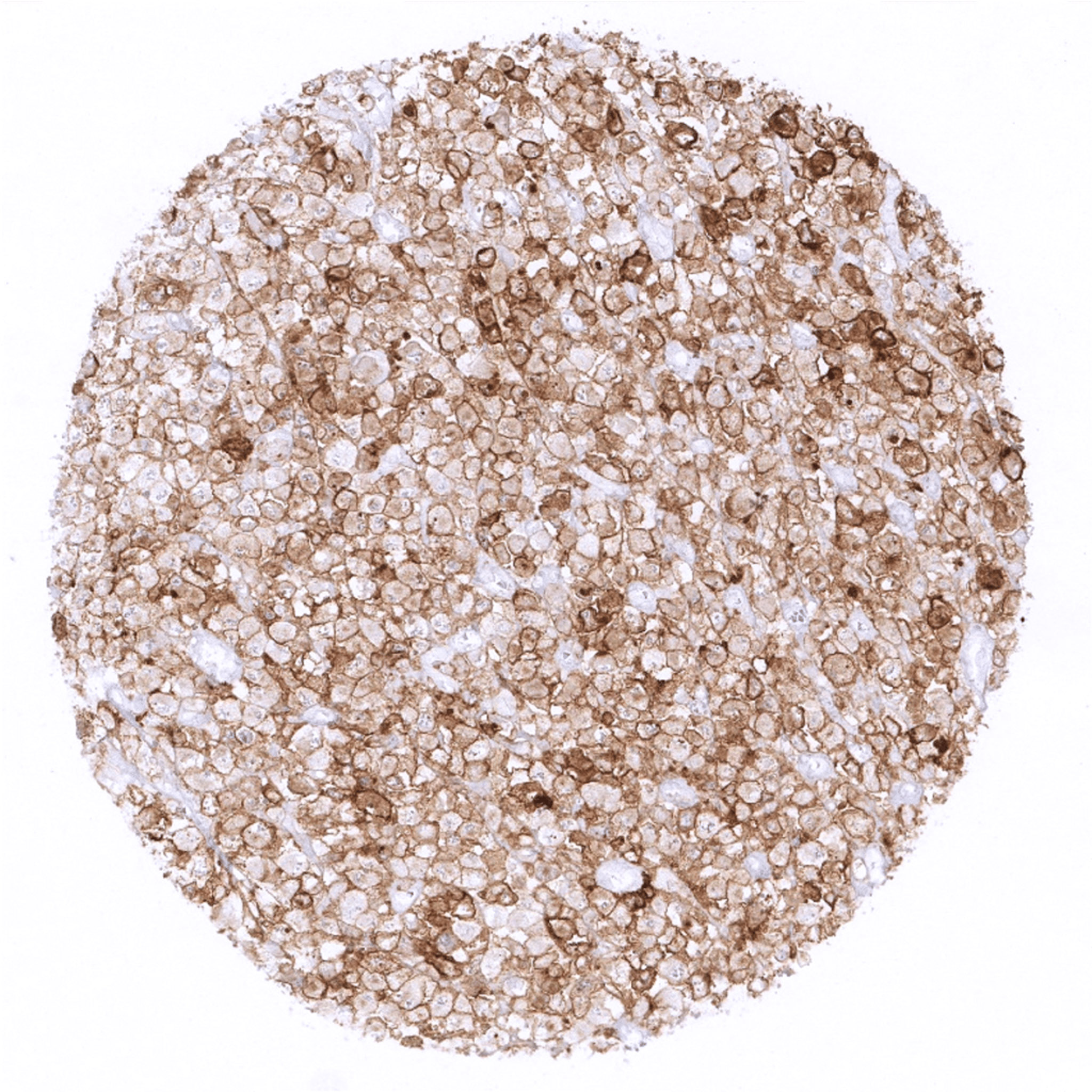
Thyroid – Follicular thyroid cancer with moderate to strong membranous PD-L1 staining.
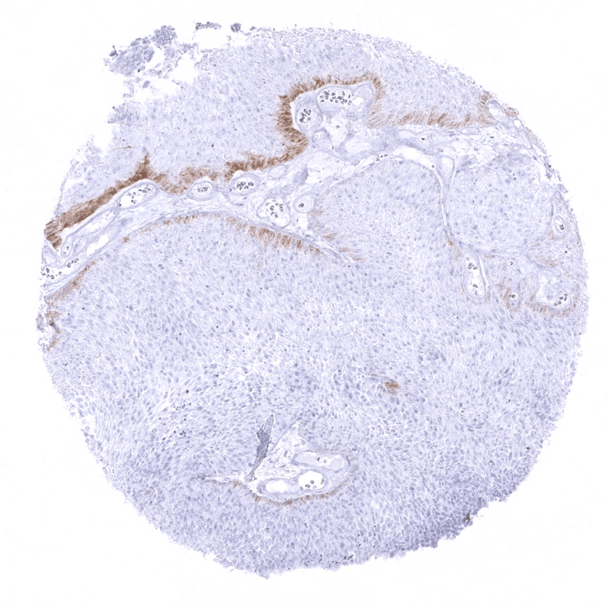
Urinary bladder – Non-invasive papillary urothelial carcinoma with moderate PD-L1 staining which is strictly limited to the basal cell layer. Absence of tumor associated inflammatory cells.
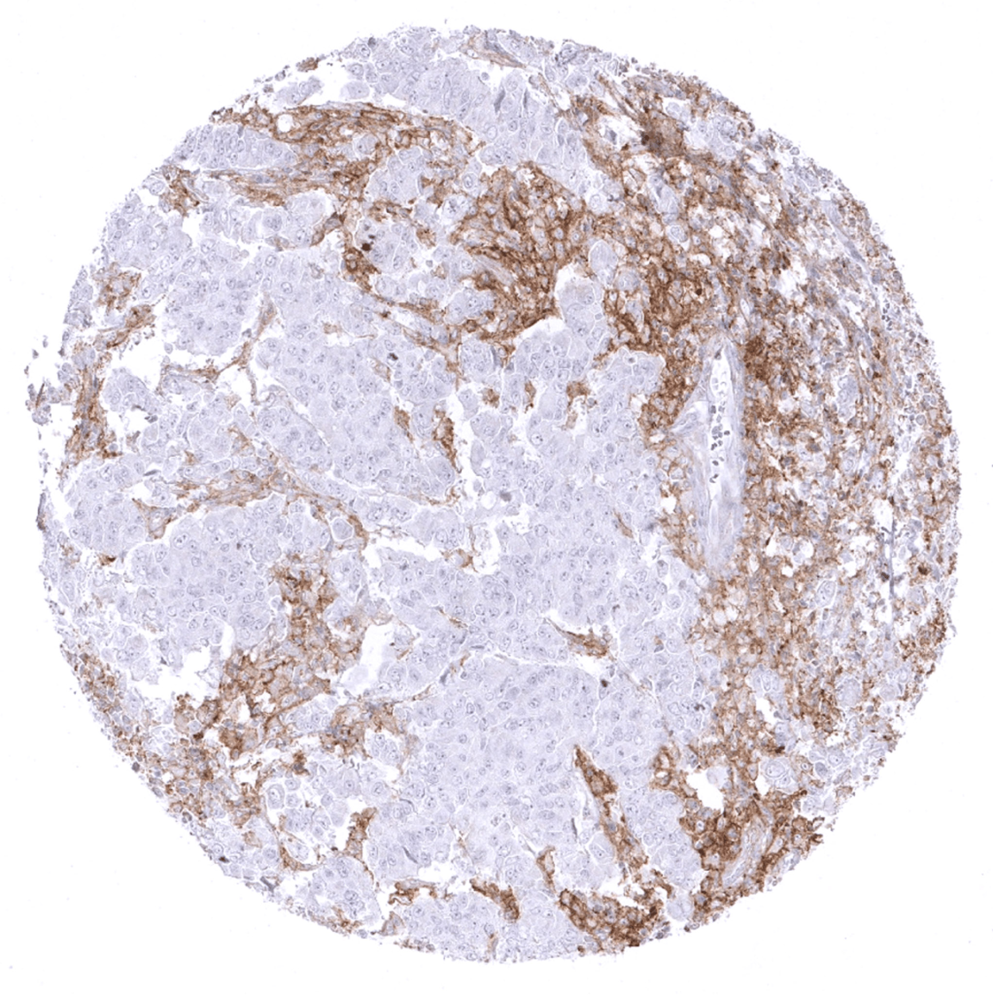
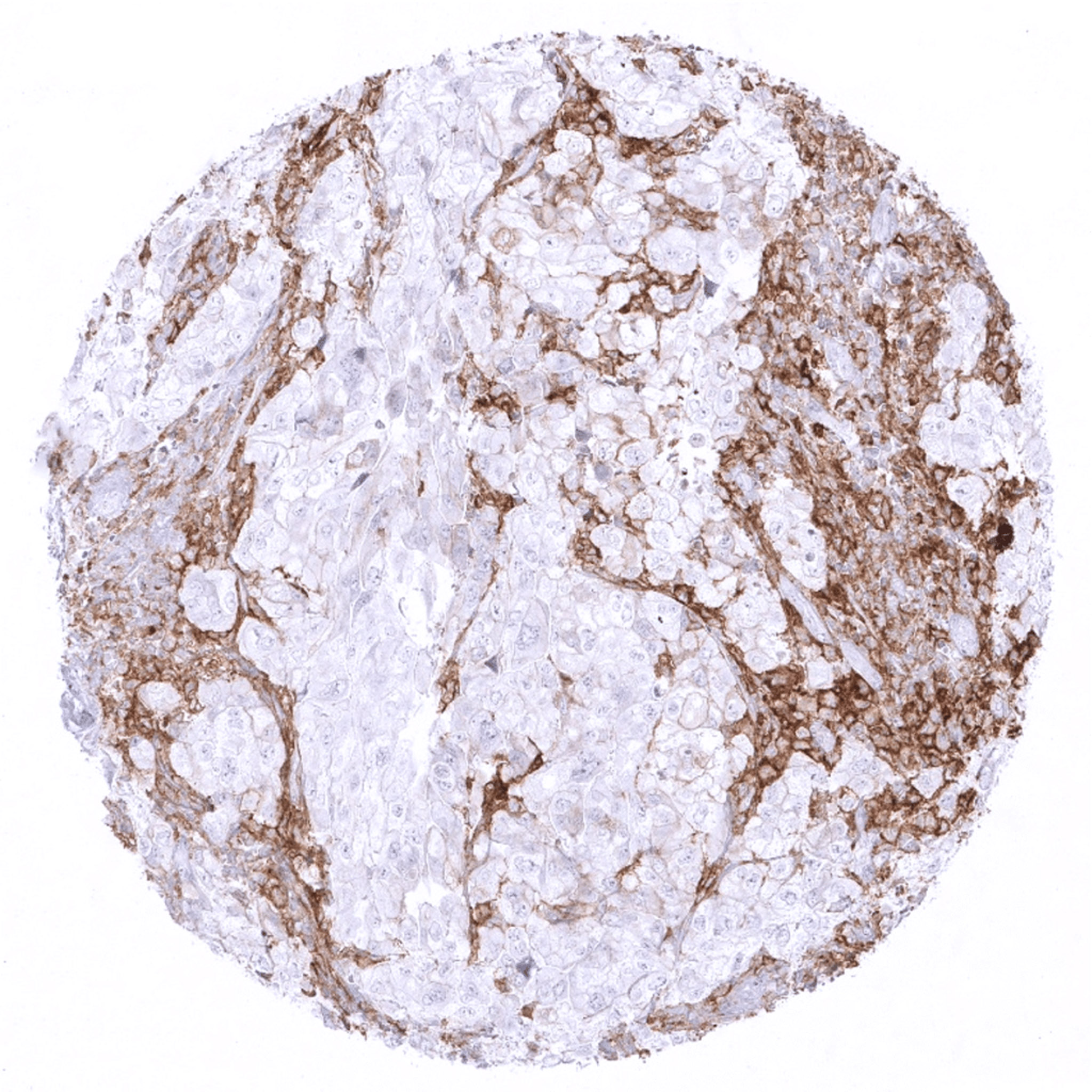
Urinary bladder – Urothelial carcinoma with PD-L1 negative tumor cells containing large numbers of PD-L1 positive tumor associated inflammatory cells.
Urinary bladder – Urothelial carcinoma with prominent PD-L1 staining in tumor associated inflammatory cells. In addition, few tumor cells show faint membranous staining.
Vulva – Weak to moderate membranous PD-L1 positivity in the majority of tumor cells of a squamous cell carcinoma of the vulva.