GeneTex
  • Country / Location Selection

United States (US)

Normal Tissue Gallery- GTX04425

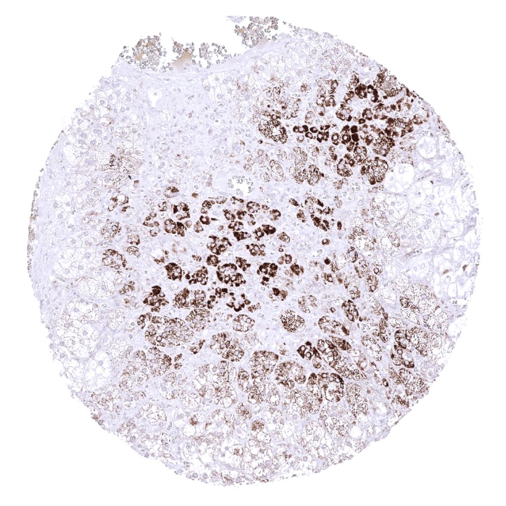
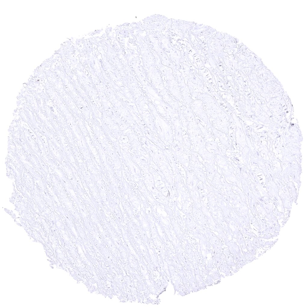
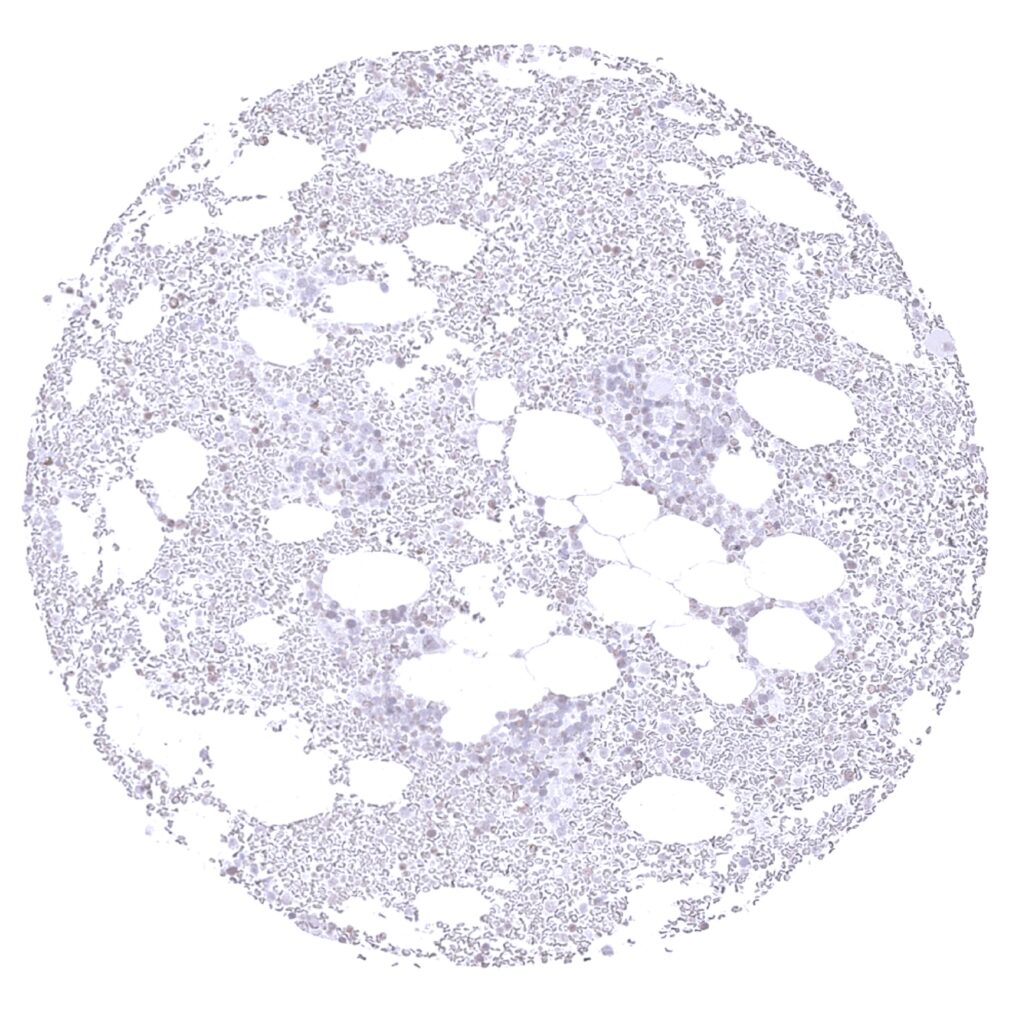
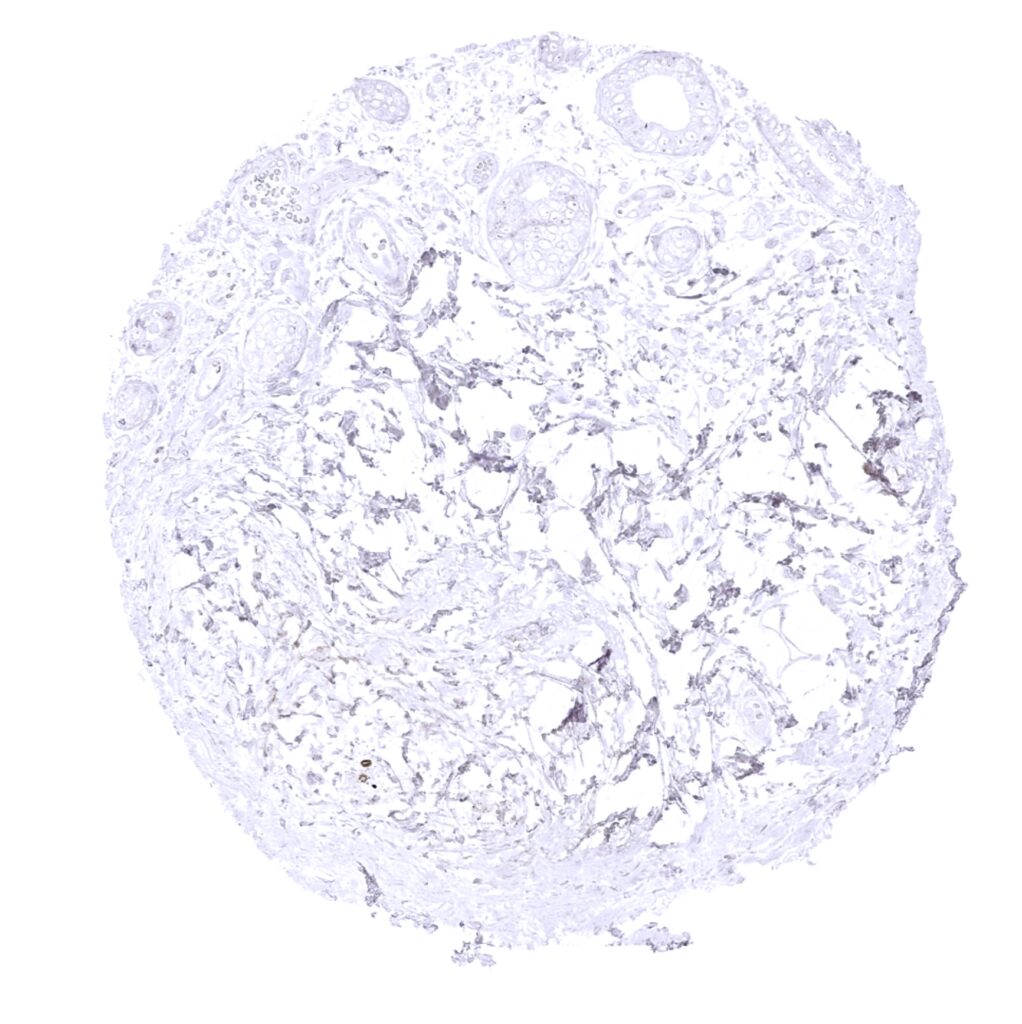
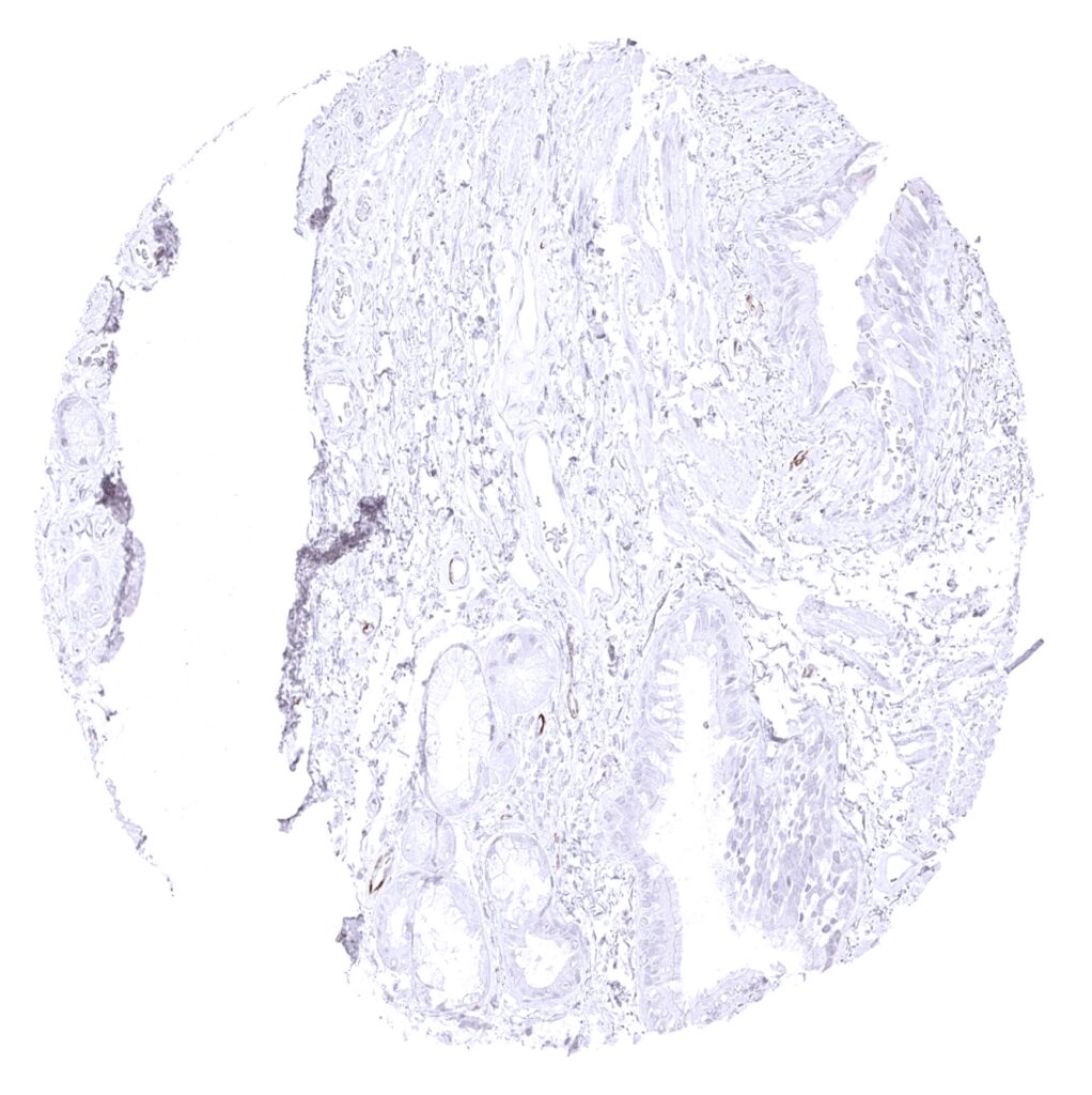
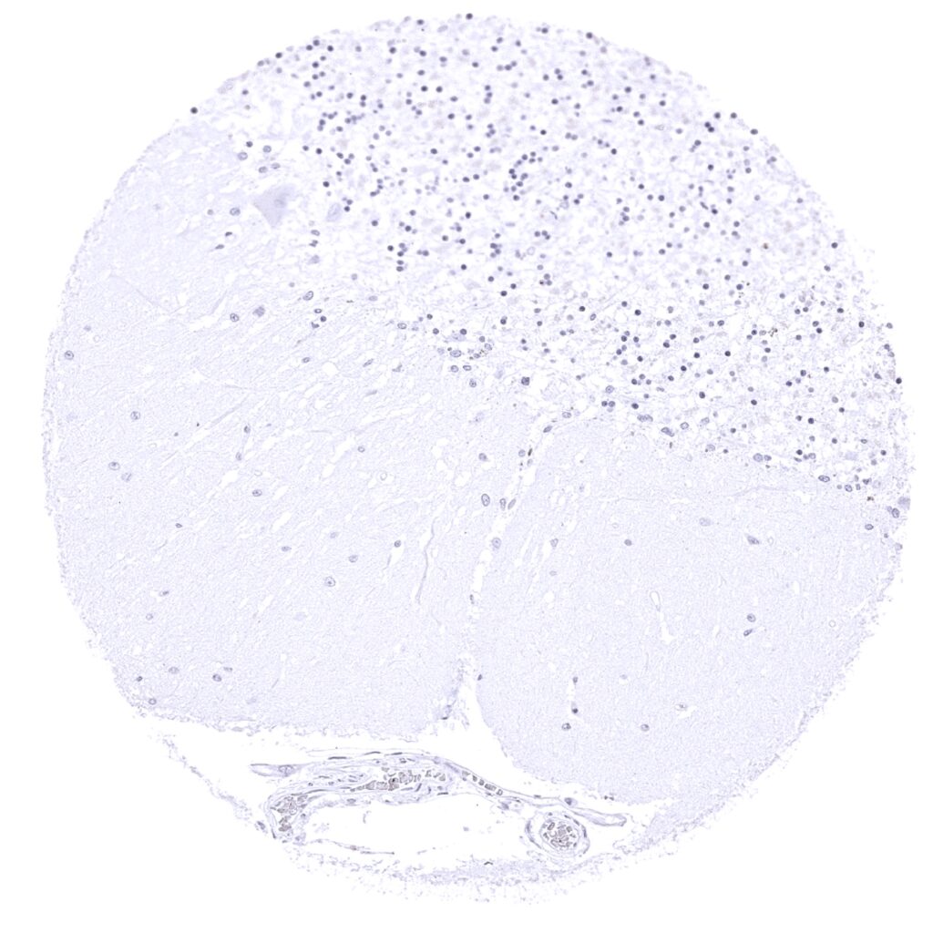
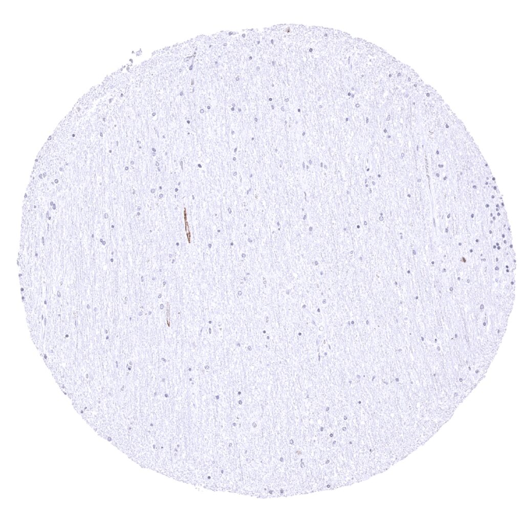
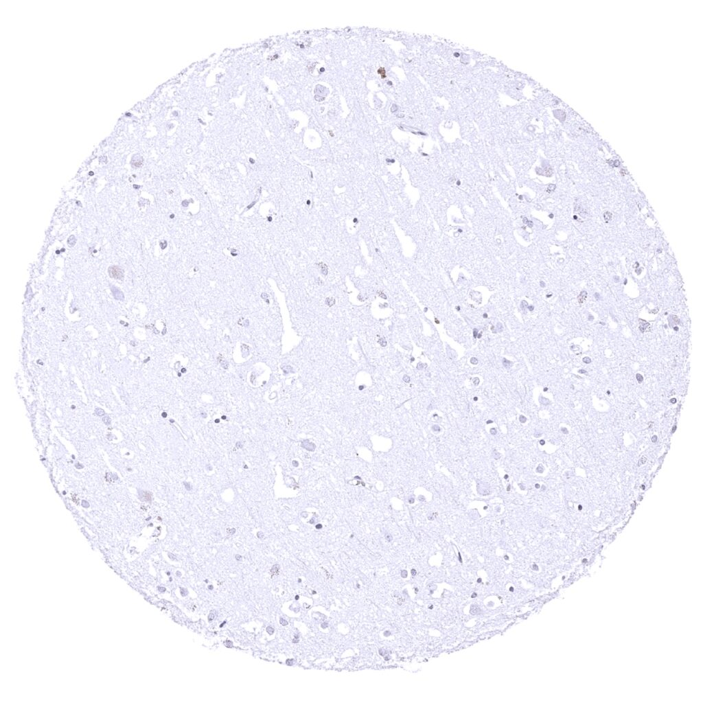
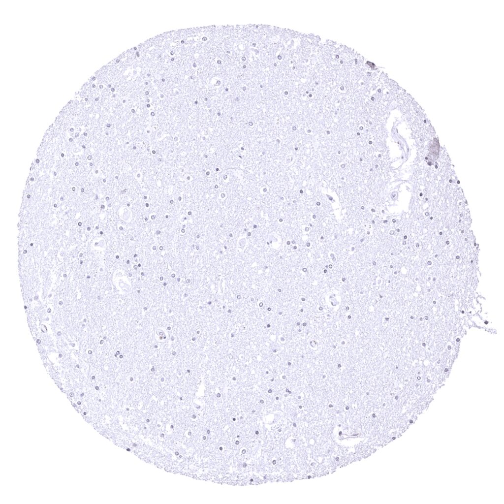
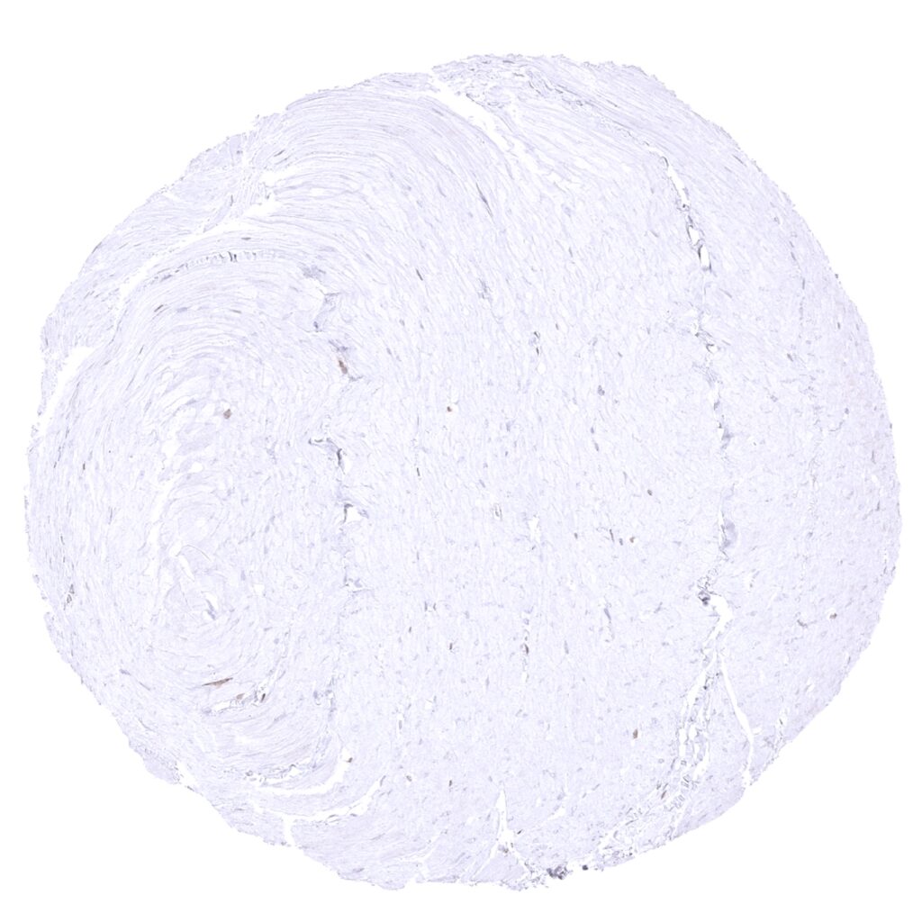
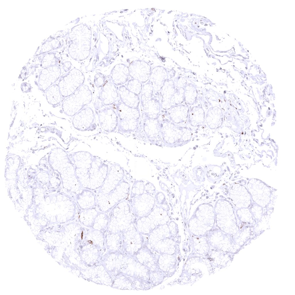
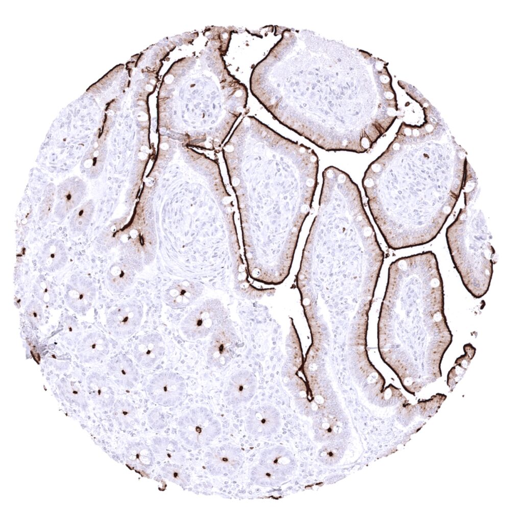
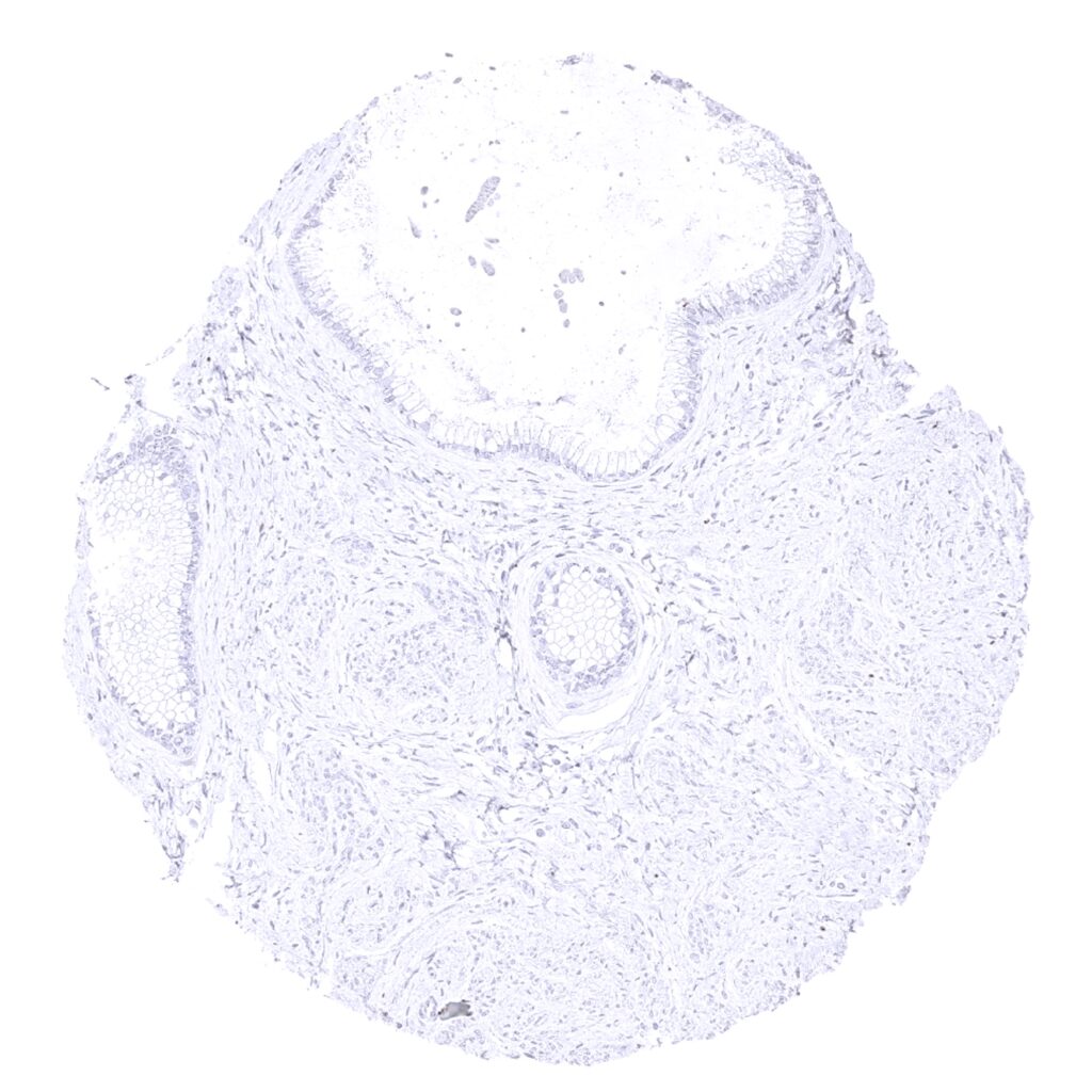
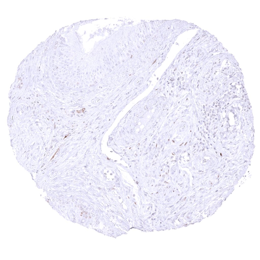
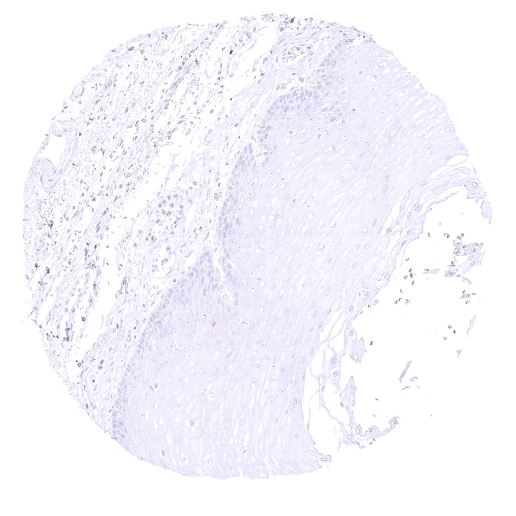
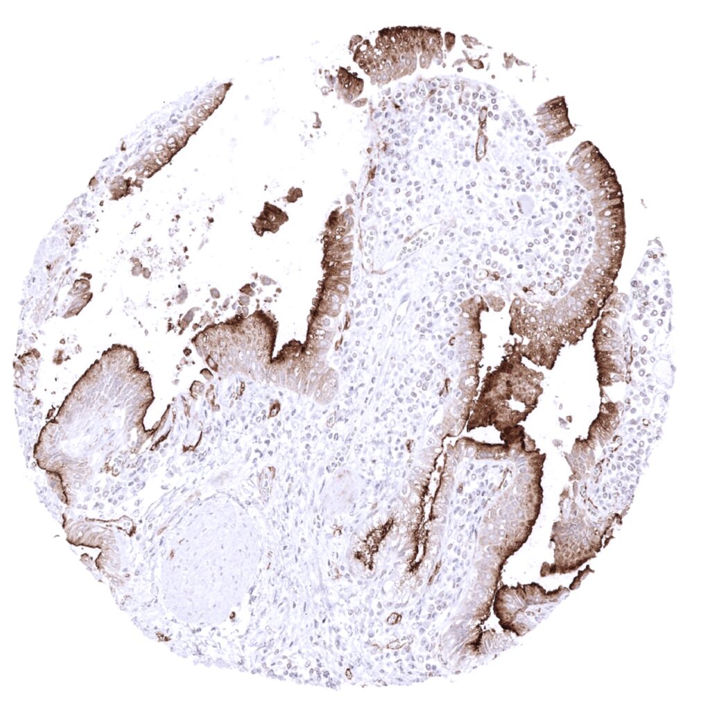
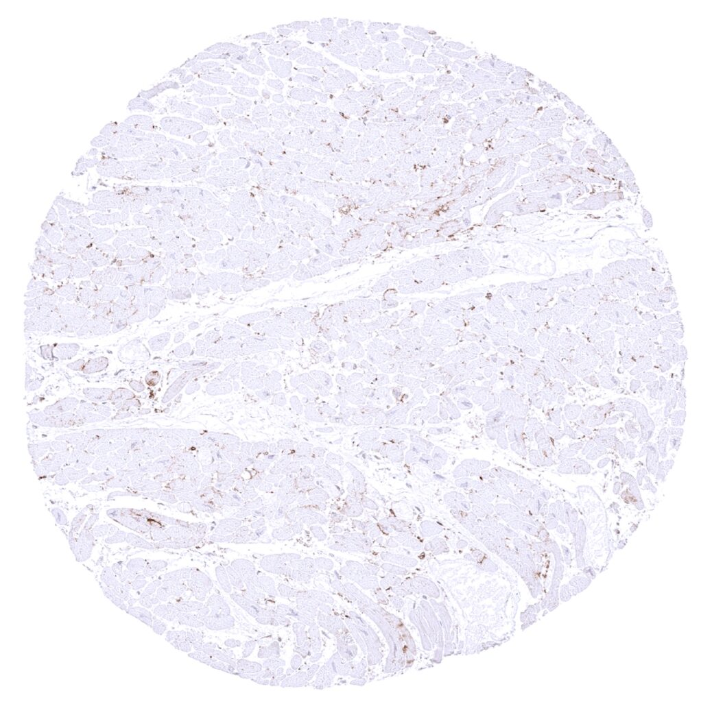
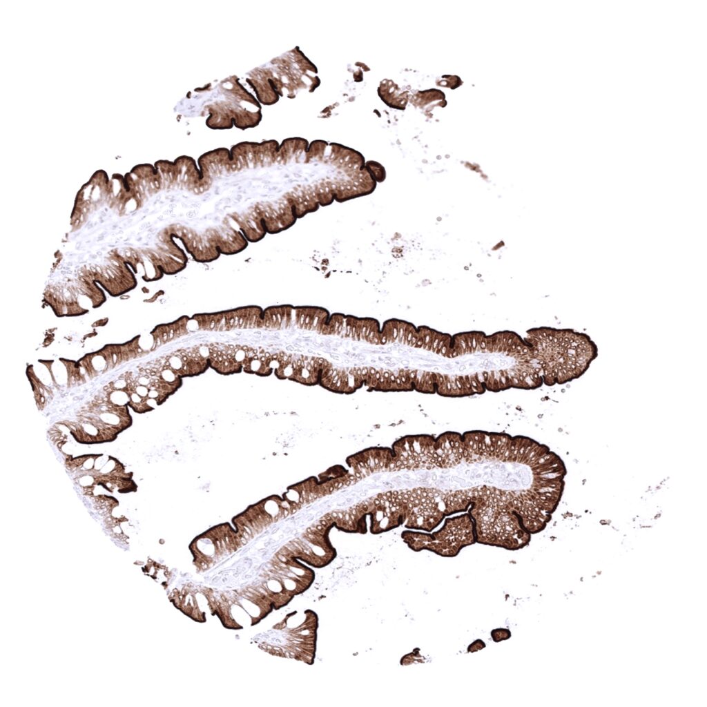
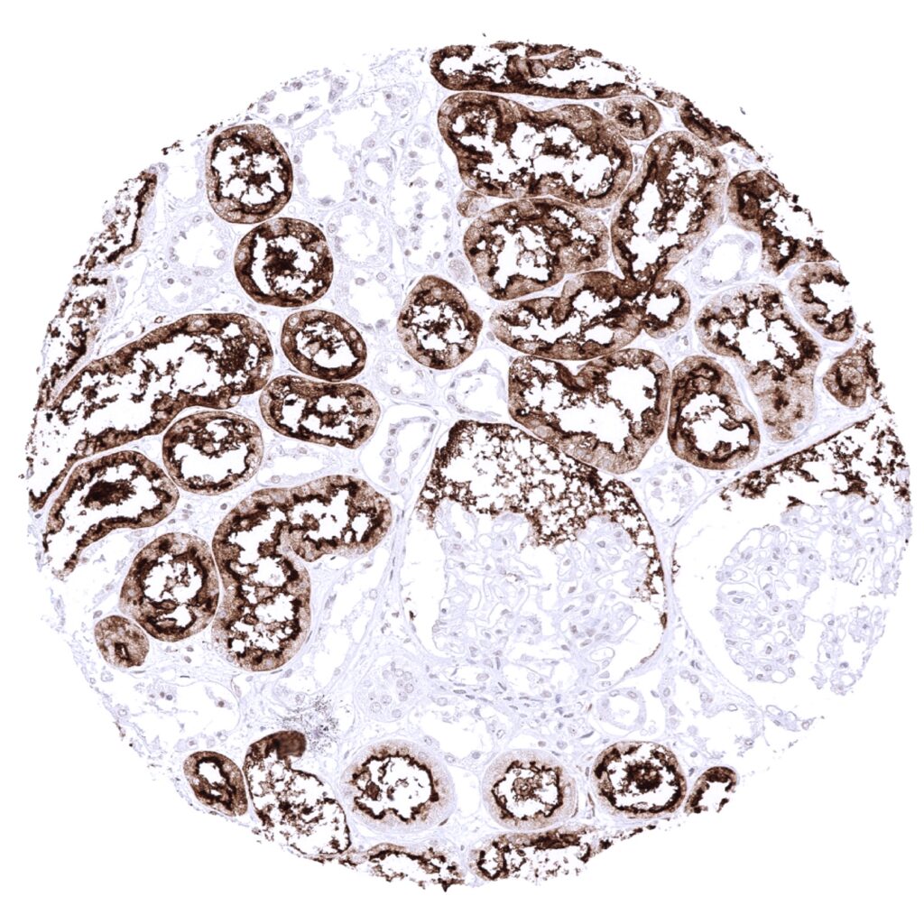
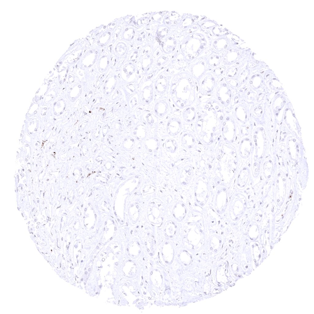
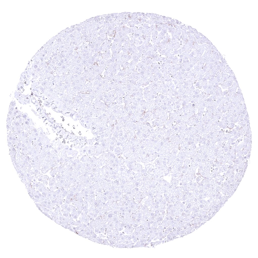
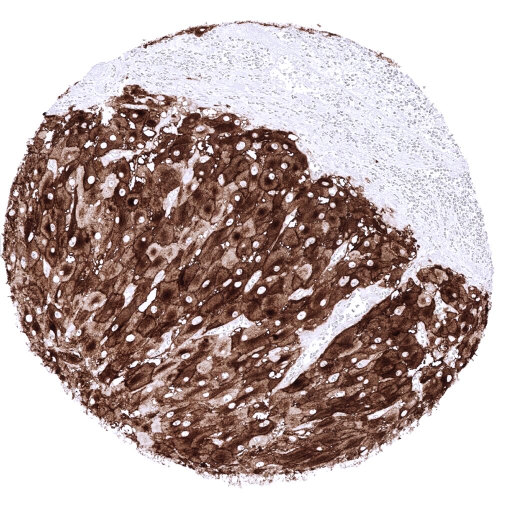
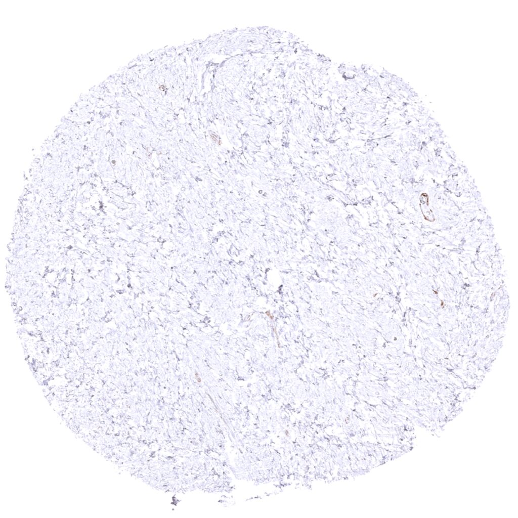
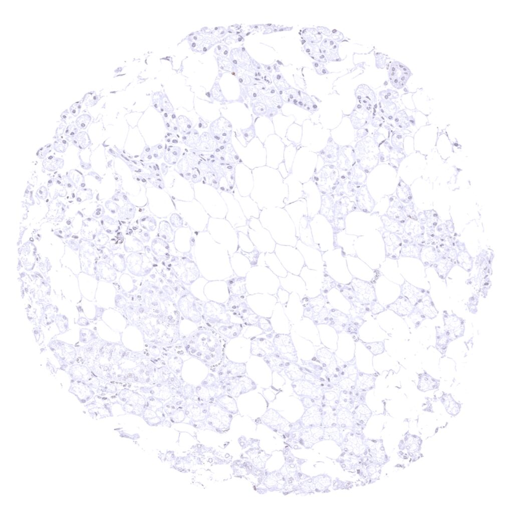
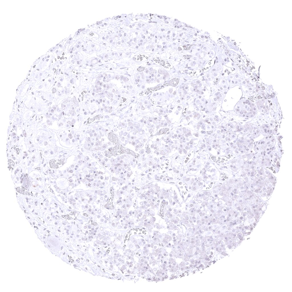
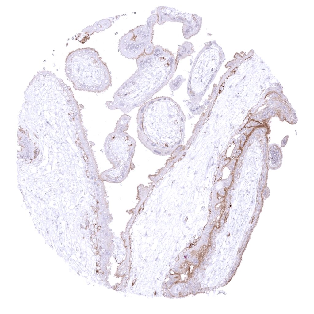
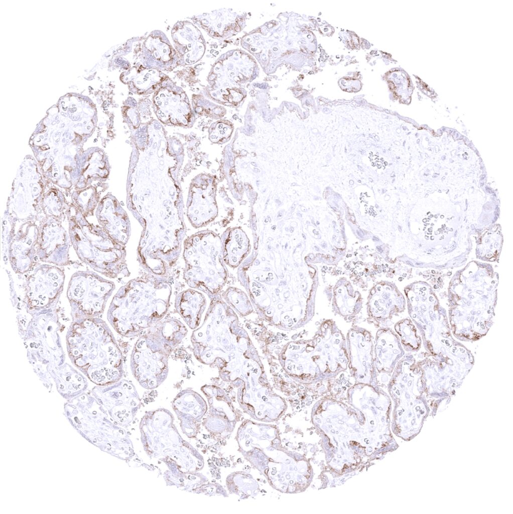
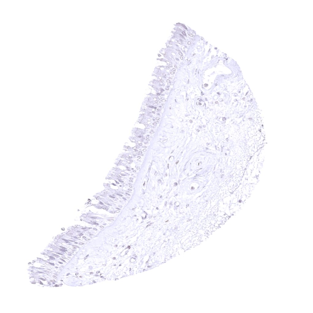
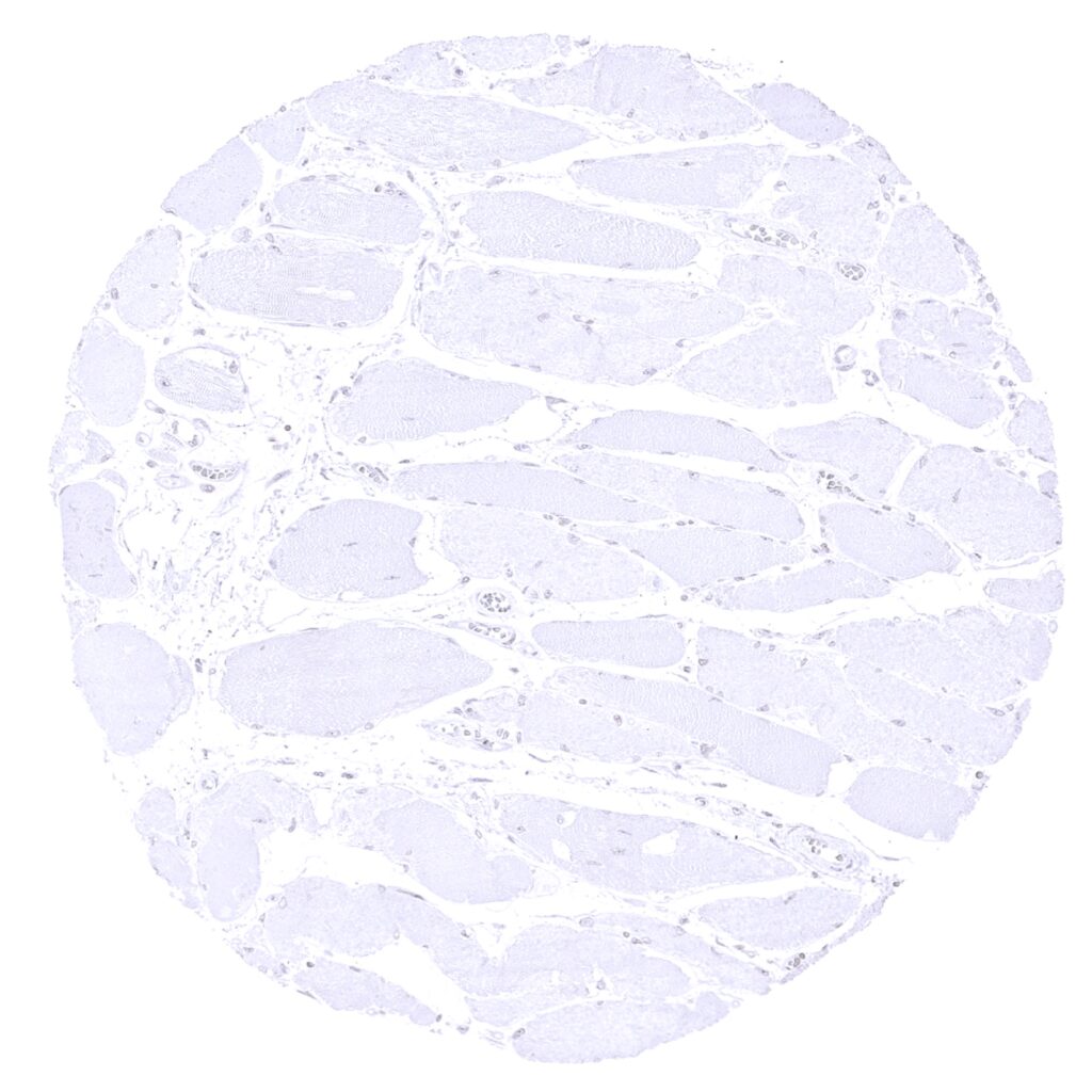
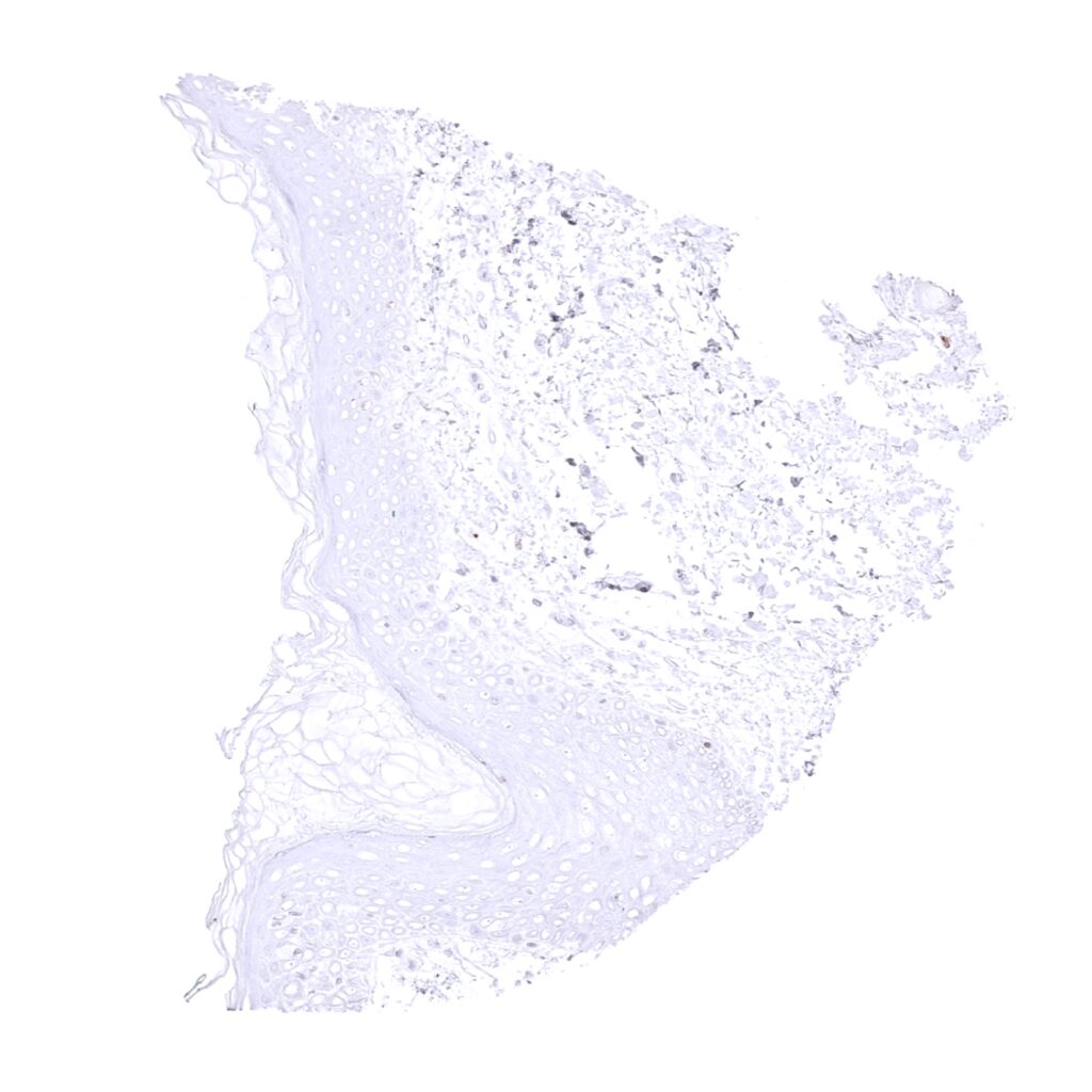
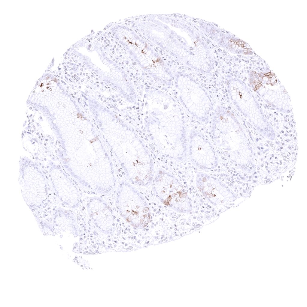
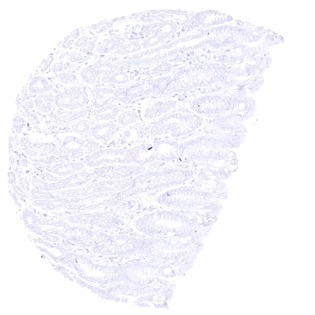
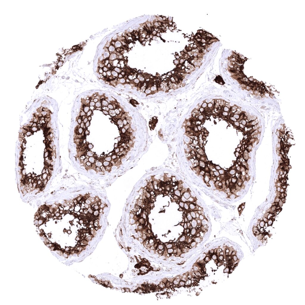
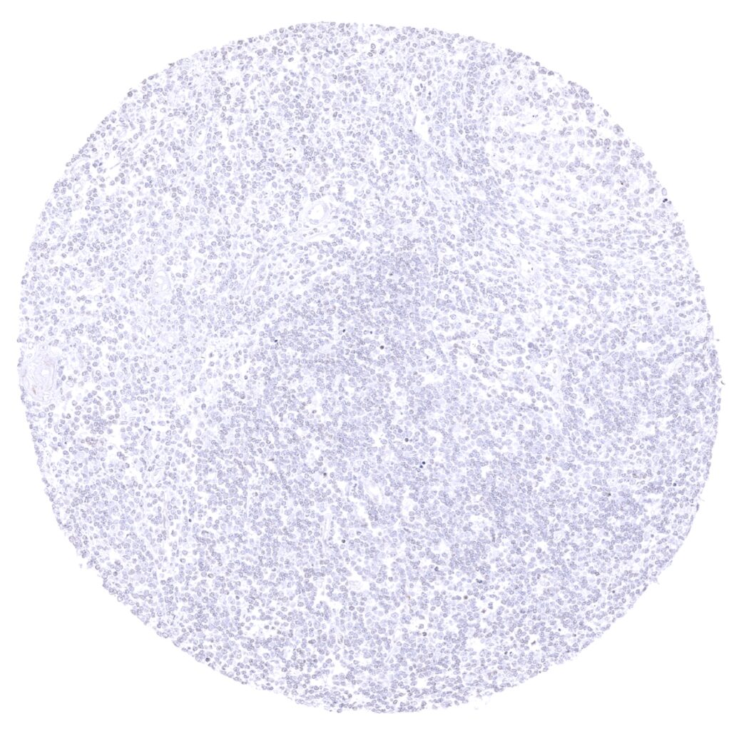
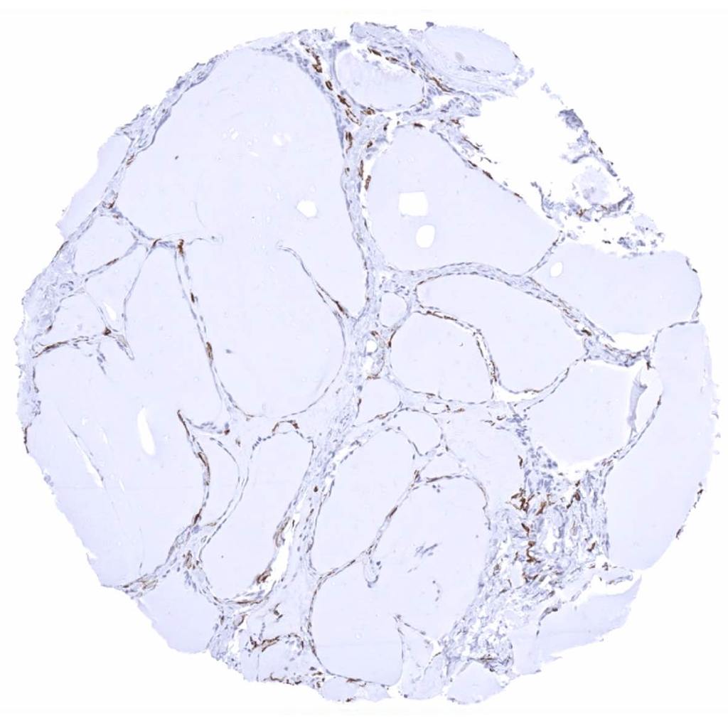
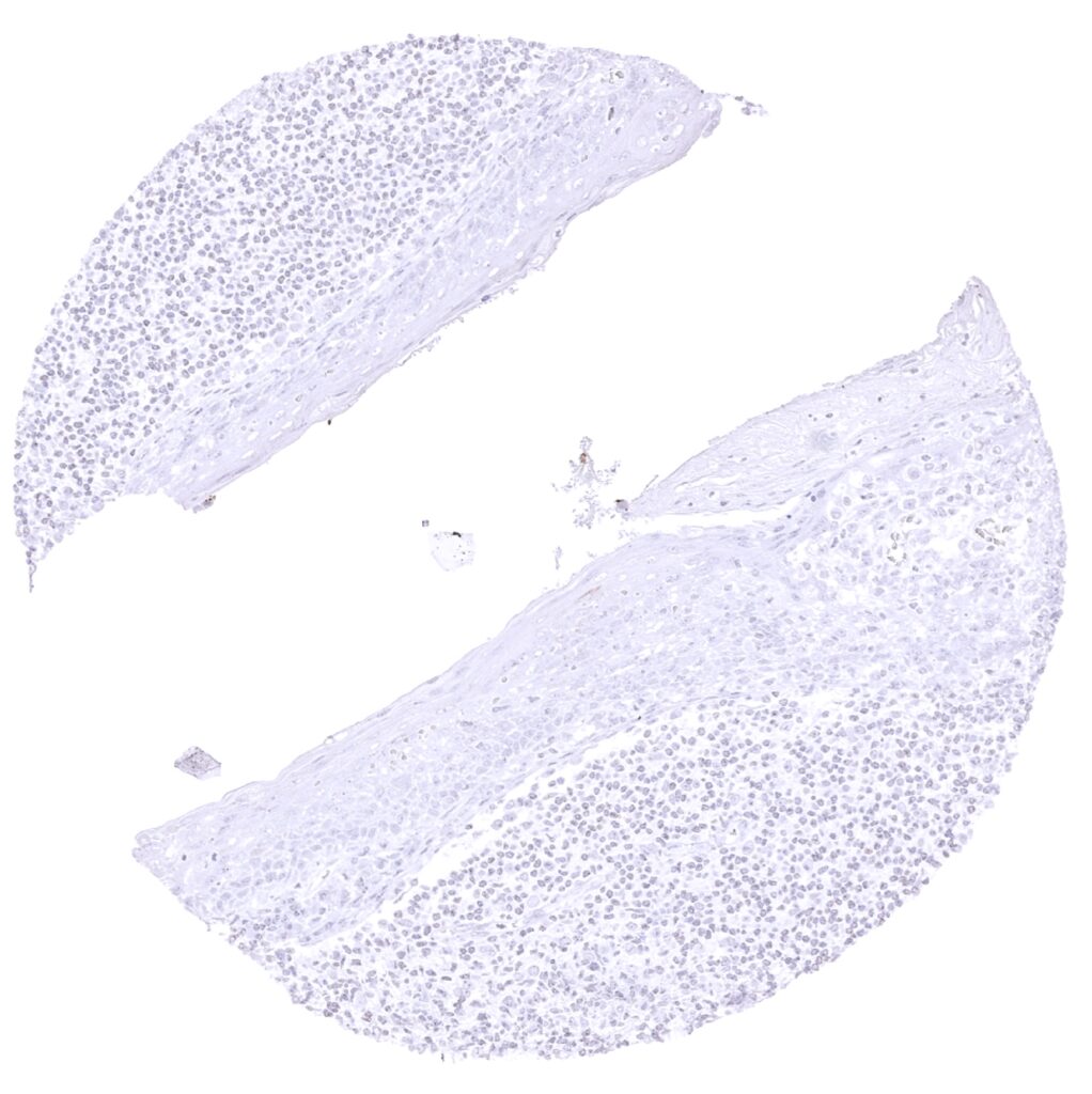
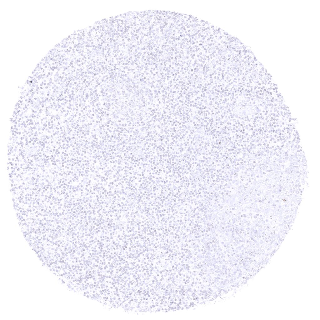
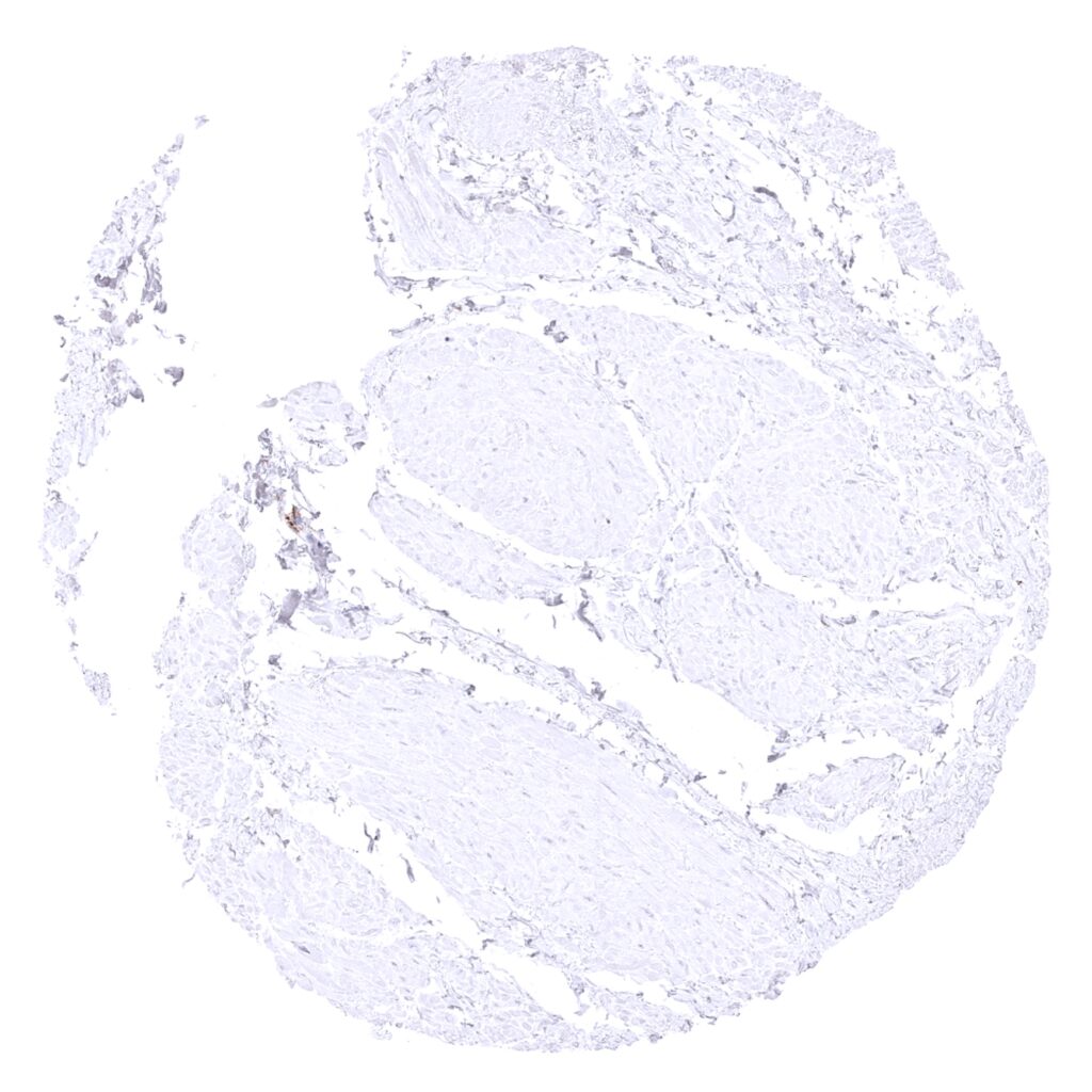
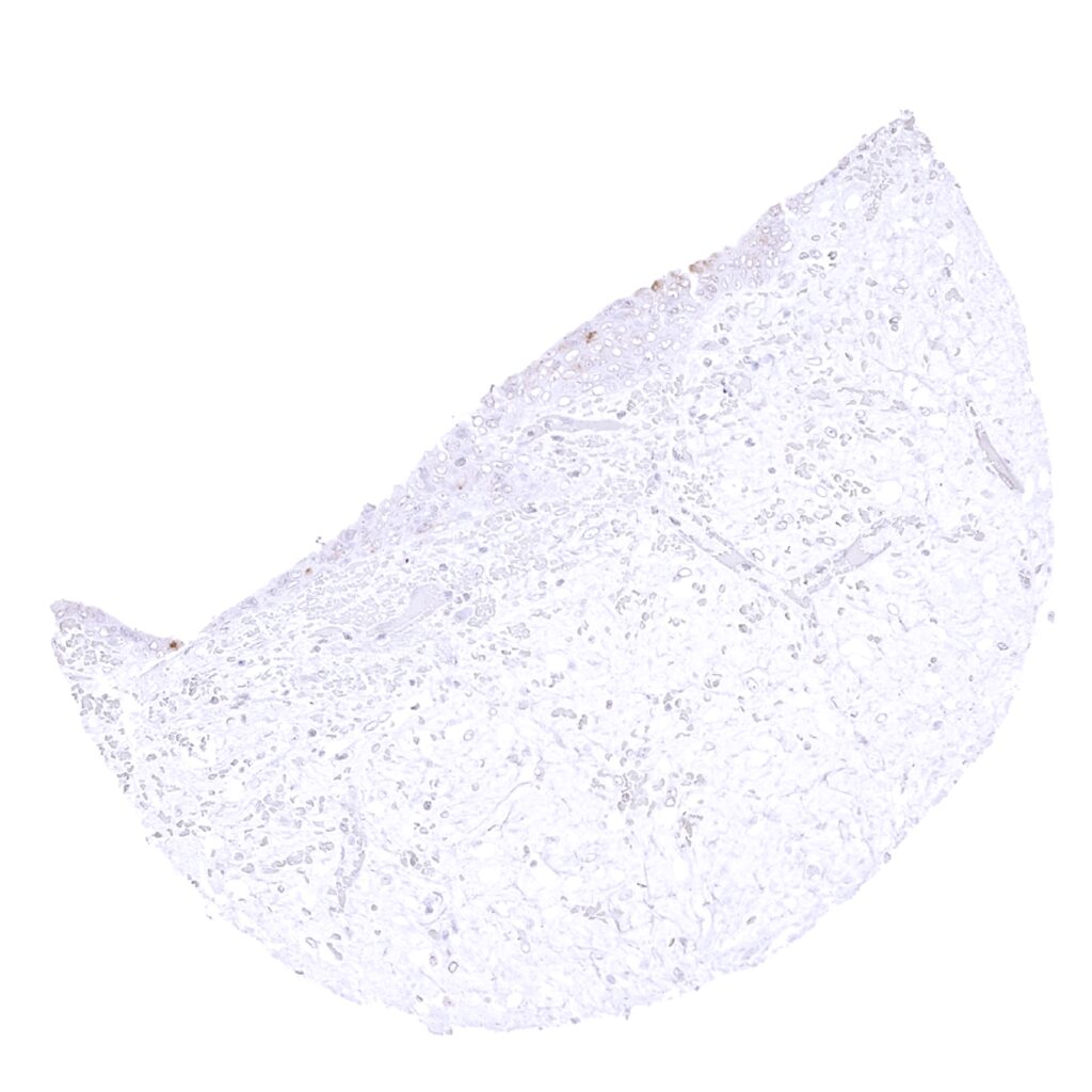
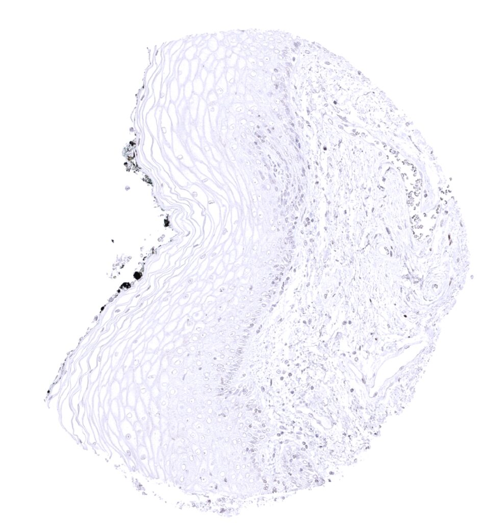
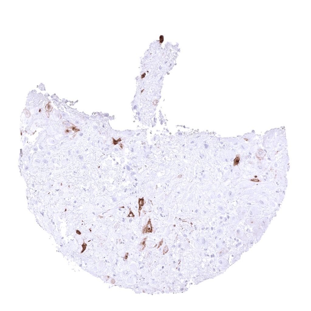
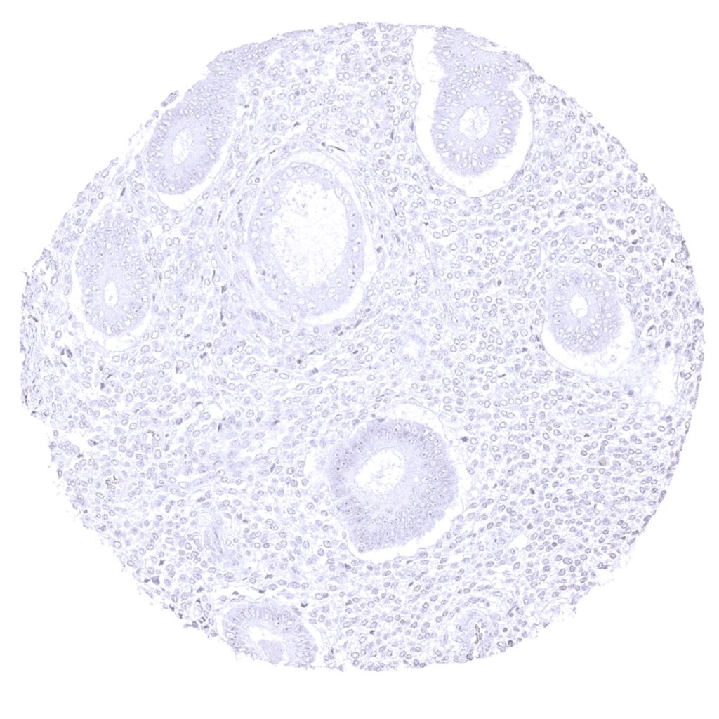
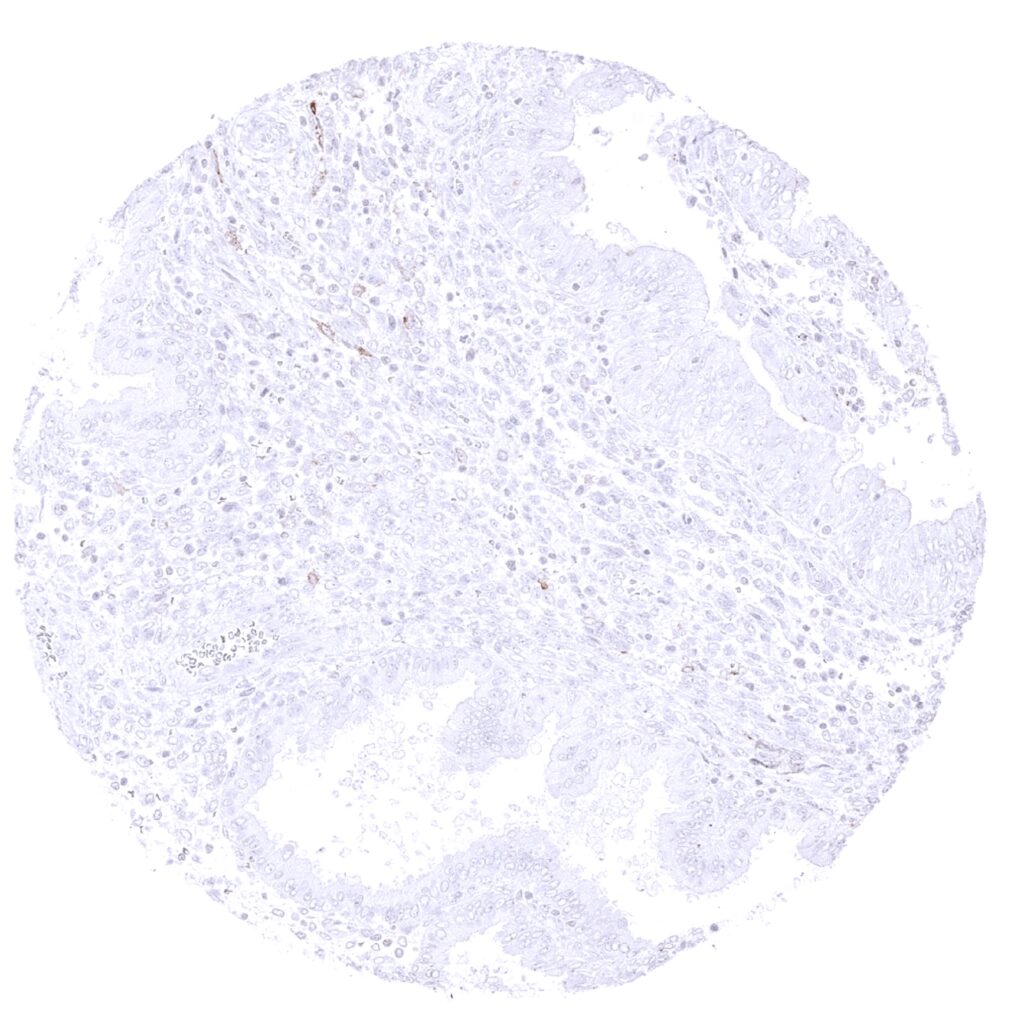
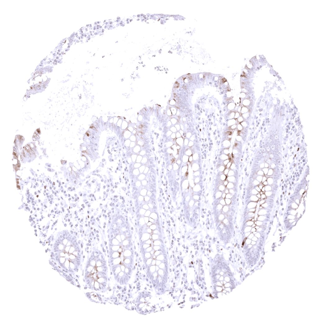
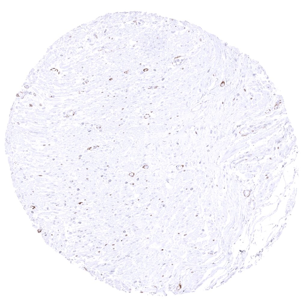
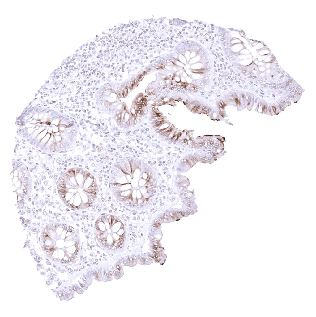
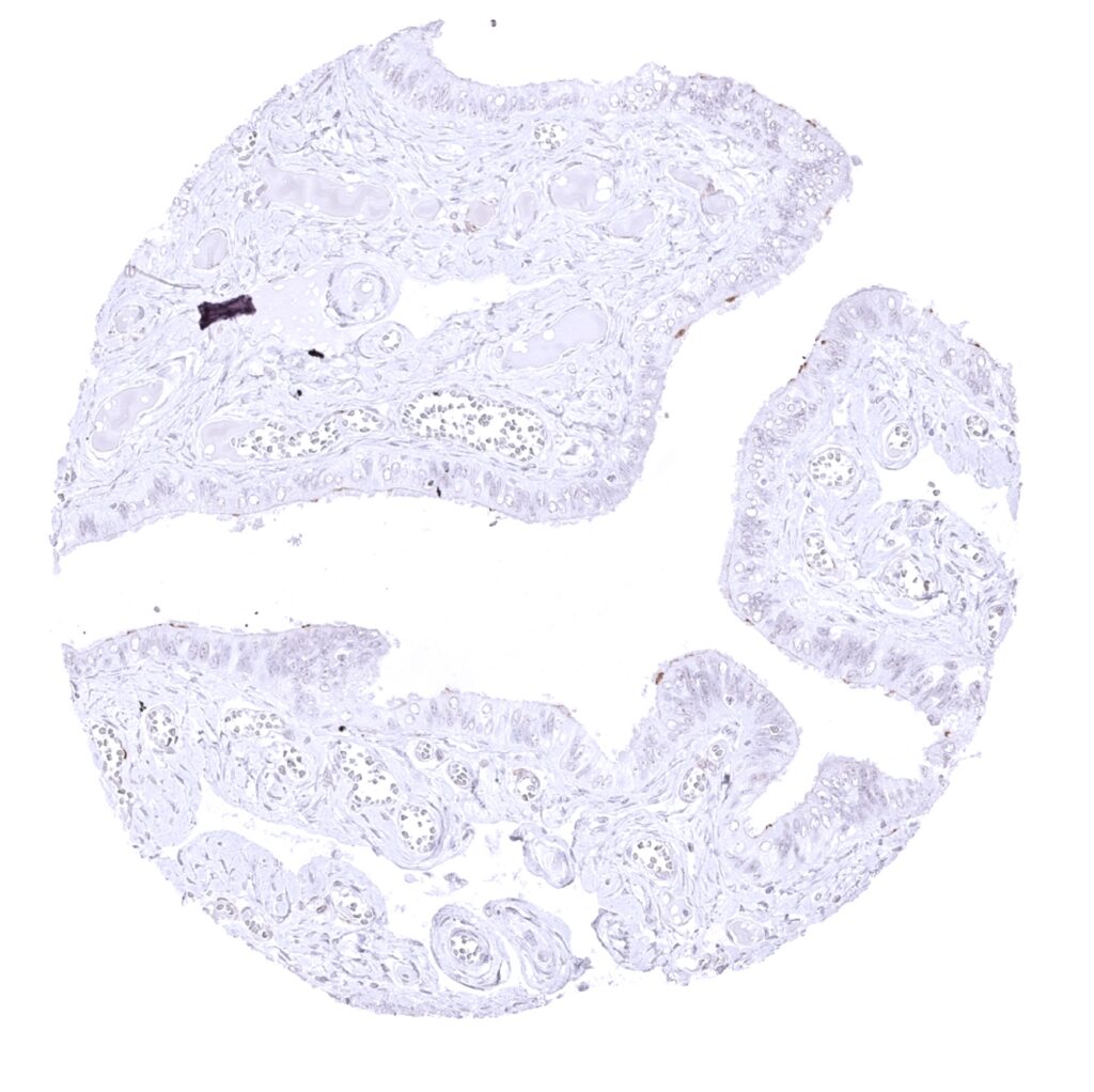
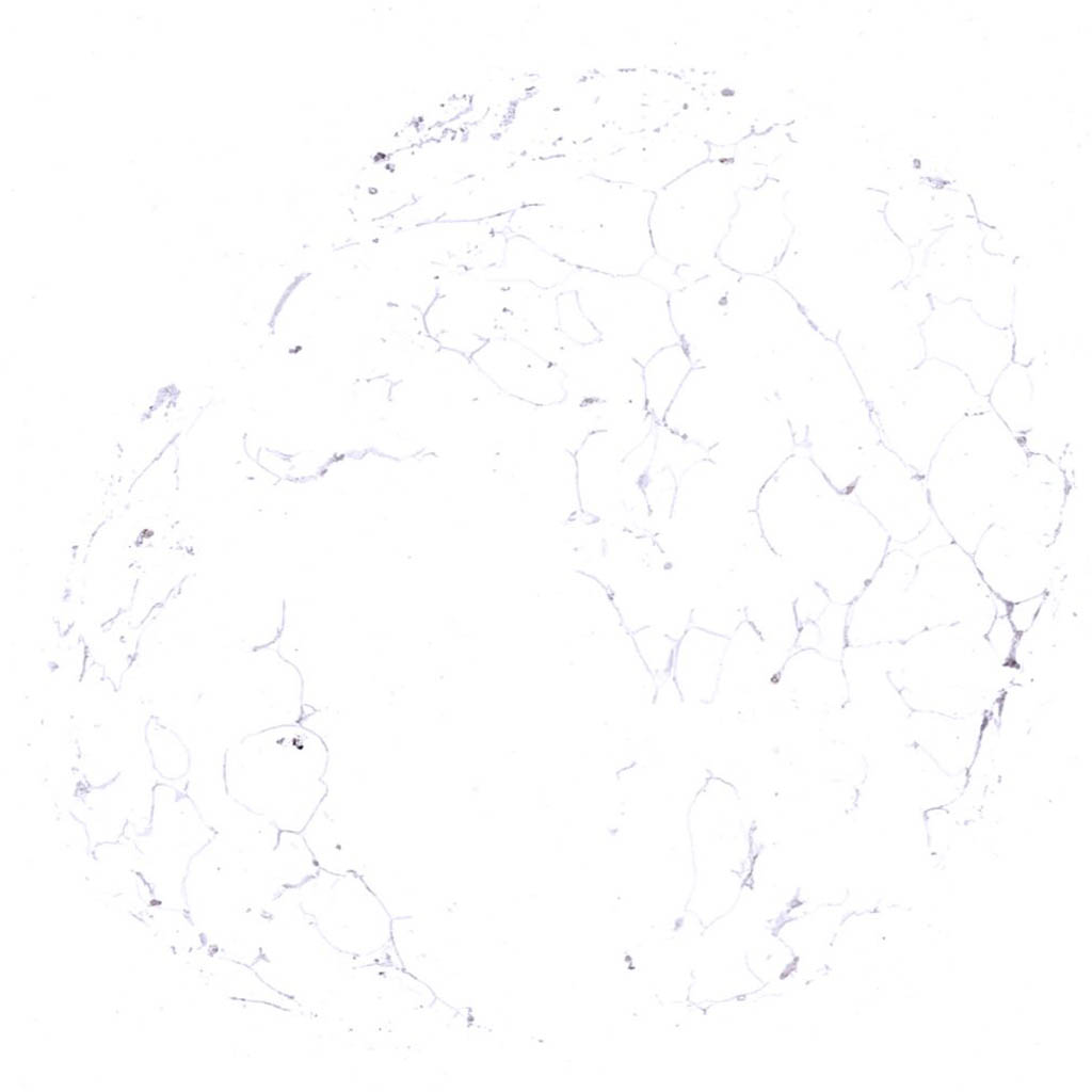
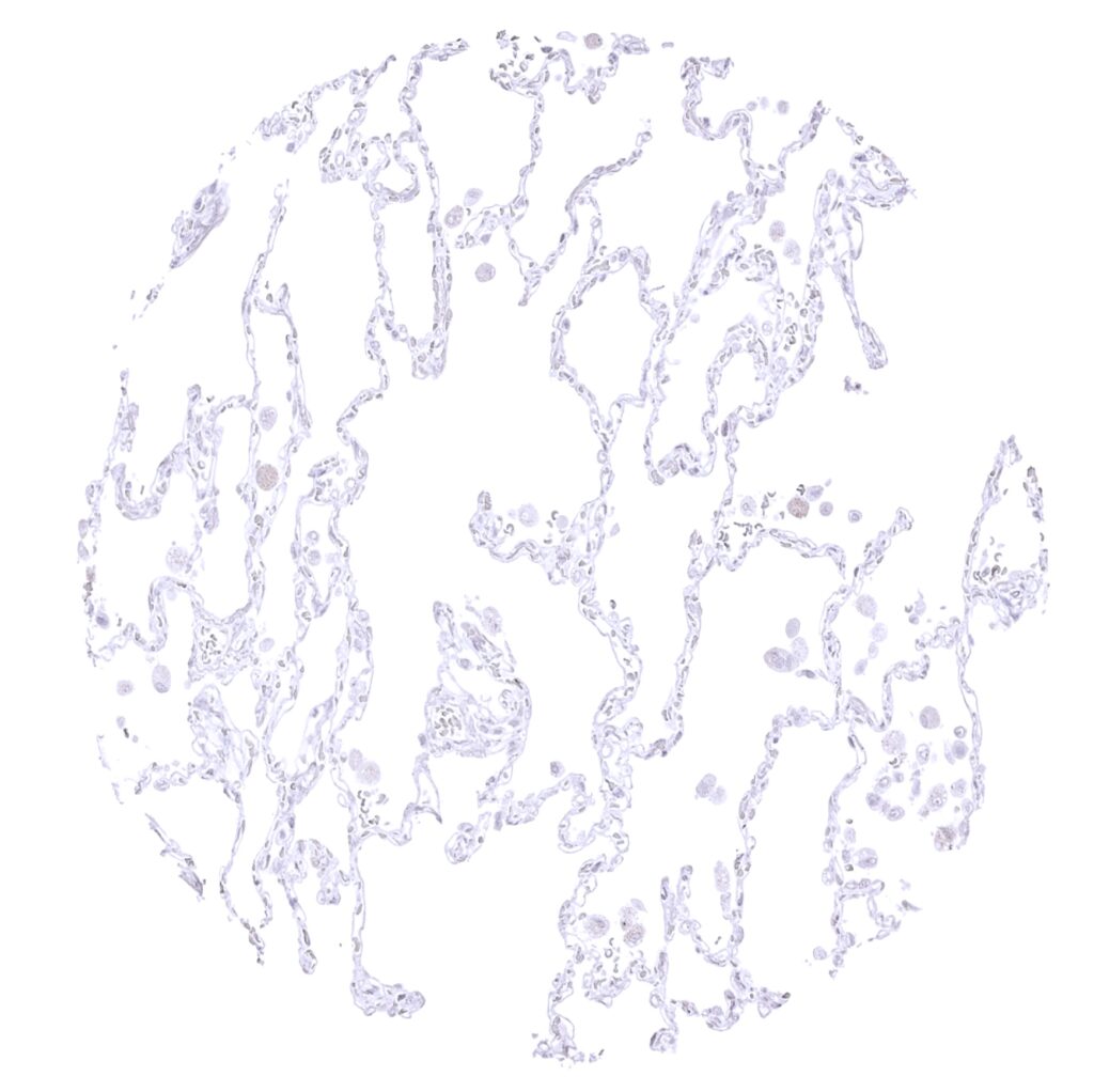
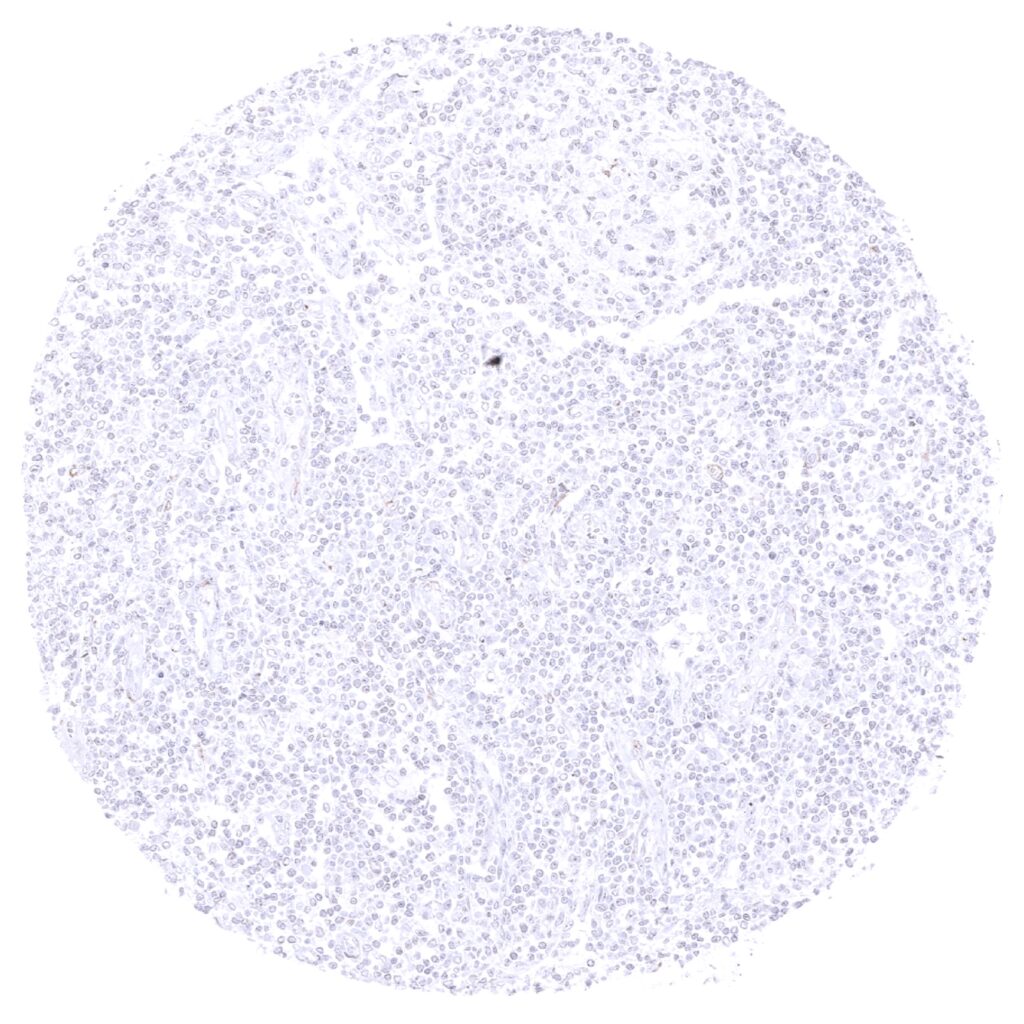
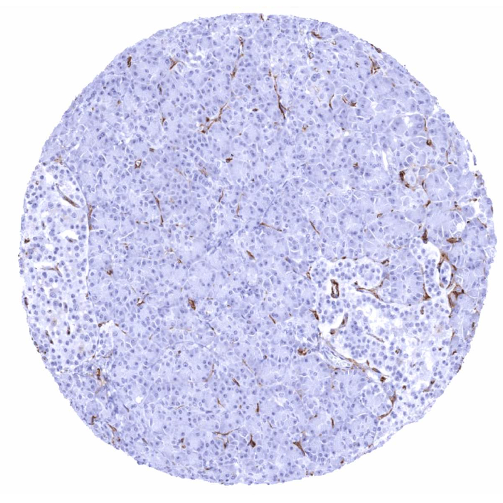
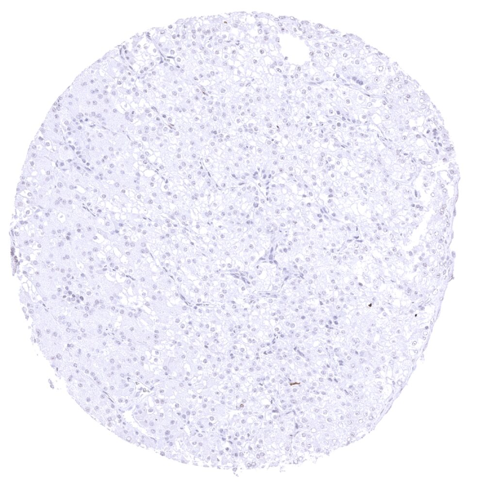
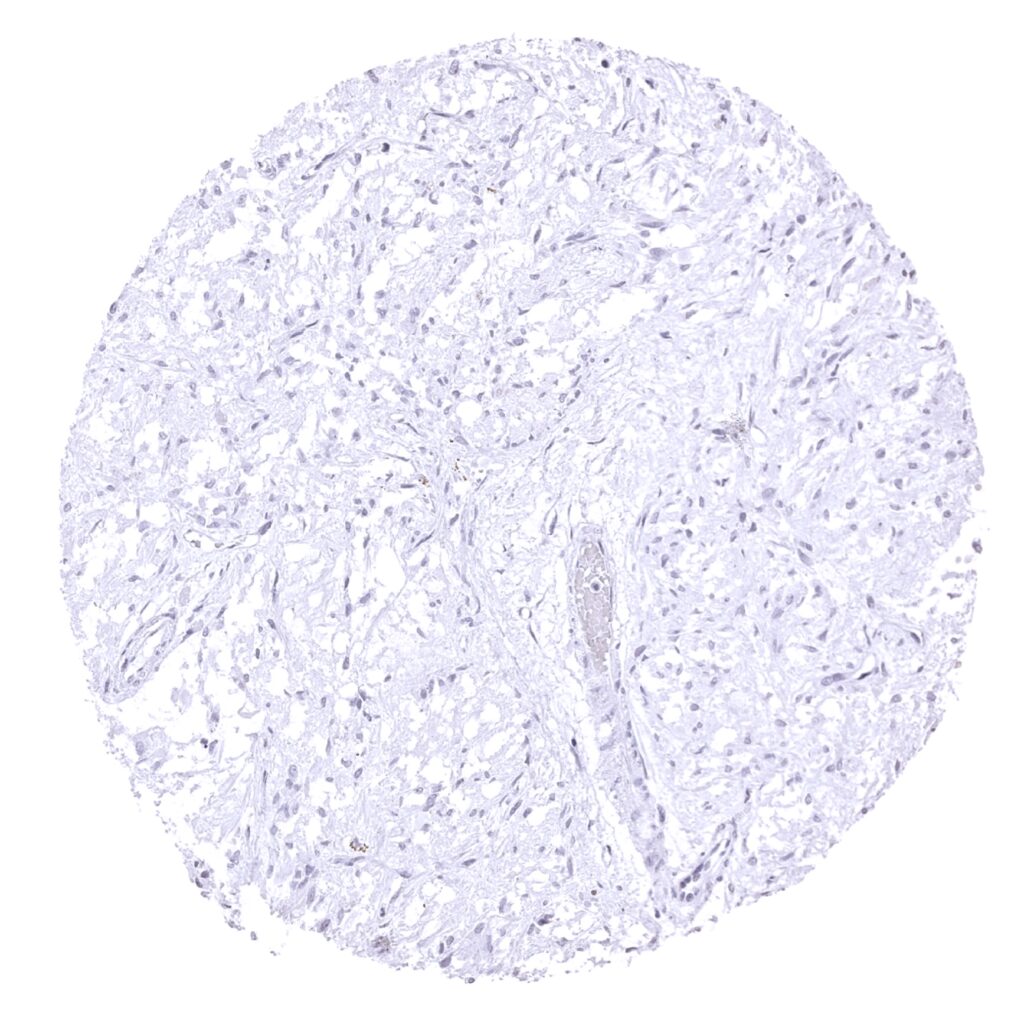
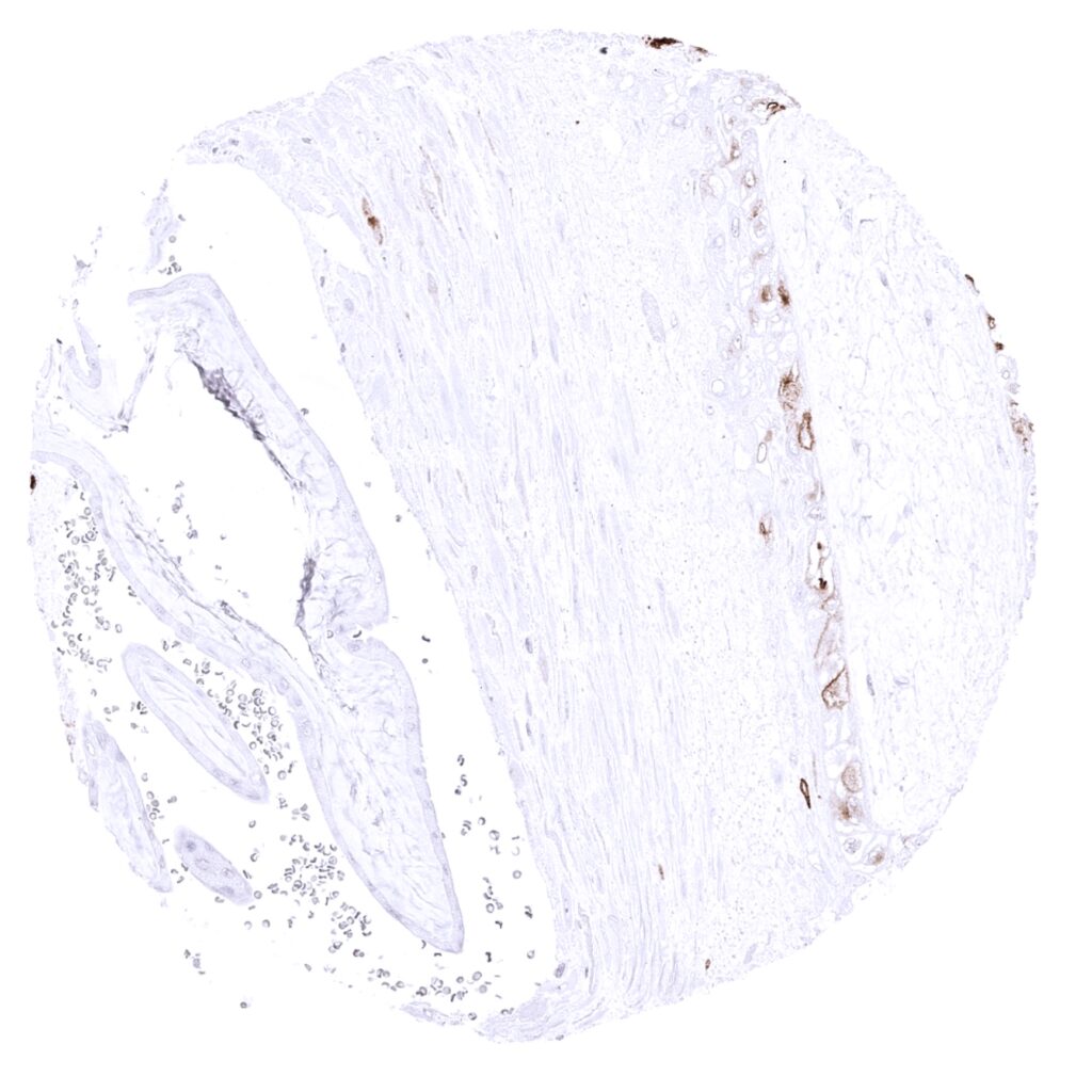
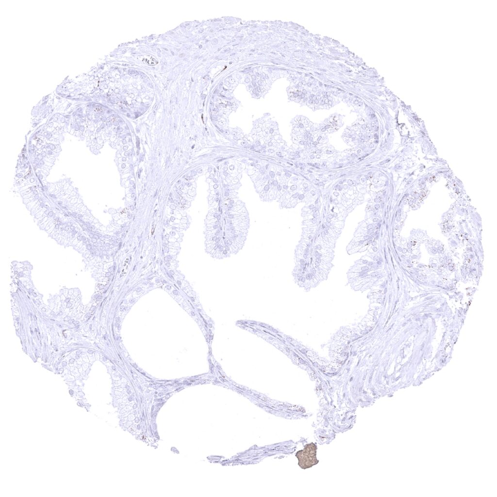
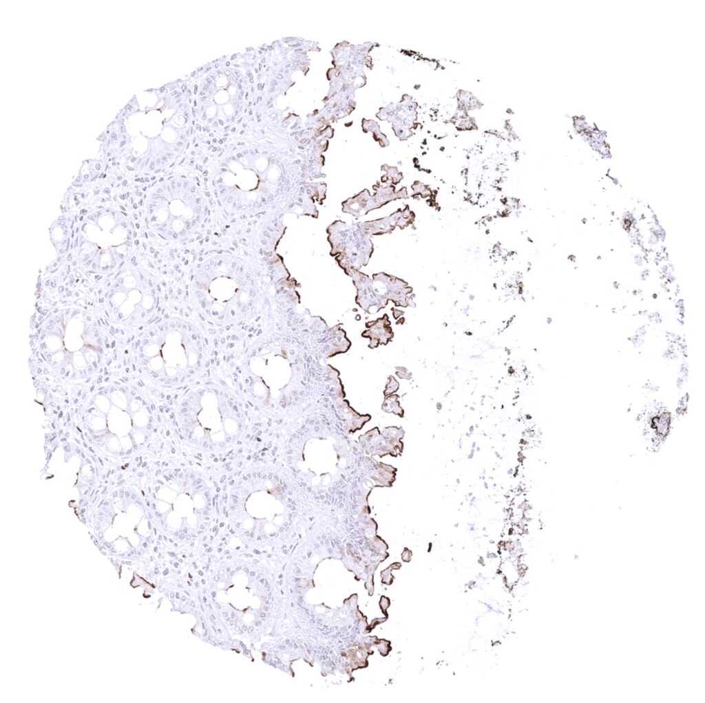
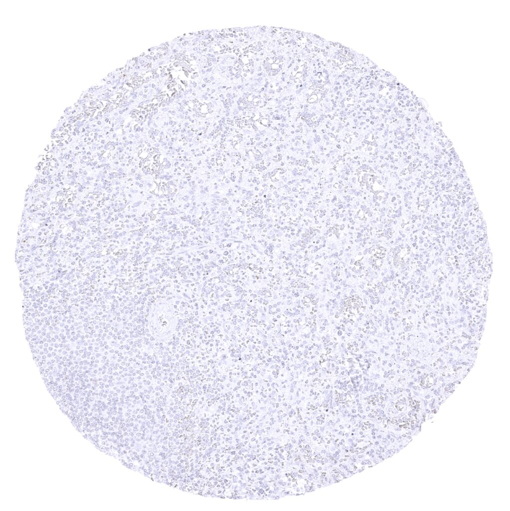
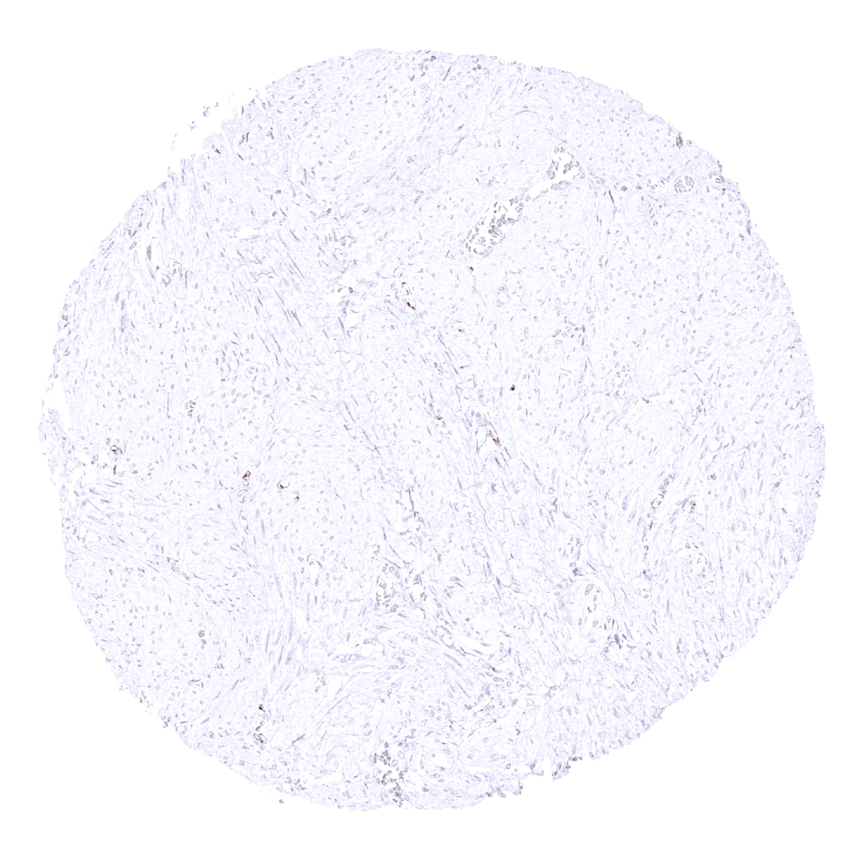
ACE2 antibody [MSVA-919R] HistoMAX


Go to Cancer Tissue Gallery Go back to product page Go to HistoMAX page


Adrenal gland – As ACE2 RNA expression is lacking in the adrenal gland, this antibody binding is considered a (tolerable) cross-reactivity (ACE2 immunohistochemistry)
Aorta, media (ACE2 immunohistochemistry)
Appendix, mucosa – A weak to moderate ACE2 immunostaining occurs focally at the surface epithelium of the appendix (ACE2 immunohistochemistry)
Appendix, muscular wall - A fraction of small capillares shows a weak ACE2 staining (ACE2 immunohistochemistry)
Bone marrow (ACE2 immunohistochemistry)
Breast (ACE2 immunohistochemistry)
Bronchus, mucosa - In the respiratory system, a weak ACE2 immunostaining can be seen in a and a subset of respiratory epithelial cells of the bronchus (not in all samples). (ACE2 immunohistochemistry)
Cerebellum (molecular layer, Purkinje cell layer, granule cell layer) (ACE2 immunohistochemistry)
Cerebellum (white matter) (ACE2 immunohistochemistry)
Cerebrum, grey matter (ACE2 immunohistochemistry)
Cerebrum, white matter (ACE2 immunohistochemistry)
Colon descendens, mucosa - A weak to moderate ACE2 immunostaining occurs focally at the surface epithelium of the colorectum (ACE2 immunohistochemistry)
Colon descendens, muscular wall (ACE2 immunohistochemistry)
Duodenum, Brunner gland (ACE2 immunohistochemistry)
Duodenum, mucosa - A strong ACE2 immunostaining occurs at the surface epithelium of the small intestine. (ACE2 immunohistochemistry)
Endocervix (ACE2 immunohistochemistry)
Epididymis (ACE2 immunohistochemistry)
Esophagus, squamous epithelium (ACE2 immunohistochemistry)
Fallopian tube, mucosa - A weak to moderate apical membrane ACE2 staining of a small subset of cells occurs in the fallopian tube. (ACE2 immunohistochemistry)
Fat (ACE2 immunohistochemistry)
Gallbladder, epithelium - A strong ACE2 immunostaining is seen in the surface epithelium of the gallbladder. (ACE2 immunohistochemistry)
Heart muscle - Membranous ACE2 immunostaining is seen in some heart muscle cells. A fraction of small capillares is also ACE2 positive. (ACE2 immunohistochemistry)
Ileum, mucosa - A strong ACE2 immunostaining occurs at the surface epithelium of the small intestine. (ACE2 immunohistochemistry)
Kidney, cortex - A strong ACE2 immunostaining occurs in proximal tubuli of the kidney. (ACE2 immunohistochemistry)
Kidney, medulla (ACE2 immunohistochemistry)
Liver (ACE2 immunohistochemistry)
Lung - In the respiratory system, a weak ACE2 immunostaining can be seen at apical cell membranes of few alveolar pneumocytes. (ACE2 immunohistochemistry)
Lymph node (ACE2 immunohistochemistry)
Ovary, corpus luteum - Strong ACE2 staining in the vast majority of cells. (ACE2 immunohistochemistry)
Ovary, stroma (ACE2 immunohistochemistry)
Pancreas - Endothelial cells of small capillaries show a more intense ACE2 positivity in or near the islets of Langerhans than in the exocrine parts of the pancreas (ACE2 immunohistochemistry)
Parathyroid gland (ACE2 immunohistochemistry)
Parotid gland (ACE2 immunohistochemistry)
Pituitary gland, anterior lobe (ACE2 immunohistochemistry)
Pituitary gland, posterior lobe (ACE2 immunohistochemistry)
Placenta (amnion and chorion) - A fraction of chorion cells shows weak to moderate ACE2 positivity (ACE2 immunohistochemistry)
Placenta, early - A moderate ACE2 membranous immunostaining is seen in the cytotrophoblast while a weaker membranous staining may also be seen of the syncytiotrophoblast. (ACE2 immunohistochemistry)
Placenta, mature - A moderate membranous ACE2 immunostaining is seen in the cytotrophoblast while a weaker membranous staining may also be seen of the syncytiotrophoblast. (ACE2 immunohistochemistry)
Prostate (ACE2 immunohistochemistry)
Rectum, mucosa - A weak to moderate ACE2 immunostaining occurs focally at the surface epithelium of the colorectum. (ACE2 immunohistochemistry)
Seminal vesicle - A weak to moderate apical membrane staining of a small subset of cells occurs in the seminal vesicle. (ACE2 immunohistochemistry)
Sinus paranasales - In the respiratory system, a weak ACE2 immunostaining can be seen in a and a subset of respiratory epithelial cells of the nasal sinuses (not in all samples). (ACE2 immunohistochemistry)
Skeletal muscle (ACE2 immunohistochemistry)
Skin (ACE2 immunohistochemistry)
Spleen (ACE2 immunohistochemistry)
Stomach, antrum (ACE2 immunohistochemistry)
Stomach, corpus - A limited number of cells of the stomach surface epithelium shows a weak to moderate ACE2 staining. (ACE2 immunohistochemistry)
Stomach, corpus (ACE2 immunohistochemistry)
Testis - Leydig cells and spermatocytes of the testis show a particularly strong ACE2 immunostaining in the testis. (ACE2 immunohistochemistry)
Thymus (ACE2 immunohistochemistry)
Thyroid gland - A strong ACE2 staining is seen in endothelial cells of small blood vessels (ACE2 immunohistochemistry)
Tonsil, surface epithelium (ACE2 immunohistochemistry)
Tonsil (ACE2 immunohistochemistry)
Urinary bladder, muscular wall (ACE2 immunohistochemistry)
Urinary bladder, urothelium (ACE2 immunohistochemistry)
Uterus, ectocervix (ACE2 immunohistochemistry)
Uterus, endometrium (pregnancy) - A strong ACE2 immunostaining can be seen in a fraction of decidua cells. (ACE2 immunohistochemistry)
Uterus, endometrium (proliferation) (ACE2 immunohistochemistry)
Uterus, endometrium (secretion) (ACE2 immunohistochemistry)
Uterus, myometrium - A fraction of small capillares shows a weak ACE2 staining. (ACE2 immunohistochemistry)

| TOP |