GeneTex
  • Country / Location Selection

United States (US)

Normal Tissue Gallery- GTX04453

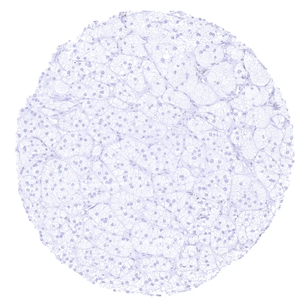
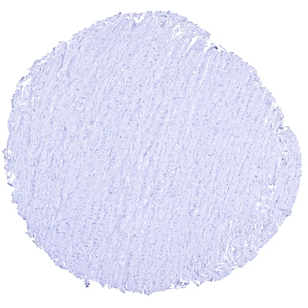
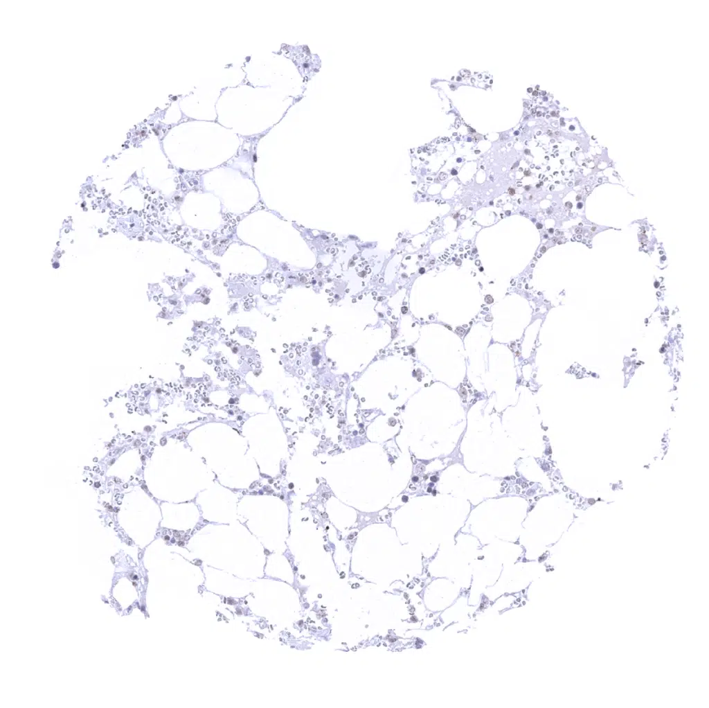
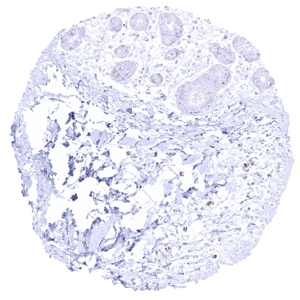
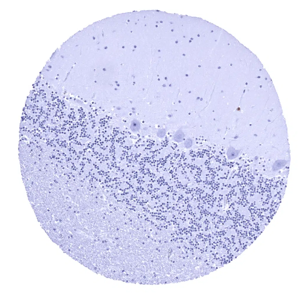
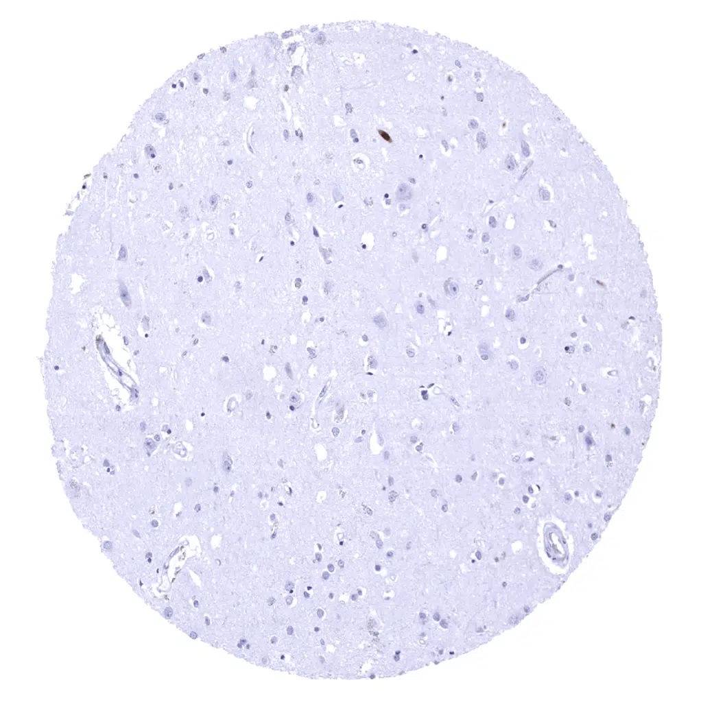
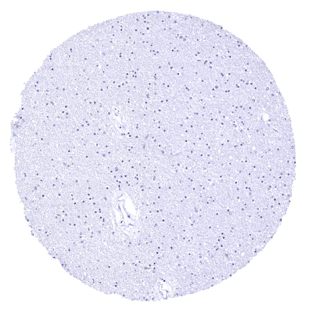
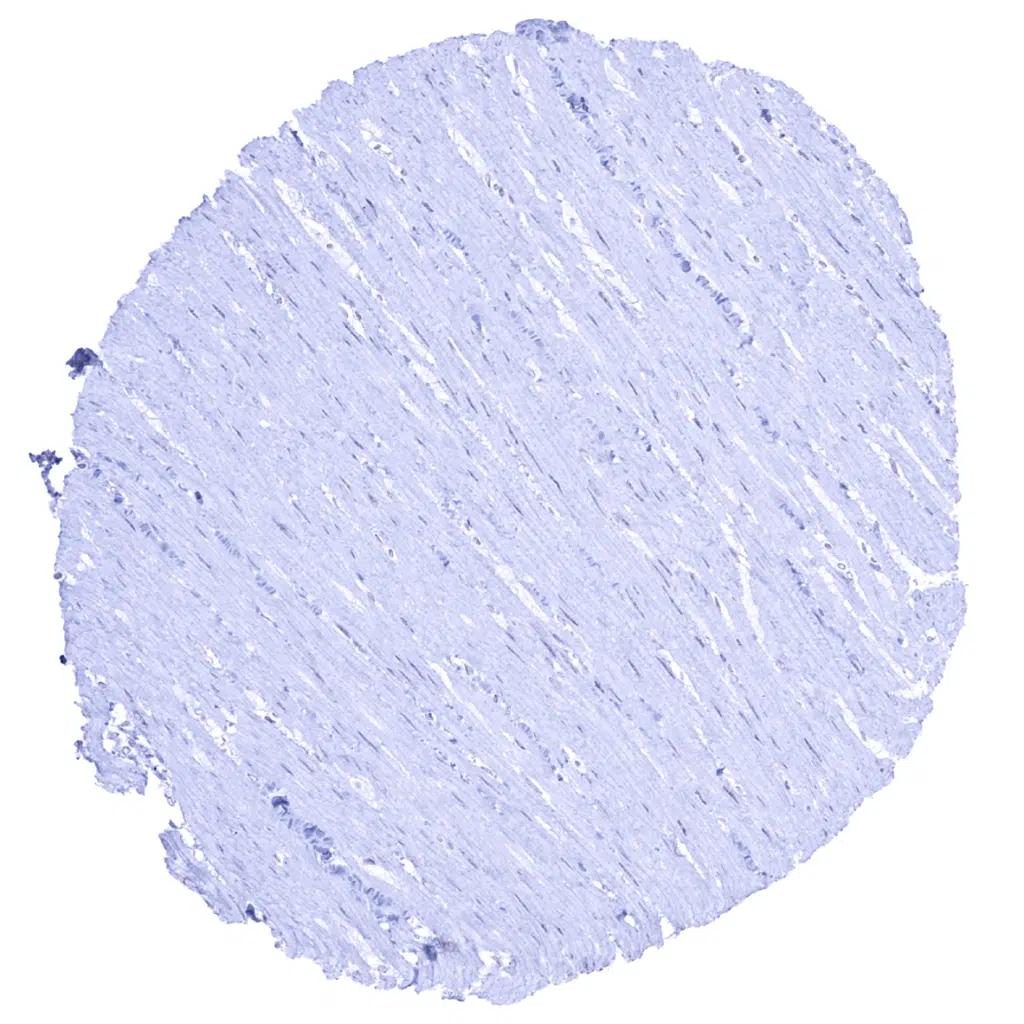
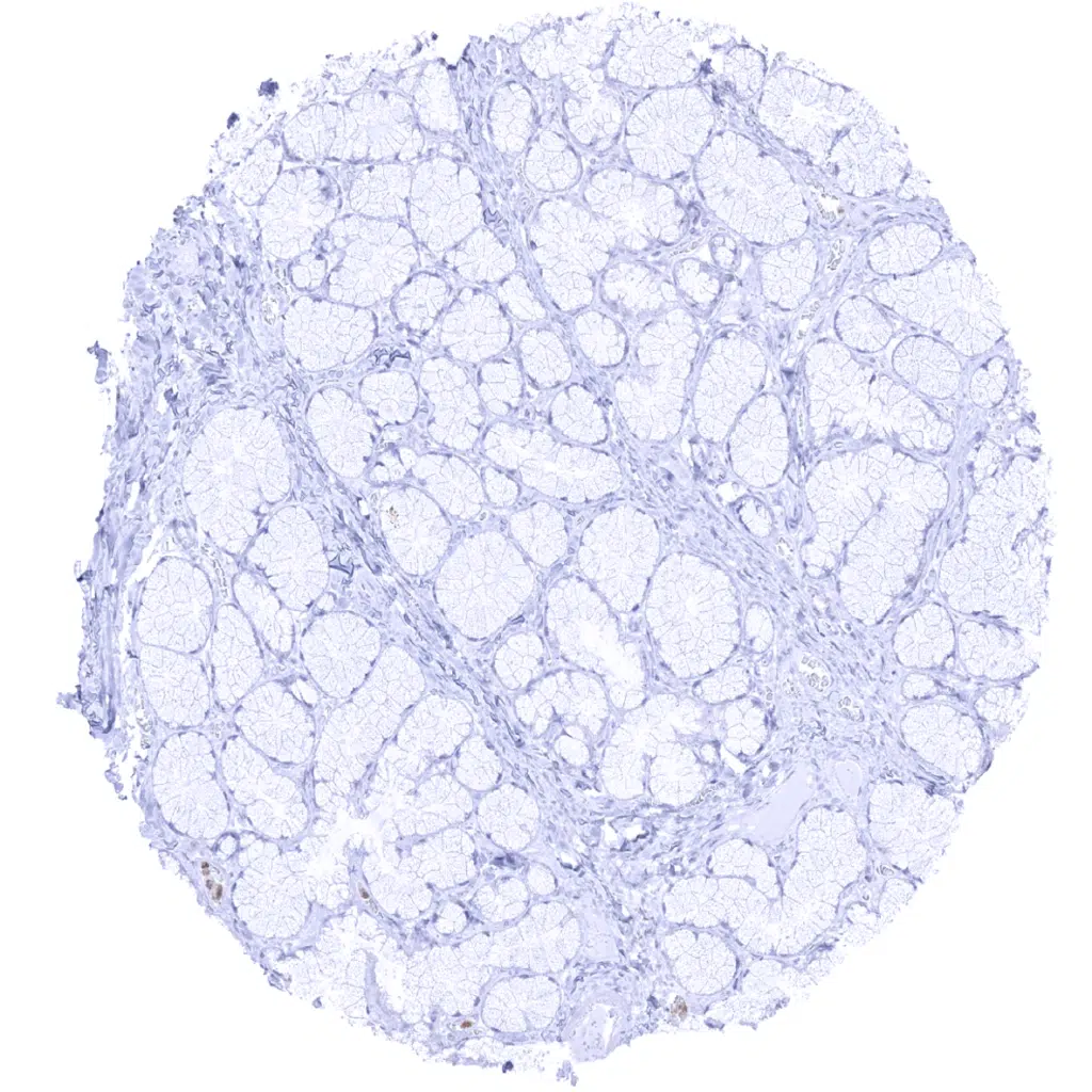
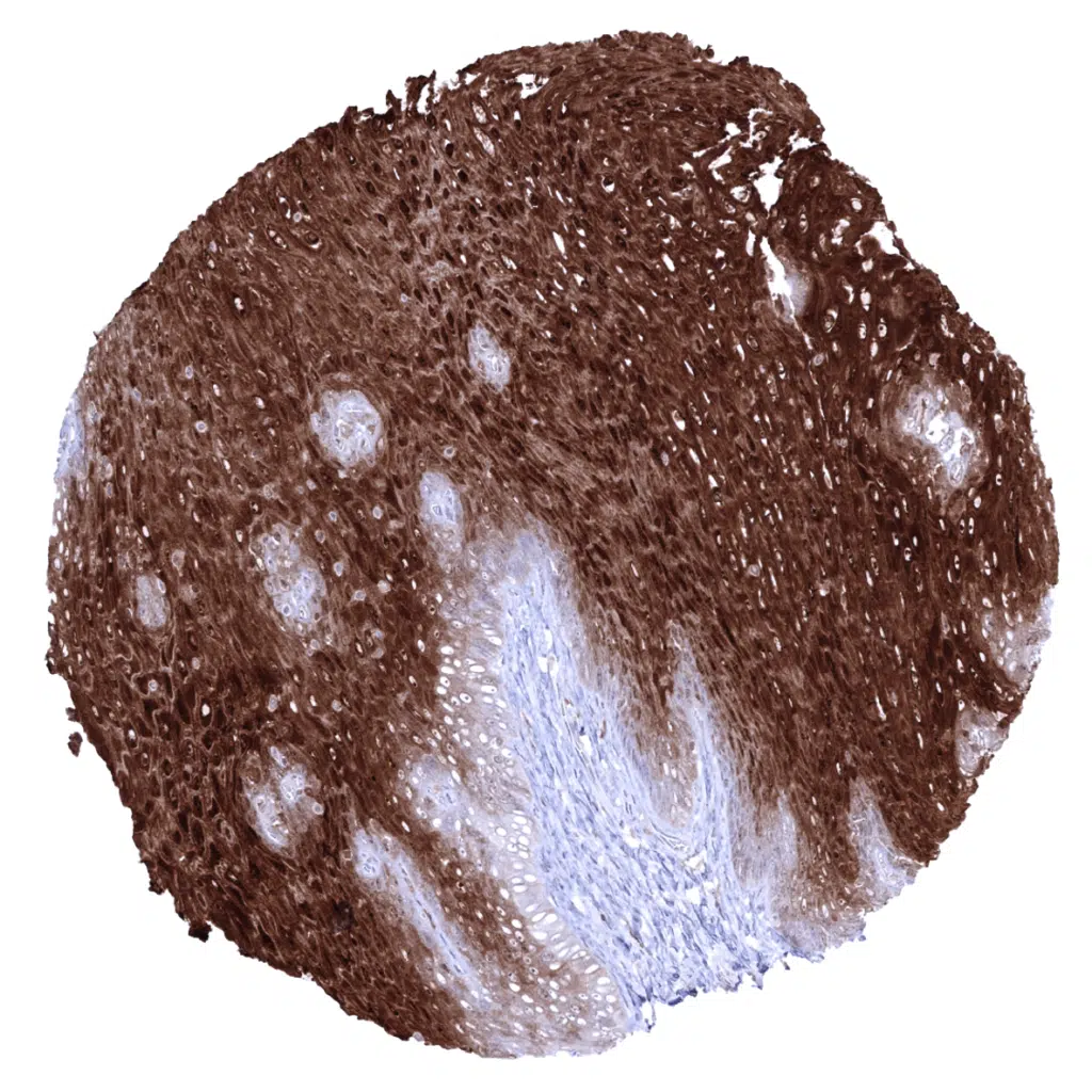
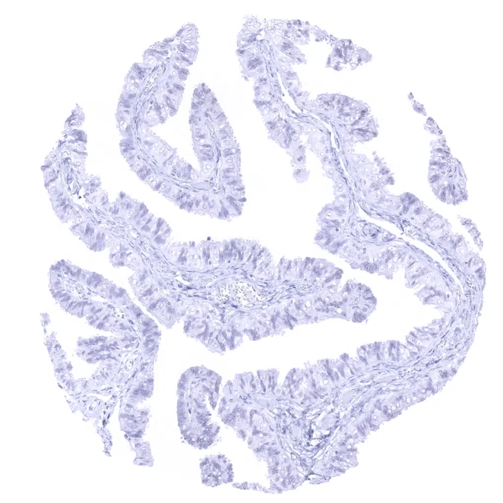
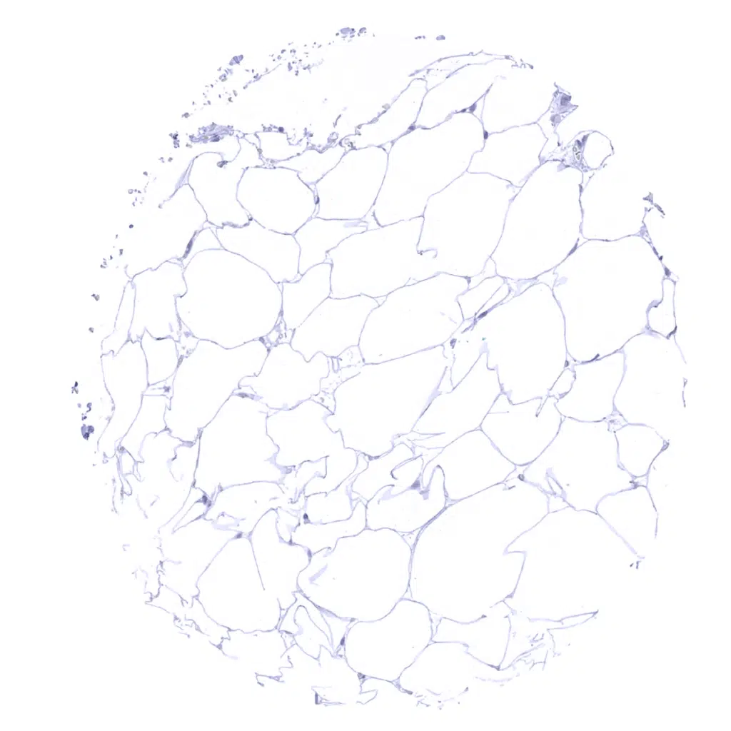
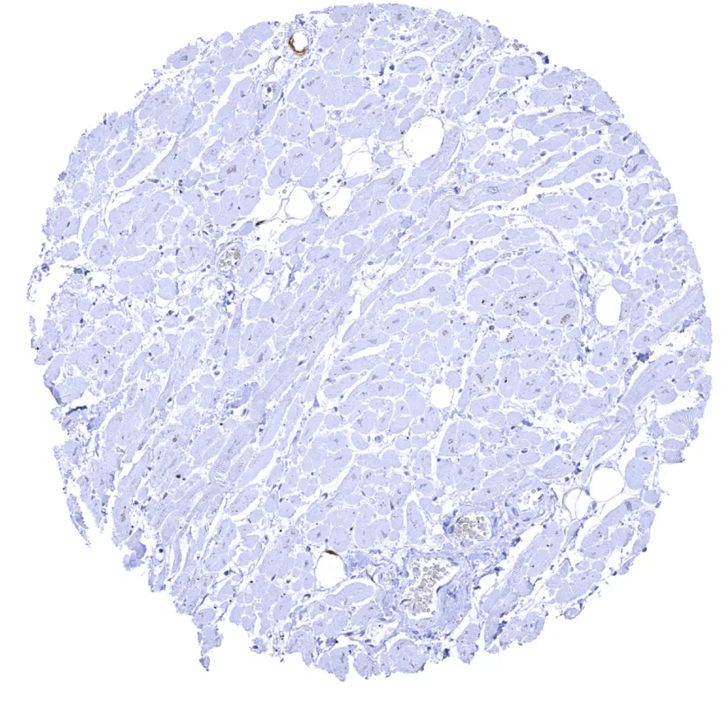
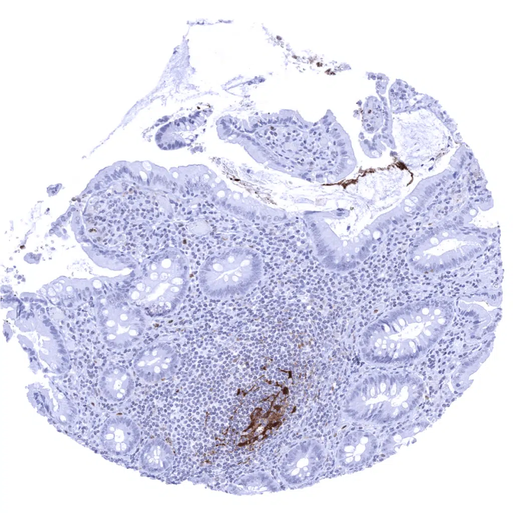
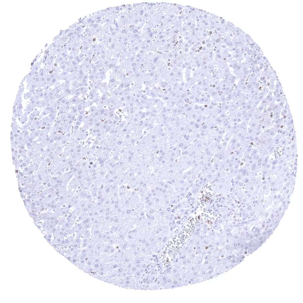
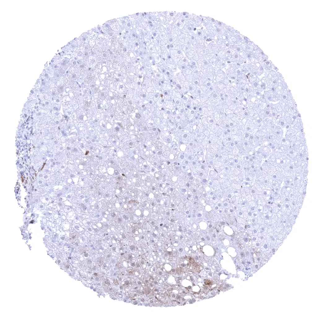
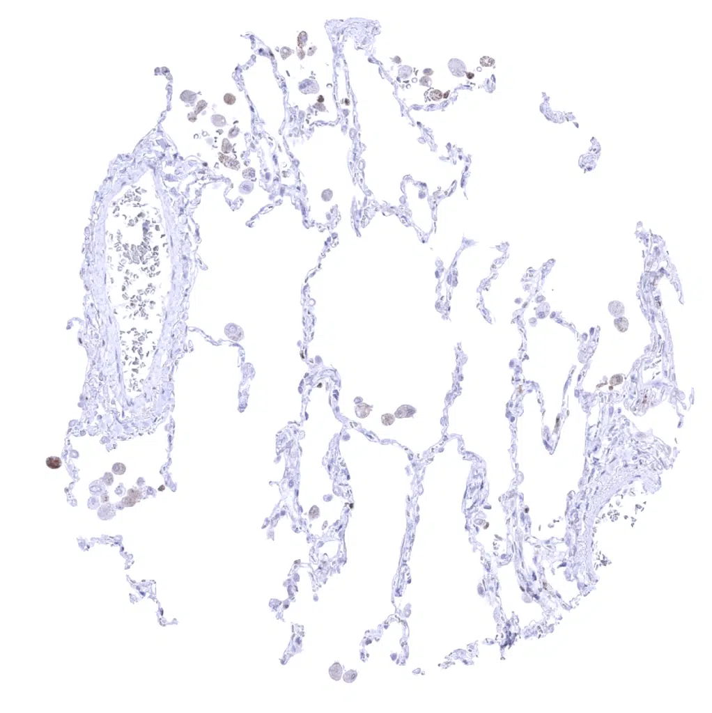
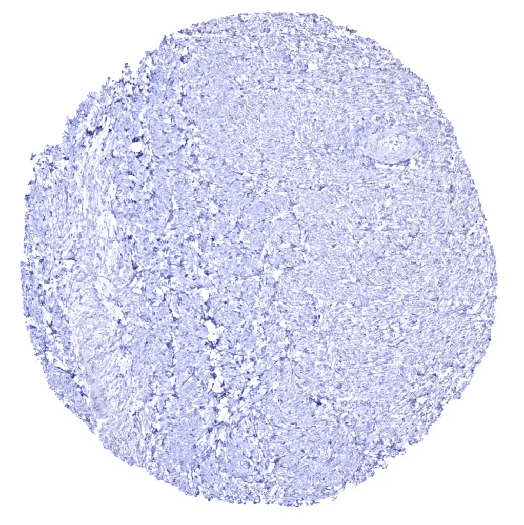
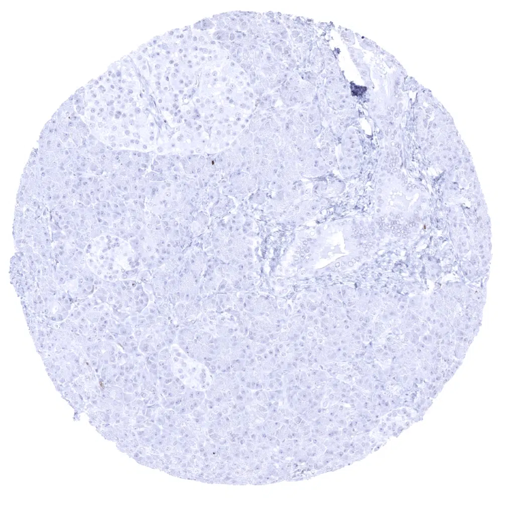
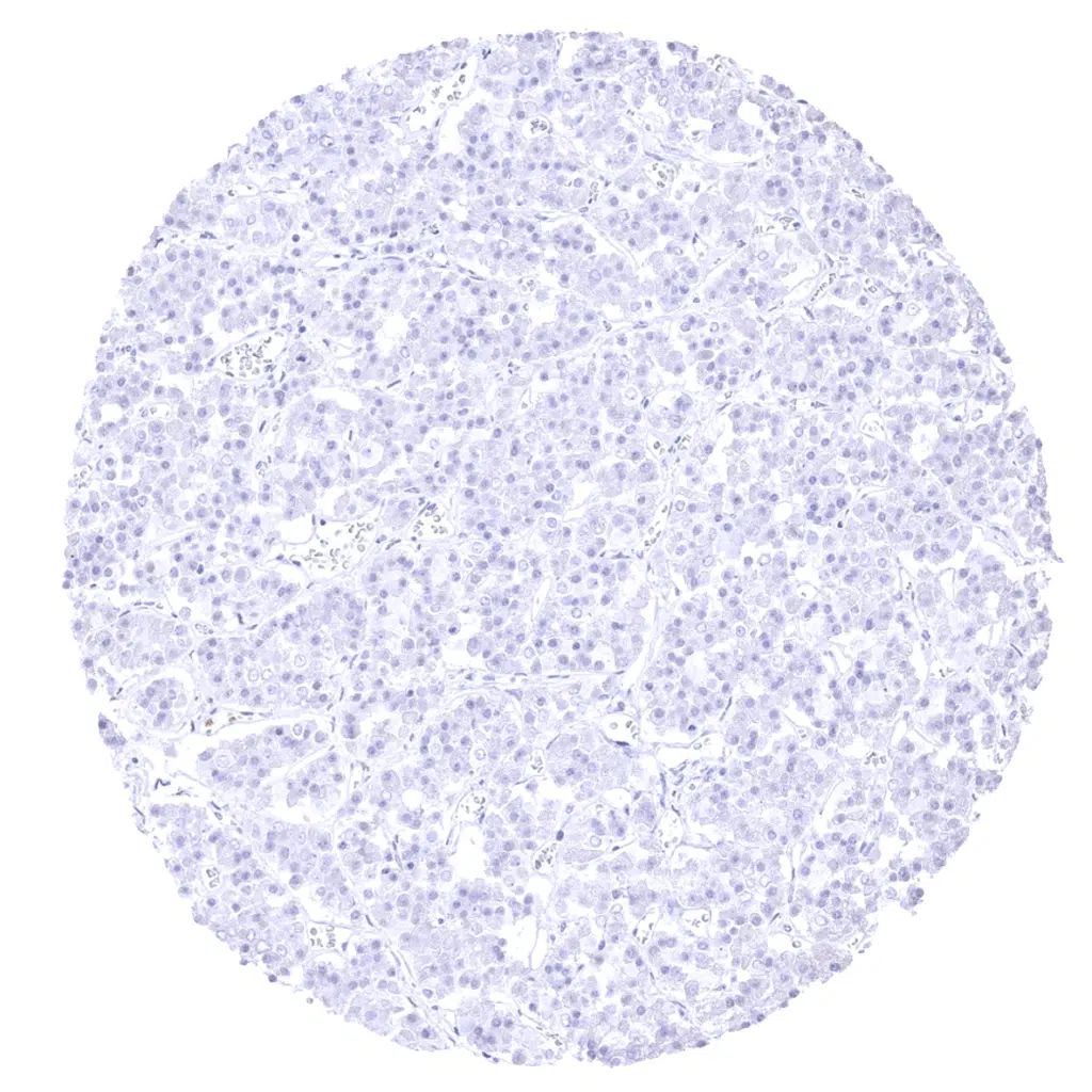
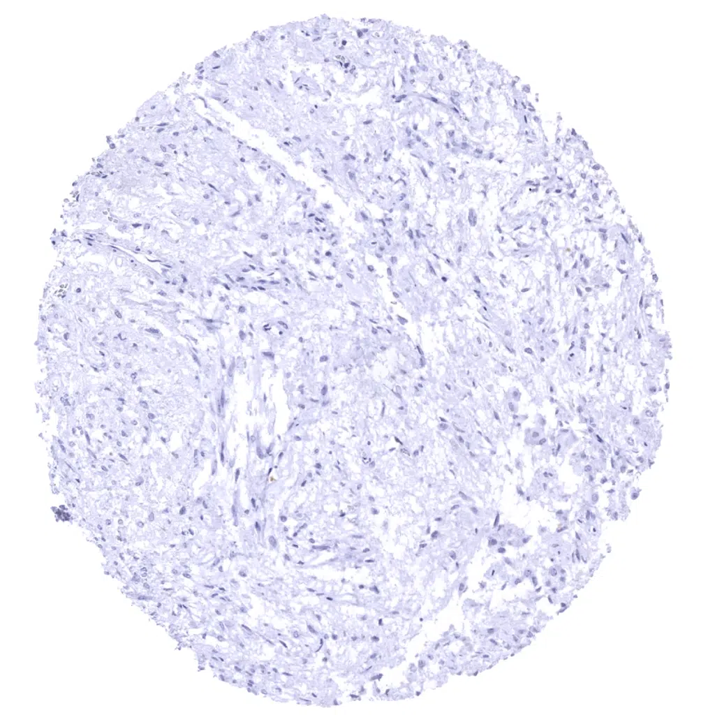
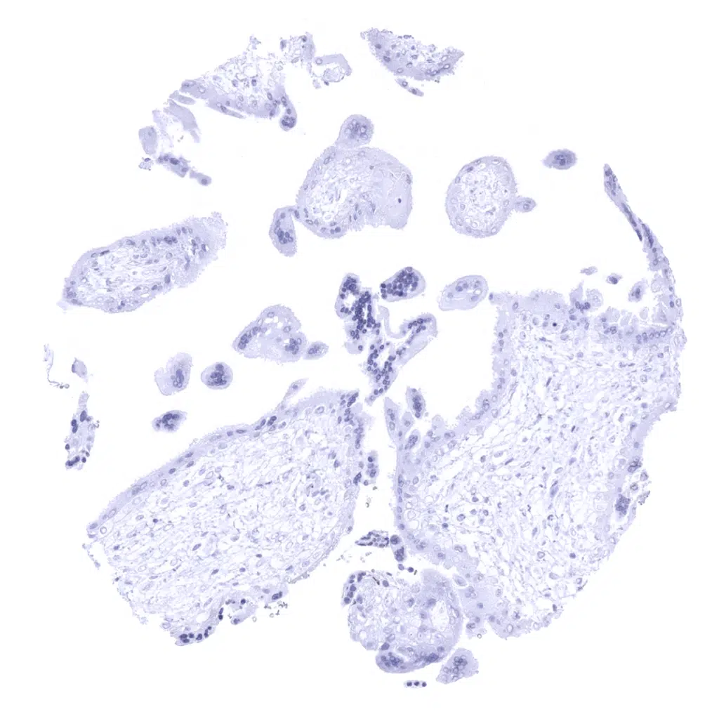
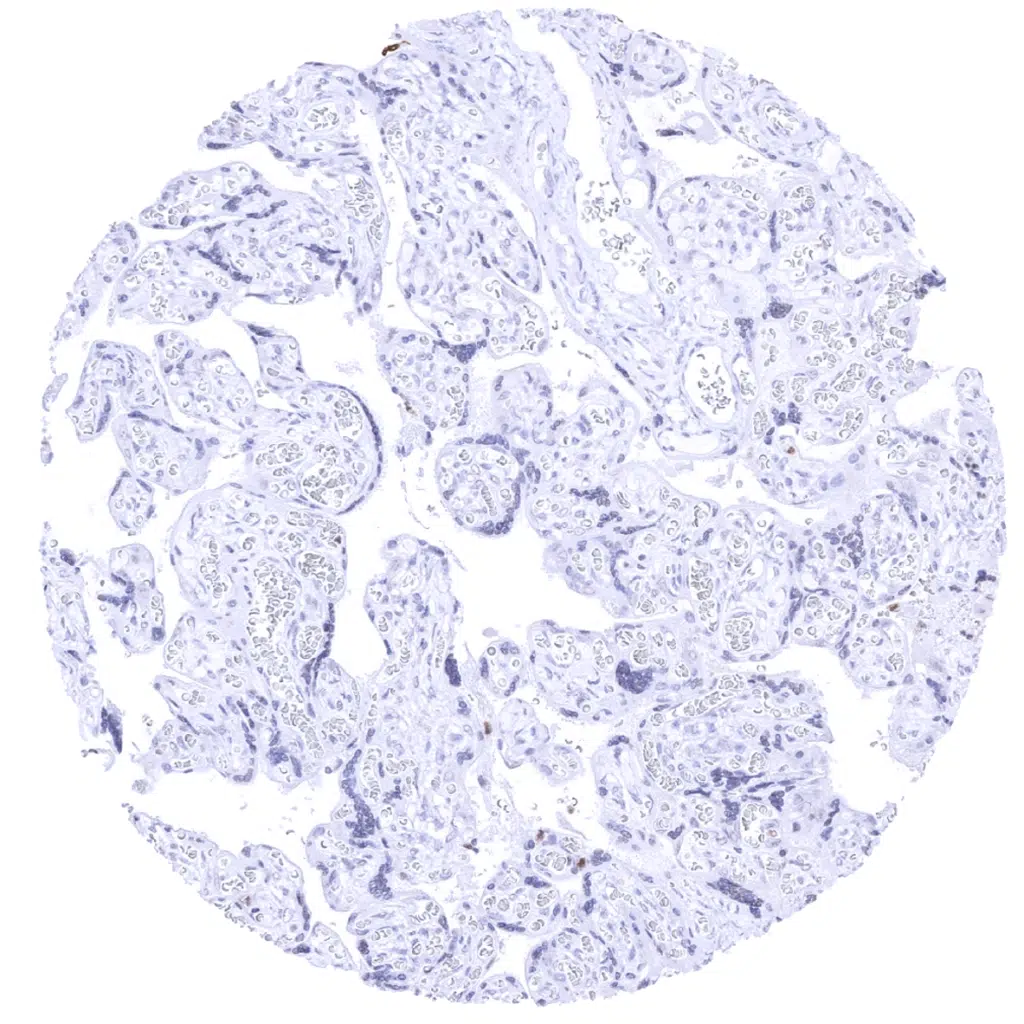
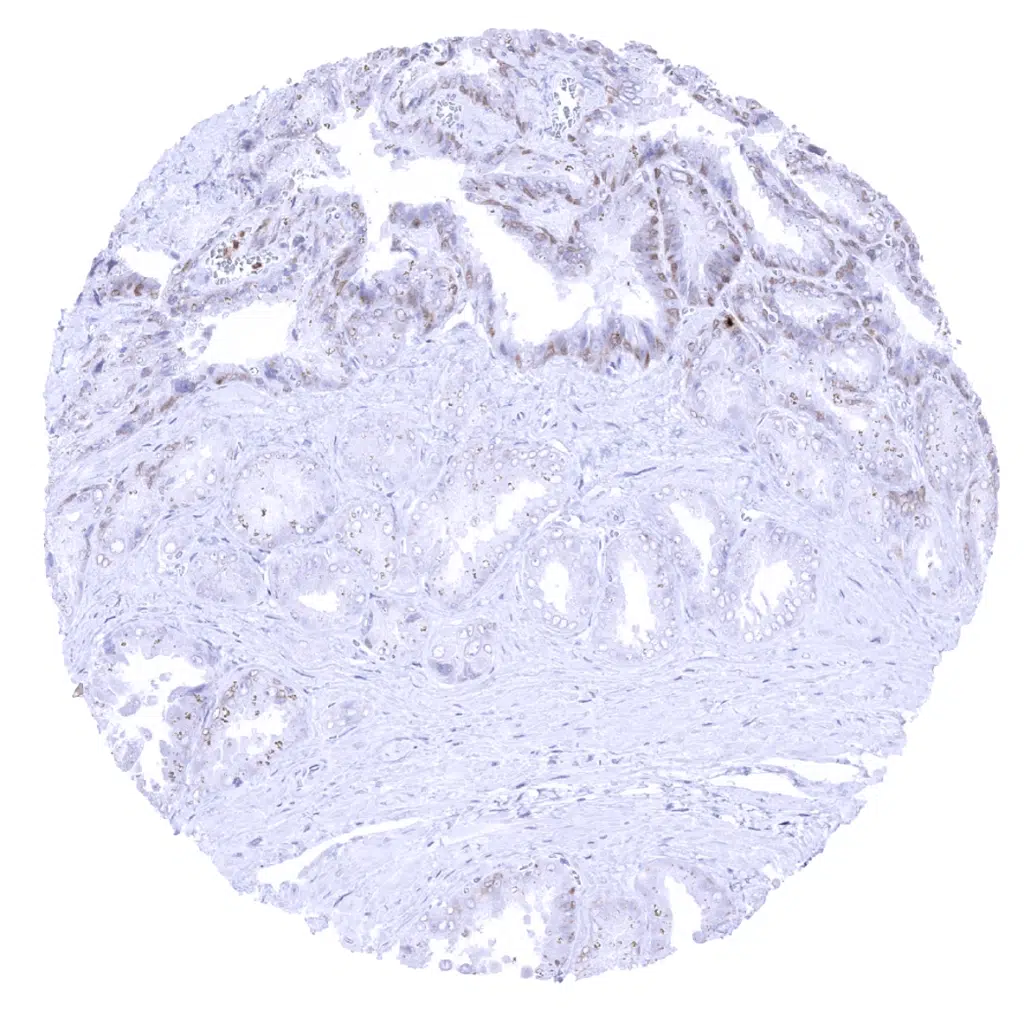
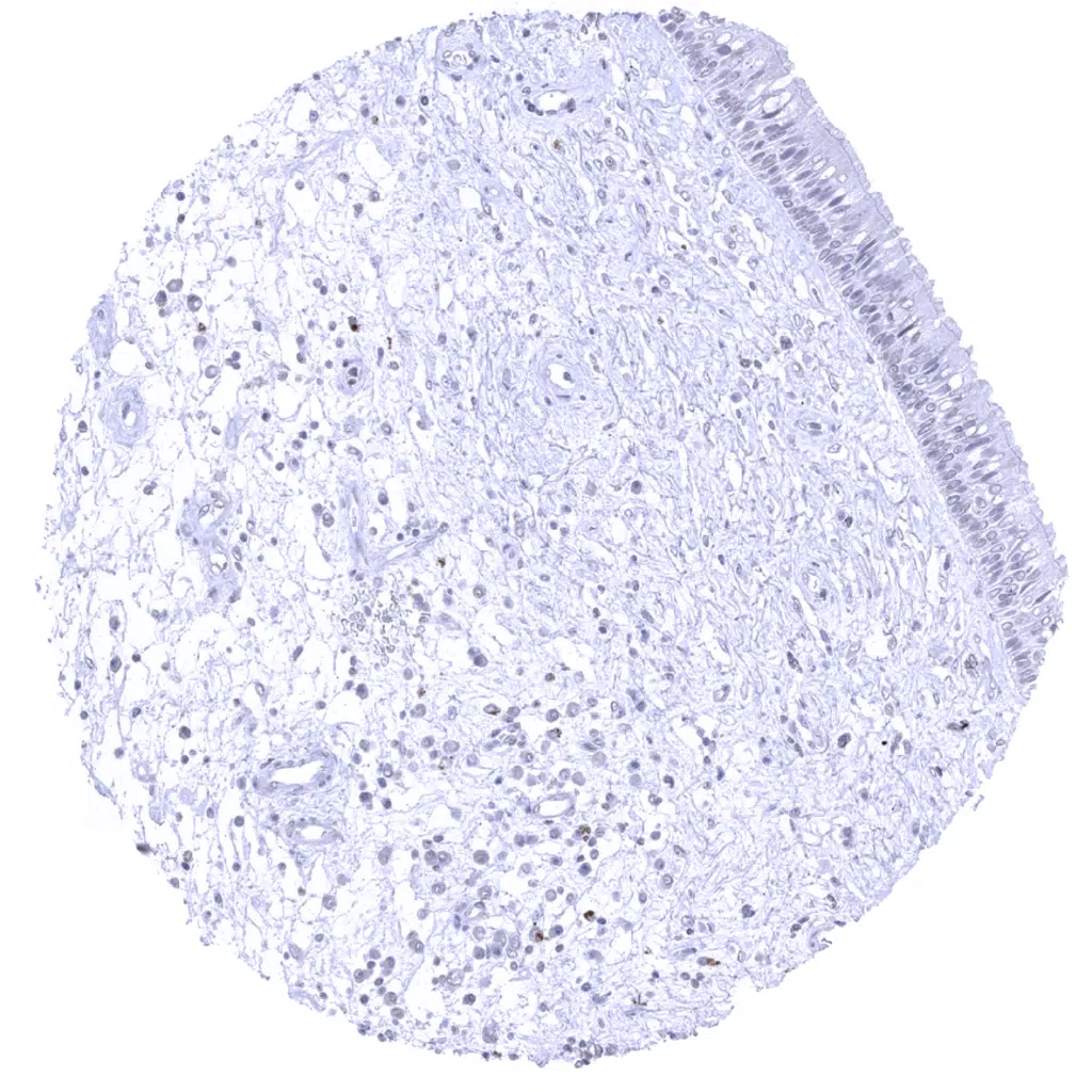
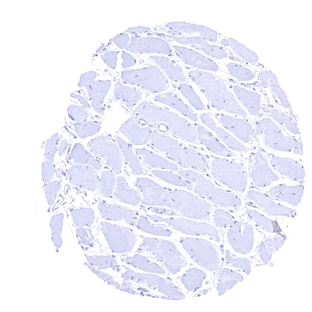
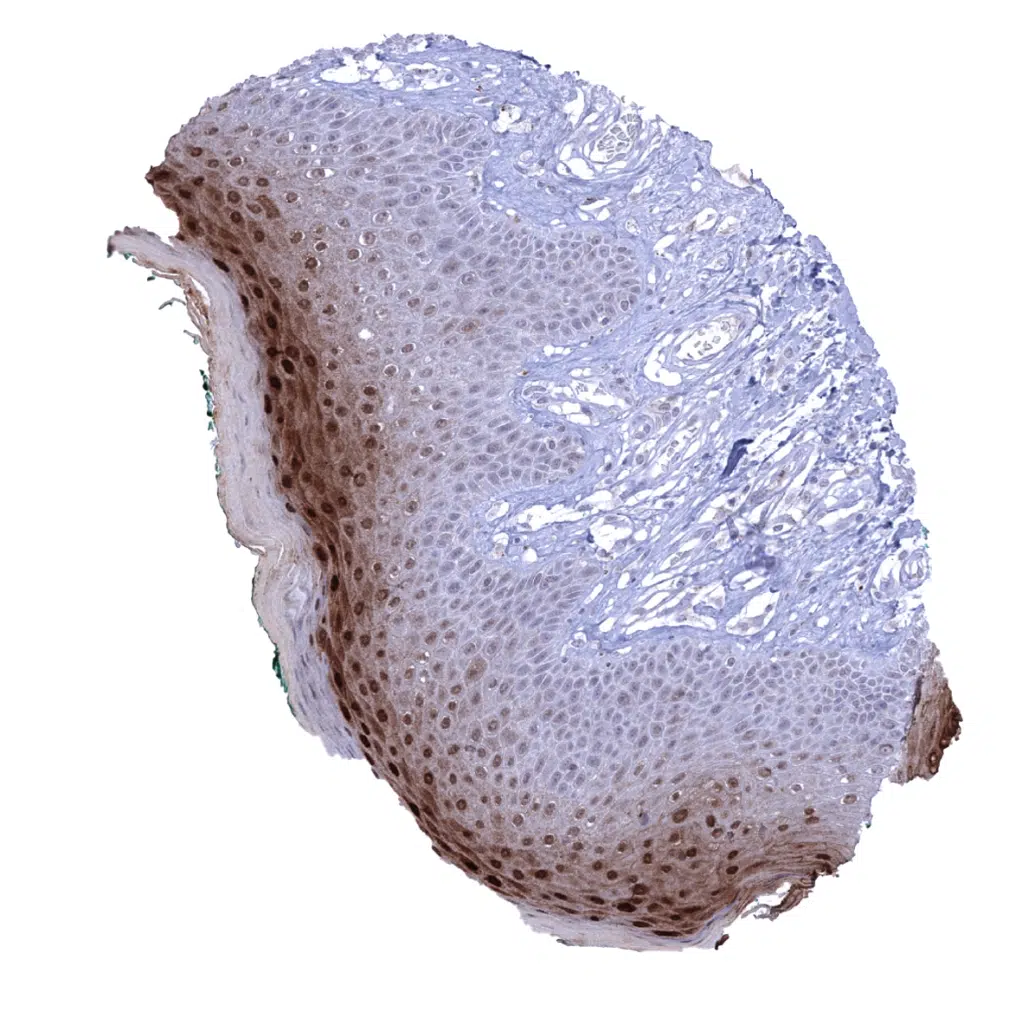
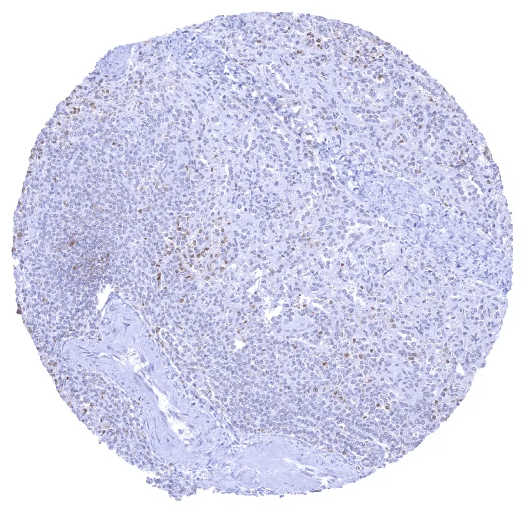
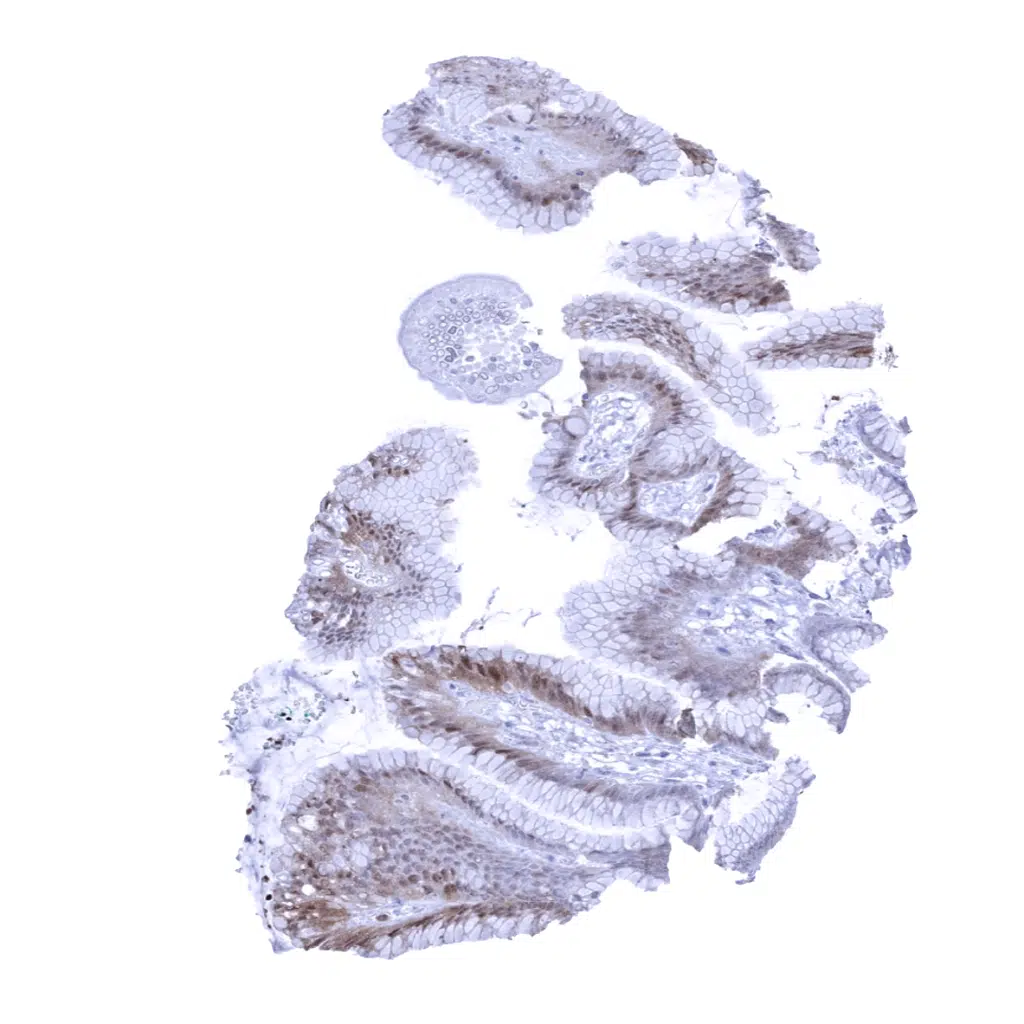
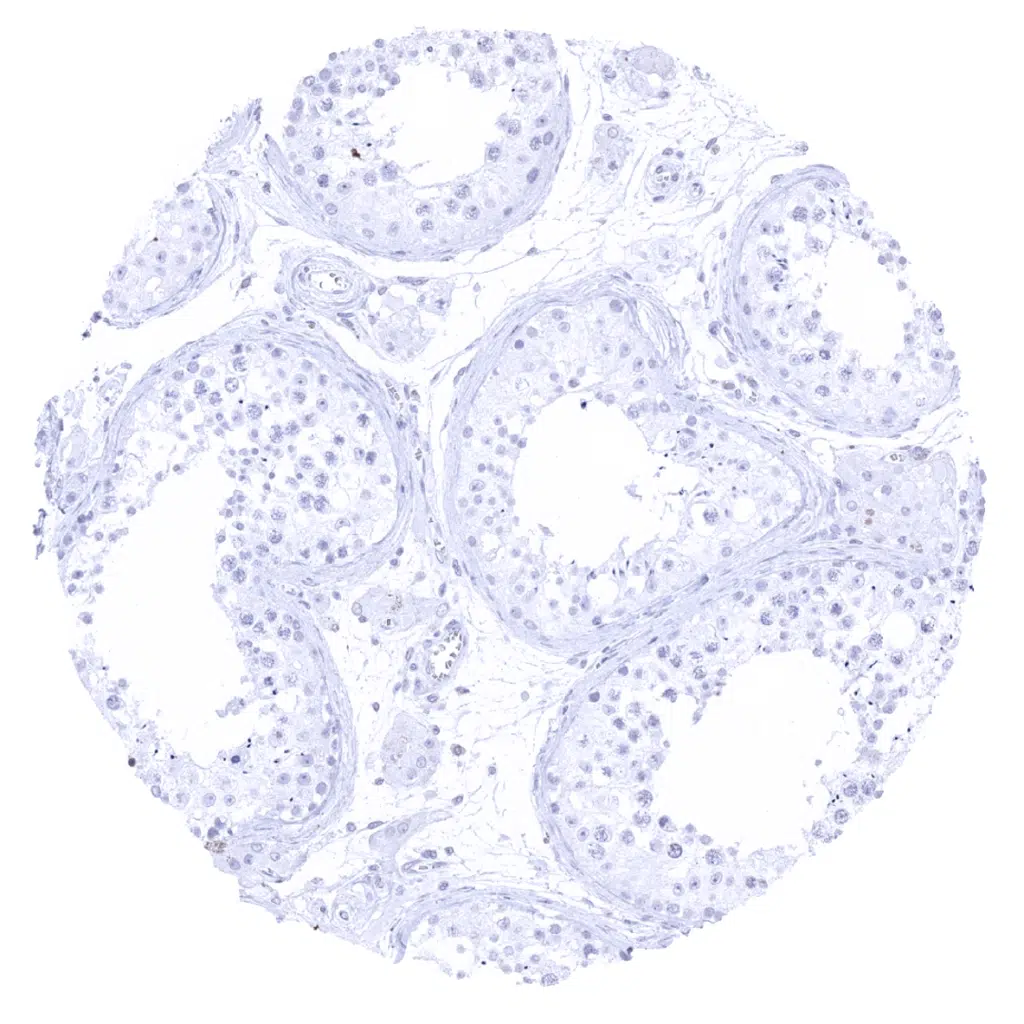
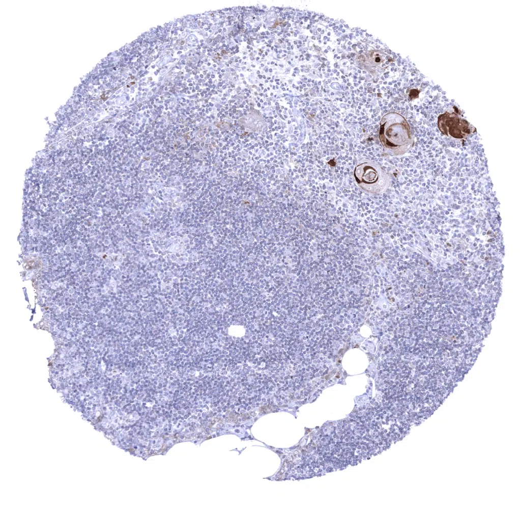
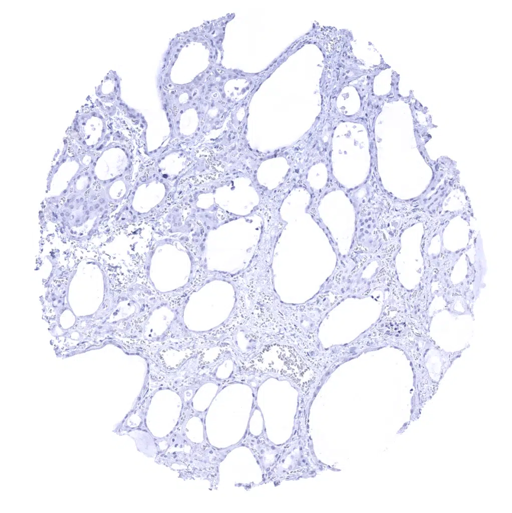
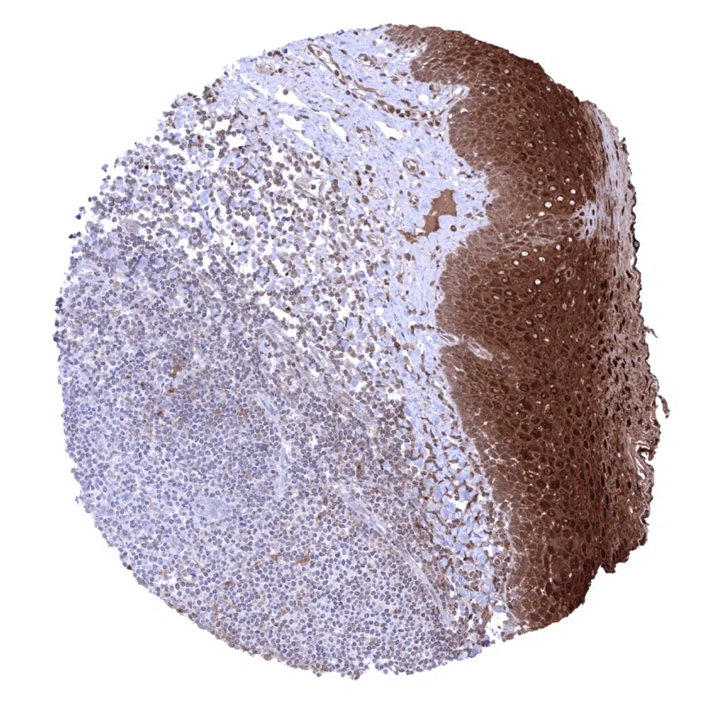
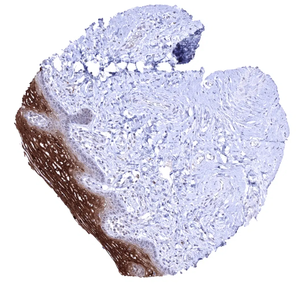
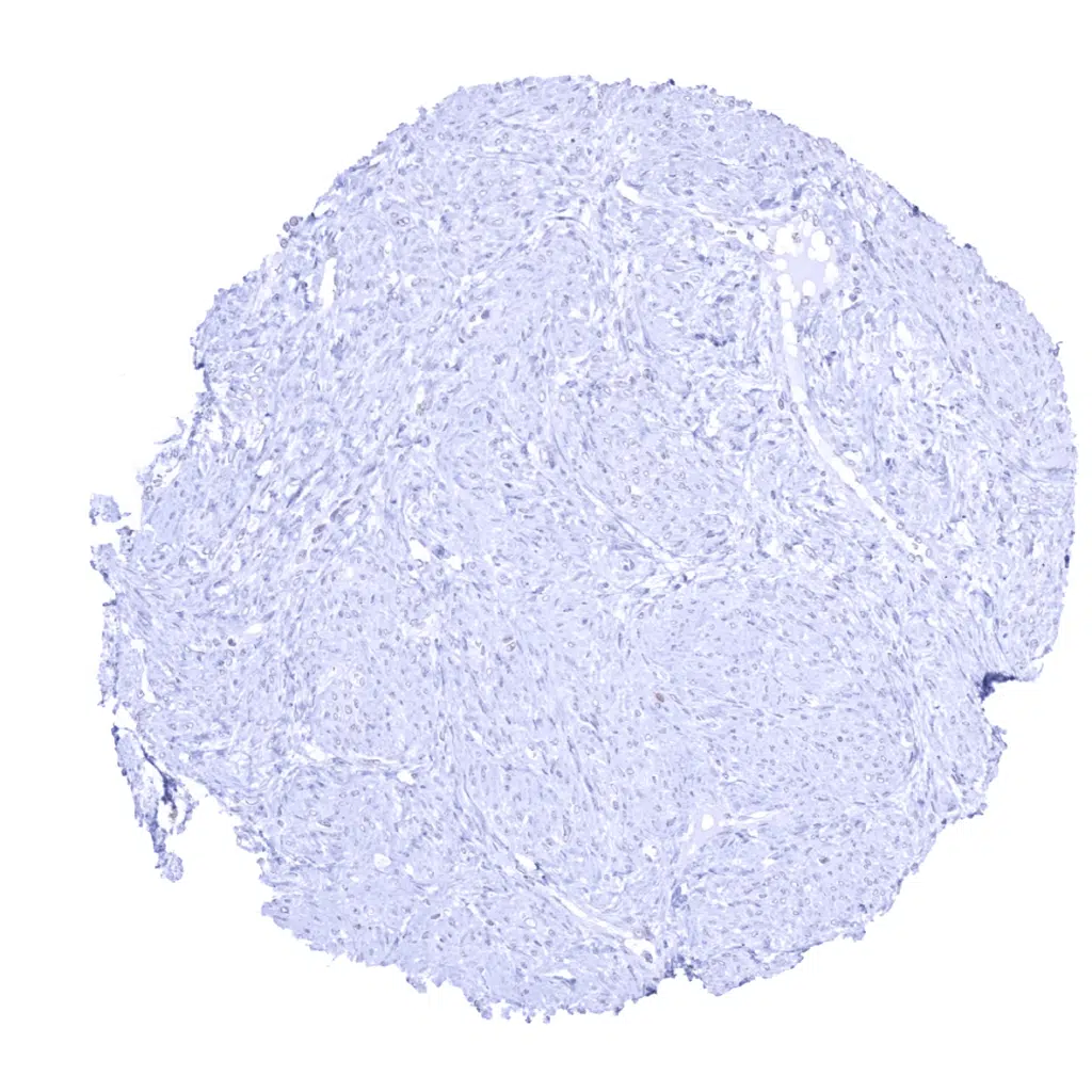
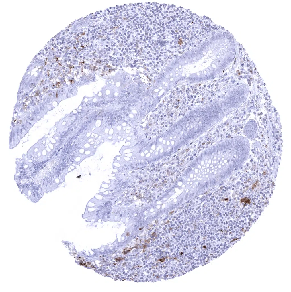
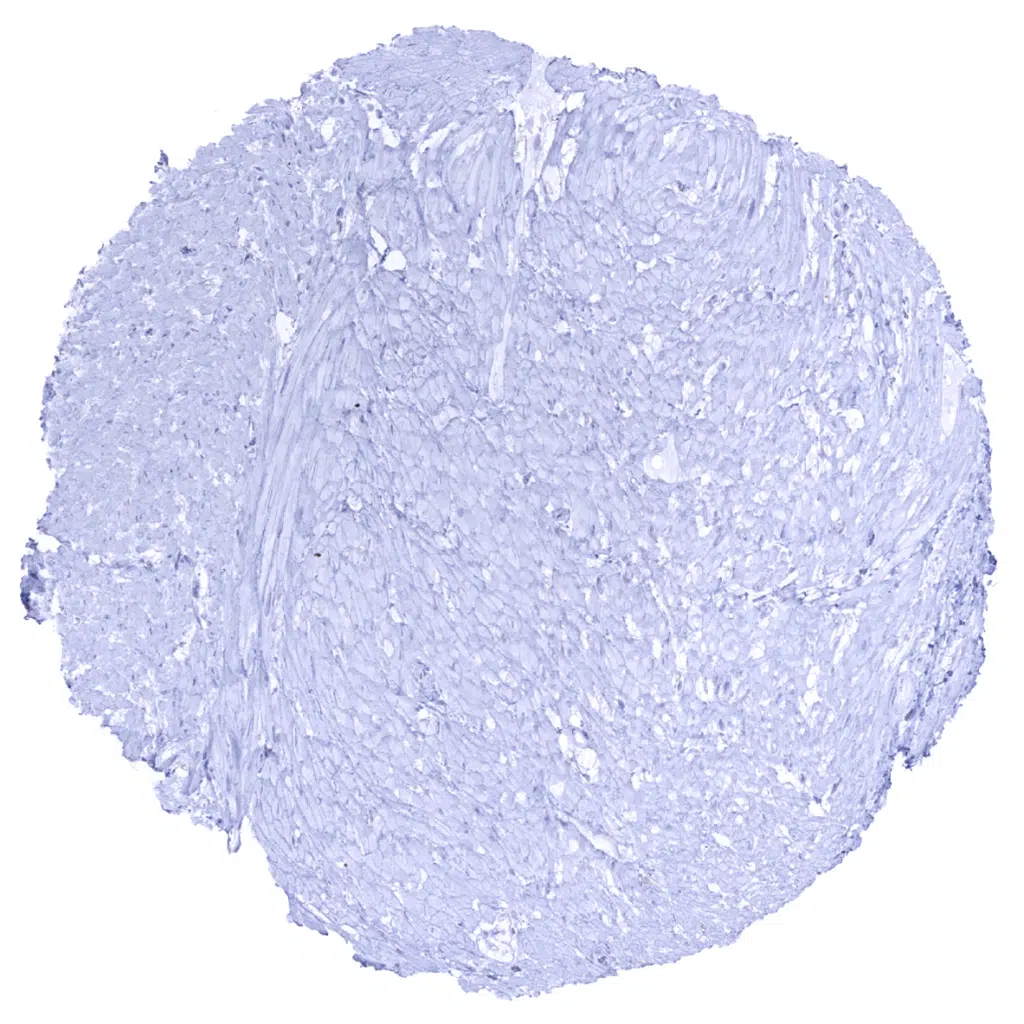
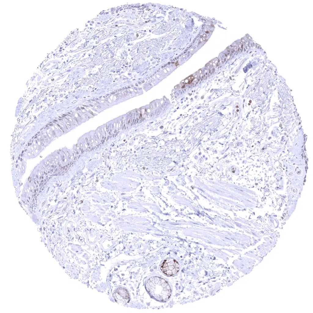
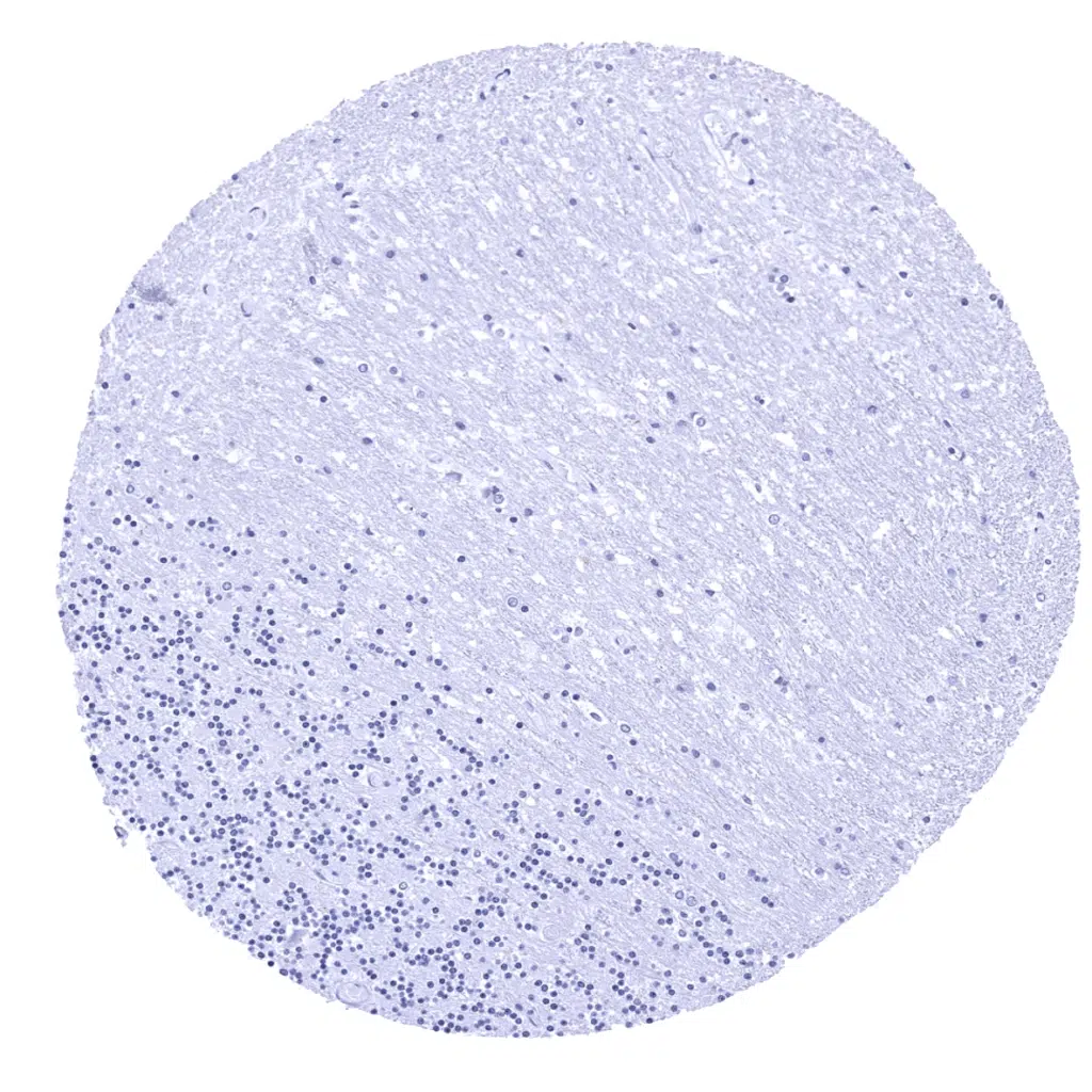
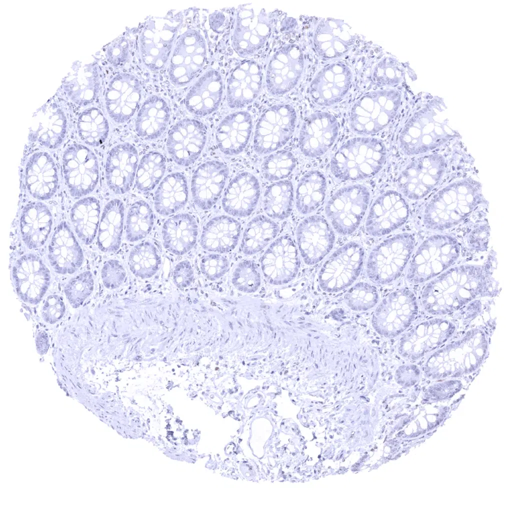
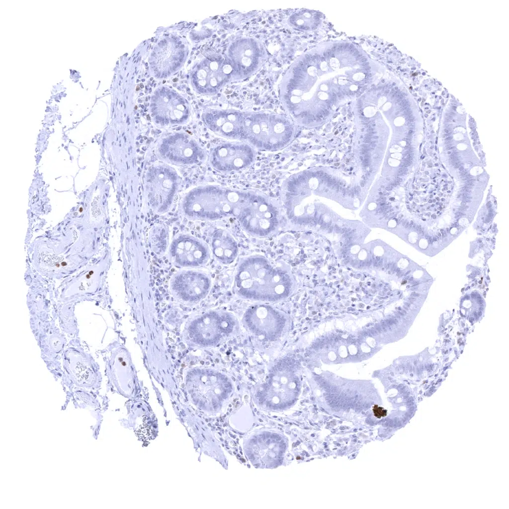
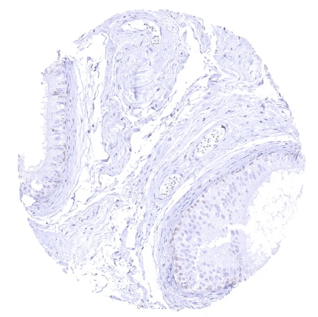
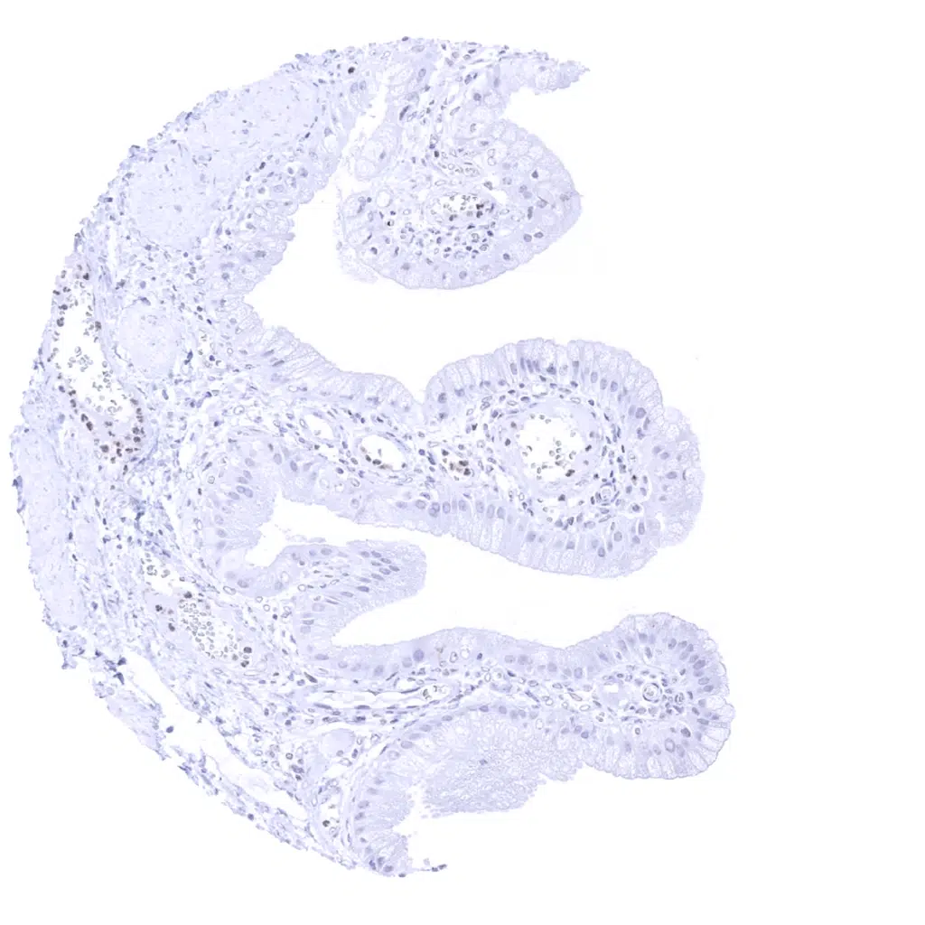
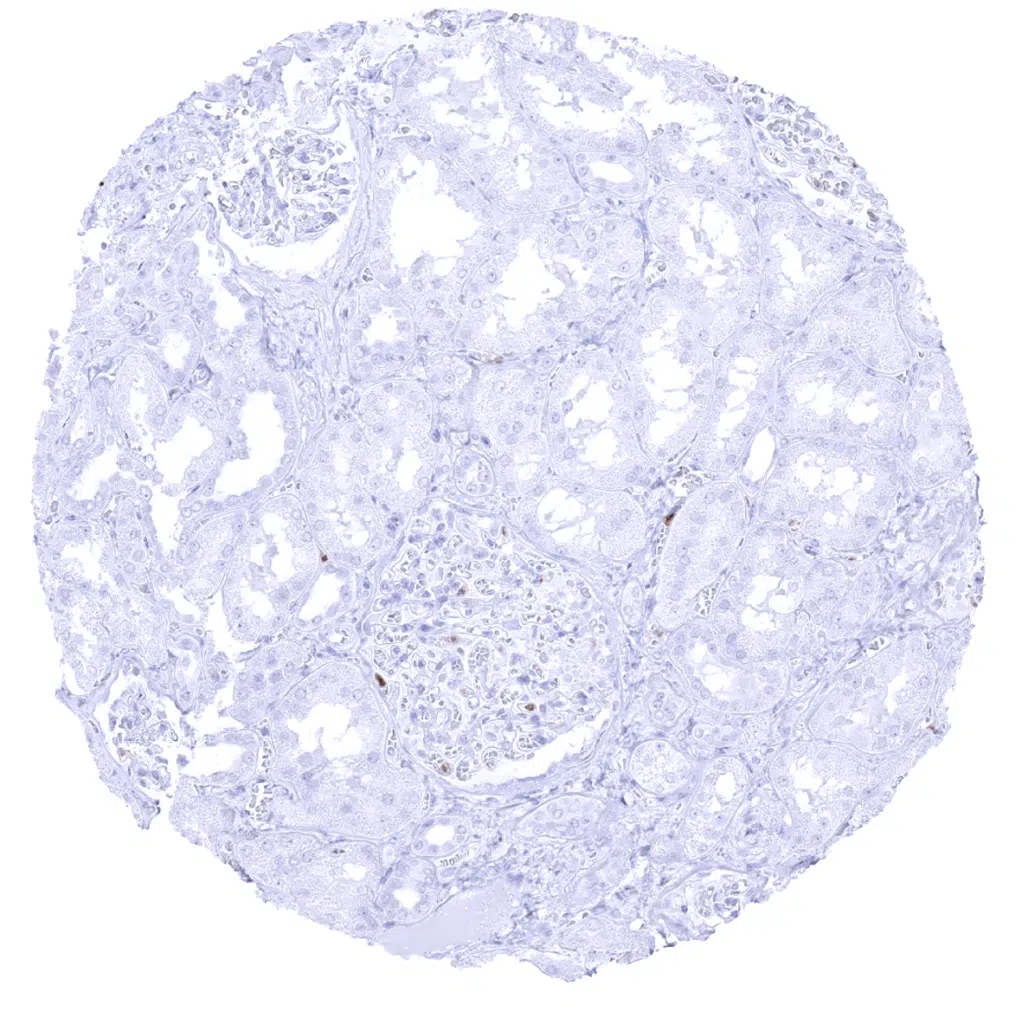
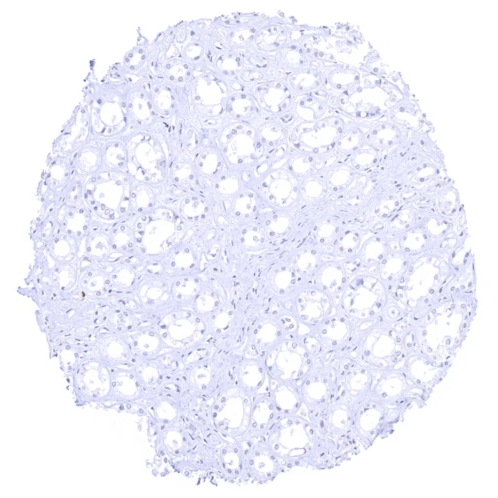
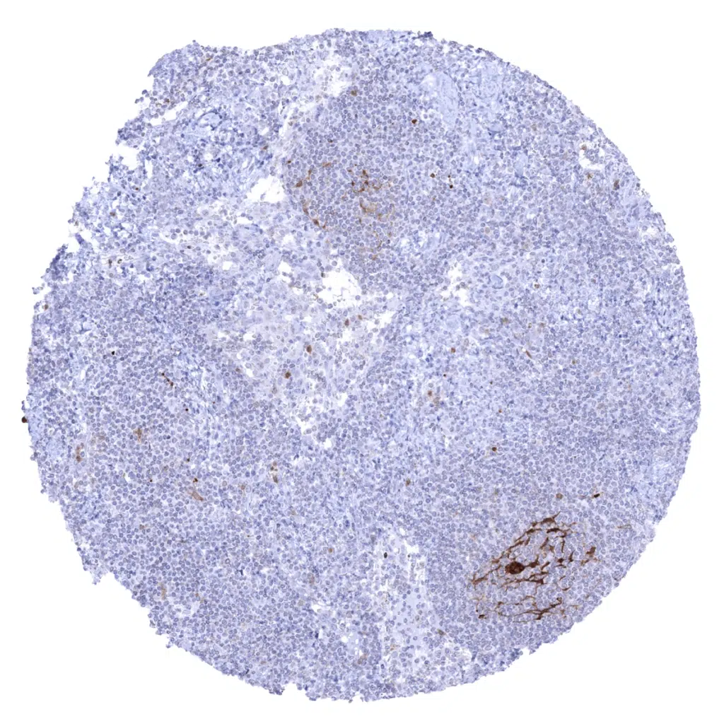
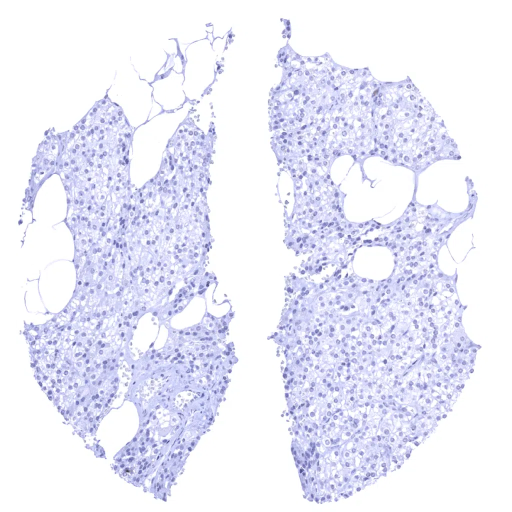
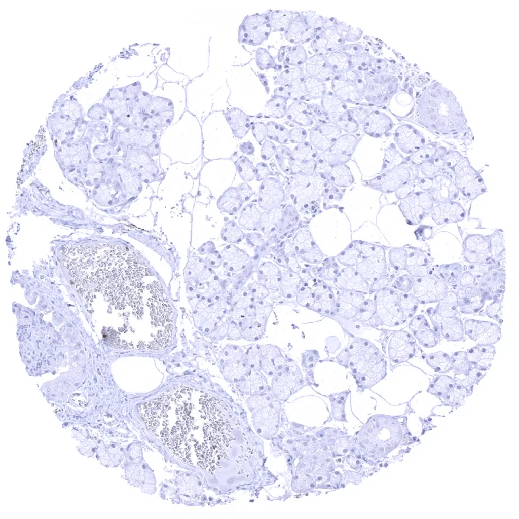
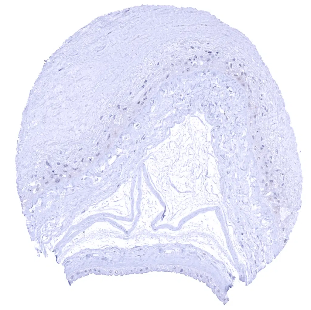
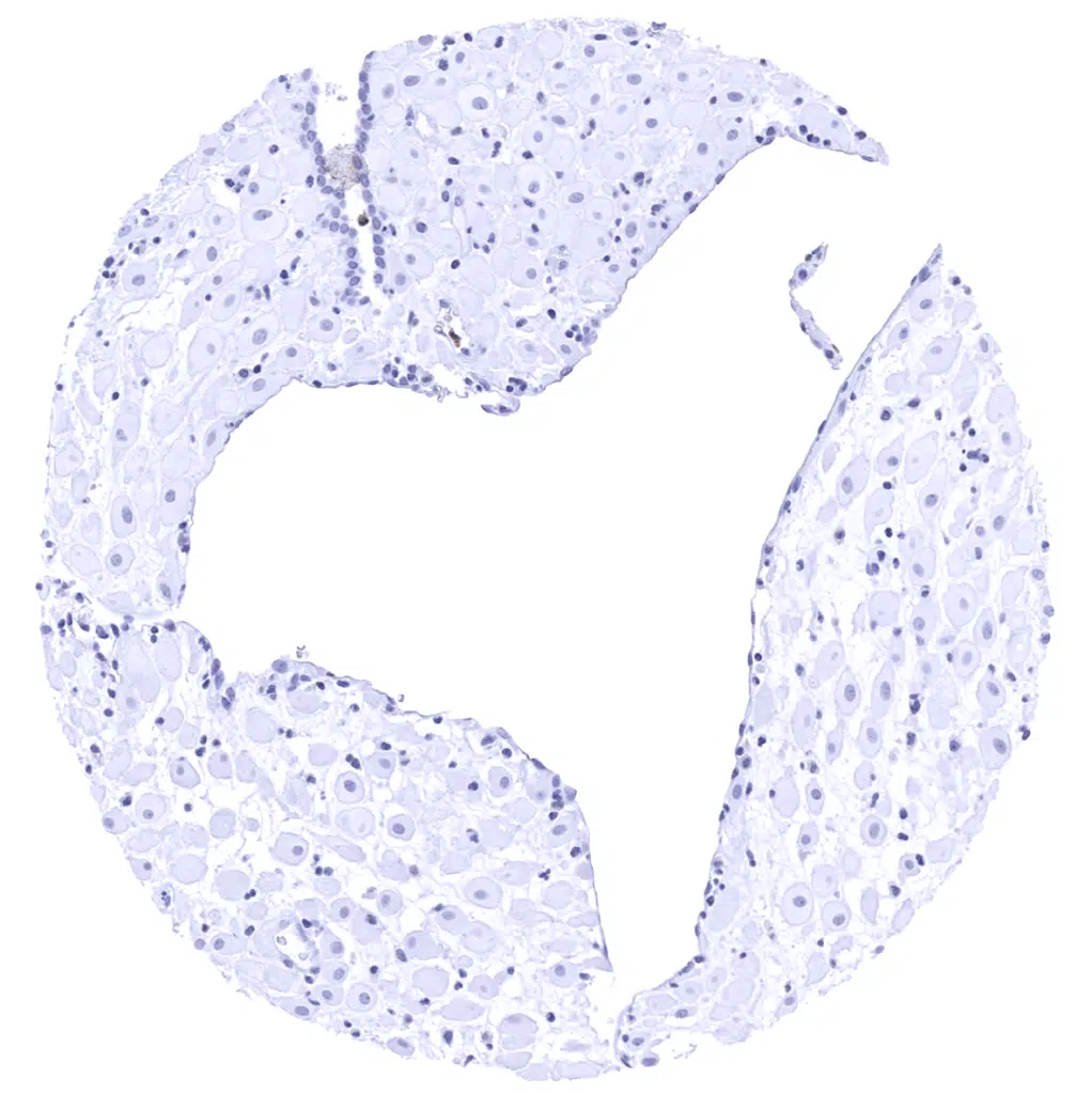
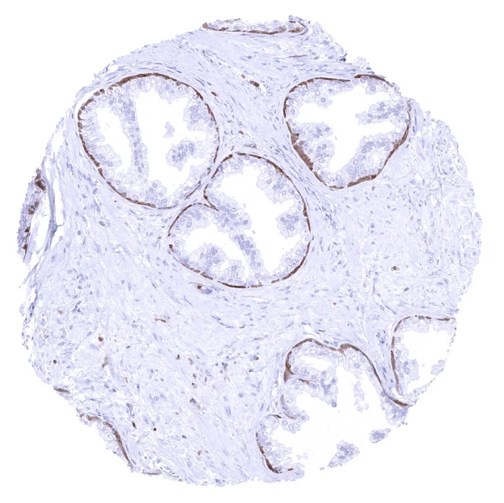
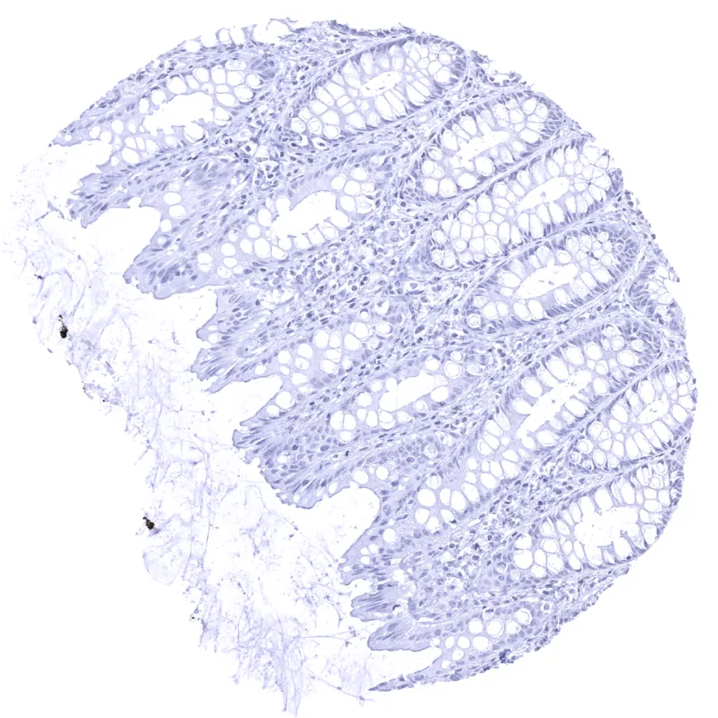
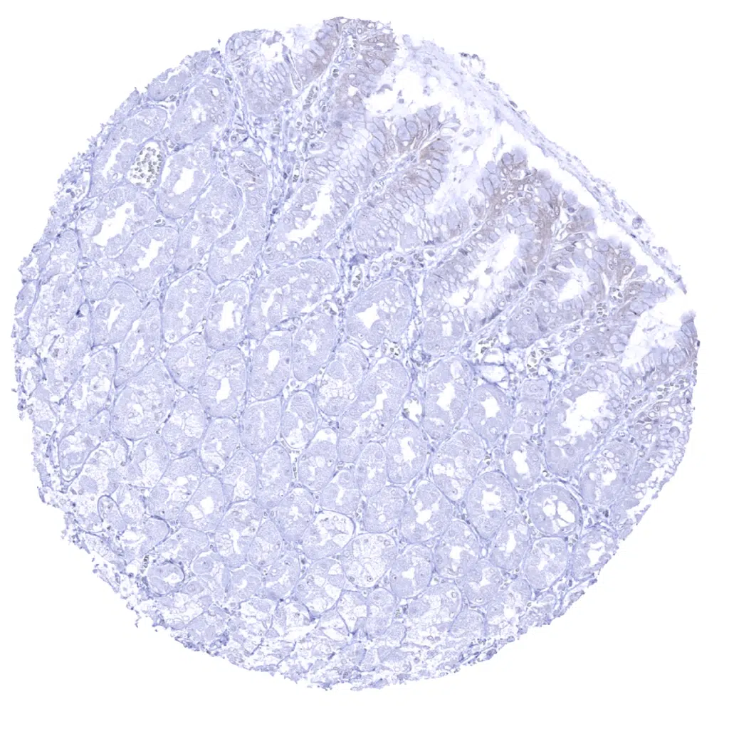
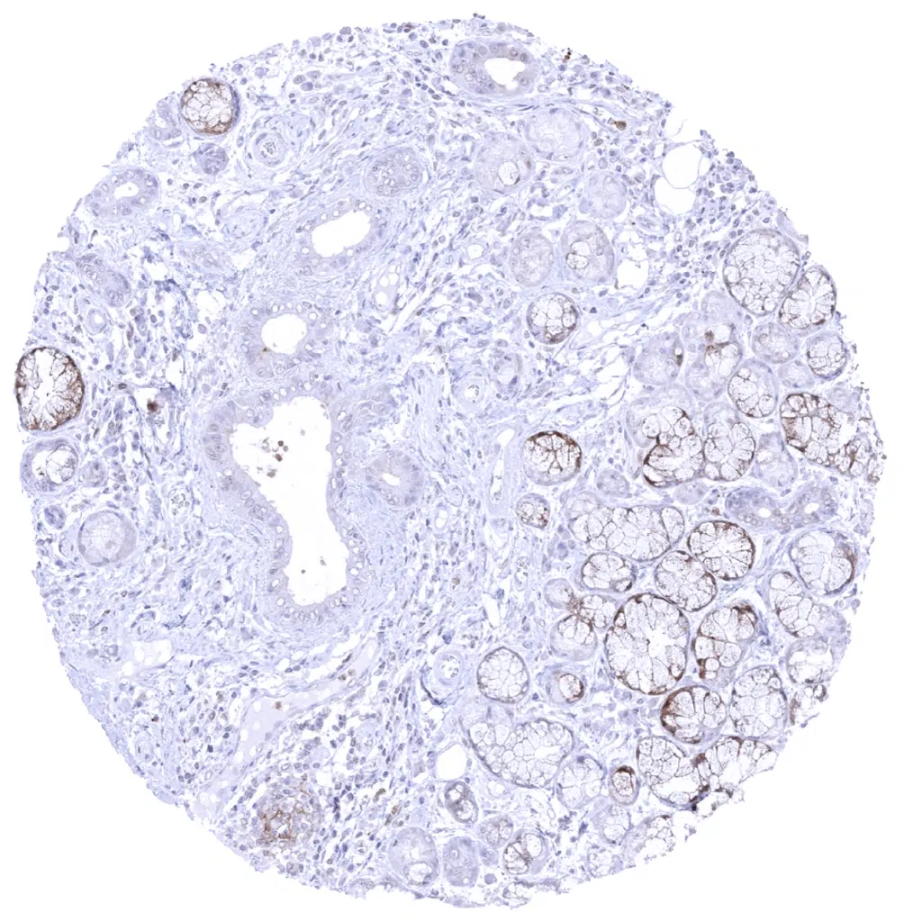
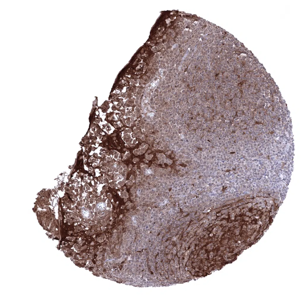
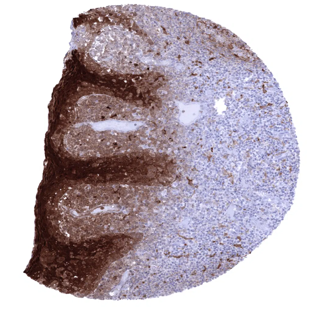
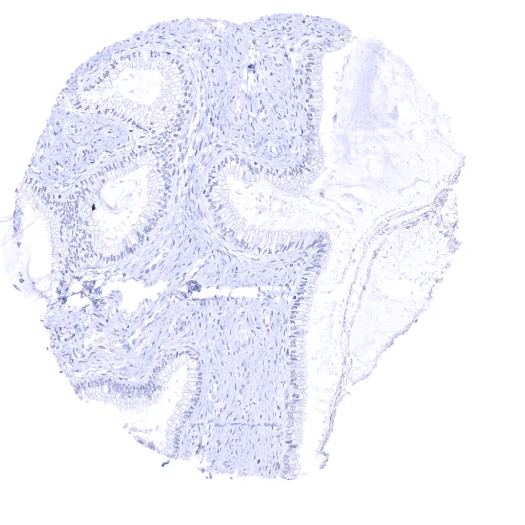
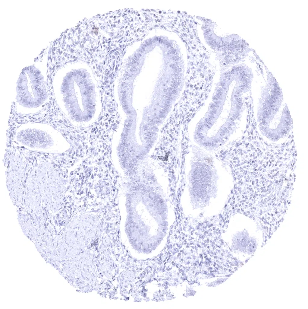
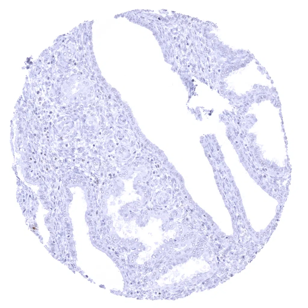
Cystatin A antibody [MSVA-461M] HistoMAX


Go to Cancer Tissue Gallery Go back to product page Go to HistoMAX page


Adrenal gland
Aorta, media
Appendix, mucosa
Appendix, muscular wall
Bone marrow - Weak to moderate Cystatin A staining of a subset of cells.
Breast
Bronchus, mucosa - Cystatin A staining is seen in mucinous cells of bronchial glands and in few cells of the respiratory epithelium
Cerebellum (granule cell layer, white matter)
Cerebellum (molecular layer, Purkinje cell layer, granule cell layer, white matter)
Cerebrum, grey matter
Cerebrum, white matter
Colon descendens, mucosa
Colon descendens, muscular wall
Duodenum, Brunner gland
Duodenum, mucosa
Epididymis - Weak to moderate intensity Cystatin A staining of basal.
Esophagus, squamous epithelium - Strong Cystatin A immunostaining in suprabasal epithelial cell layers.
Fallopian tube, mucosa
Fat
Gallbladder, epithelium
Heart muscle
Ileum, mucosa - Strong Cystatin A immunostaining in dendritic cells of a germinal centre.
Kidney, cortex
Kidney, medulla
Liver - Cystatin A immunostaining is not seen in this liver sample.
Liver - Faint Cystatin A immunostaining of hepatocytes showing a zonal distribution.
Lung
Lymph node - Strong Cystatin A immunostaining in dendritic cells of germinal centres.
Ovary, stroma
Pancreas
Parathyroid
Parotid gland
Pituitary gland, anterior lobe
Pituitary gland, posterior lobe
Placenta (amnion and chorion)
Pregnant uterus (decidua)
Placenta, early
Placenta, mature
Prostate - Moderate intensity Cystatin A staining of basal cells.
Rectum, mucosa
Seminal vesicle - Weak to moderate intensity Cystatin A staining of basal cells.
Sinus paranasales
Skeletal muscle
Skin - Cystatin A immunostaining is nuclear and cytoplasmic. It predominates in the granular cell layer and decreases towards the basal cell layer
Spleen - Weak Cystatin A immunostaining in granulocytes.
Stomach, antrum - Faint basal cytoplasmic Cystatin A immunostaining of surface epithelial cells.
Stomach, corpus - Faint basal cytoplasmic Cystatin A immunostaining of surface epithelial cells
Sublingual gland - Weak to moderate Cystatin A staining in myoepithelial cells and some mucinous glands.
Testis
Thymus - Strong Cystatin A immunostaining of a fraction of cells in corpuscles of Hassall's.
Thyroid gland
Tonsil - Strong Cystatin A immunostaining of crypt epithelium and of dendritic cells. Adjacent lymphocytes also show weak staining. This may represent a tissue contamination due to the very high levels of target protein in this tissue sample.
Tonsil, surface epithelium - Intense Cystatin A immunostaining occurs across all epithelial cell layers. A weaker staining is also seen in subepithelial inflammatory cells and endothelia of subepithelial vessels. Endothelial cell staining is only seen in vessels adjacent to strongly positive epithelial tissues.
Tonsil, surface epithelium - Intense Cystatin A immunostaining occurs across all epithelial cell layers. A weaker staining is also seen in subepithelial inflammatory cells. This may represent tissue contamination in case of very high levels of target protein or cystatin A secretion.
Urinary bladder, muscular wall
Urinary bladder, urothelium
Uterus, ectocervix - Strong Cystatin A immunostaining in suprabasal epithelial cells.
Uterus, endocervix
Uterus, endometrium (proliferation)
Uterus, endometrium (secretion)
Uterus, myometrium
     

| TOP |