GeneTex
  • Country / Location Selection

United States (US)

Normal Tissue Gallery- GTX04402

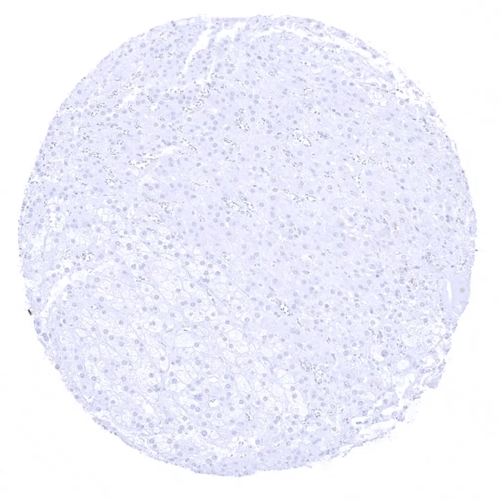
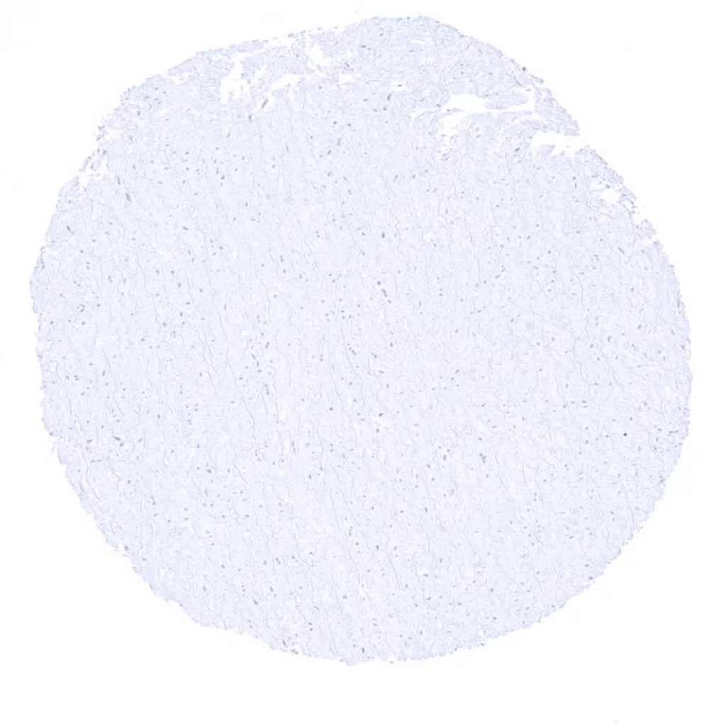
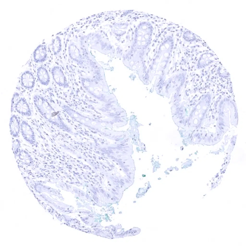
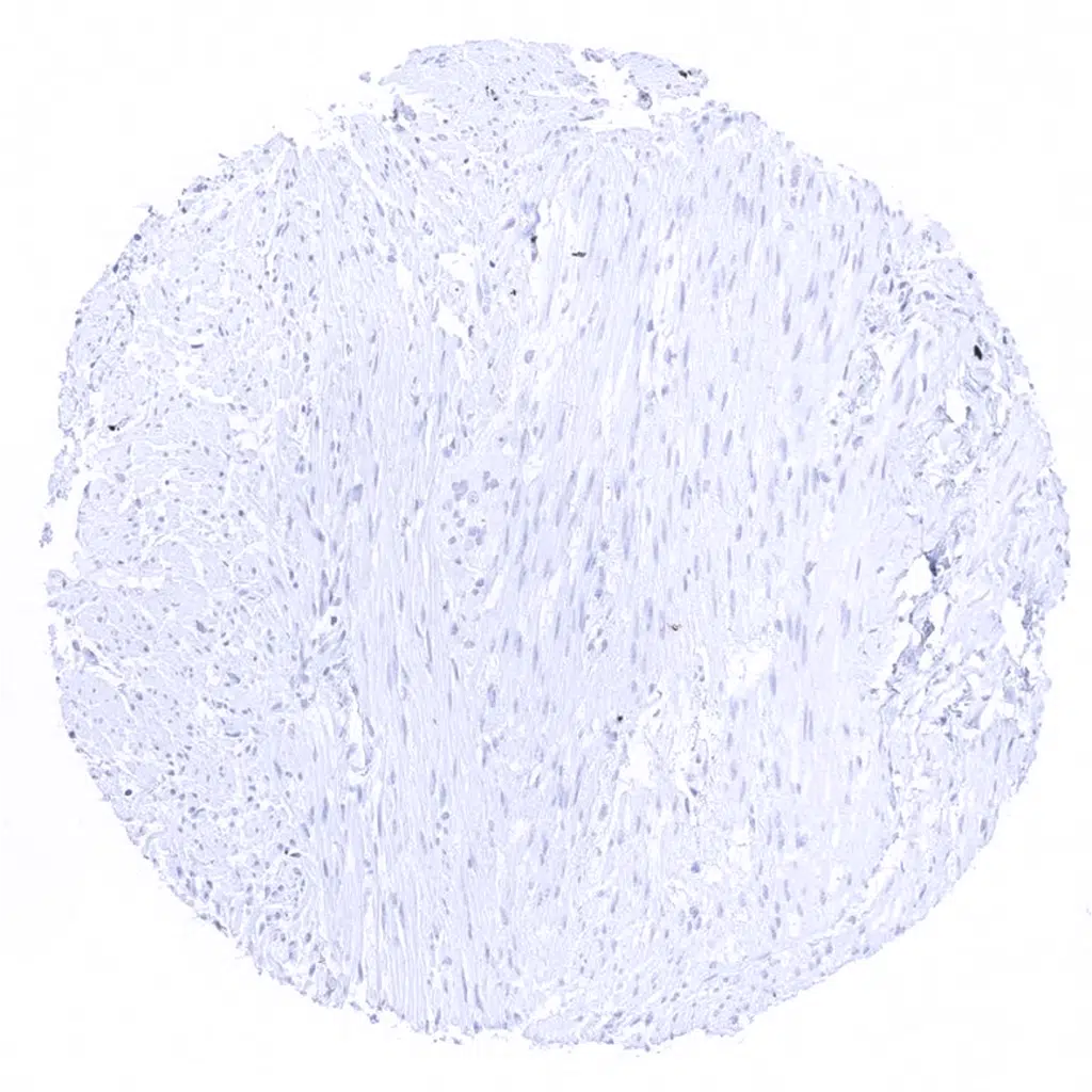
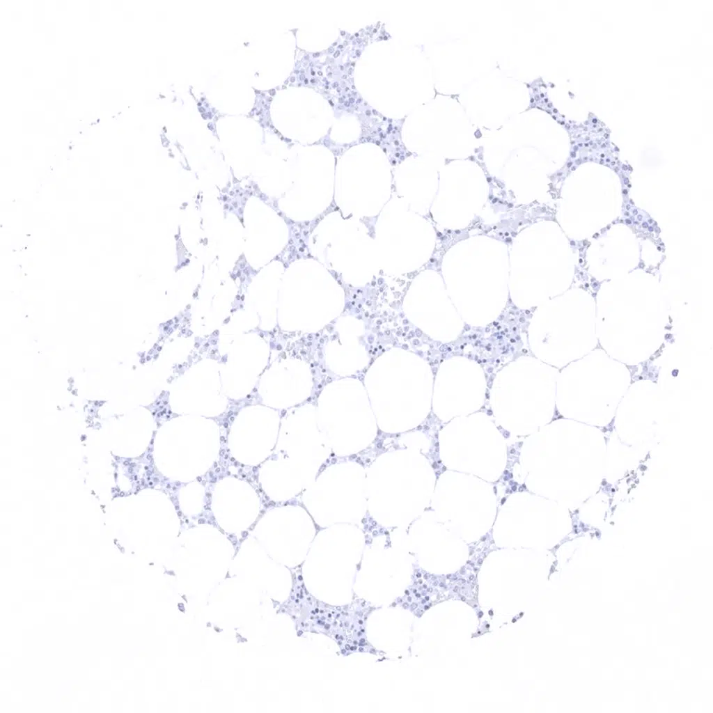
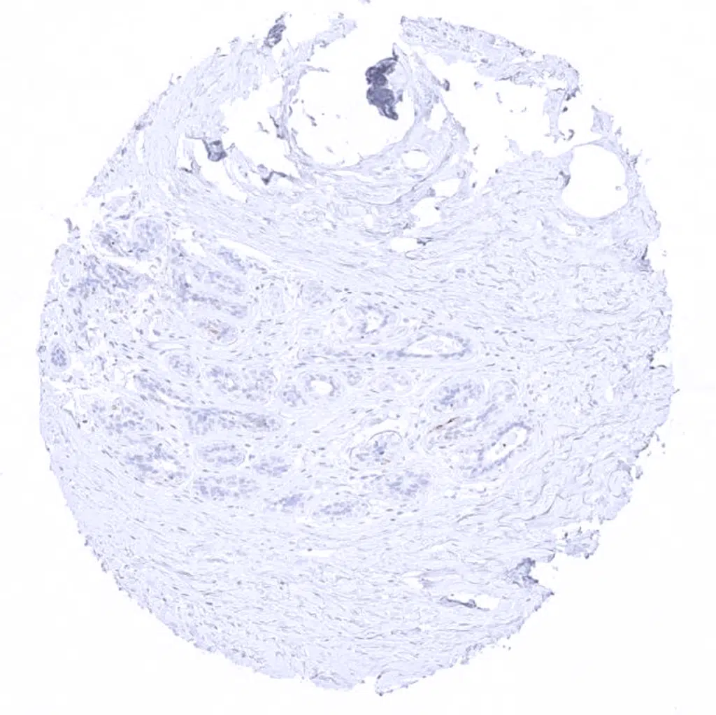
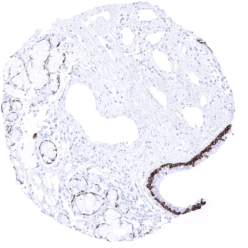
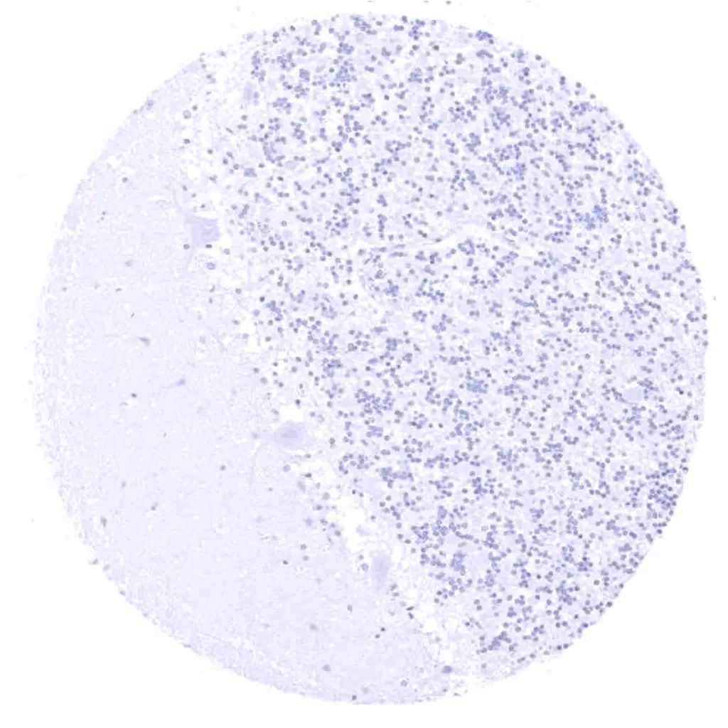
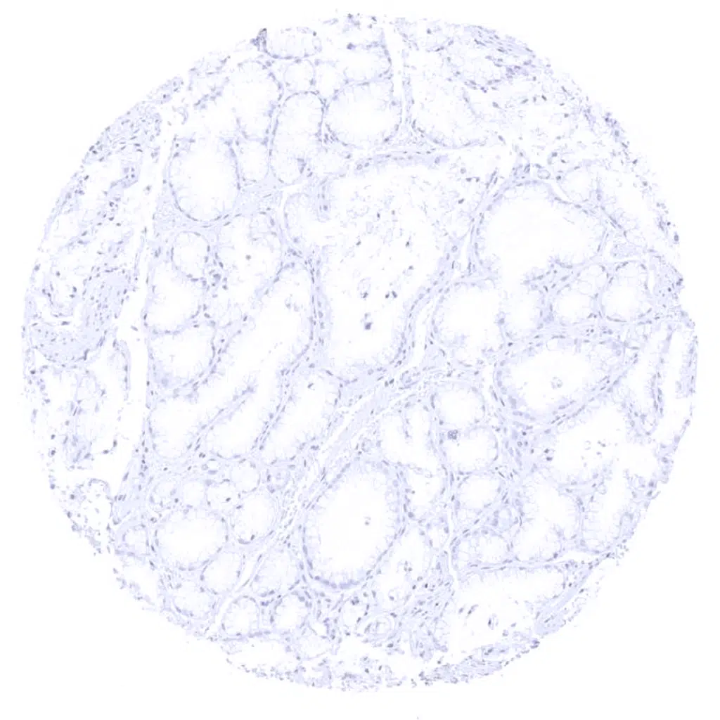
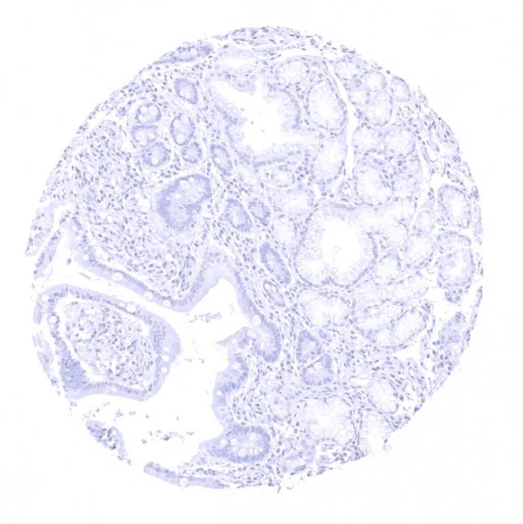
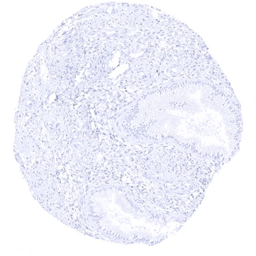
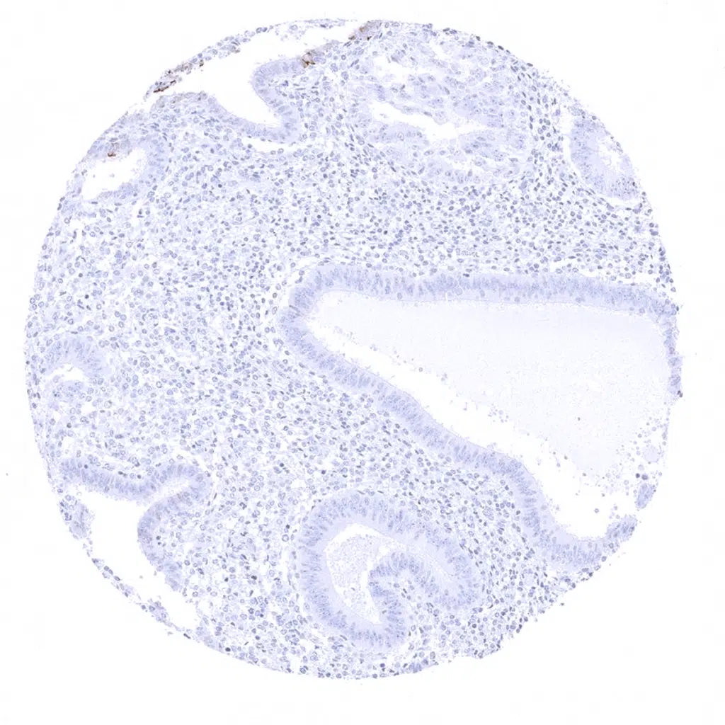
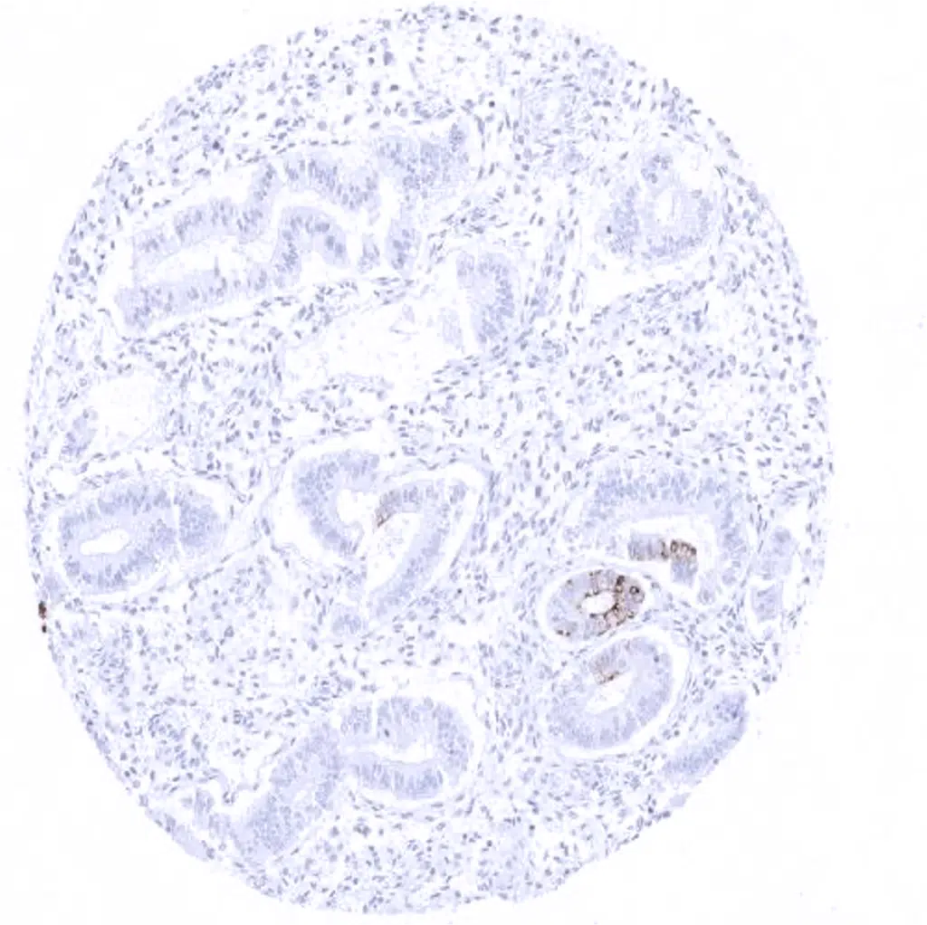
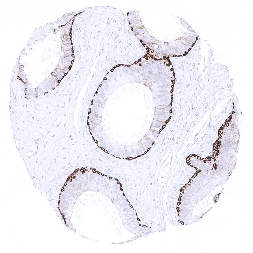
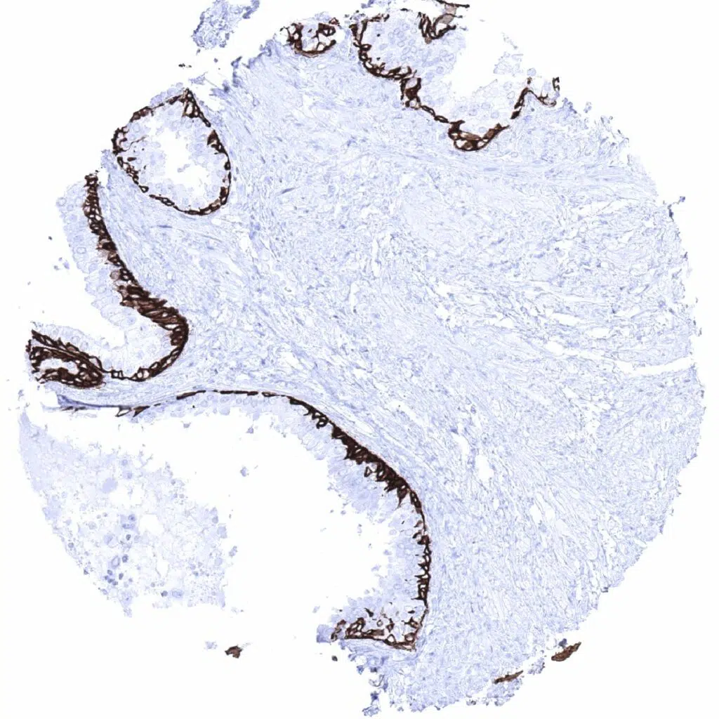
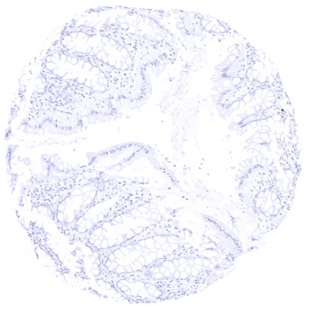
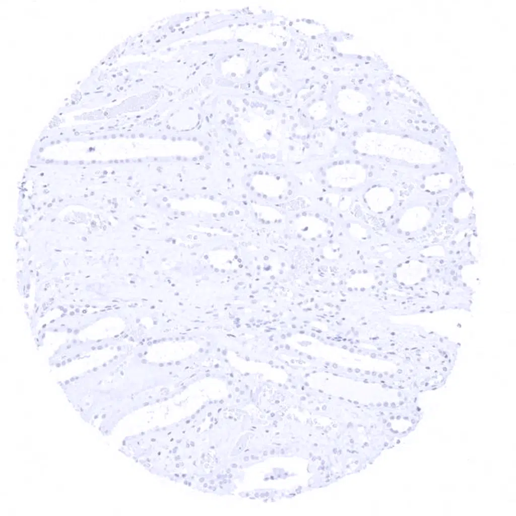
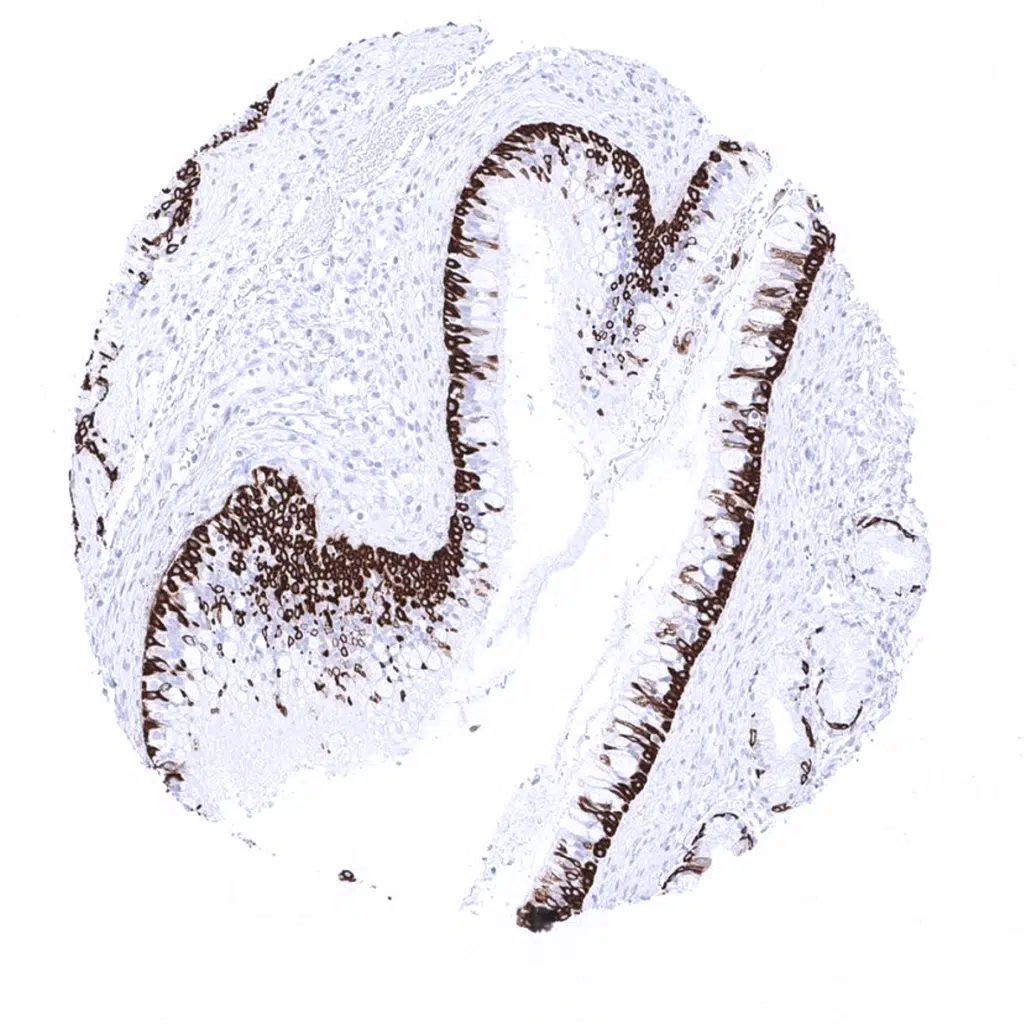
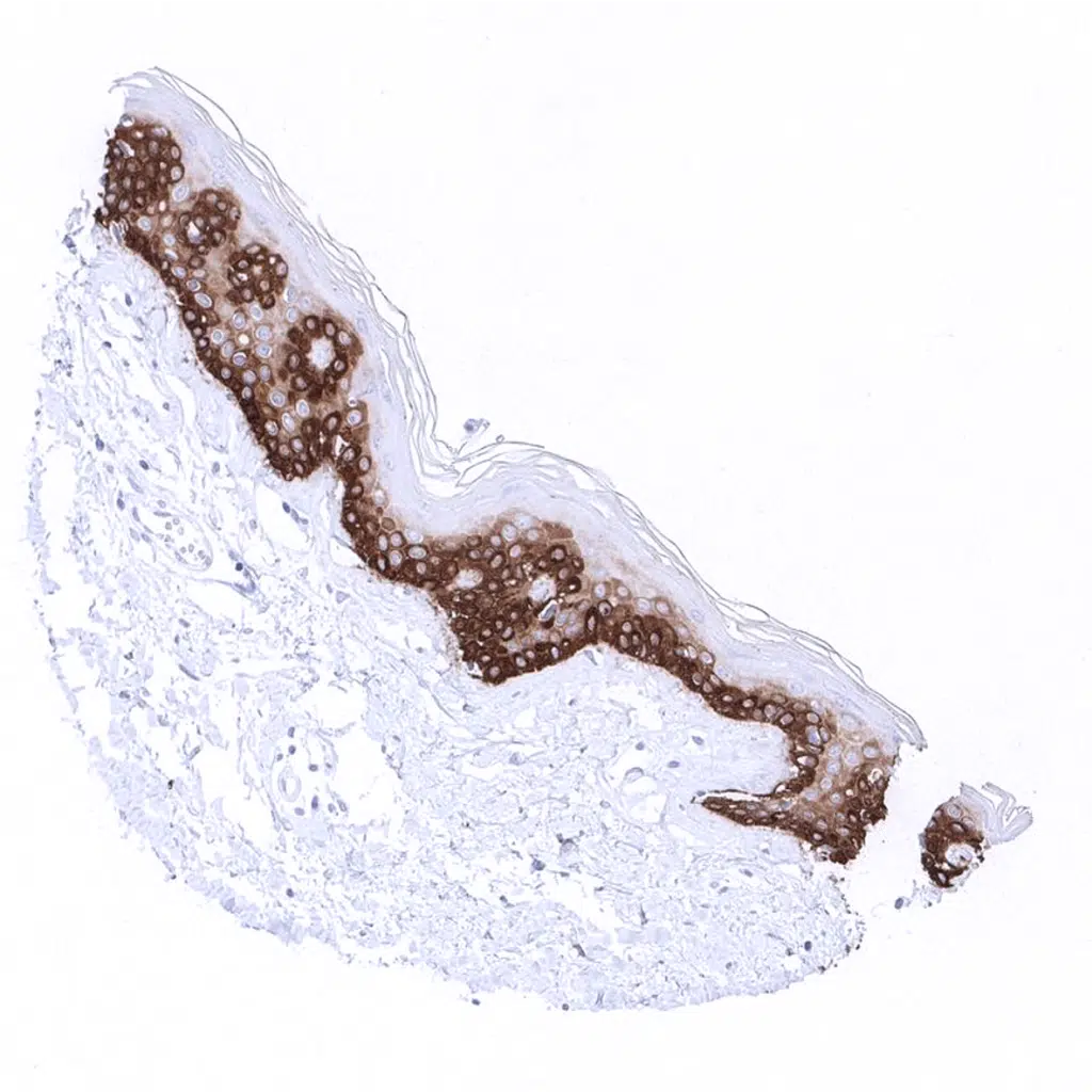
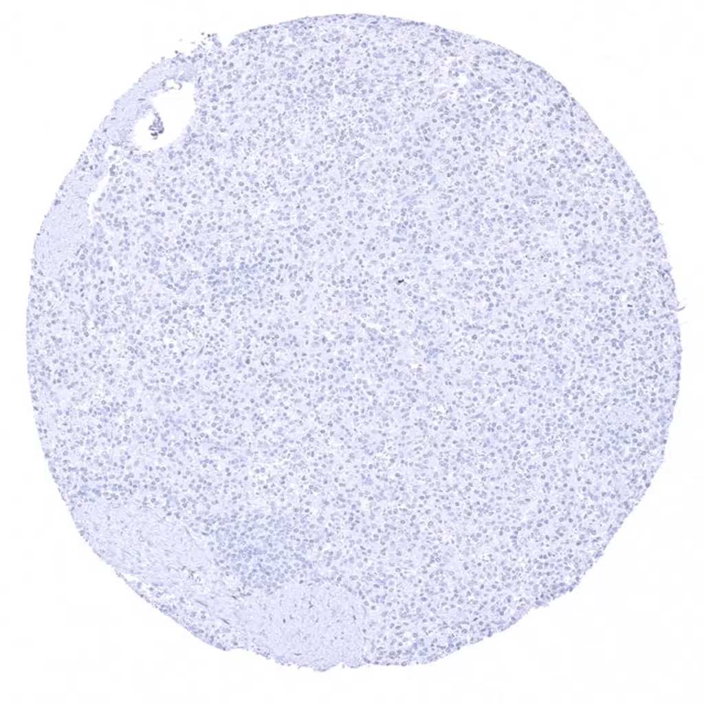
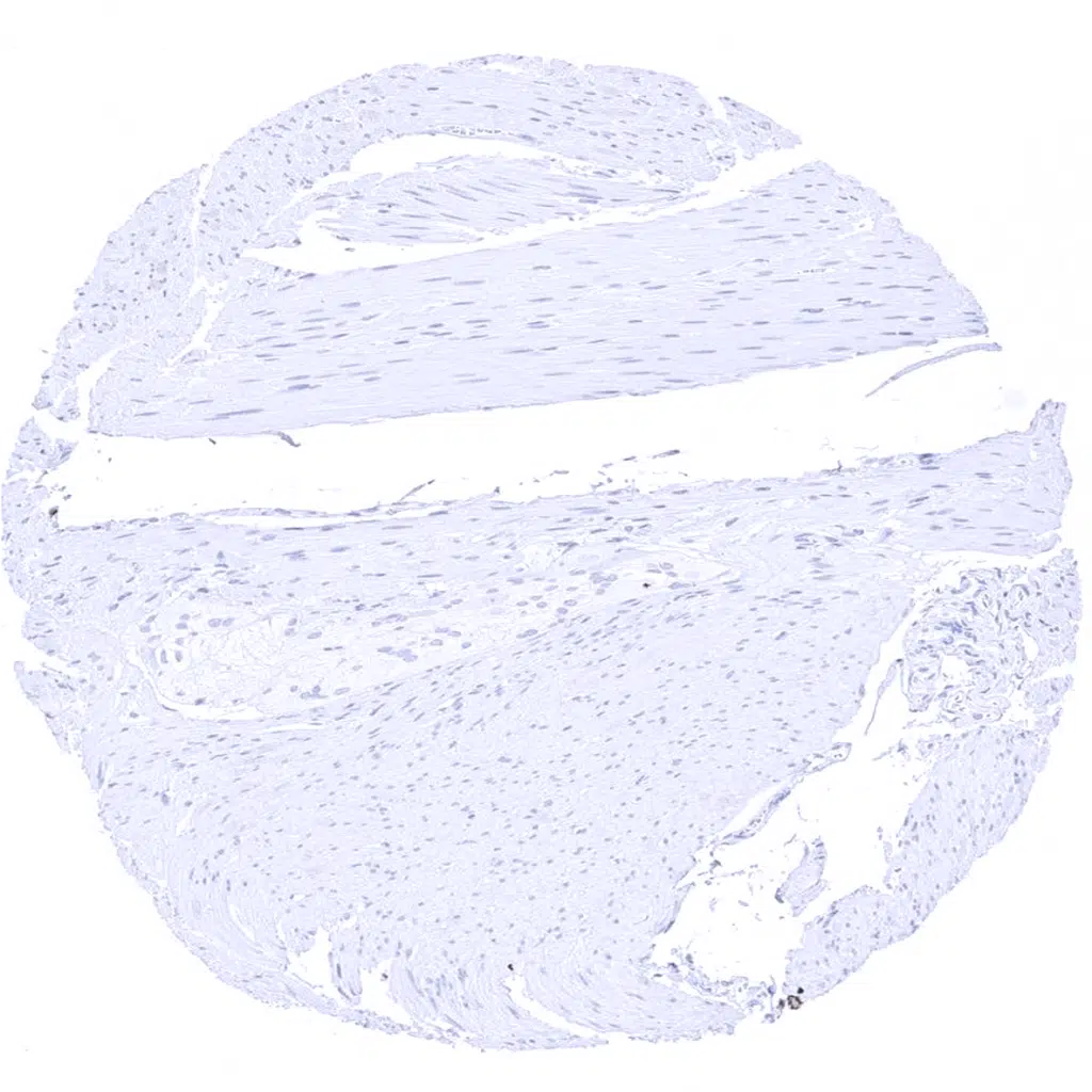
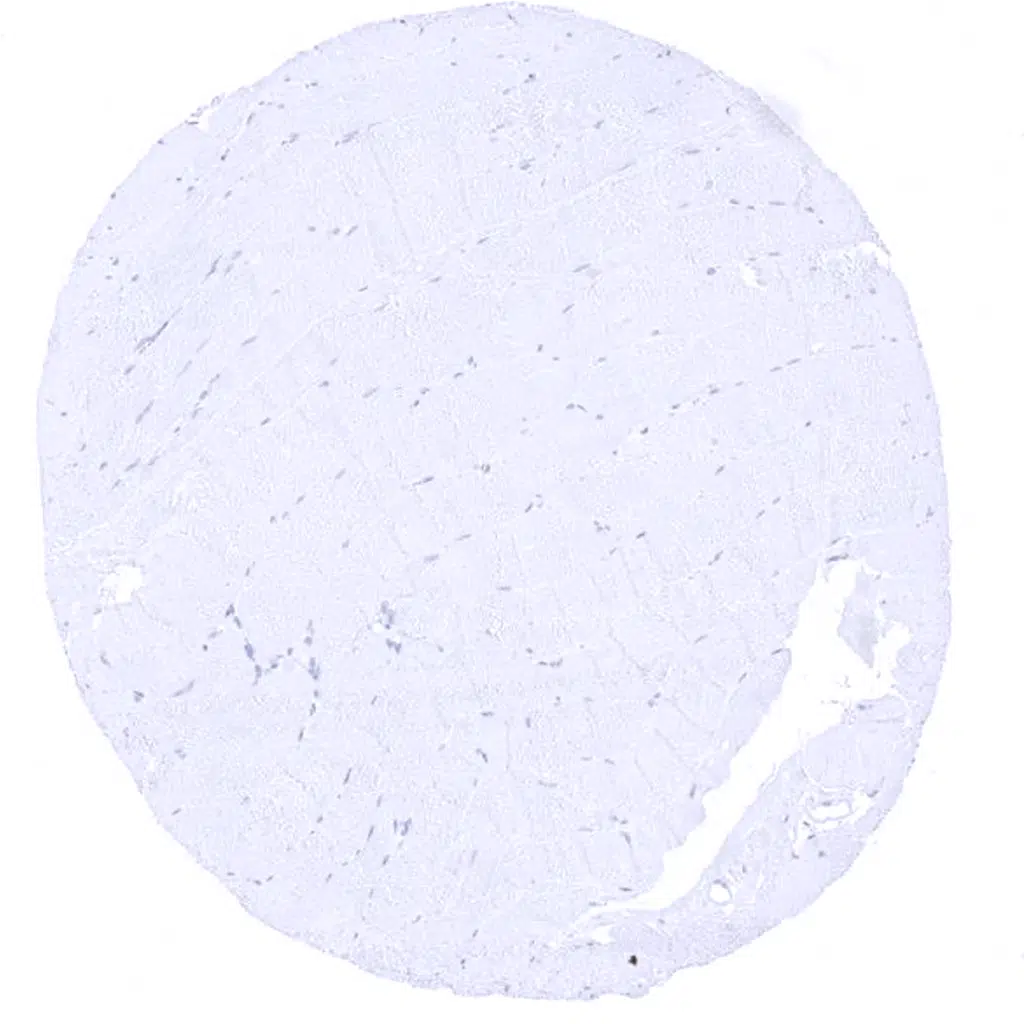
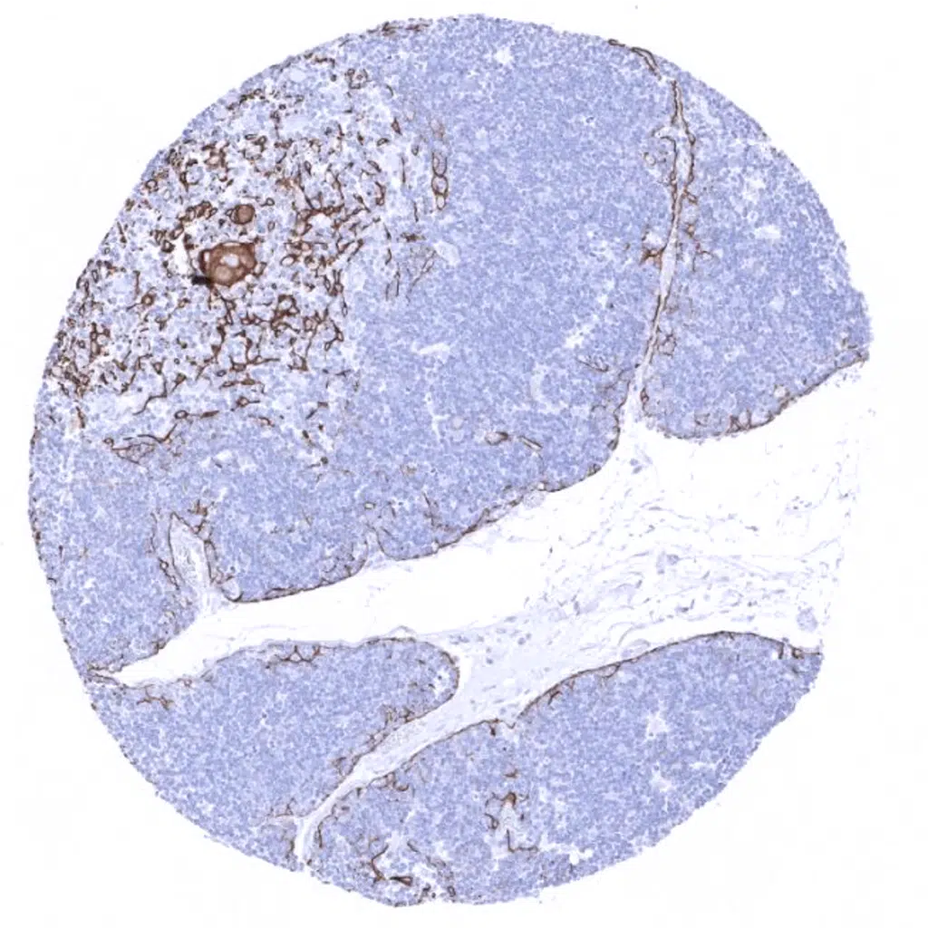
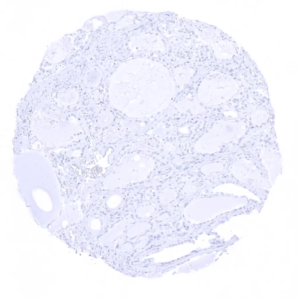
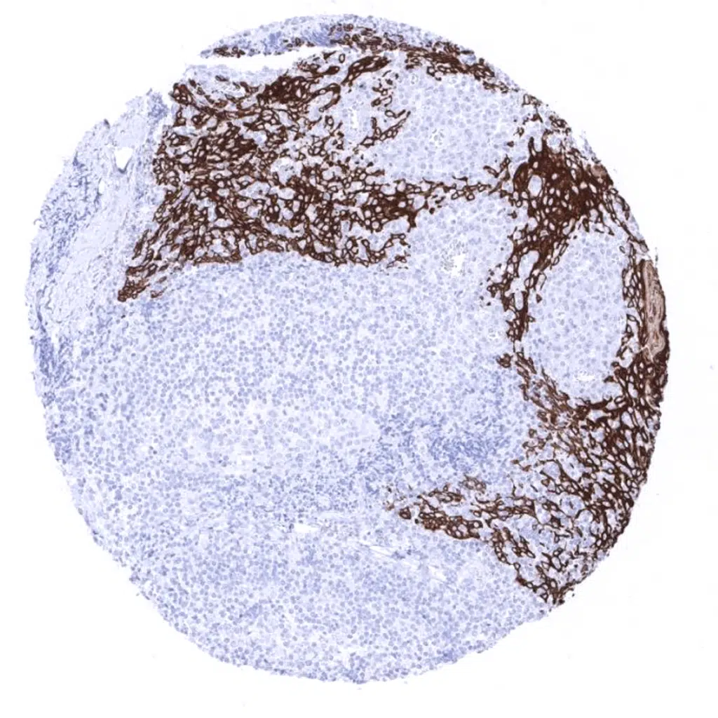
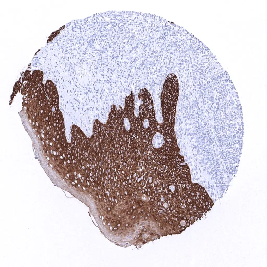
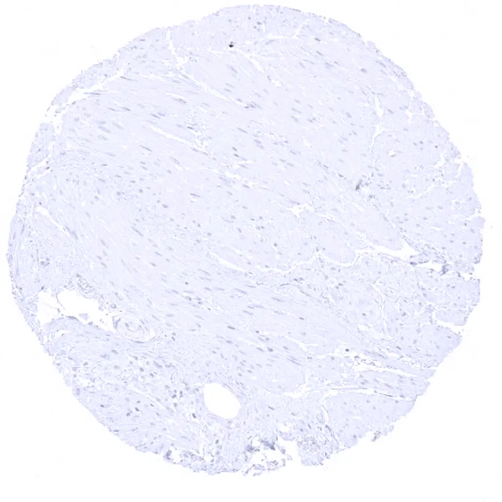
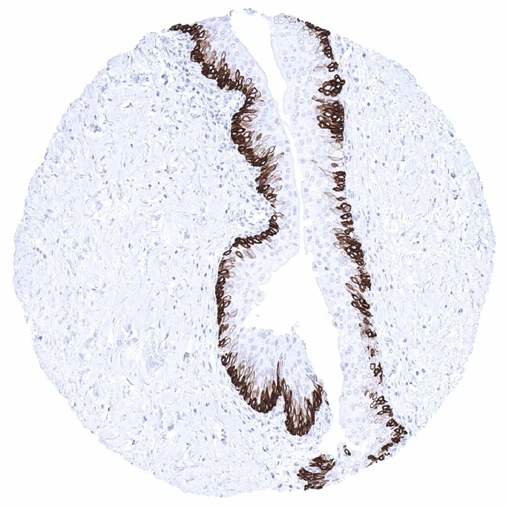
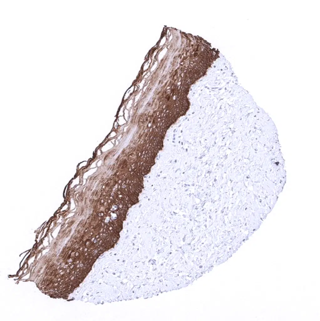
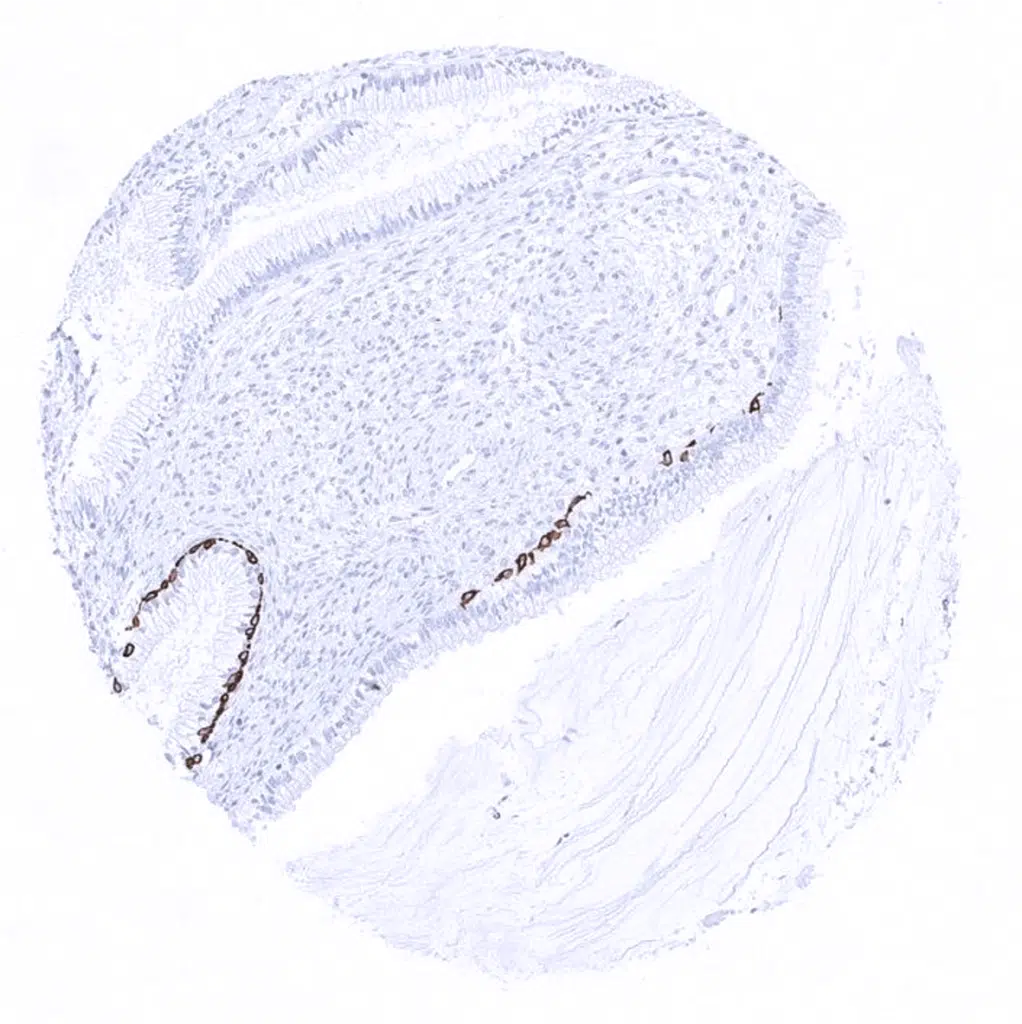
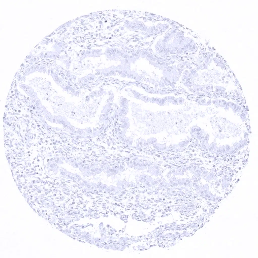
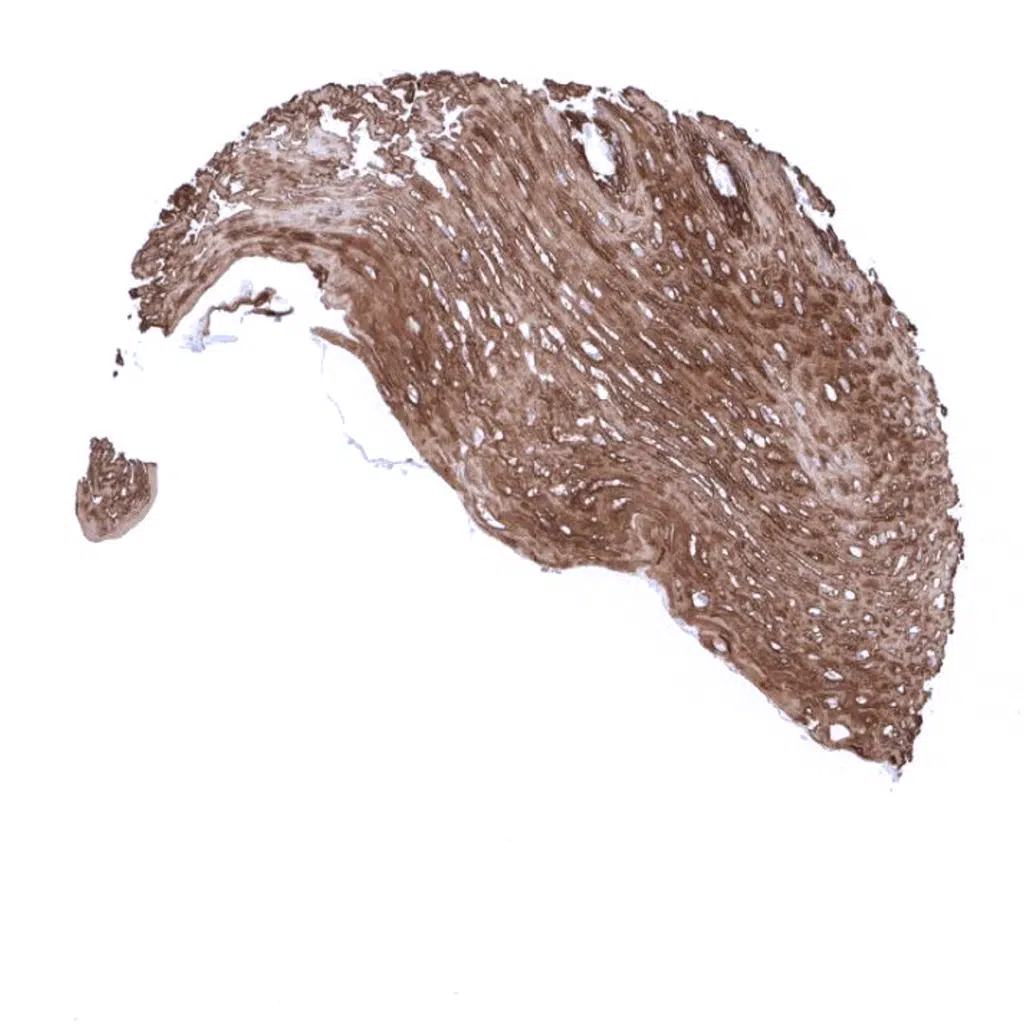
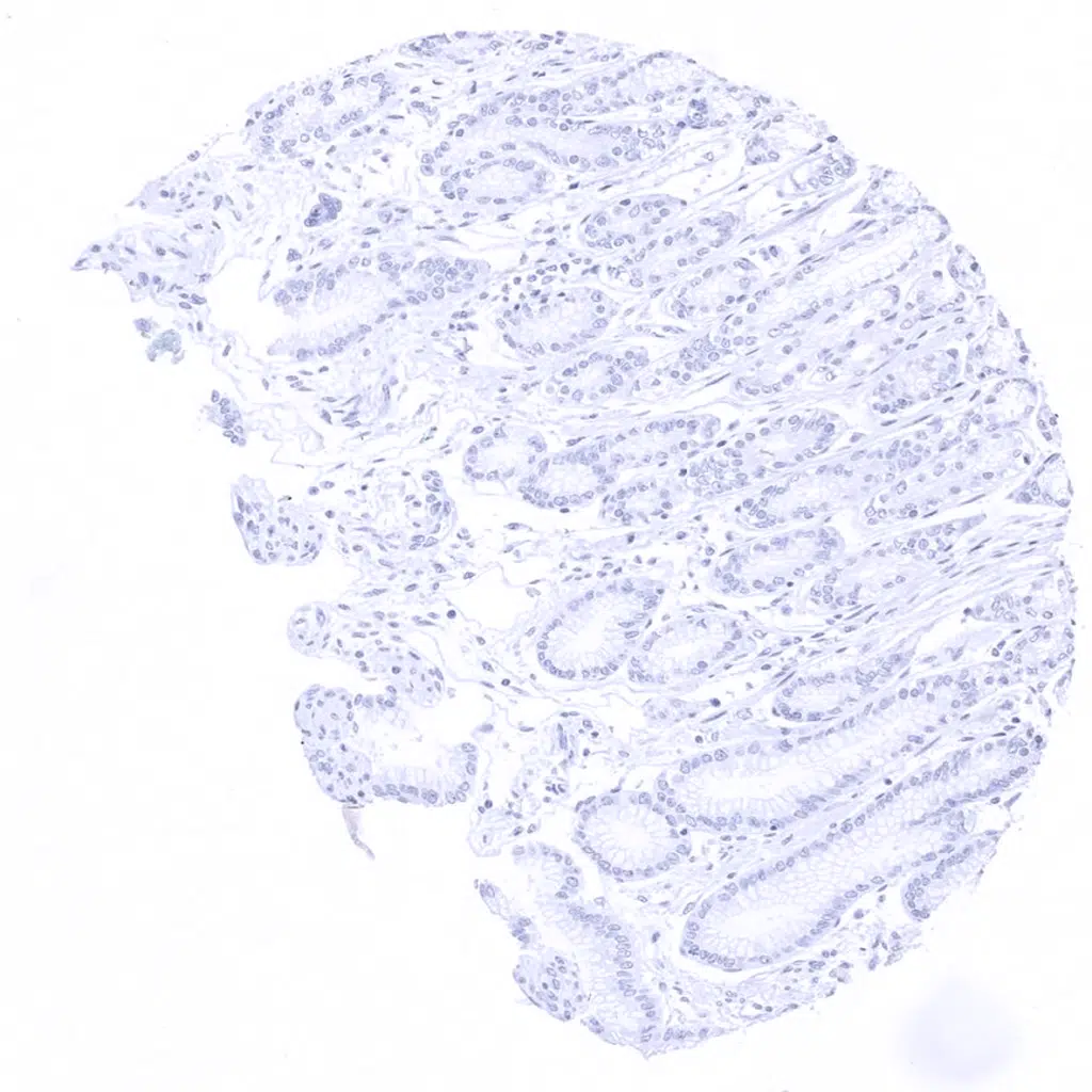
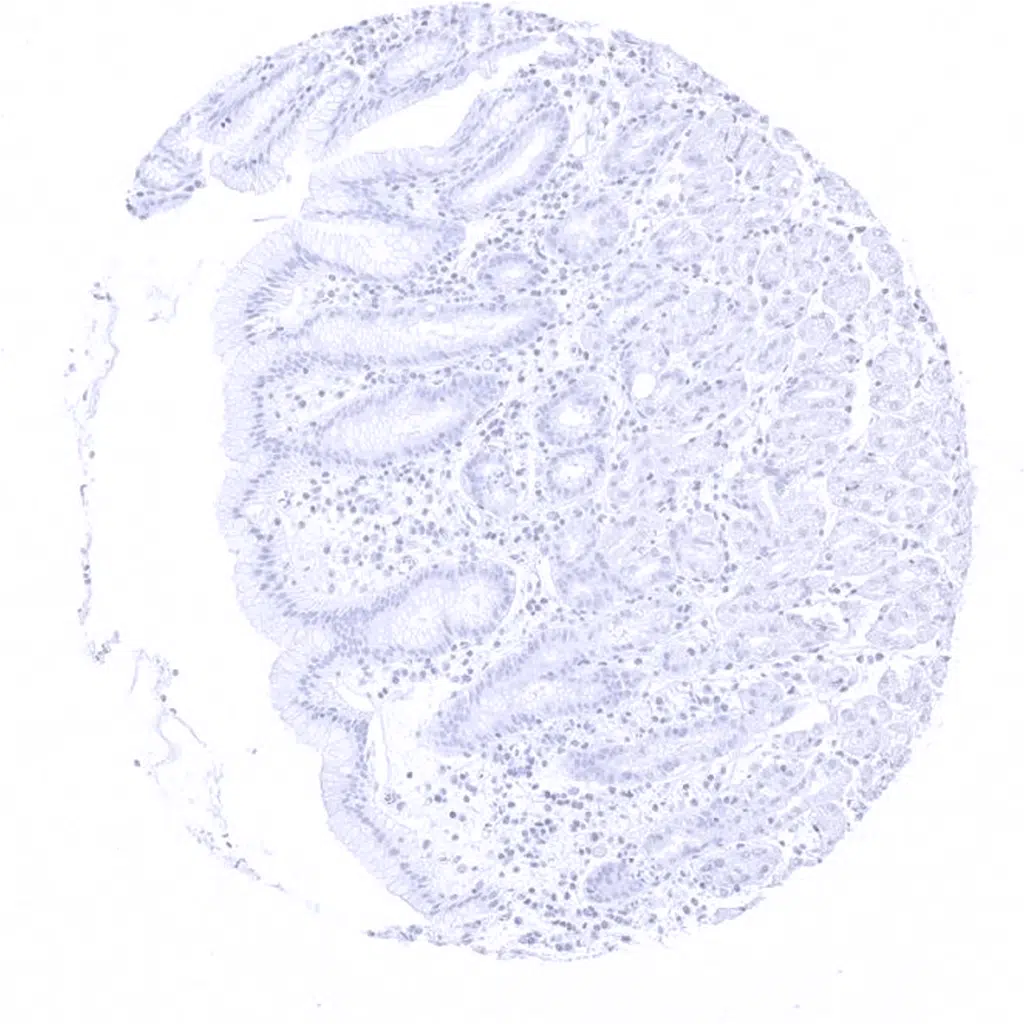
Cytokeratin 5 antibody [MSVA-605M] HistoMAX


Go to Cancer Tissue Gallery Go back to product page Go to HistoMAX page


Adrenal gland
Aorta, media
Appendix, mucosa
Appendix, muscular wall
Bone marrow
Breast - Cytokeratin 5 staining can be very weak in myoepithelial cells of the breast.
Bronchus, mucosa - Cytokeratin 5 is expressed in basal cells of respiratory epithelium as well as in myoepithelial cells of bronchial glands.
Cerebellum (molecular layer, Purkinje cell layer, granule cell layer)
Cerebellum (granule cell layer, white matter)
Cerebrum, grey matter
Cerebrum, white matter
Colon descendens, mucosa
Duodenum, Brunner gland
Duodenum, mucosa
Ectocervix - Cytokeratin 5 staining is usually seen in all layers of non-keratinizing squamous epithelium, but superficial layers stain sometimes slightly weaker.
Endocervix - Some basal cells can show Cytokeratin 5 staining in endocervical glands.
Endocervix
Endometrium, proliferation
Endometrium, proliferation - Few endometrial epithelial cells (not all glands and only in a few samples) can show Cytokeratin 5 immunostaining.
Endometrium, secretion
Epididymis - Basal cells of the epididymis show strong and columnar cells weak Cytokeratin 5 immunostaining.
Esophagus, squamous epithelium
Fallopian tube, mucosa
Fat
Gallbladder, epithelium
Heart
Ileum, mucosa
Kidney, cortex
Kidney, medulla
Liver
Lung
Ovary, stroma
Pancreas
Parathyroid
Parotid gland - Cytokeratin 5 is expressed in myoepithelial cells and basal cells of excretion ducts of salivary glands.
Pituitary gland, anterior lobe
Pituitary gland, posterior lobe
Pregnant uterus (decidua)
Placenta, early
Placenta, mature
Prostate - Basal cells of the prostatic glands show strong Cytokeratin 5 immunostaining.
Rectum, mucosa
Seminal vesicle
Sinus paranasales - Cytokeratin 5 is expressed in basal cells of respiratory epithelium as well as in myoepithelial cells of associated glands.
Skin - In the skin, CK5 staining predominantly occurs in the basal 50% of the epidermis, while the upper half stains weaker or can be completely negative.
Spleen
Stomach, antrum
Stomach, corpus
Stomach, muscular wall
Striated muscle
Testis
Thymus - All epithelial cells and corpuscles of Hassall's of the thymus stain Cytokeratin 5 positive.
Thyroid gland
Tonsil - All epithelial cells of tonsil crypts stain Cytokeratin 5 positive.
Tonsil, surface epithelium - Squamous epithelium of the tonsil surface shows strong cytokeratin 5 immunostaining of all cell layers.
Urinary bladder, muscular wall
Urinary bladder, urothelium - Strong Cytokeratin 5 staining of basal cells.
Uterus, myometrium
   

| TOP |