GeneTex
  • Country / Location Selection

United States (US)

Normal Tissue Gallery- GTX04397

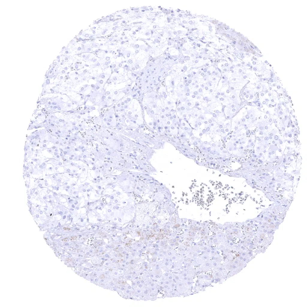
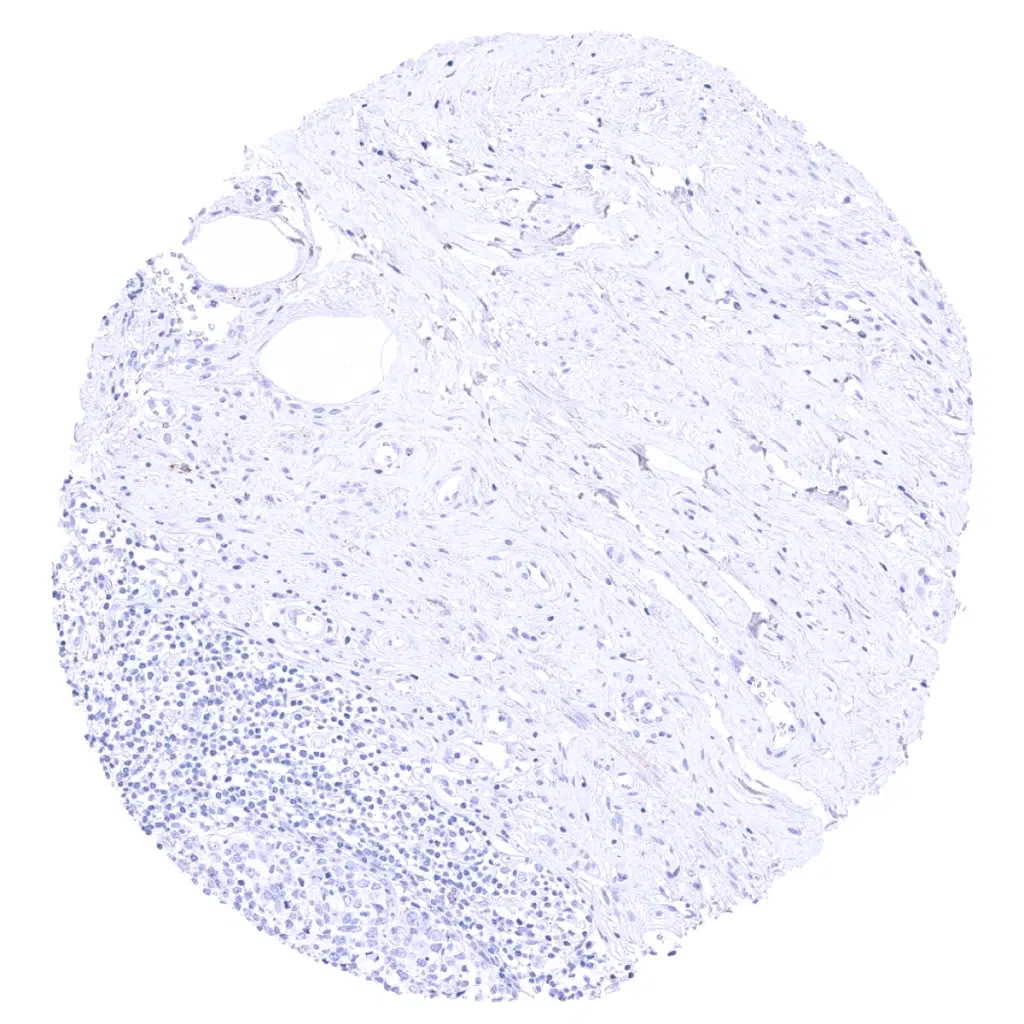
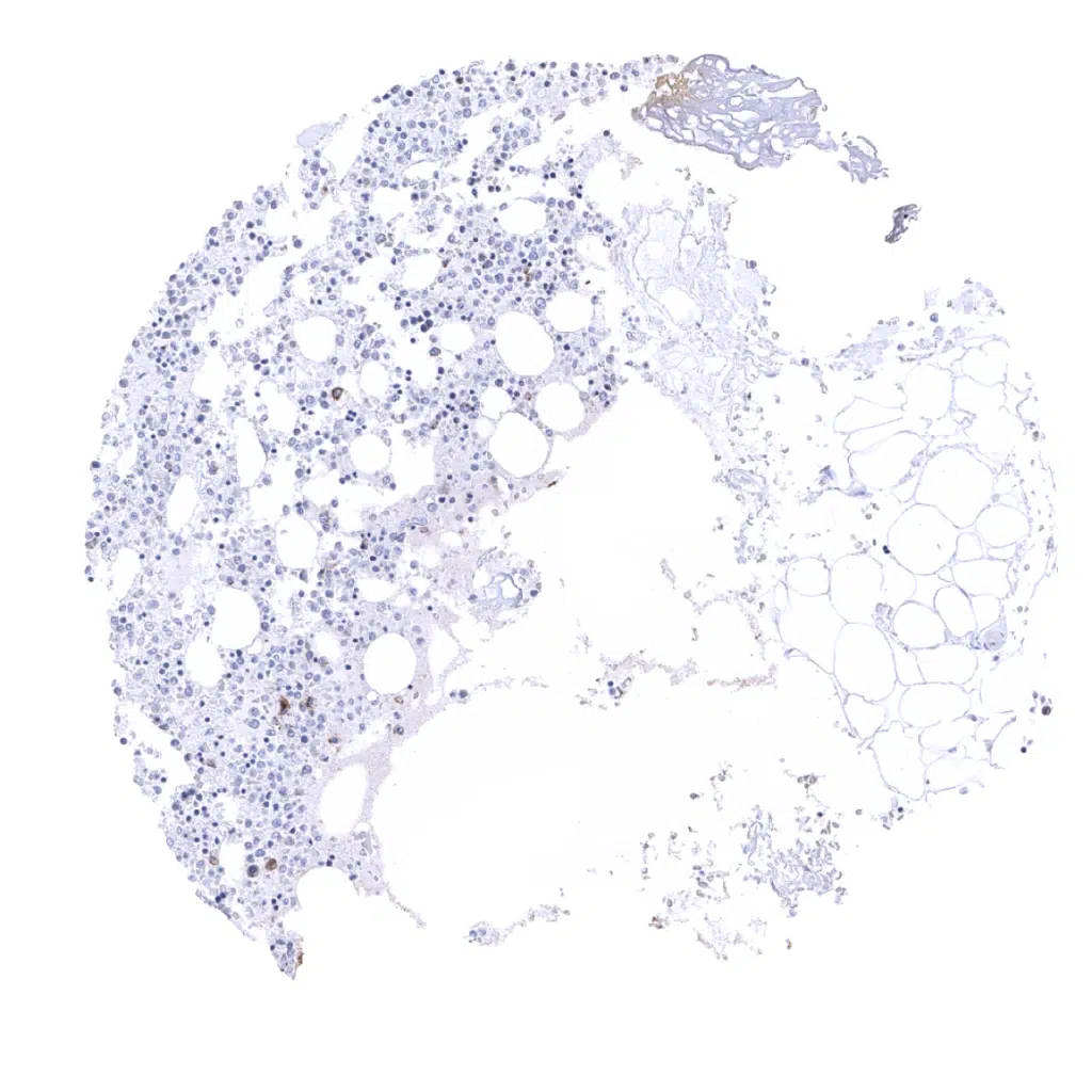
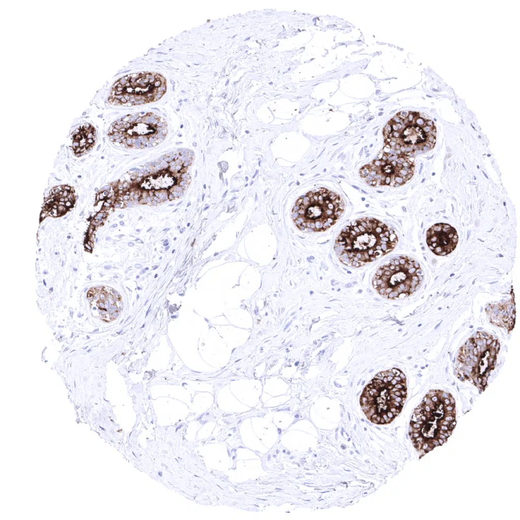
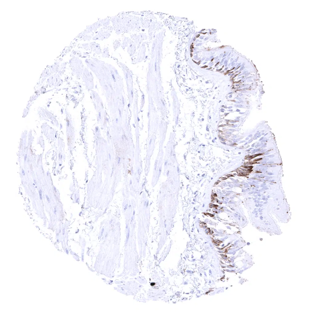
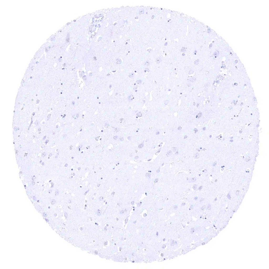
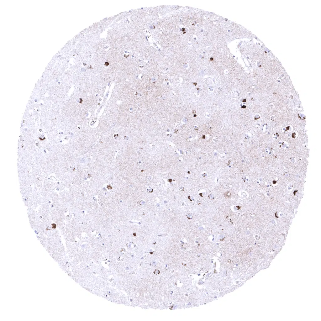
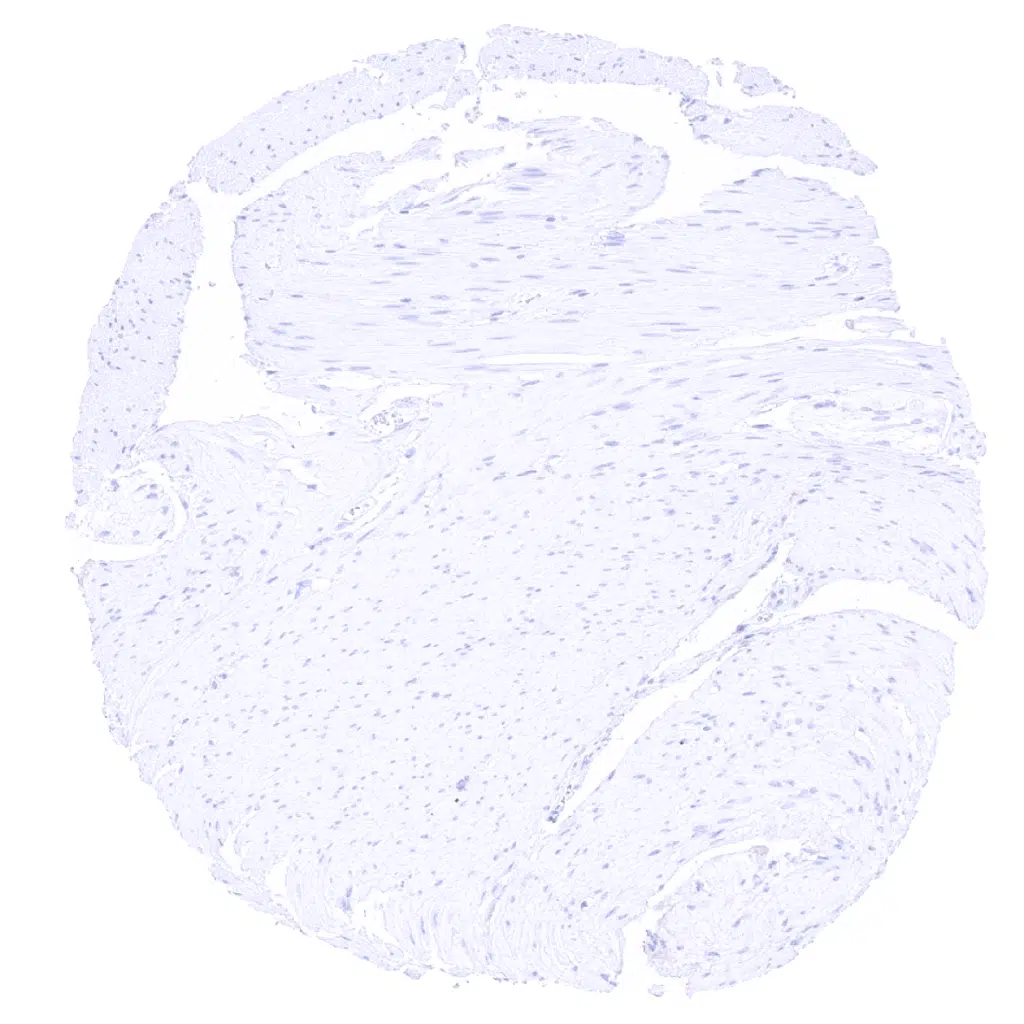
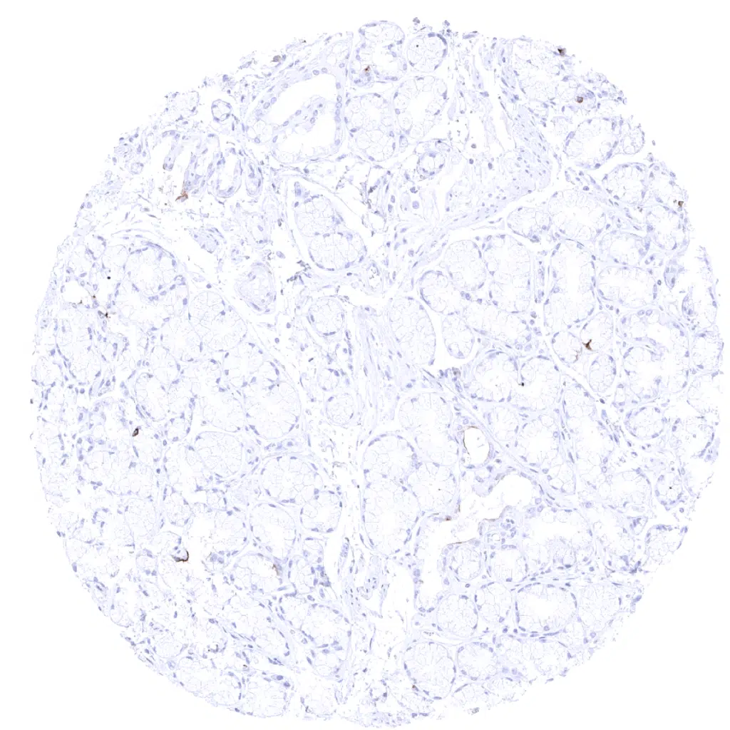
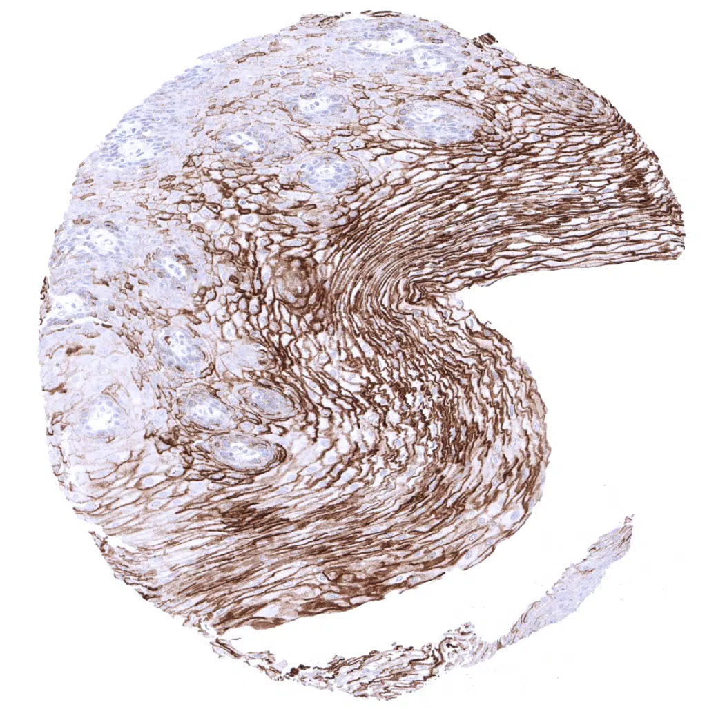
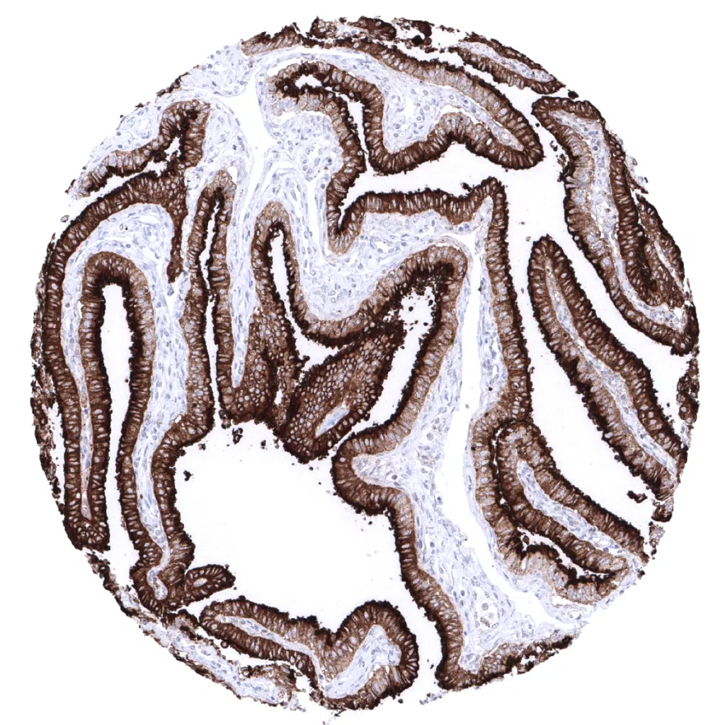
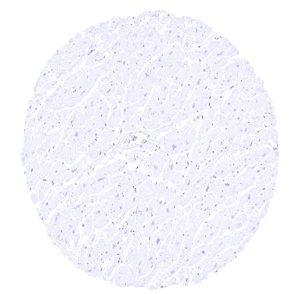
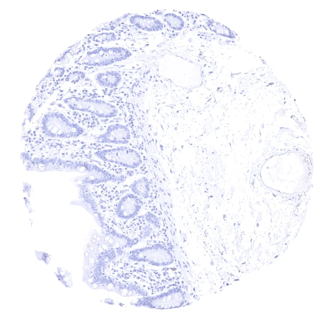
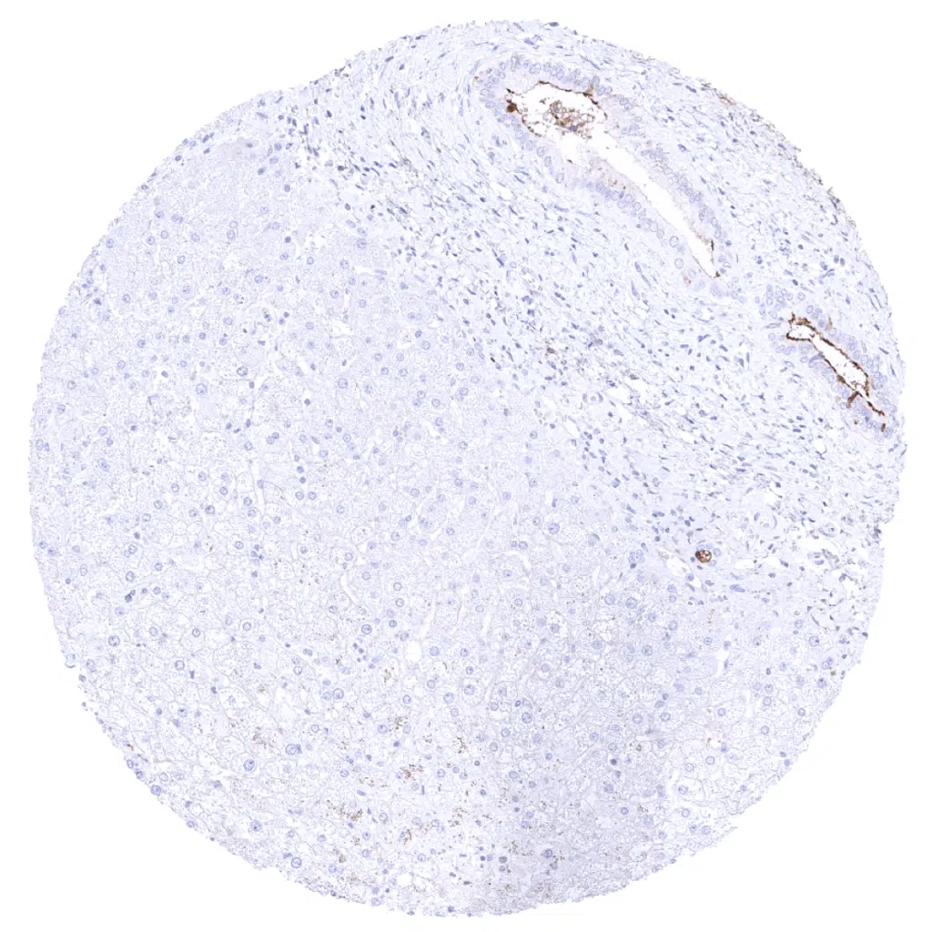
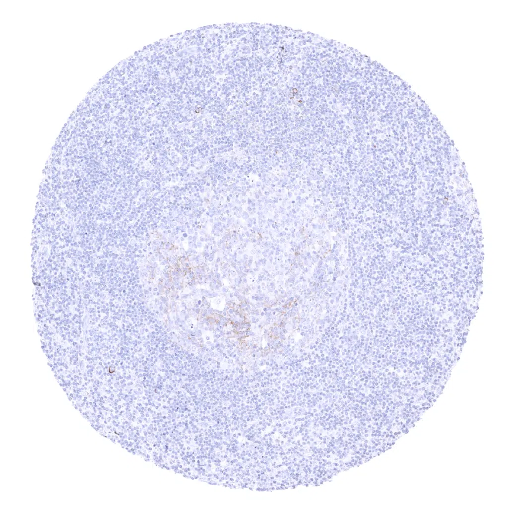
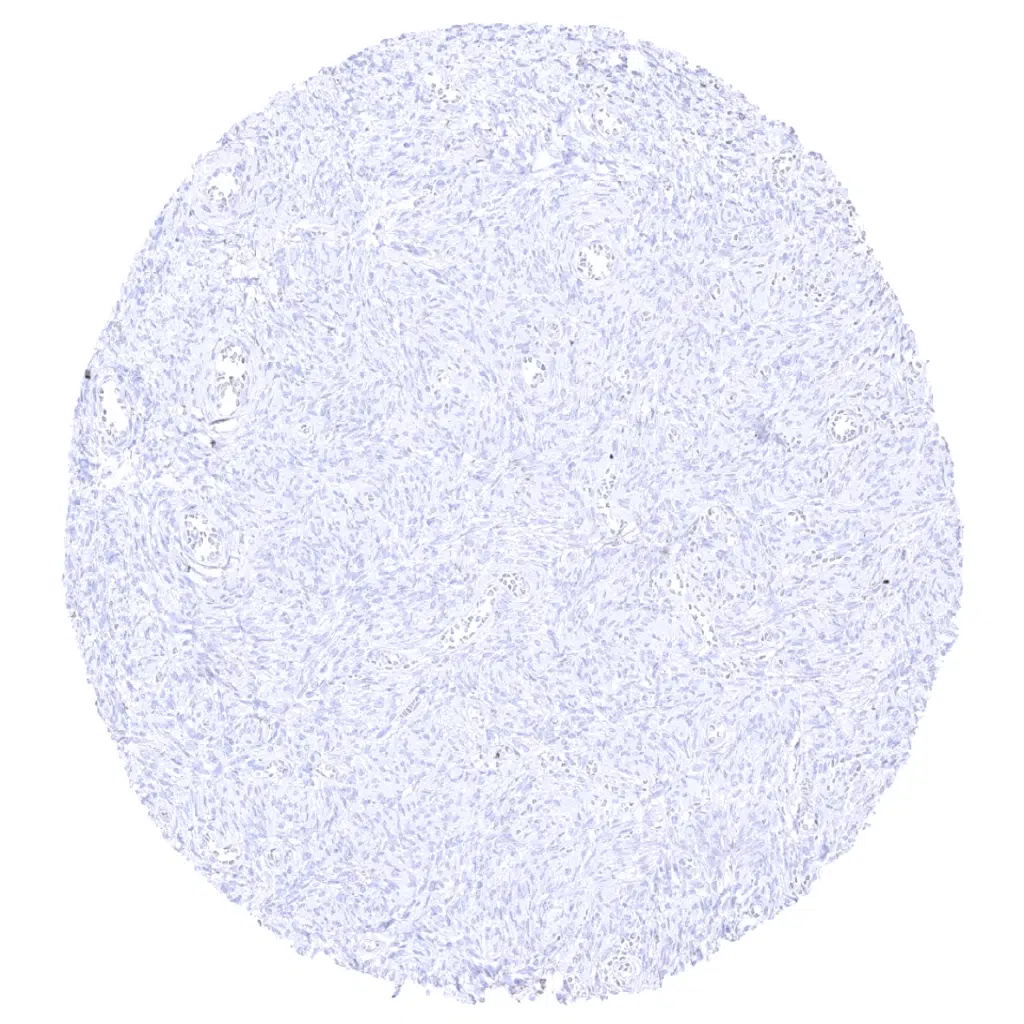
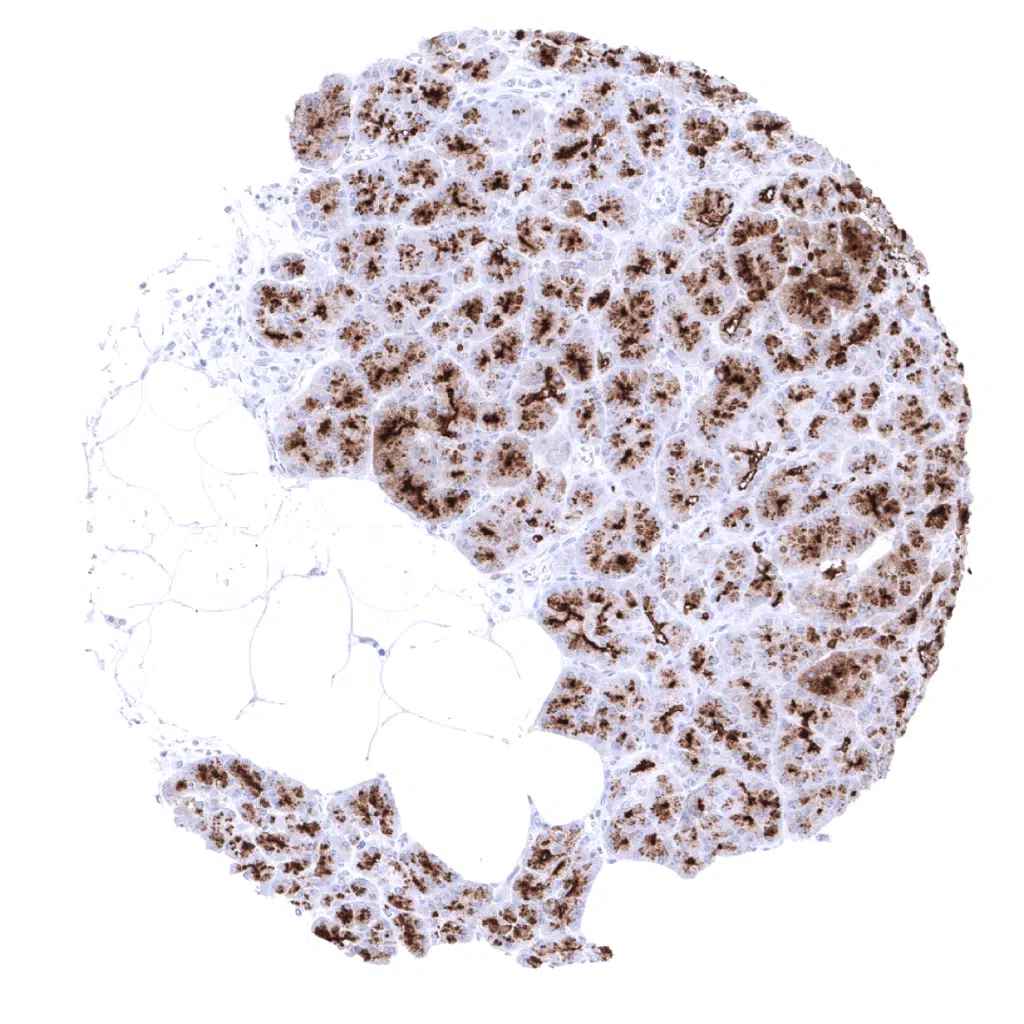
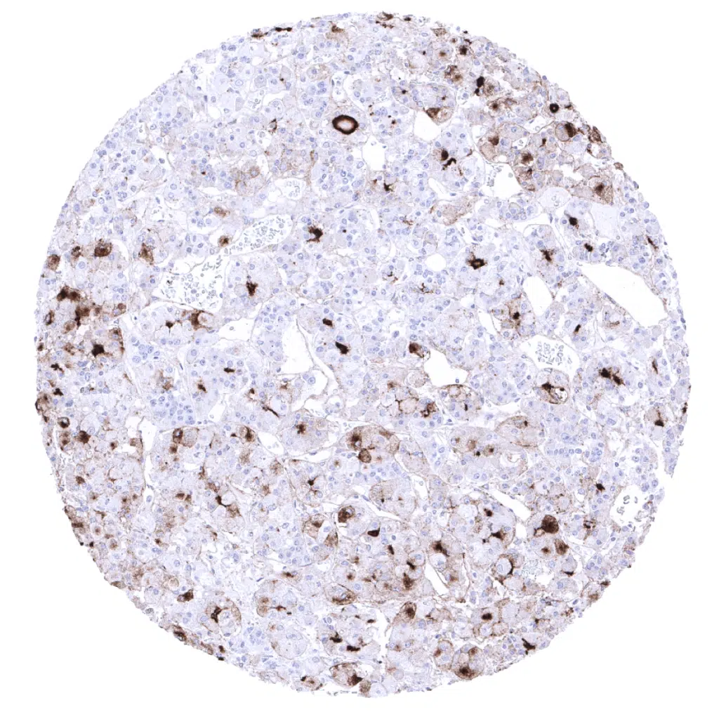
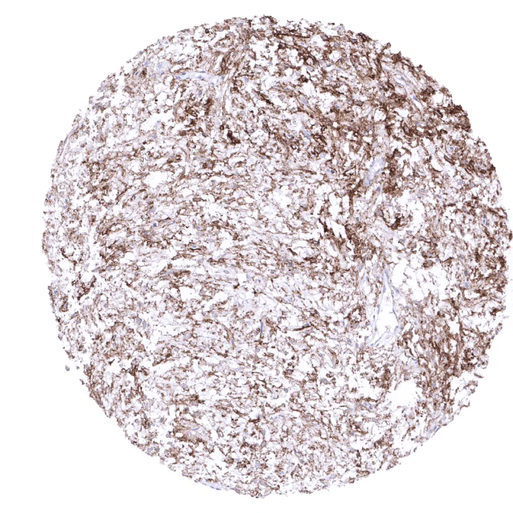
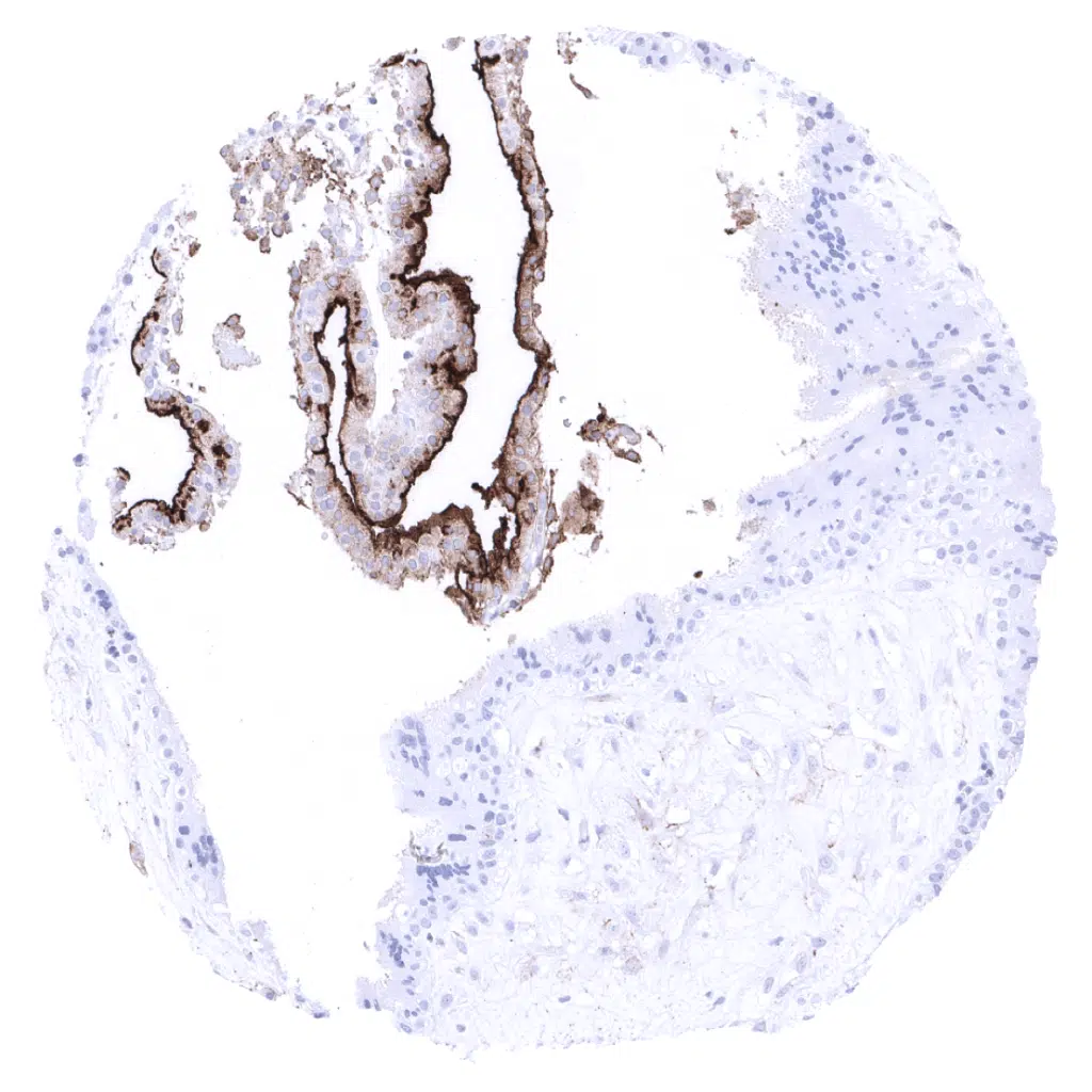
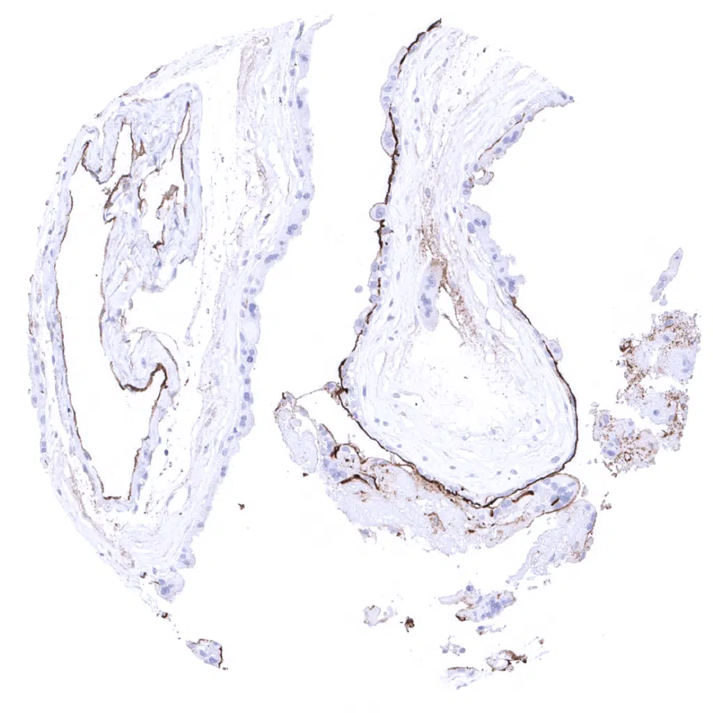
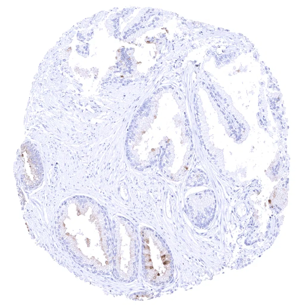
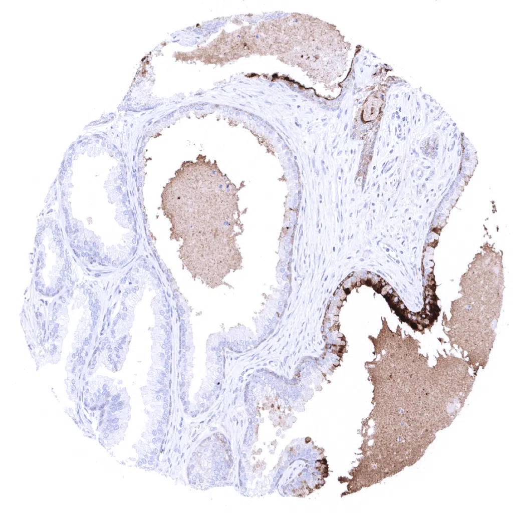
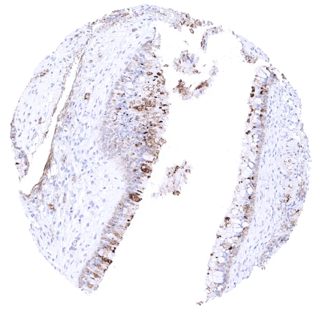
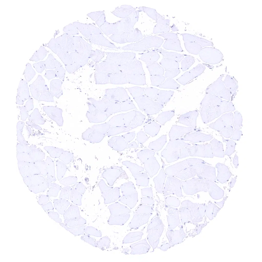
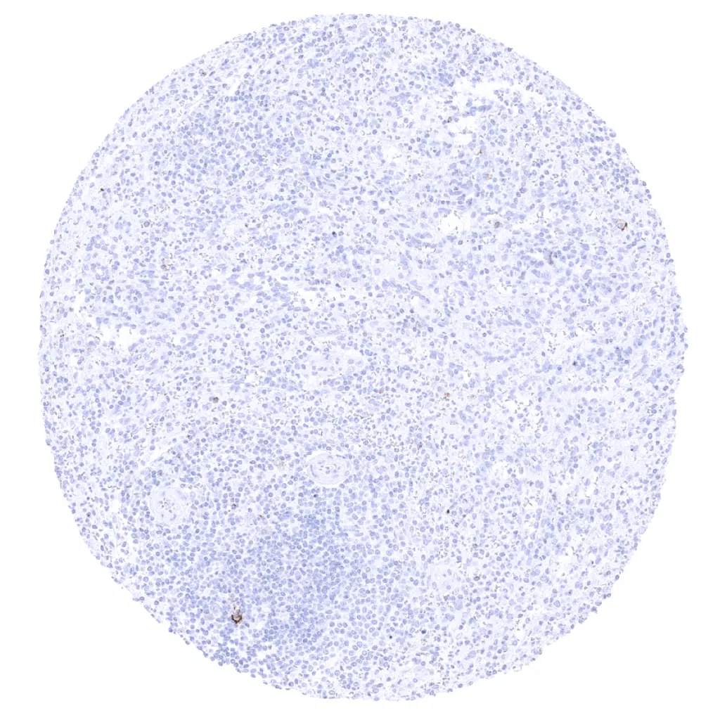
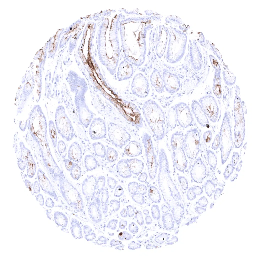
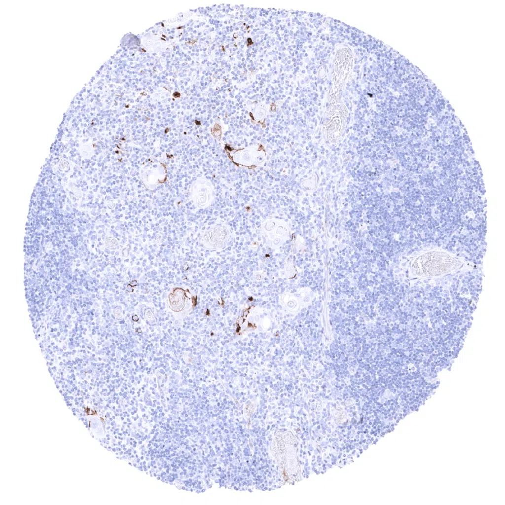
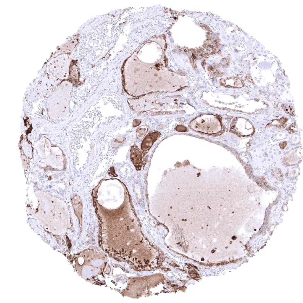
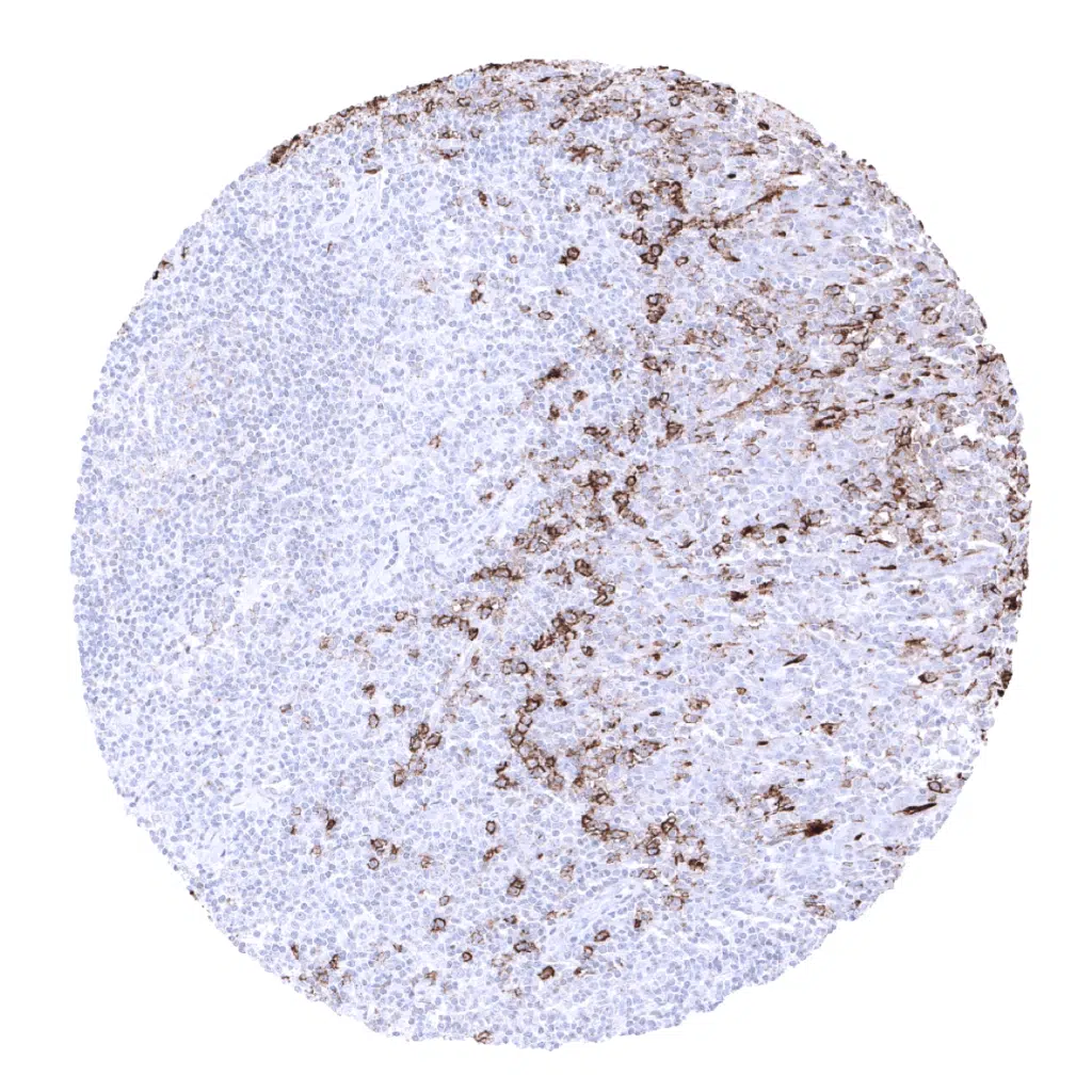
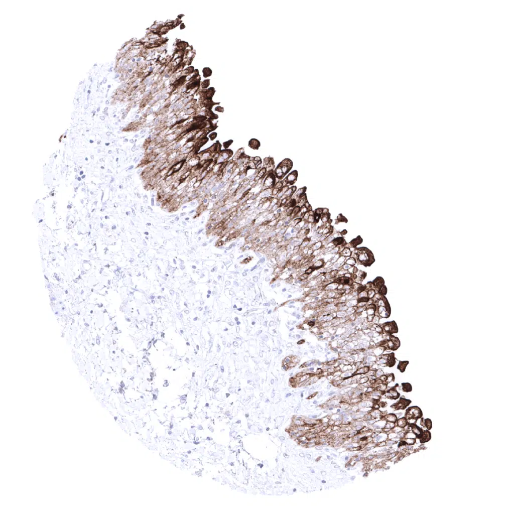
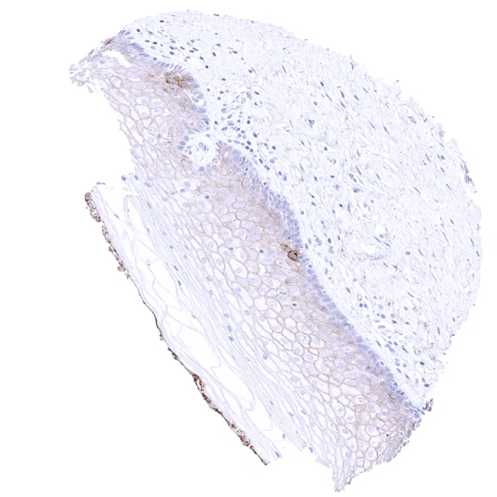
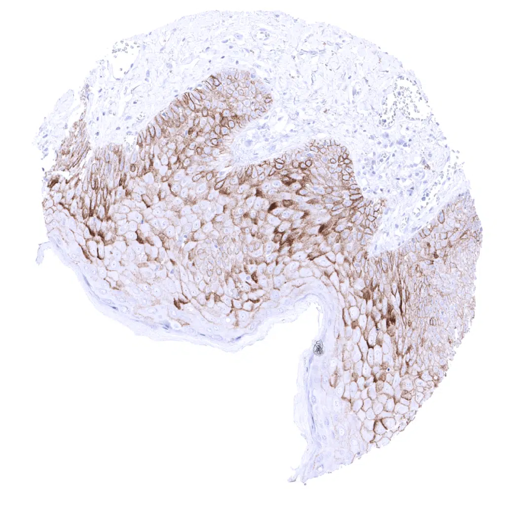
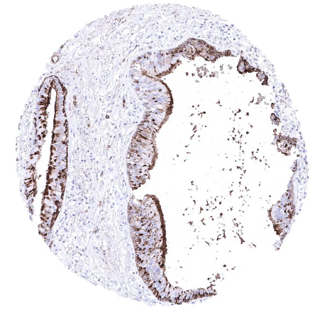
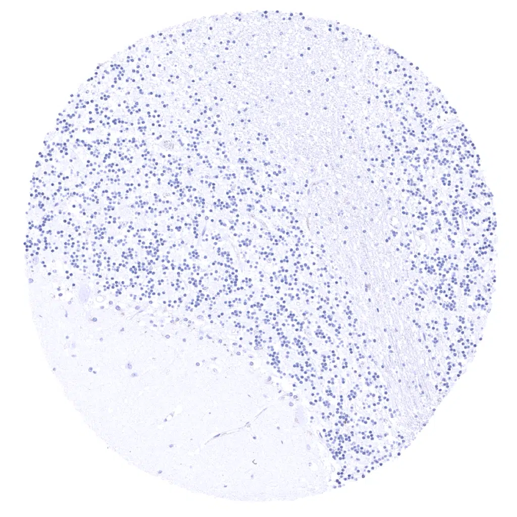
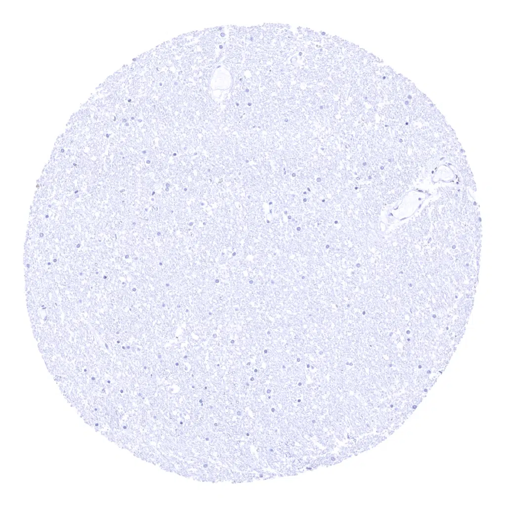
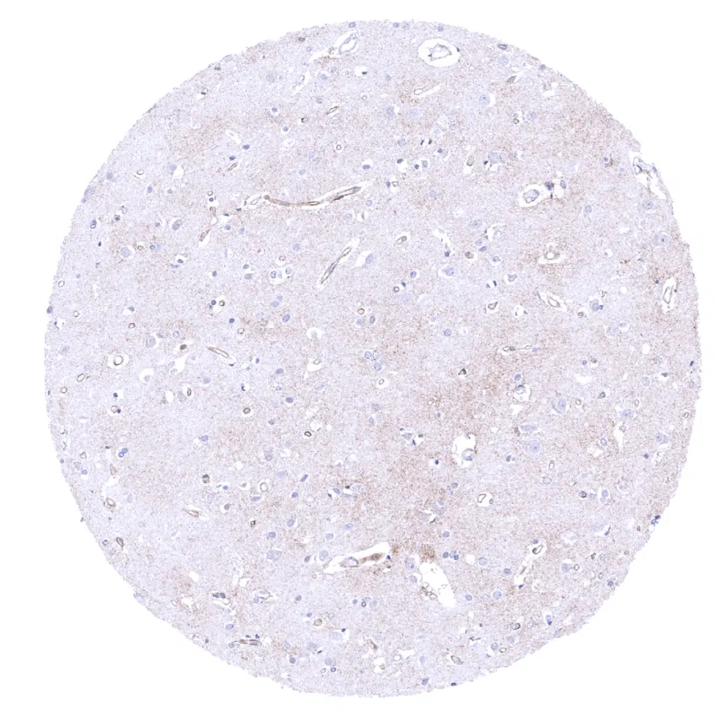
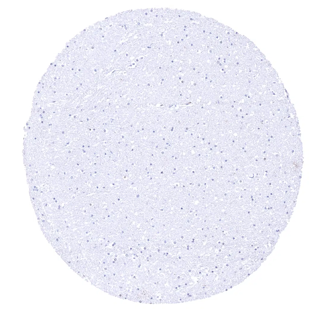
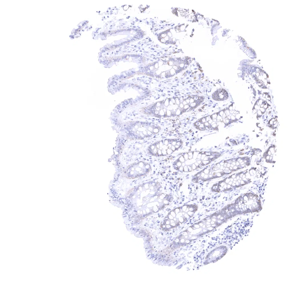
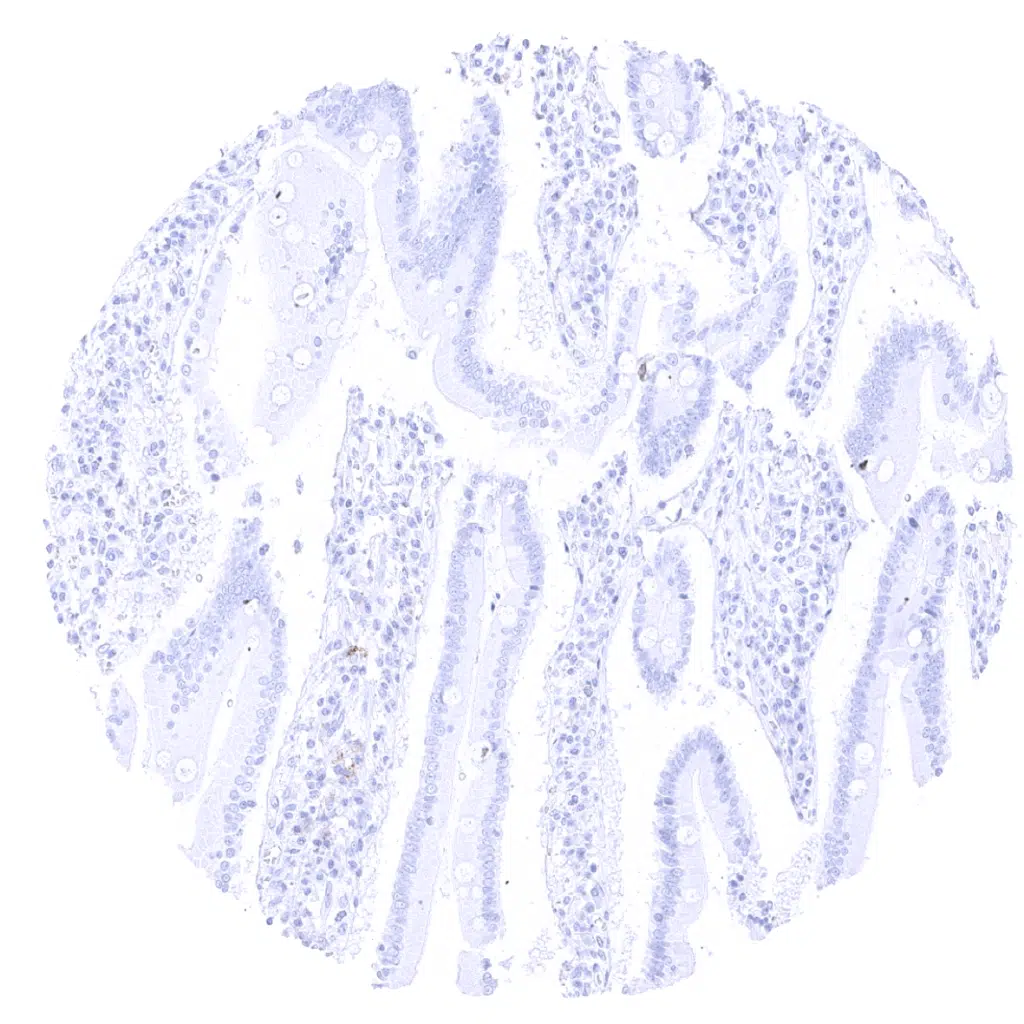
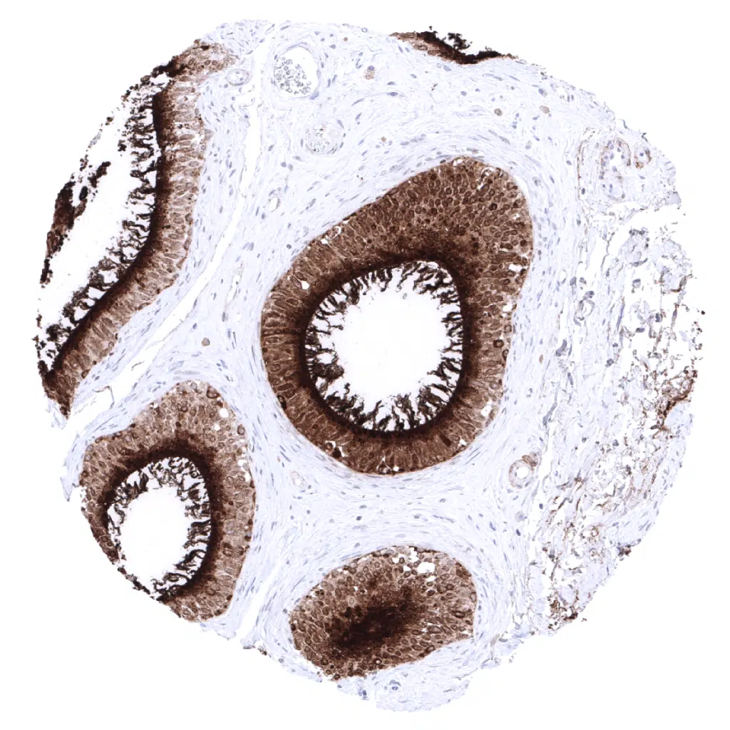
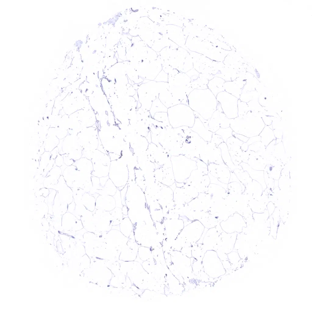
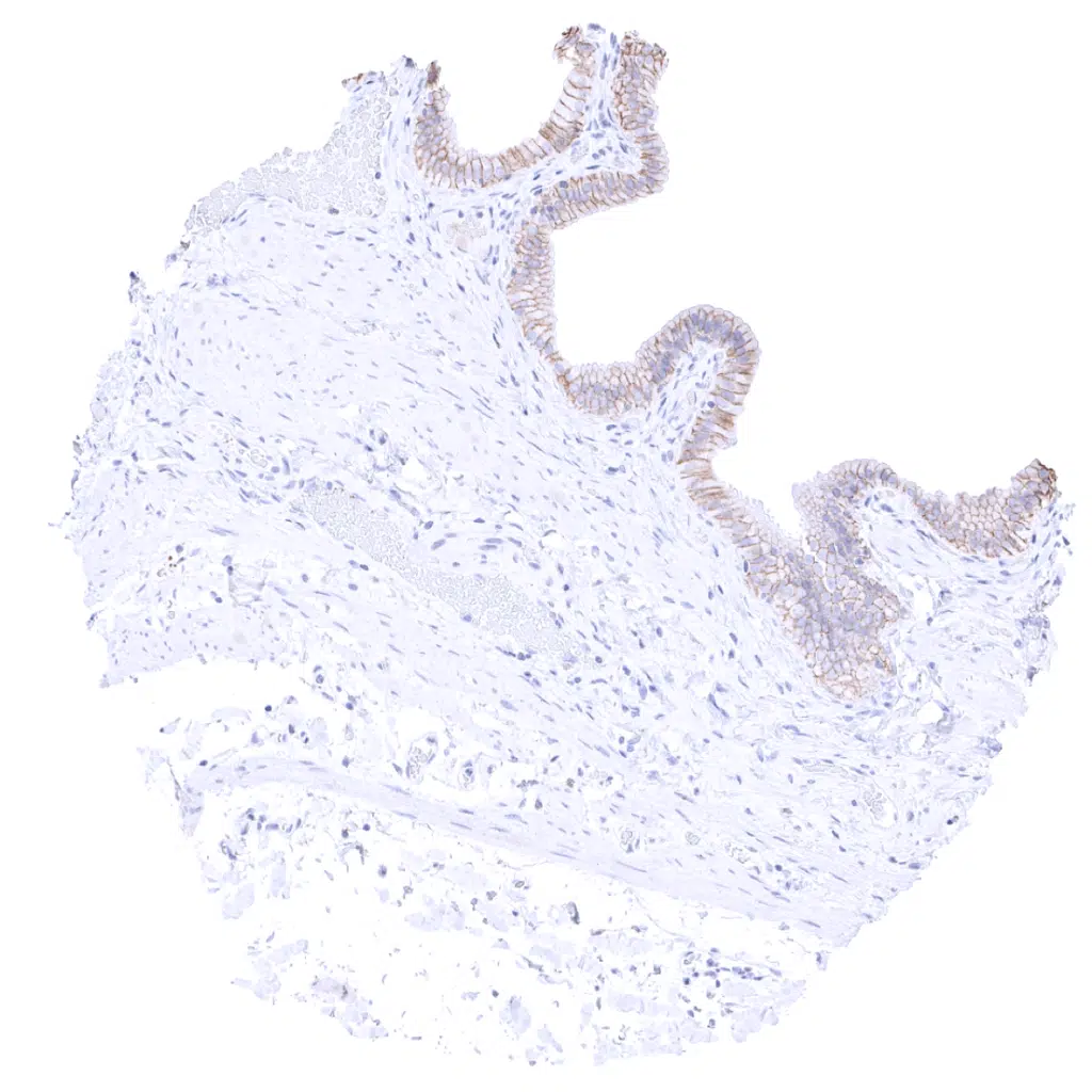
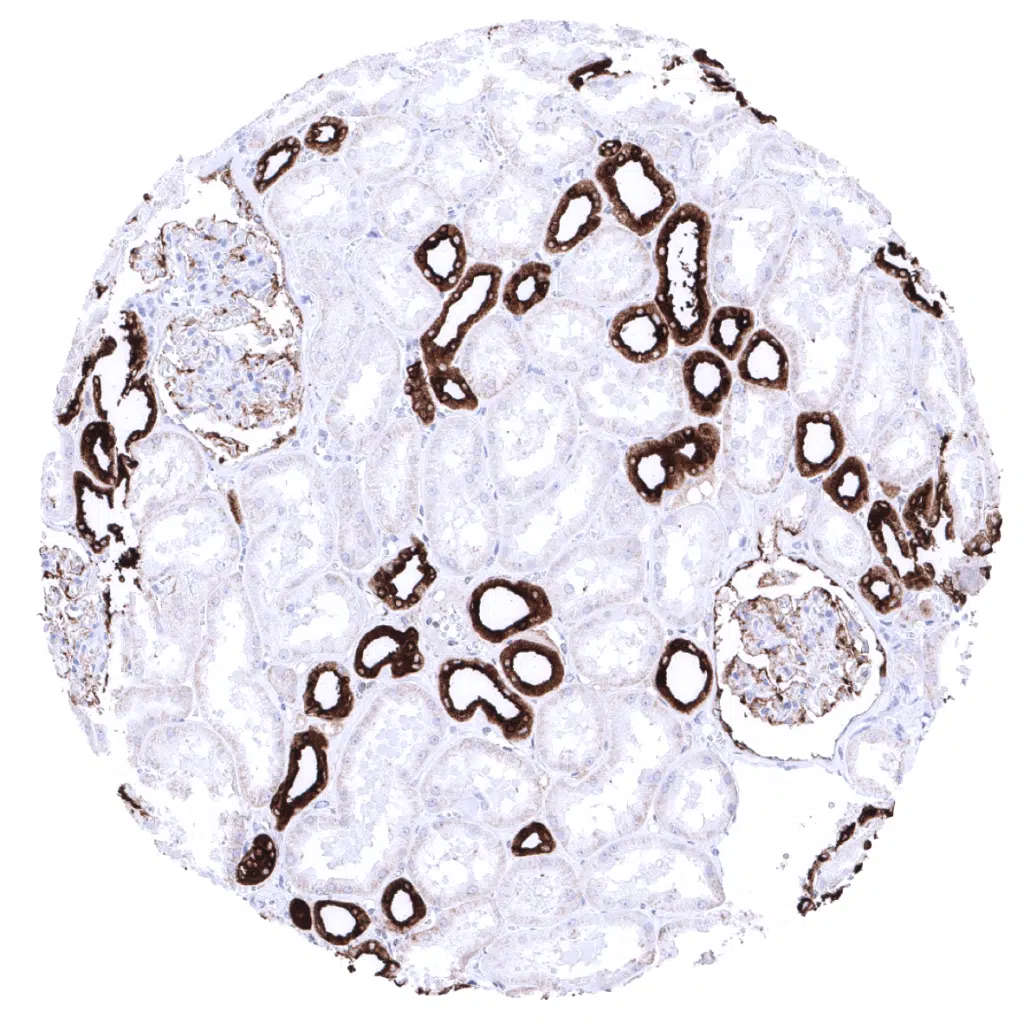
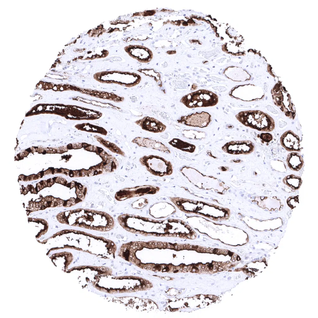
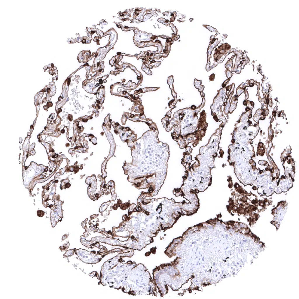
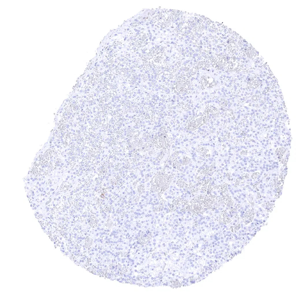
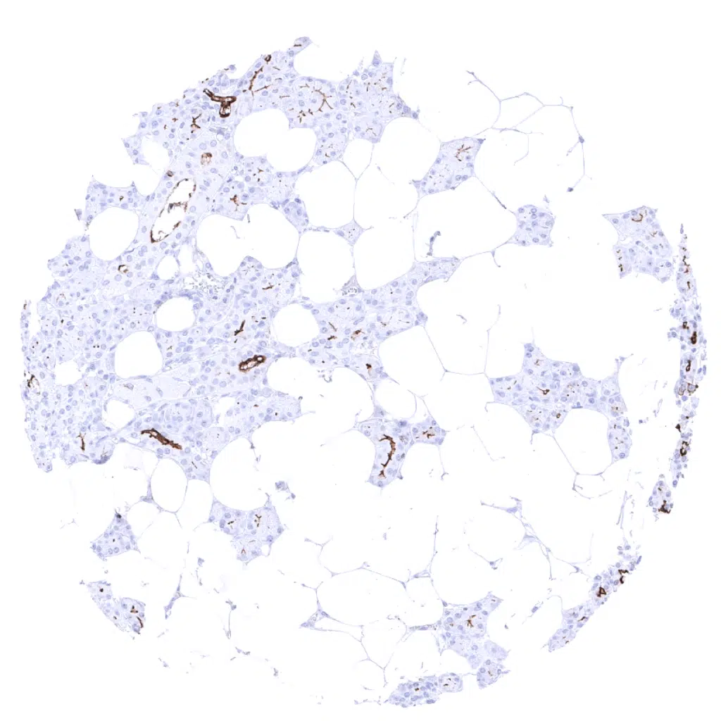
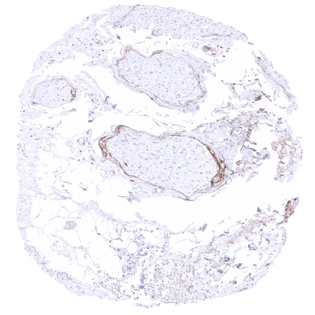
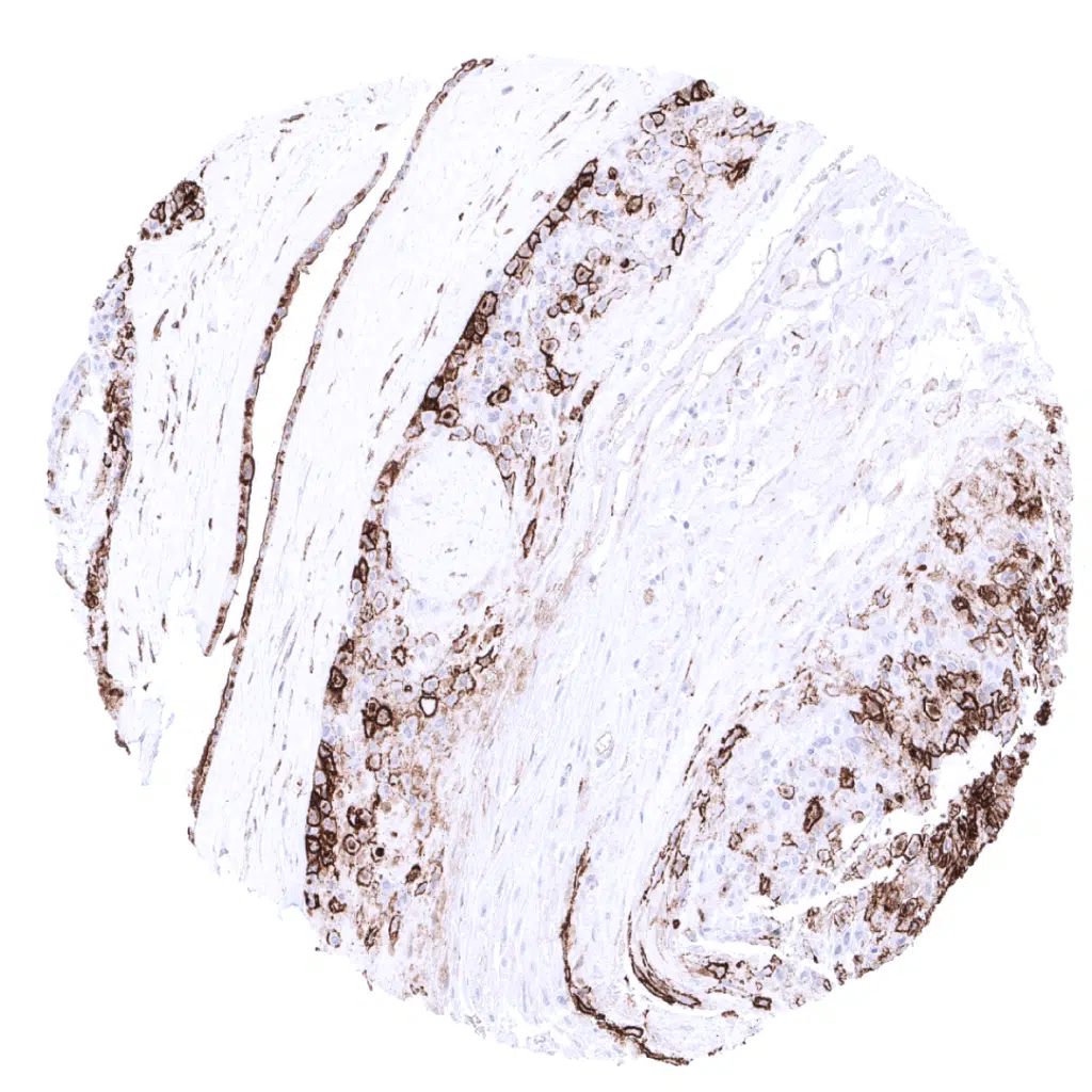
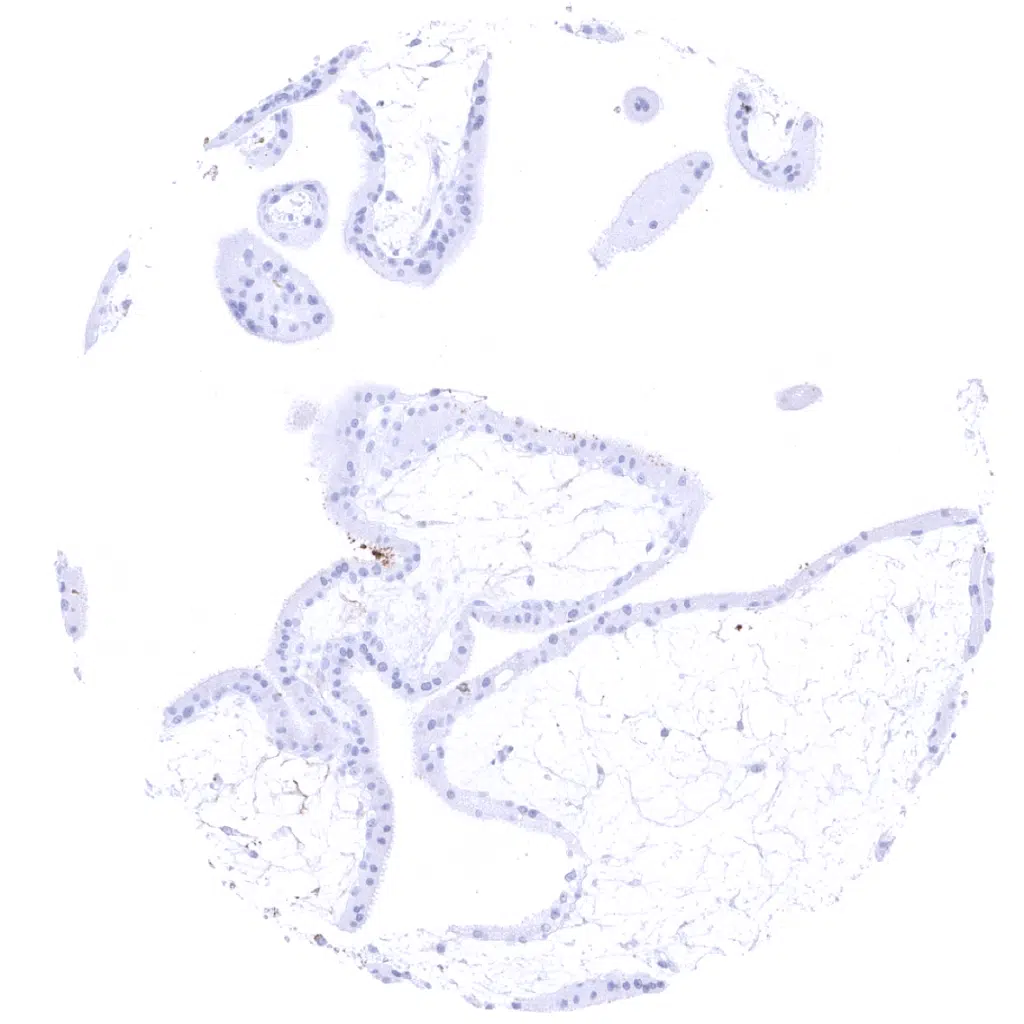
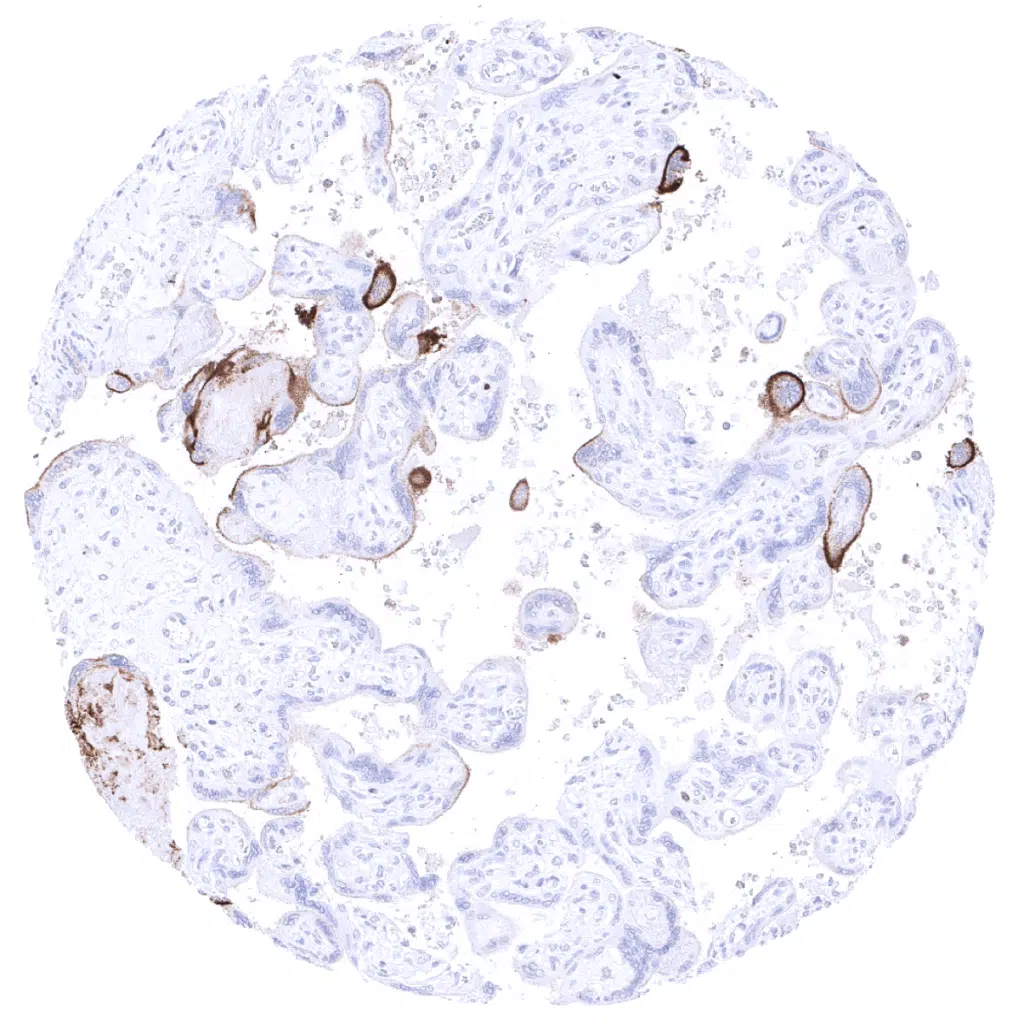
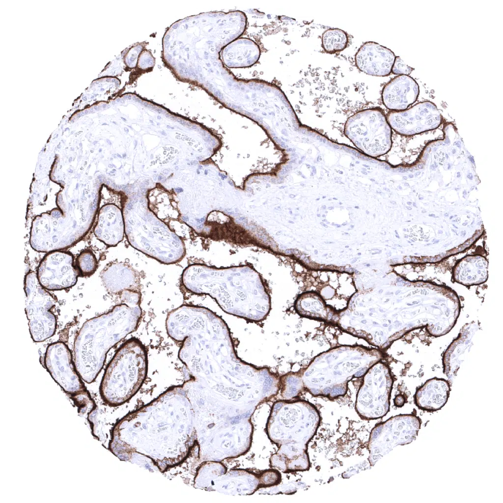
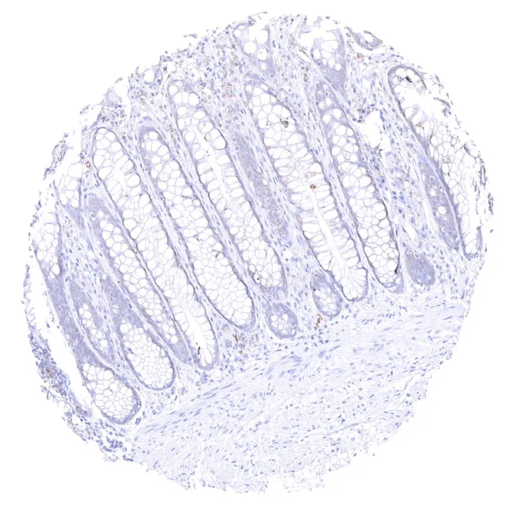
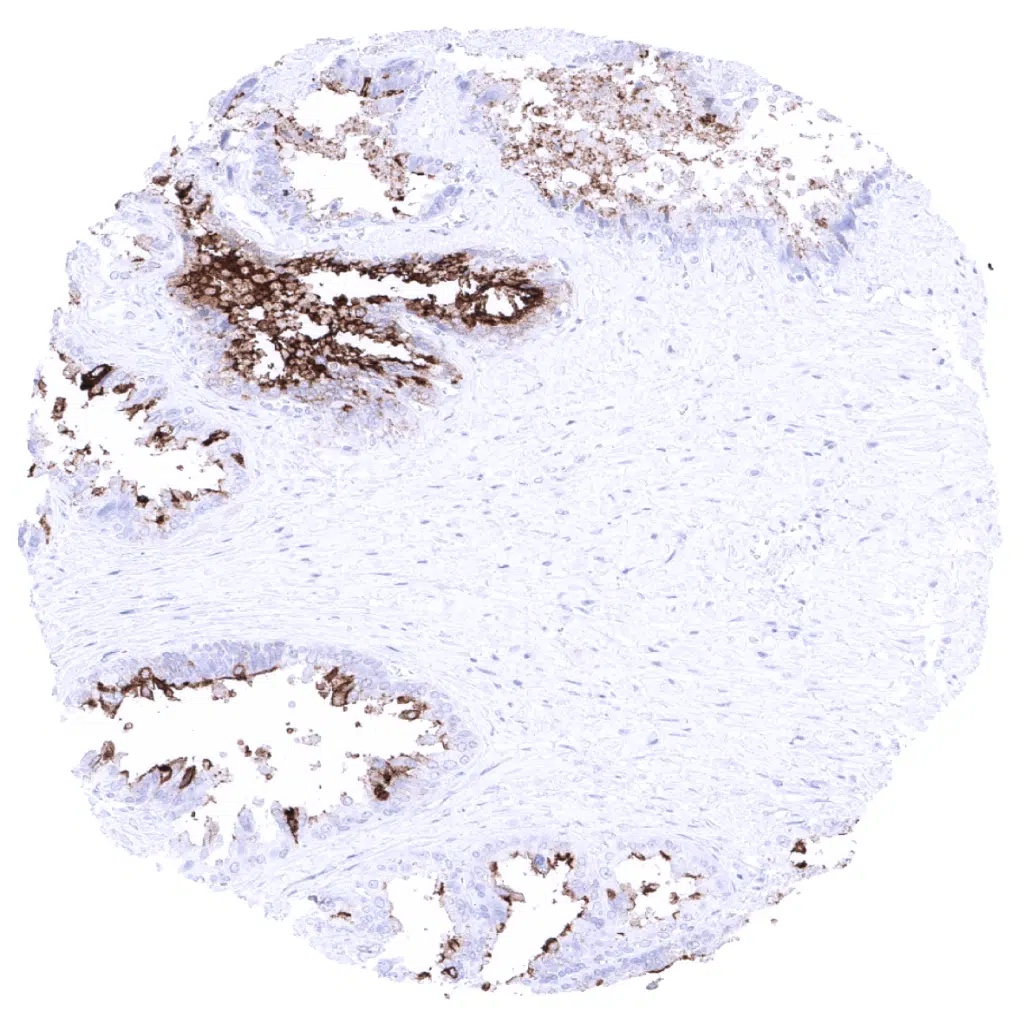
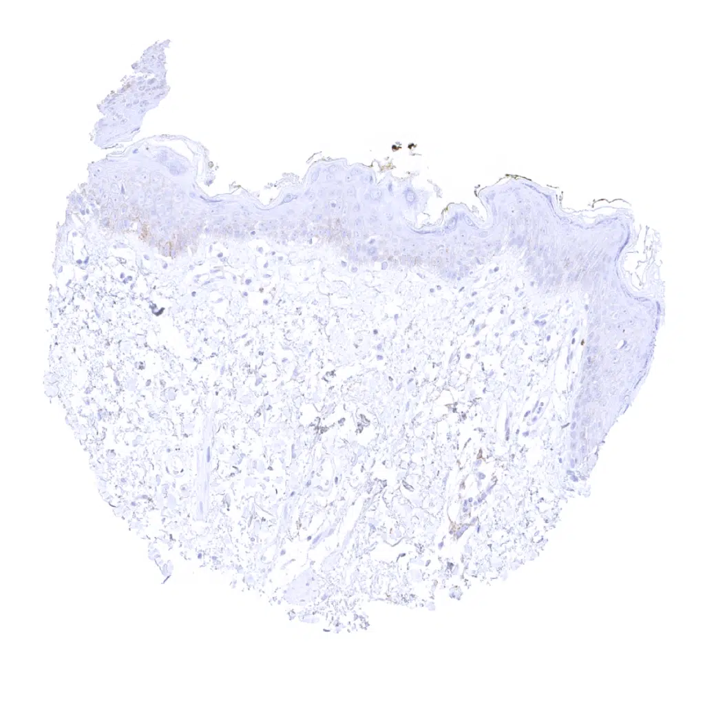
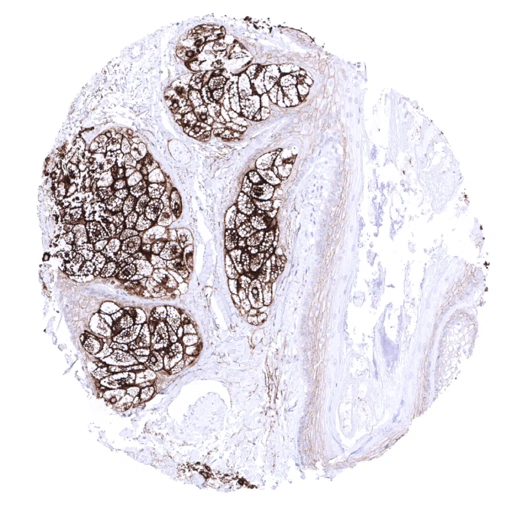
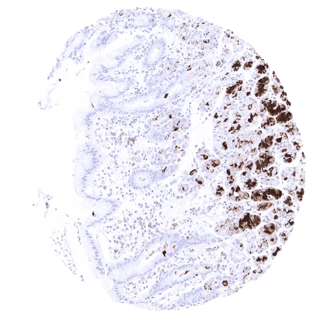
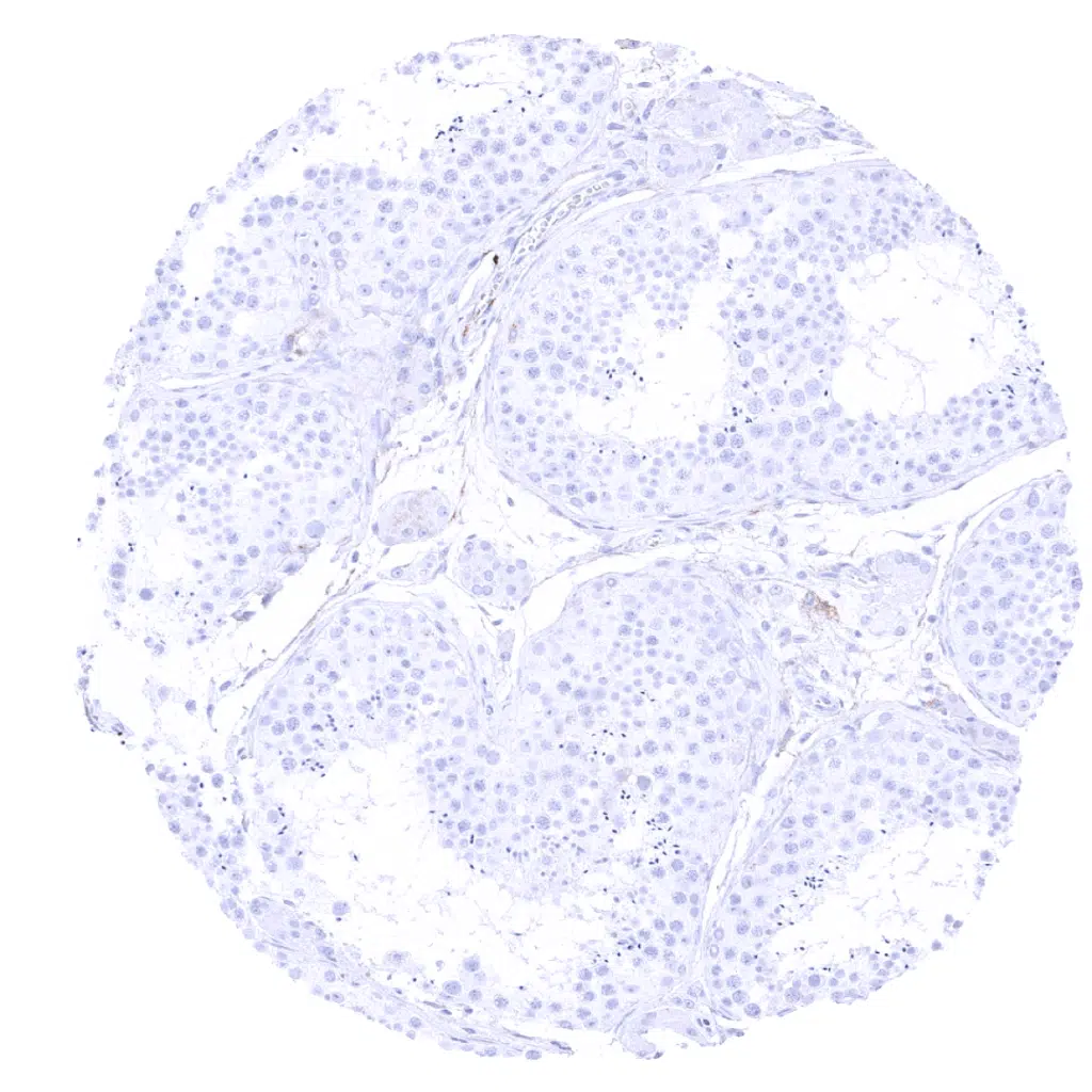
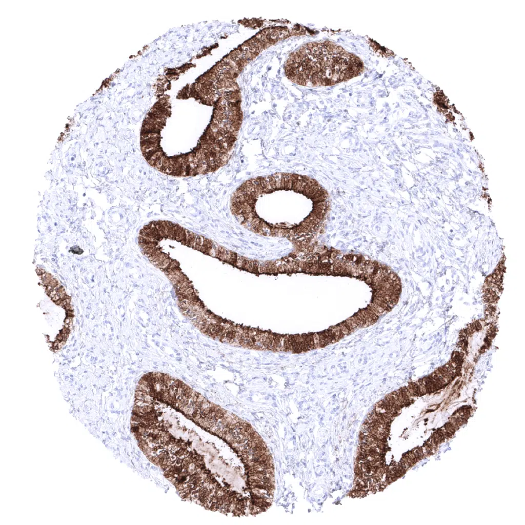
MUC1 antibody [MSVA-672R] HistoMAX


Go to Cancer Tissue Gallery Go back to product page Go to HistoMAX page


Adrenal gland
Anal canal, skin – A weak to moderate, mostly membranous MUC1 EMA immunostaining is seen in this keratinizing squamous epithelium from the anal skin.
Aorta, media
Appendix, mucosa
Appendix, muscular wall
Bone marrow
Breast – A moderate to strong cytoplasmic MUC1 EMA immunostaining with apical predominance is seen in breast glands. MUC1 EMA staining may be weaker in basal cells.
Bronchus, mucosa – A variable, moderate to strong MUC1 EMA staining occurs in respiratory epithelium.
Cerebellum (molecular layer, Purkinje cell layer, granule cell layer, white matter)
Cerebellum (white matter)
Cerebrum, grey matter – A weak fibrillar MUC1 EMA staining can be seen in samples from the cerebrum.
Cerebrum, grey matter
Cerebrum, white matter – A weak fibrillar MUC1 EMA staining can be seen in samples from the cerebrum.
Cerebrum, white matter
Colon descendens, mucosa
Colon descendens, muscular wall
Duodenum, Brunner gland – A weak apical membranous staining is occasionally seen in Brunner glands.
Duodenum, mucosa
Epididymis – In the epididymis the staining varies from strong diffuse cytoplasmic with apical predominance to only apical membranous staining. This sample shows a particularly strong staining.
Esophagus, squamous epithelium – A strong MUC1 EMA immunostaining is seen in this non-keratinizing squamous epithelium of the esophagus.
Fallopian tube, mucosa – A strong cytoplasmic MUC1 EMA immunostaining with apical predominance is seen in the epithelium of the fallopian tube.
Fat
Gallbladder, epithelium – A weak to moderate membranous MUC1 EMA staining is seen in gallbladder epithelium.
Heart muscle
Ileum, mucosa
Kidney, cortex – Distal tubuli and collecting ducts show a strong cytoplasmic MUC1 EMA staining while proximal tumuli are completely negative and cells of the Bowman’s capsule show weak to moderate positivity.
Kidney, medulla – A strong cytoplasmic MUC1 EMA staining is seen in collecting ducts.
Liver – A weak to moderate, predominantly apical MUC1 EMA staining is seen in intrahepatic bile ducts, while hepatocytes are negative.
Lung – In the lung, a strong MUC1 EMA staining is seen in all pneumocytes.
Lymph node – Faint MUC1 EMA staining of few cells in the germinal centre.
Ovary, stroma
Pancreas – Pancreatic acinar cells show a strong MUC1 EMA immunostaining of the apical membranes and of granular (Golgi like) structures below the apical surface.
Parathyroid
Parotid gland – In salivary glands, a moderate MUC1 EMA immunostaining of apical membranes of serous glandular cells and of excretory ducts is seen.
Peripheral nerves – Sheaths of peripheral nerves show moderate MUC1 EMA staining.
Pituitary gland, anterior lobe – In the adenohypophysis a moderate staining with apical predominance is seen in a fraction of cells/acini.
Pituitary gland, posterior lobe – A strong fibrillar MUC1 EMA staining occurs in the neurohypophsis.
Placenta (amnion and chorion) – A strong cytoplasmic staining is seen in amnion and chorion cells of the placenta.
Placenta, early – In the first trimenon, a predominantly membranous MUC1 EMA staining can occur in a fraction of trophoblastic cells.
Placenta, early – In the first trimenon, a predominantly membranous MUC1 EMA staining can occur in a fraction of trophoblast cells.
Placenta, early – A predominantly membranous MUC1 EMA staining can occur in a fraction of trophoblast cells, especially in the first trimenon.
Placenta, mature – In the mature placenta, a predominantly membranous MUC1 EMA staining can occur in a fraction of trophoblastic cells.
Placenta, mature – A predominantly membranous MUC1 EMA staining can occur in a fraction of trophoblast cells. In this sample, the staining is strong.
Prostate – MUC1 EMA immunostaining is variable in the prostate. In this sample, a weak MUC1 EMA staining of luminal cells is seen in few glands while others are completely negative.
Prostate – MUC1 EMA immunostaining is variable in the prostate. In this sample, a moderate to strong MUC1 EMA staining of luminal and basal cells is seen in a fraction of cells of some glands.
Rectum, mucosa
Seminal vesicle – A strong apical MUC1 EMA staining focally occurs in seminal vesicle epithelium.
Sinus paranasales – A variable, moderate to strong MUC1 EMA staining occurs in respiratory epithelium.
Skeletal muscle
Skin – MUC1 EMA immunostaining is usually absent in the skin.
Skin (sebaceous glands) – A strong MUC1 EMA immunostaining is seen in sebaceous glands, while the skin is usually MUC1 EMA negative.
Spleen
Stomach, antrum – Mucous producing glands of the stomach show variable, sometimes also strong MUC1 EMA positivity while the surface epithelial cells of the stomach are largely MUC1 EMA negative.
Stomach, corpus – Mucous producing glands of the stomach show a strong MUC1 EMA positivity in this sample while the surface epithelial cells are largely MUC1 EMA negative.
Testis
Thymus – Some elements of corpuscles of Hassall's show a weak to moderate MUC1 EMA staining.
Thyroid gland – A moderate apical MUC1 EMA staining is seen in follicles, sometimes accompanied by a moderate to strong cytoplasmic staining.
Tonsil – A fraction of squamous epithelial cells of tonsil crypts show a moderate to strong MUC1 EMA staining. Plasma cells are also MUC1 EMA positive.
Tonsil, surface epithelium – A moderate to strong MUC1 EMA immunostaining is seen in this non-keratinizing squamous epithelia of the tonsil surface. The staining is more prominent in the upper than in the basal layers
Urinary bladder, muscular wall
Urinary bladder, urothelium – A moderate to strong MUC1 EMA immunostaining can be seen in the urothelium. The staining is most prominent in the umbrella cells.
Uterus, ectocervix – A very faint membranous MUC1 EMA immunostaining is seen in this non-keratinizing squamous epithelium from the ectocervix.
Uterus, endocervix – A strong cytoplasmic MUC1 EMA immunostaining with apical predominance is seen in the endocervical epithelium.
Uterus, endometrium (pregnancy) – A strong cytoplasmic MUC1 EMA immunostaining with apical predominance is seen in the pregnant endometrium. Decidua cells are MUC1 EMA negative.
Uterus, endometrium (proliferation) – A very strong MUC1 EMA immunostaining is seen in the endometrium, where the stroma is also often MUC1 EMA positive.
Uterus, endometrium (secretion) – A very strong MUC1 EMA immunostaining is seen in the endometrium, where the stroma is also often MUC1 EMA positive.
Uterus, myometrium

| TOP |