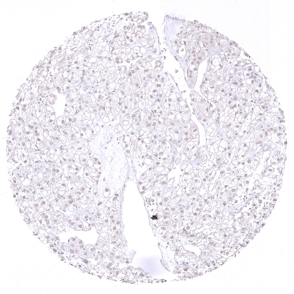
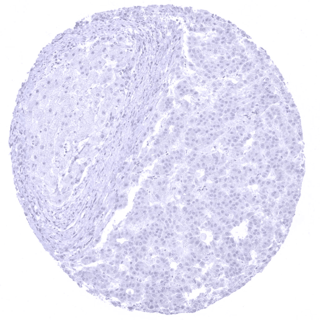
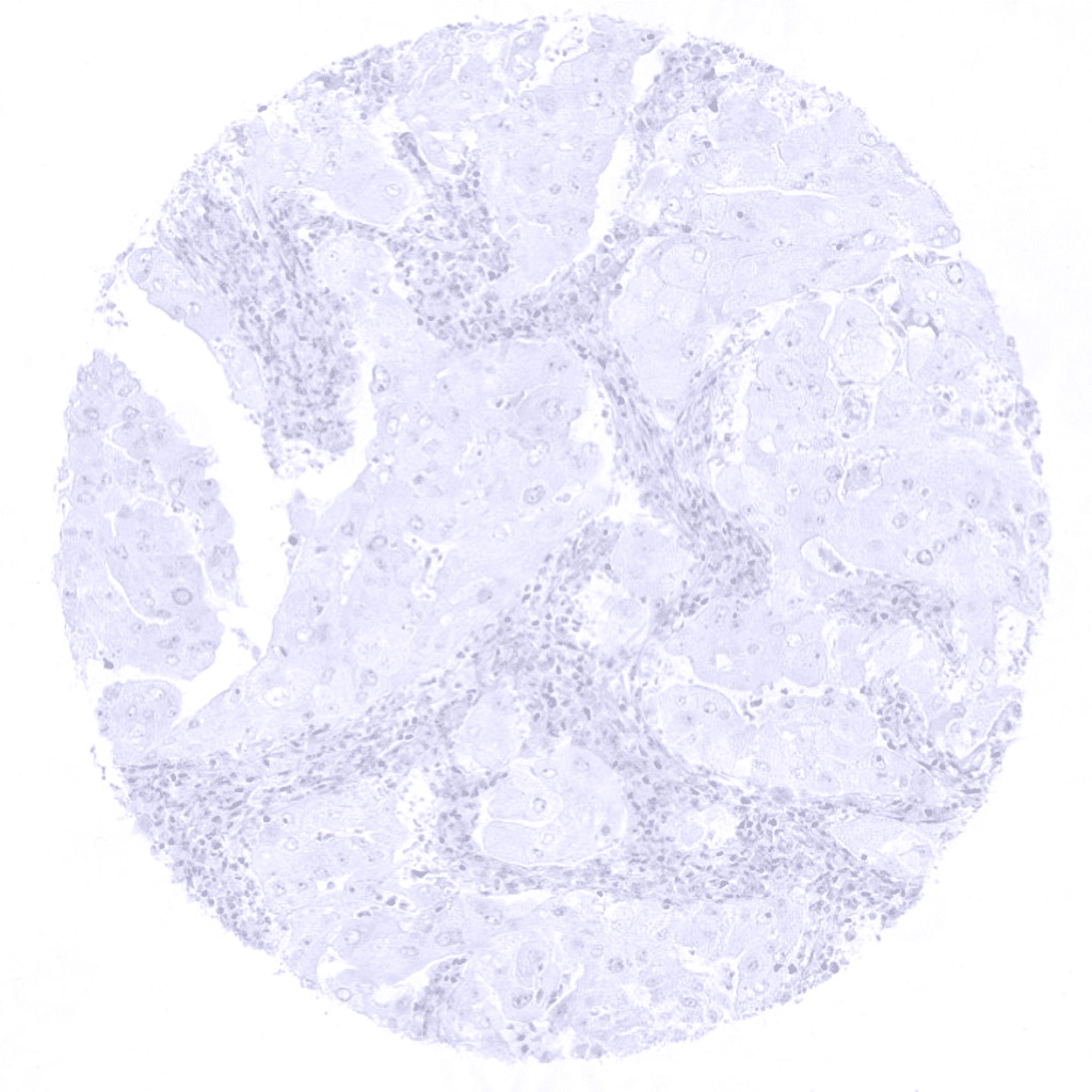
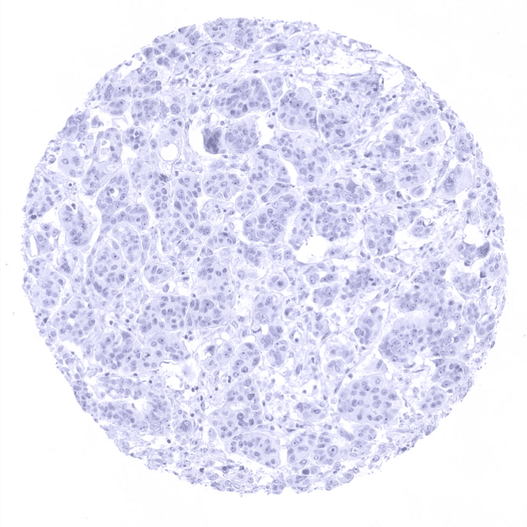
Kidney- Clear cell renal cell carcinoma without any Napsin A immunostaining.
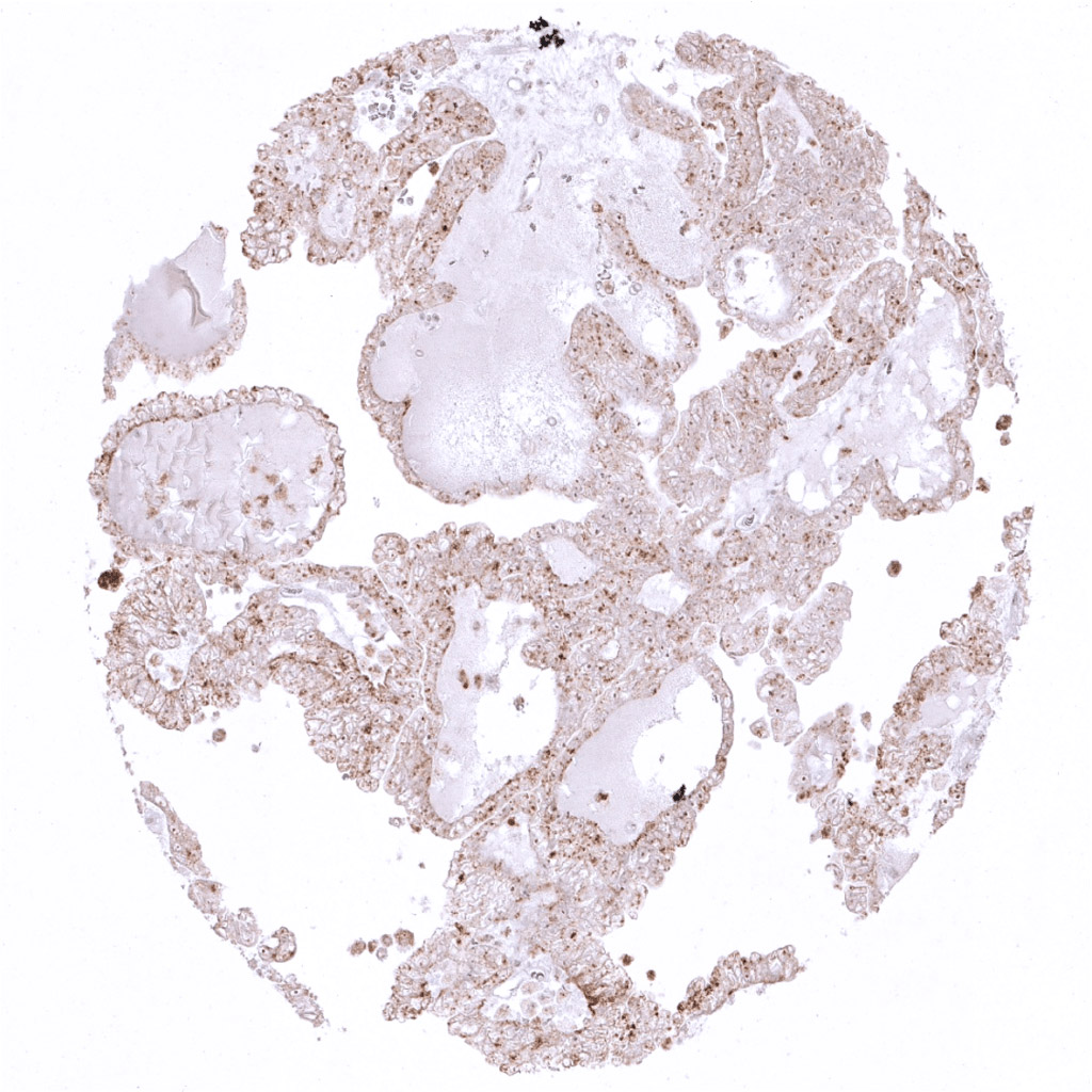
Kidney- Moderate Napsin A positivity in a papillary thyroid cancer.
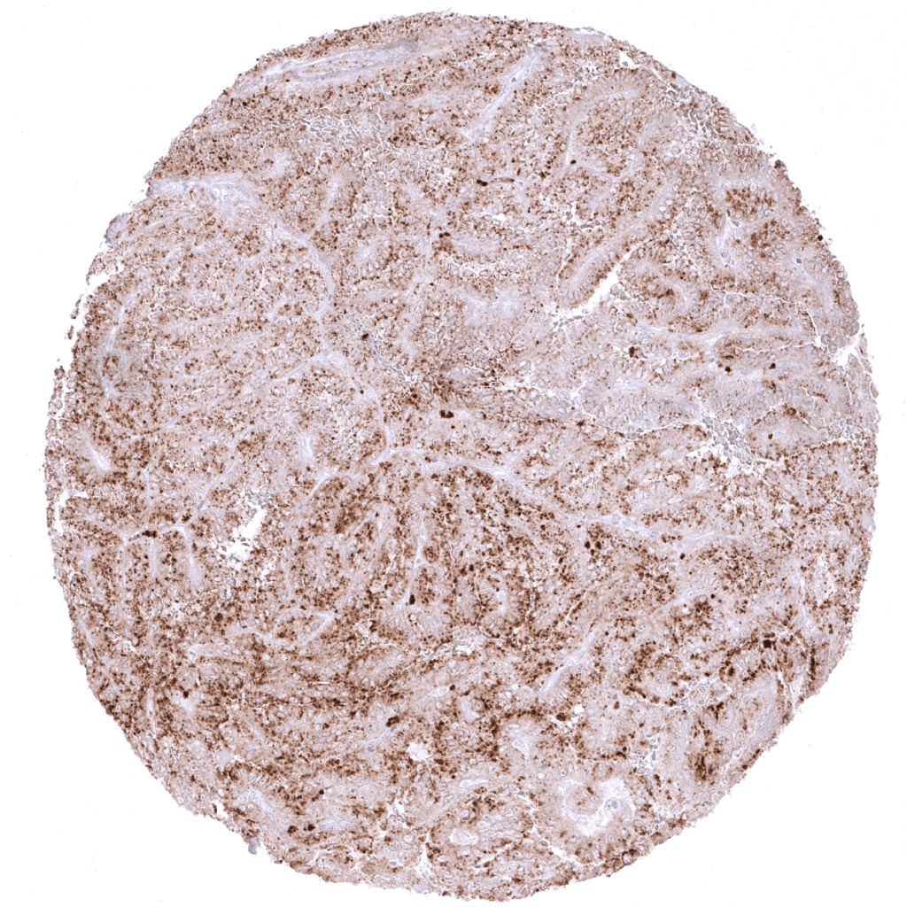
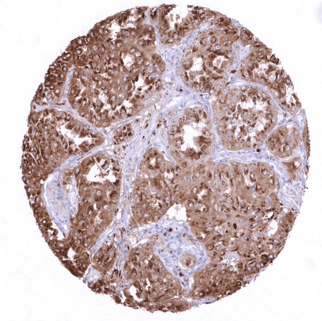
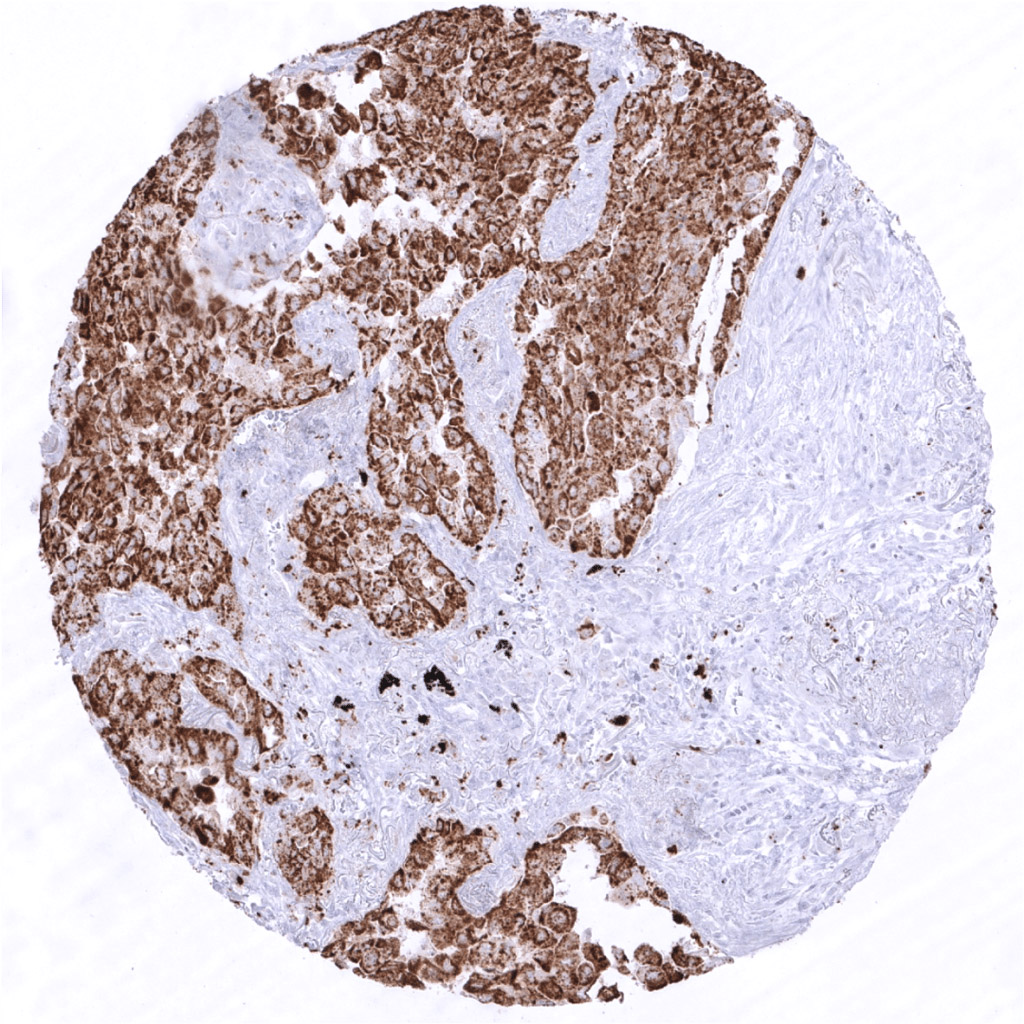
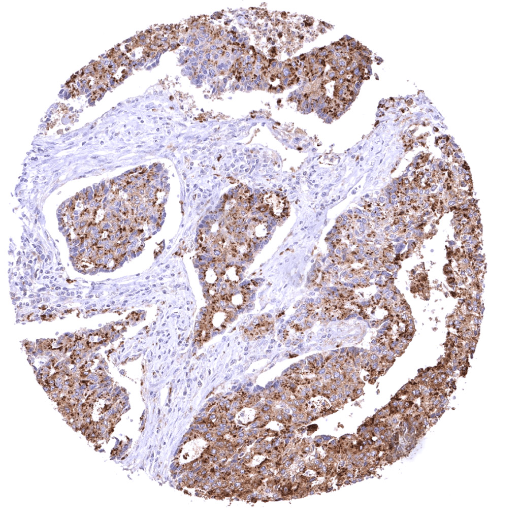
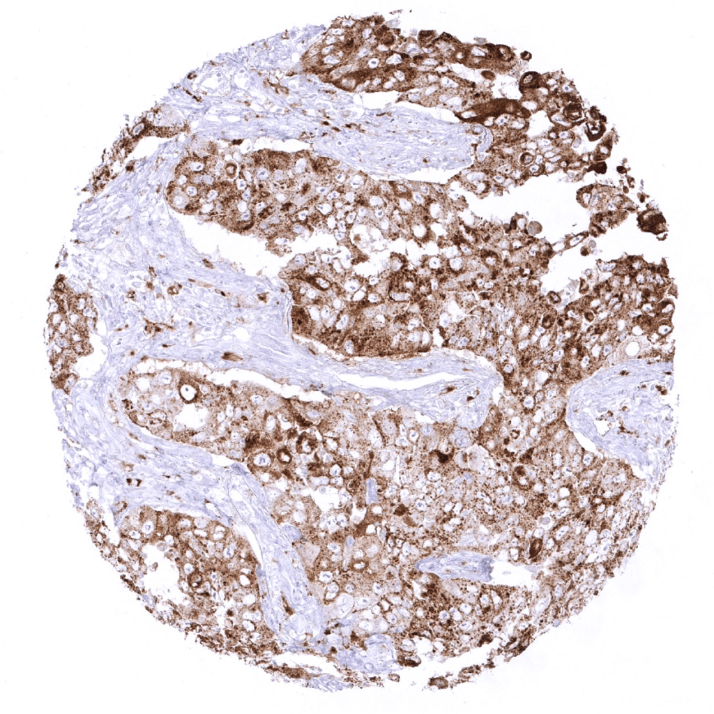
Kidney- Strong Napsin A positivity in a papillary renal cell carcinoma.
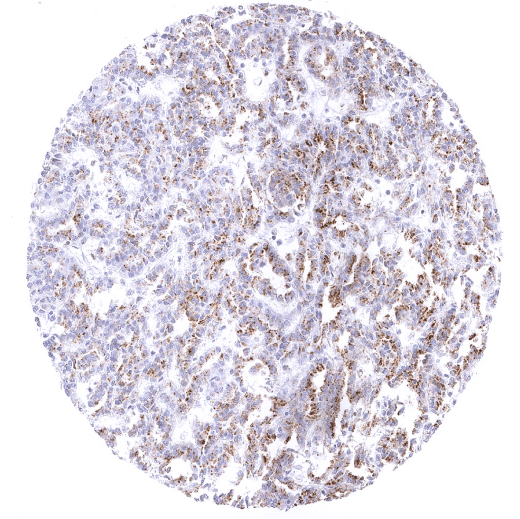
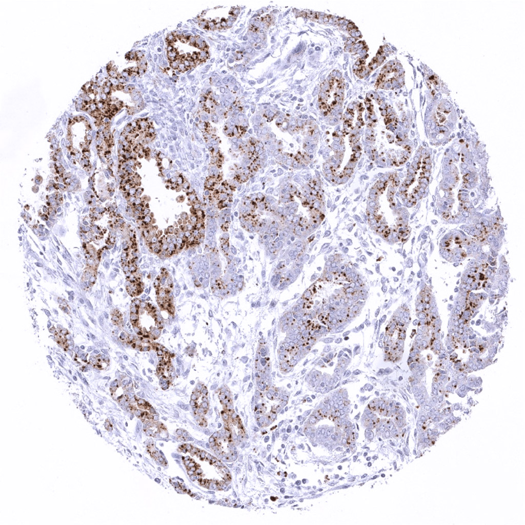
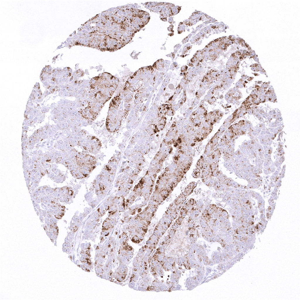
Kidney- Weak Napsin A positivity in a papillary renal cell carcinoma.
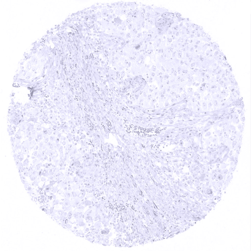
Liver- Hepatocellular carcinoma without Napsin A staining.
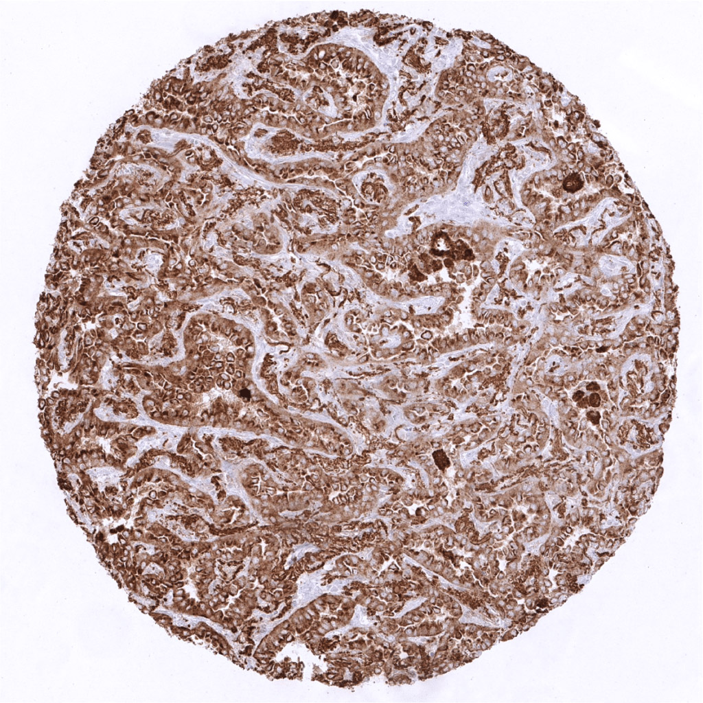
Lung- Adenocarcinoma of the lung with moderate to strong Napsin A immunostaining.
Lung- Adenocarcinoma of the lung with strong Napsin A positivity.
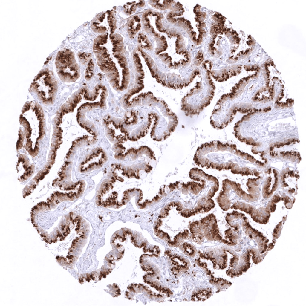
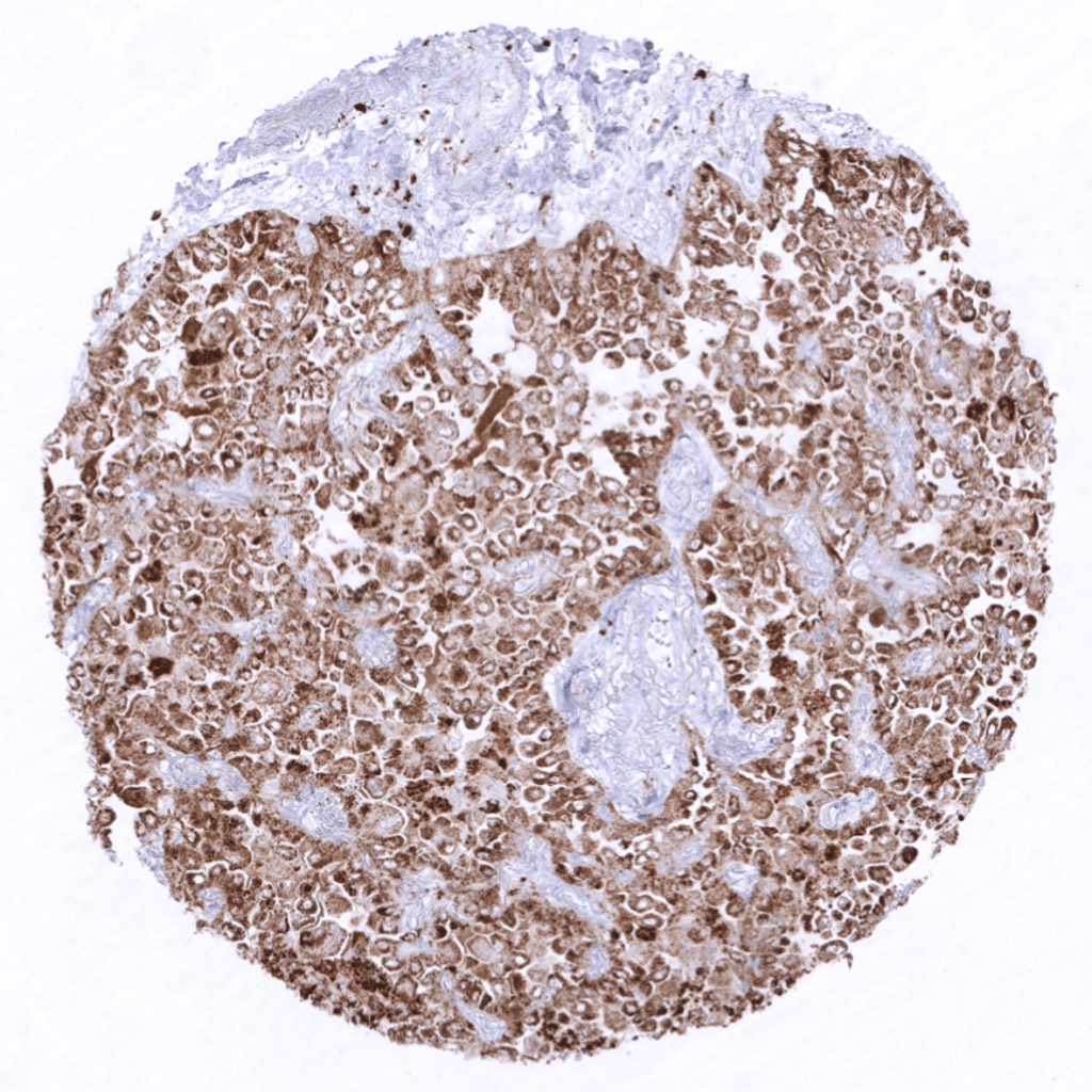
Lung- Lepidic adenocarcinoma of the lung with intense Napsin A immunostaining.
Lung- Lung adenocarcinoma with only faint focal Napsin A positivity.
Lung- Napsin A negative adenocarcinoma of the lung.
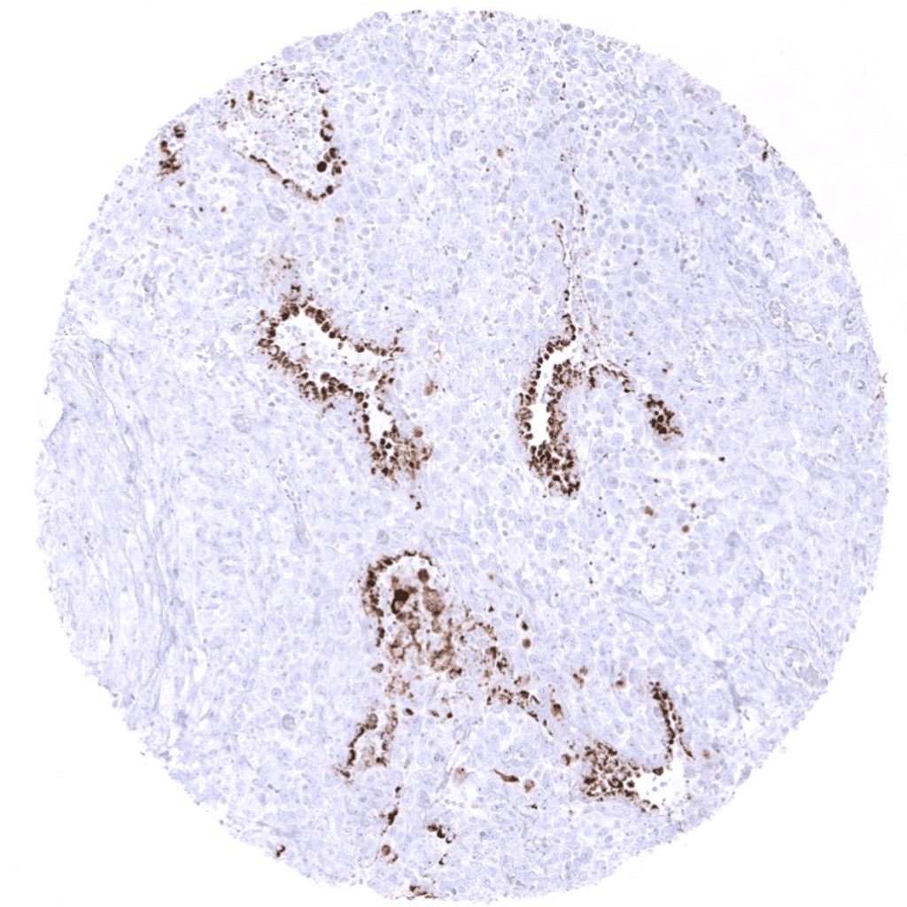
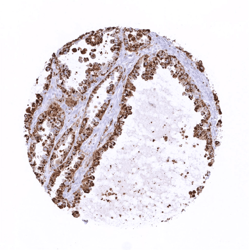
Lung- Napsin A negative lung cancer. Strong Napsin A immunostaining of remnant non-neoplastic lung tissue and/or alveolar macrophages.
Lung- Napsin A negative lung cancer. Strong Napsin A immunostaining is seen in remnant non-neoplastic lung tissue and/or alveolar macrophages.
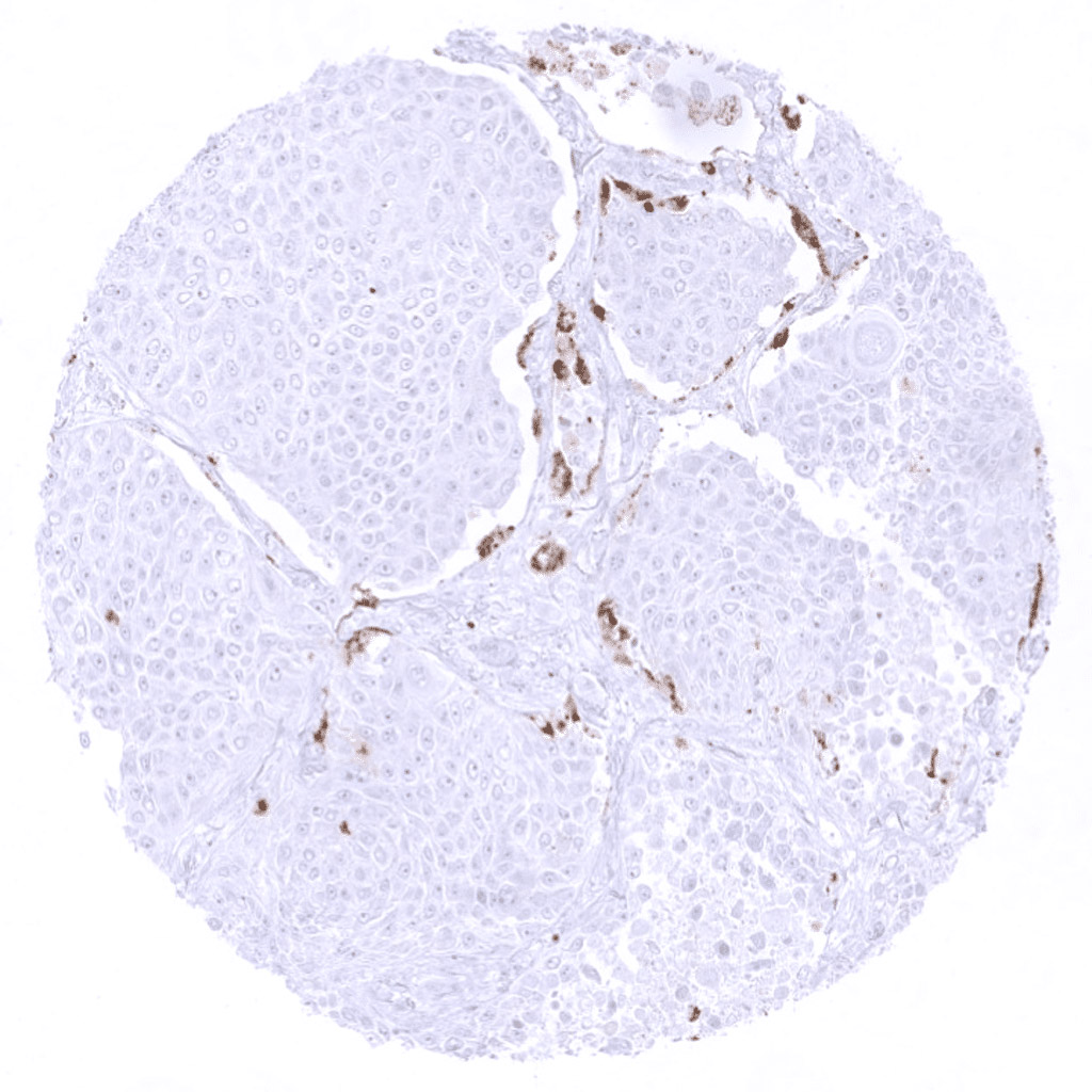
Lung- Napsin A negative squamous cell carcinoma of the lung.
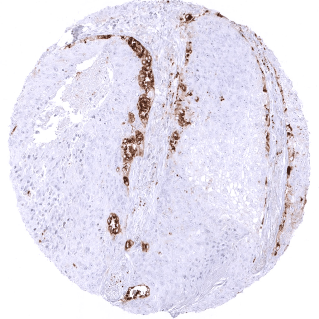
Lung- Napsin A negative squamous cell carcinoma of the lung. Strong Napsin A immunostaining of remnant non-neoplastic lung tissue and/or alveolar macrophages.
Lung- Squamous cell carcinoma of the lung without Napsin A expression. Strong focal Napsin A immunostaining of remnant non-neoplastic lung tissue.
Lung- Strong Napsin A immunostaining in an adenocarcinoma of the lung.
Lung- Strong Napsin A immunostaining in an adenocarcinoma of the lung.
Lung- Strong Napsin A positivity of an adenocarcinoma of the lung.
Lunge- Moderate to strong Napsin A positivity in an adenocarcinoma of the lung.
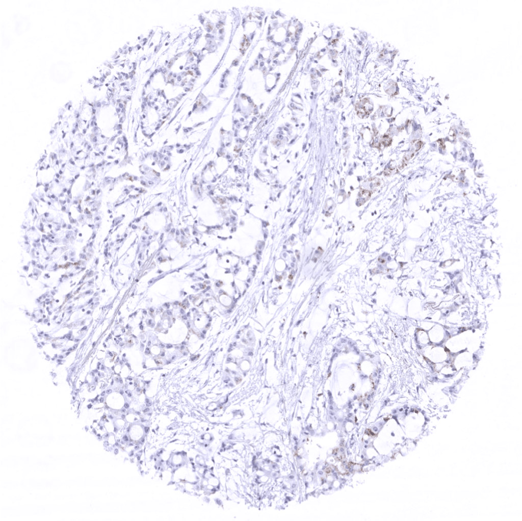
Ovary- Napsin A negative ovarian carcinoma.
Ovary- Strong Napsin A immunostaining in a clear cell carcinoma of the ovary.
Uterus- Malignant mixed Mullerian tumor with moderate Napsin A immunostaining in glandular tumor cells.
Uterus- Moderate Napsin A immunostaining in a serous endometrium carcinoma.
Uterus- Strong Napsin A immunostain in a malignant mixed Mullerian tumor of the uterus. Epithelial tumor cells are predominantly affected.