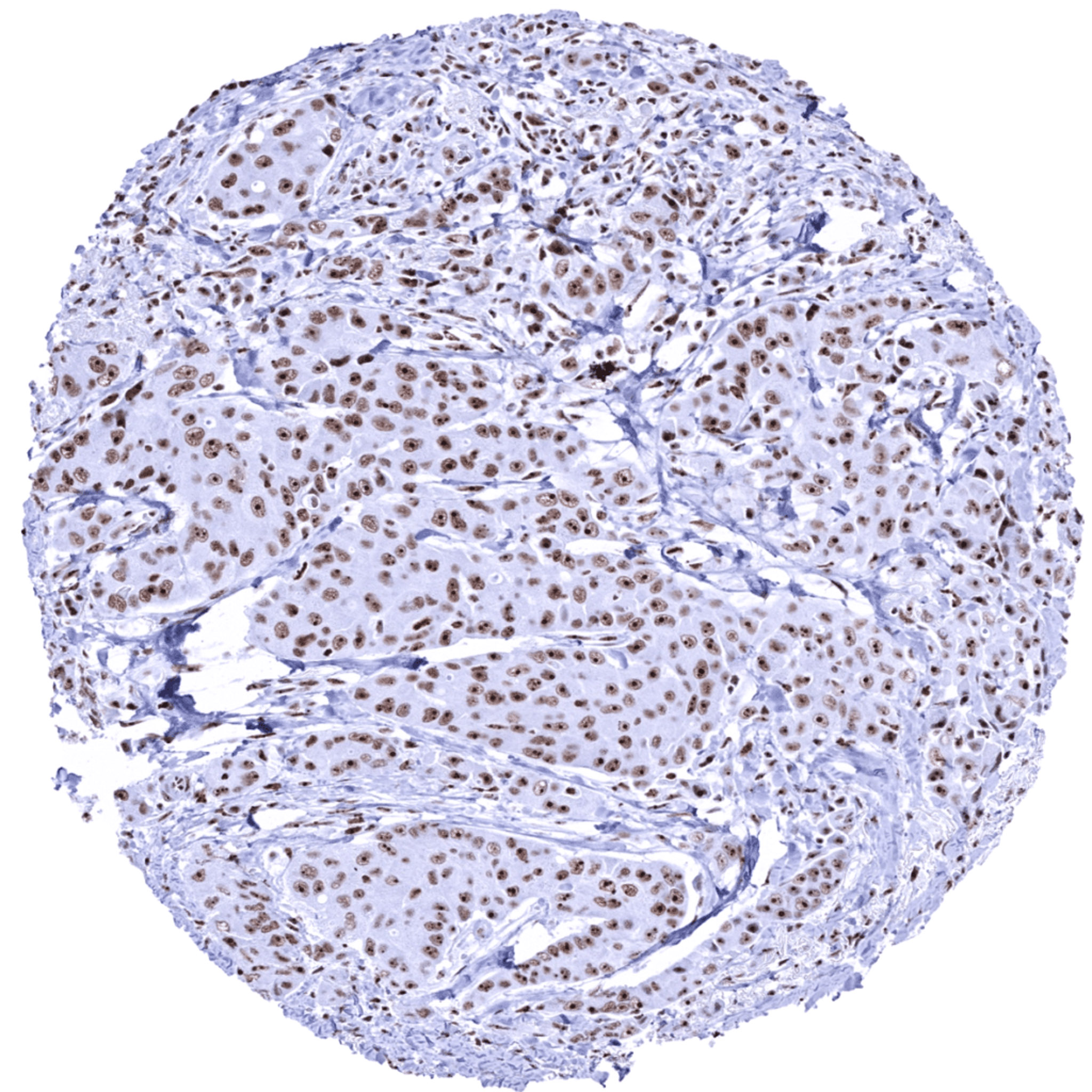
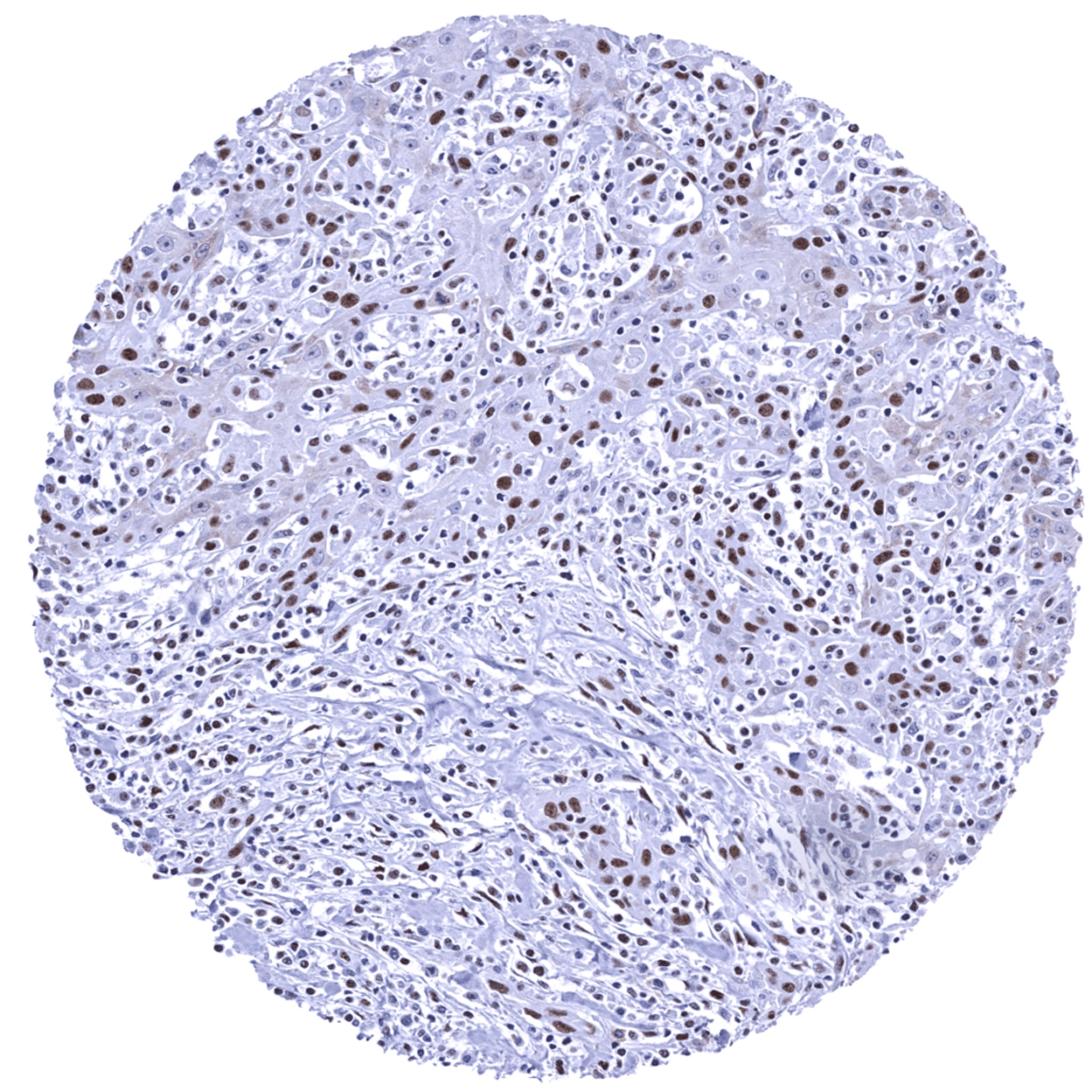
Breast- Invasive breast cancer of no special type (NST) displaying a strong nuclear nucleolin immunostaining with a particularly strong staining in the nucleoli.
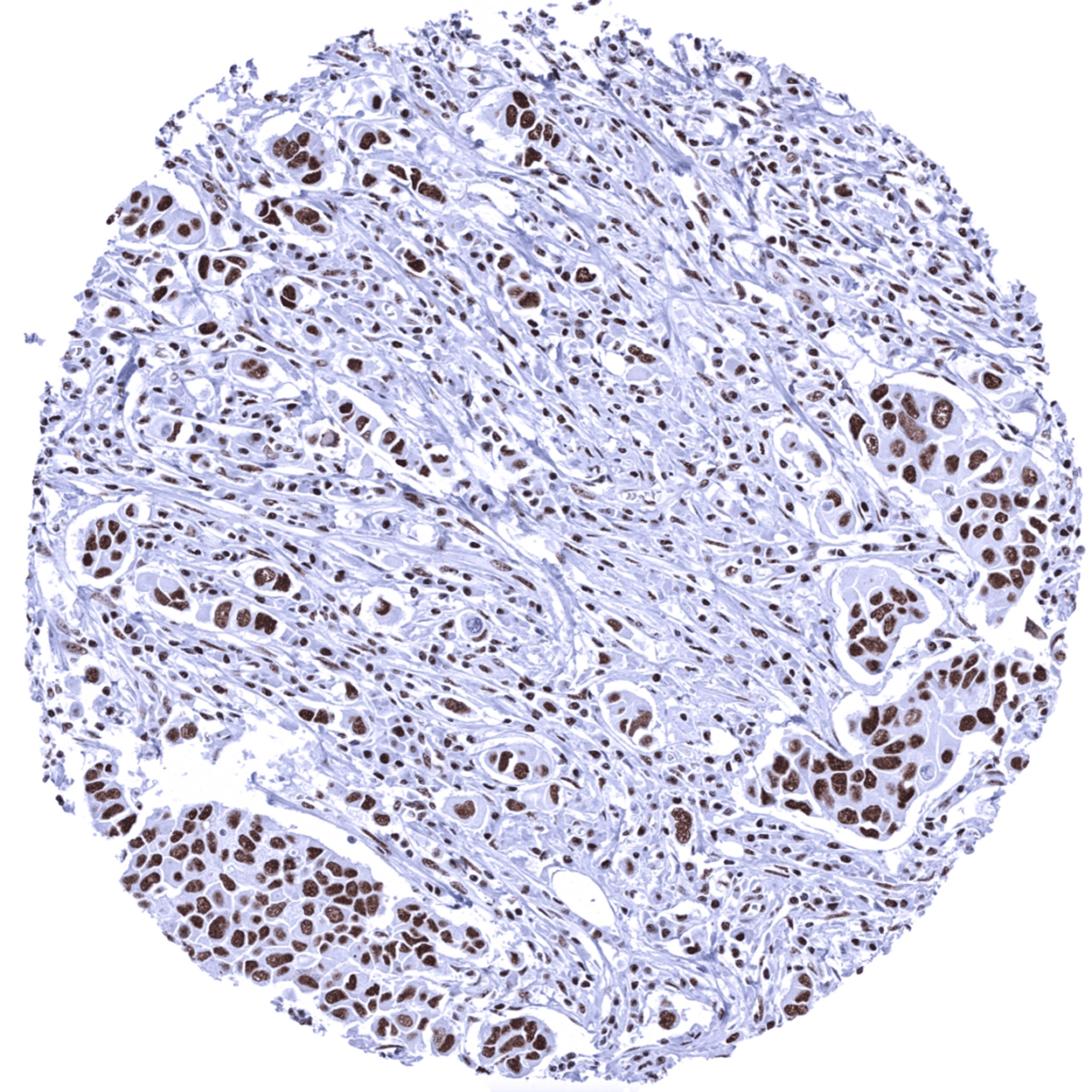
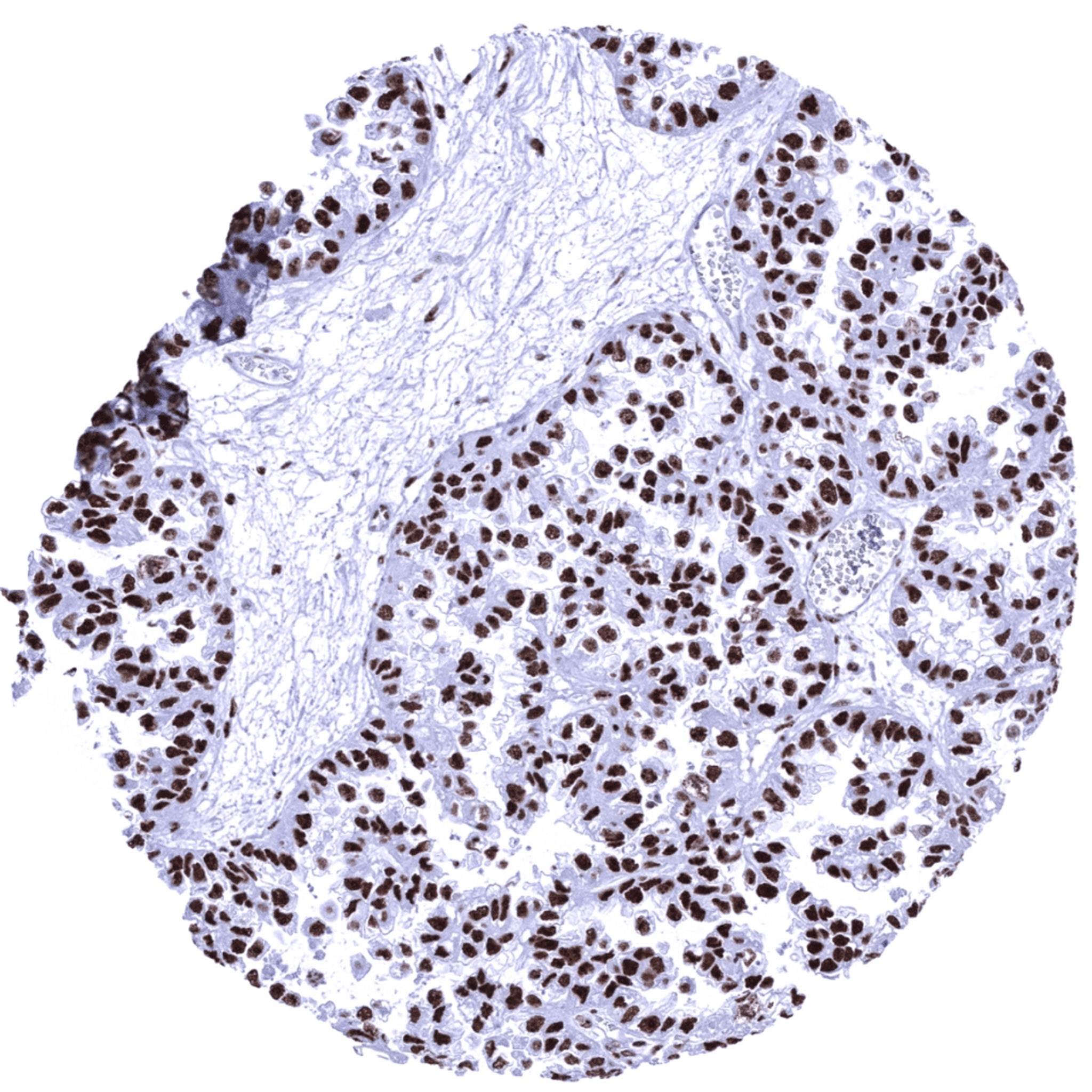
Breast- Invasive lobular breast cancer displaying a strong nuclear nucleolin positivity.
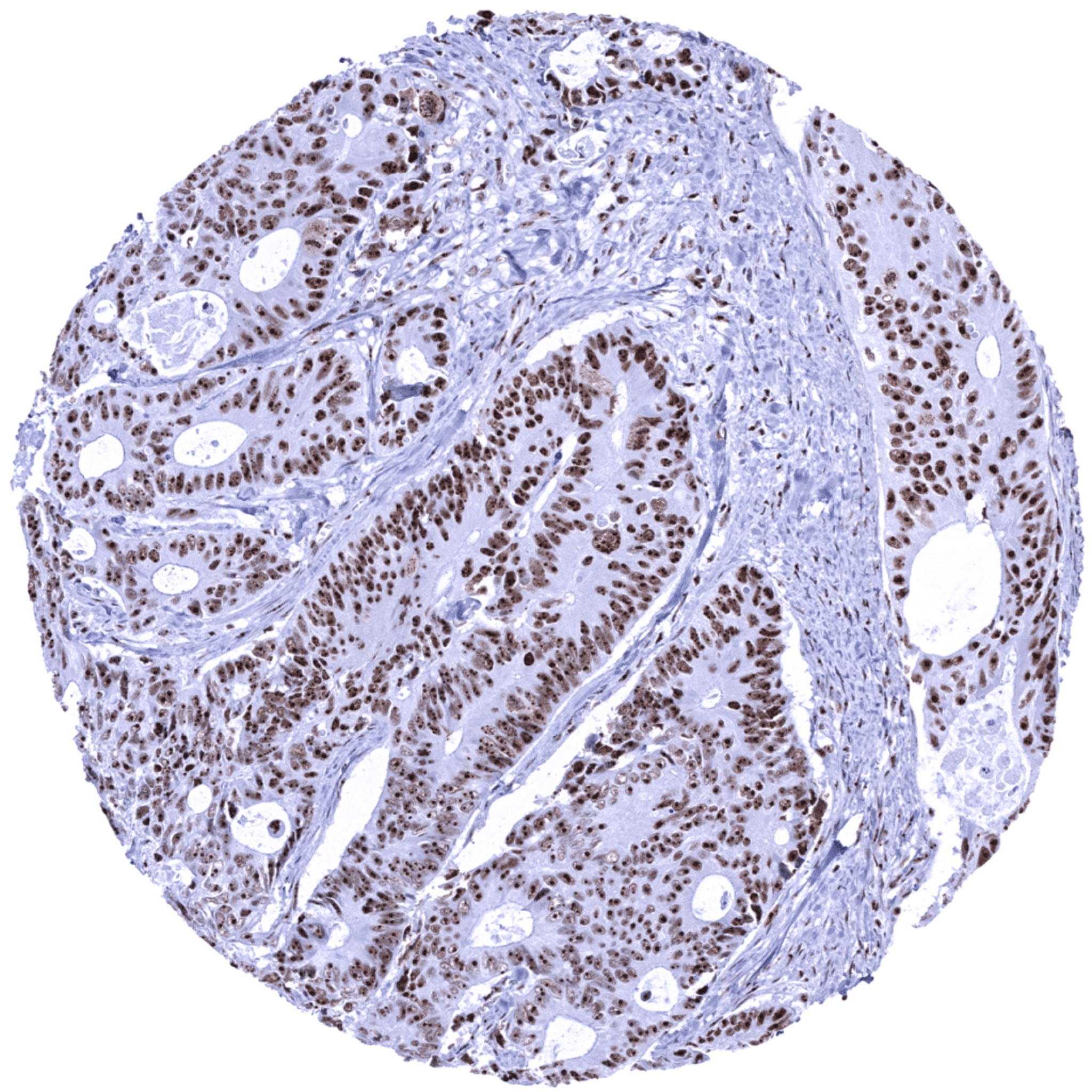
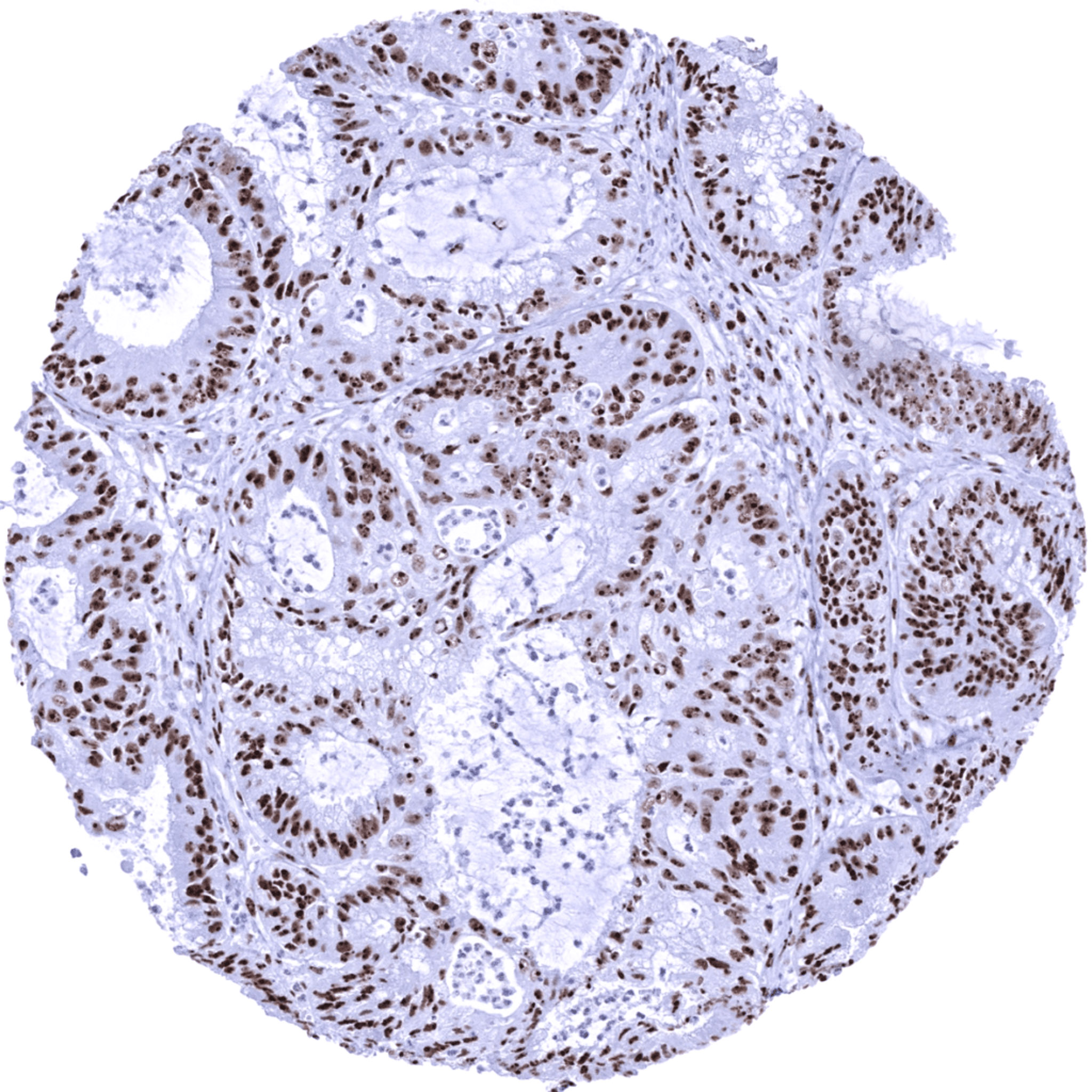
Colon- Colorectal adenocarcinoma showing strong nuclear immunostaining for nucleolin with a particularly strong intensity in nucleoli.
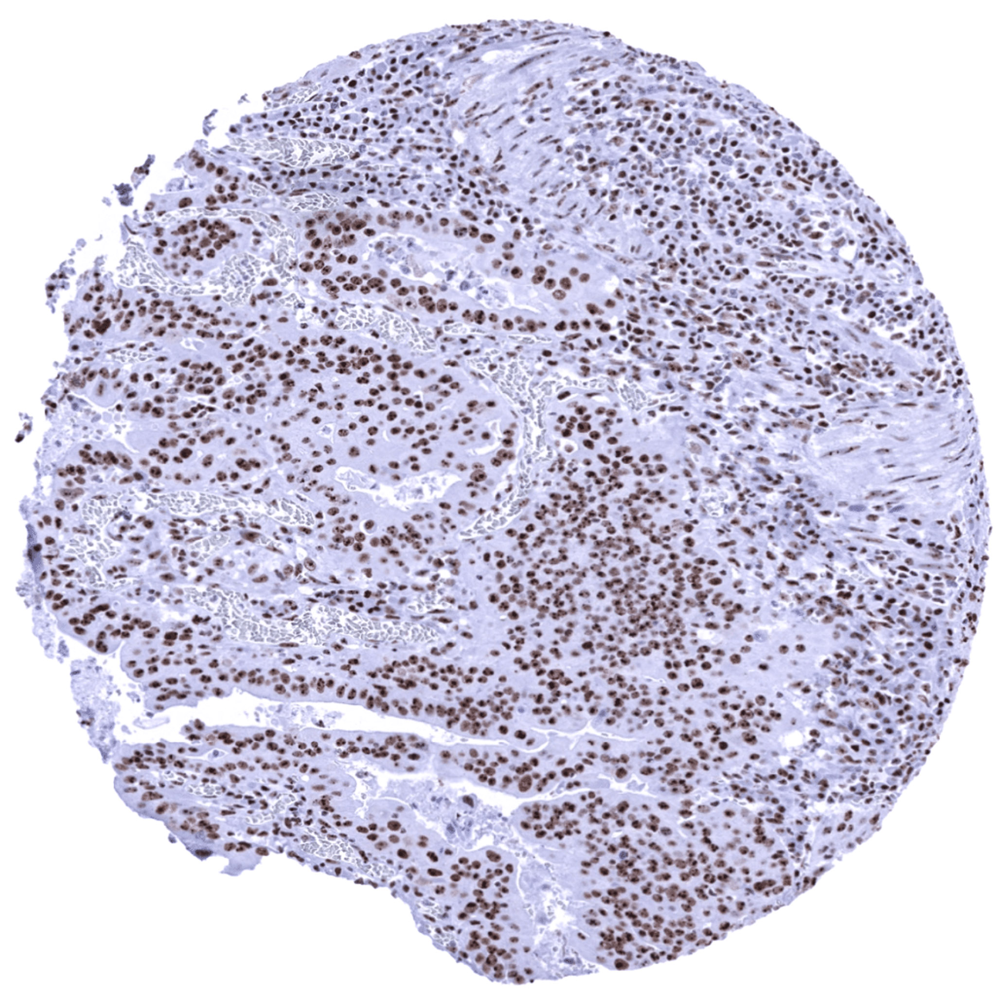
Esophagus- Adenocarcinoma with strong nuclear immunostaining for nucleolin with a particularly strong intensity in nucleoli.
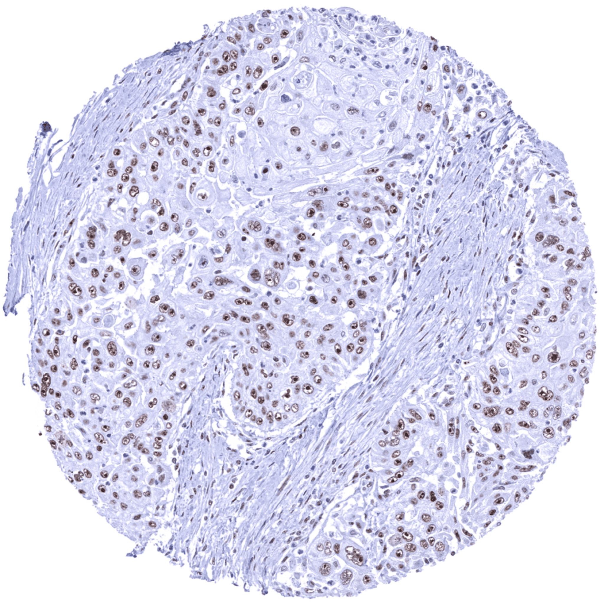
Esophagus- Squamous cell carcinoma exhibiting a distinct nucleolar nucleolin immunostaining. Staining is markedly weaker in other areas of the nuclei.
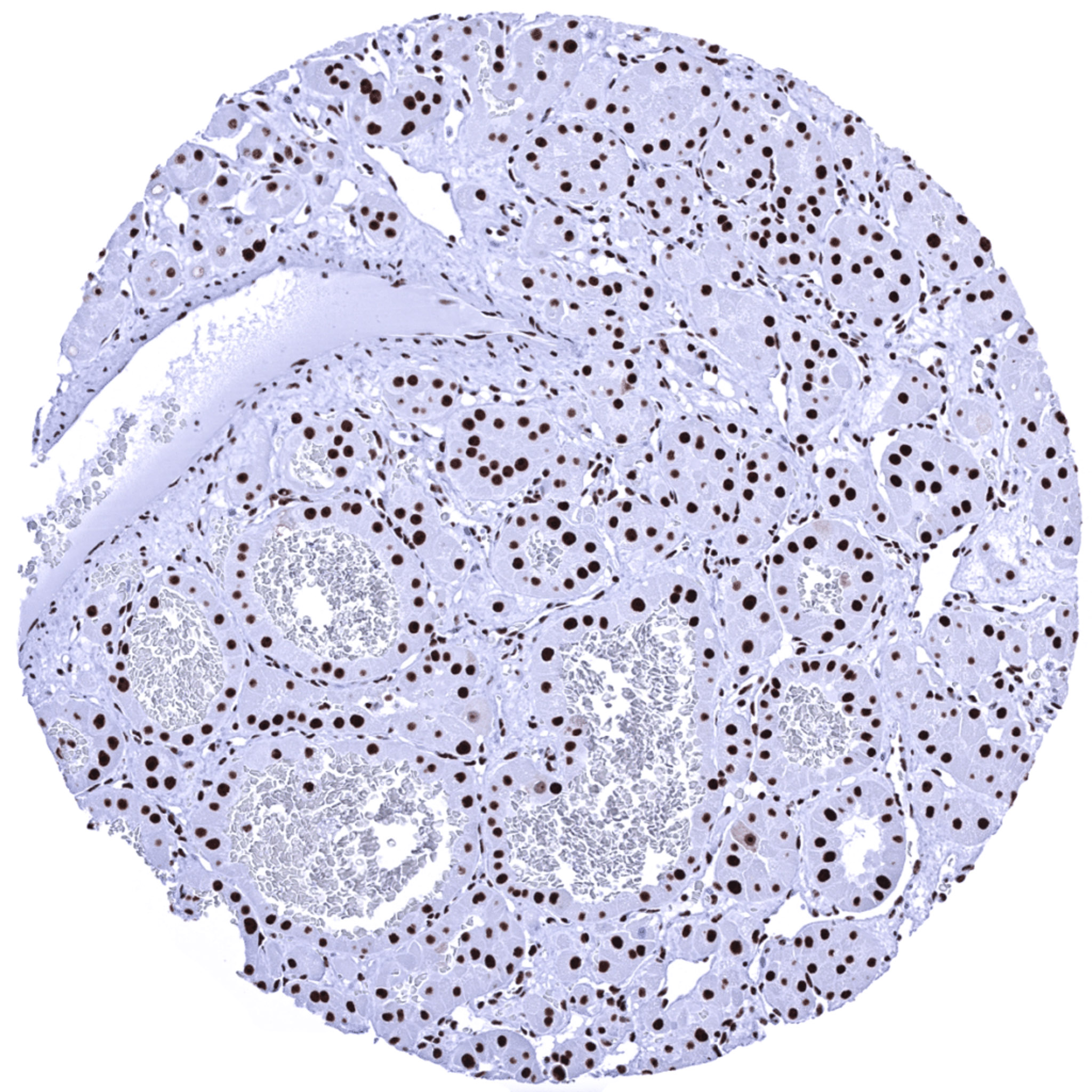
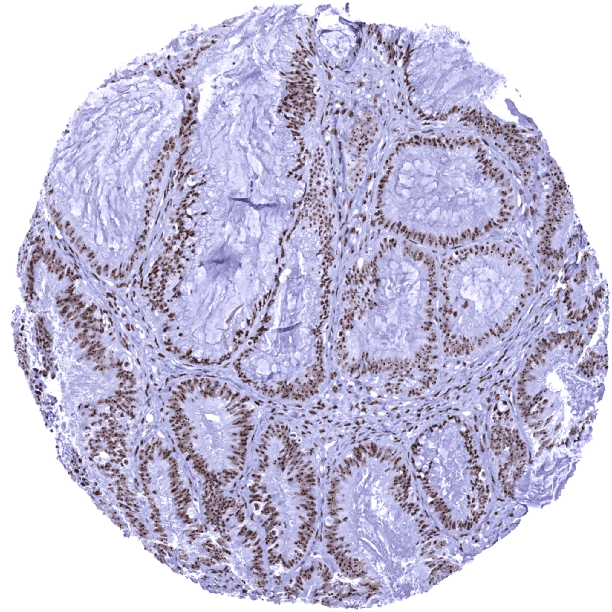
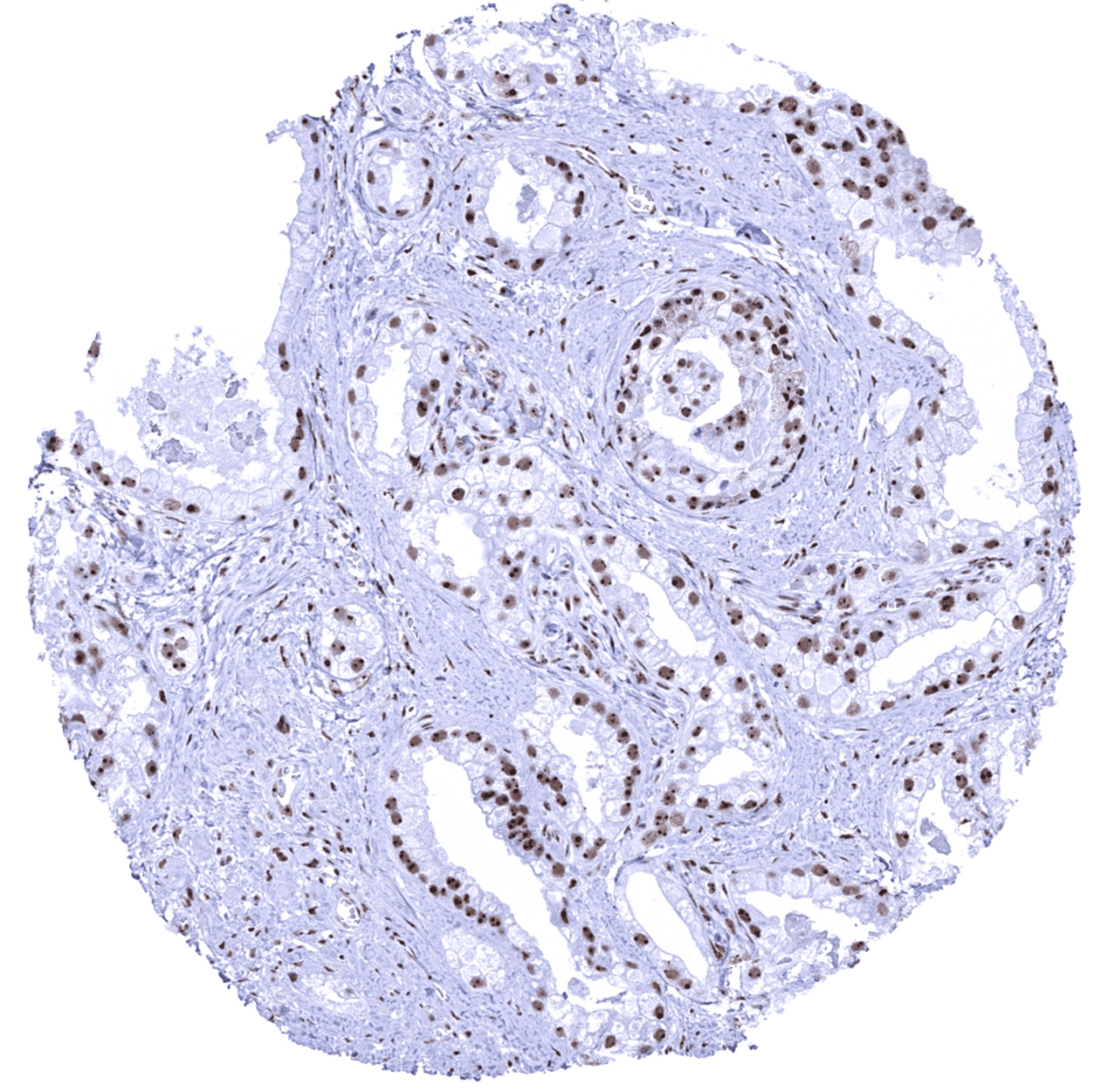
Prostate- Adenocarcinoma (Gleason 3+3=6) showing strong nuclear nucleolin immunostaining with a predominance of the staining in nucleoli.
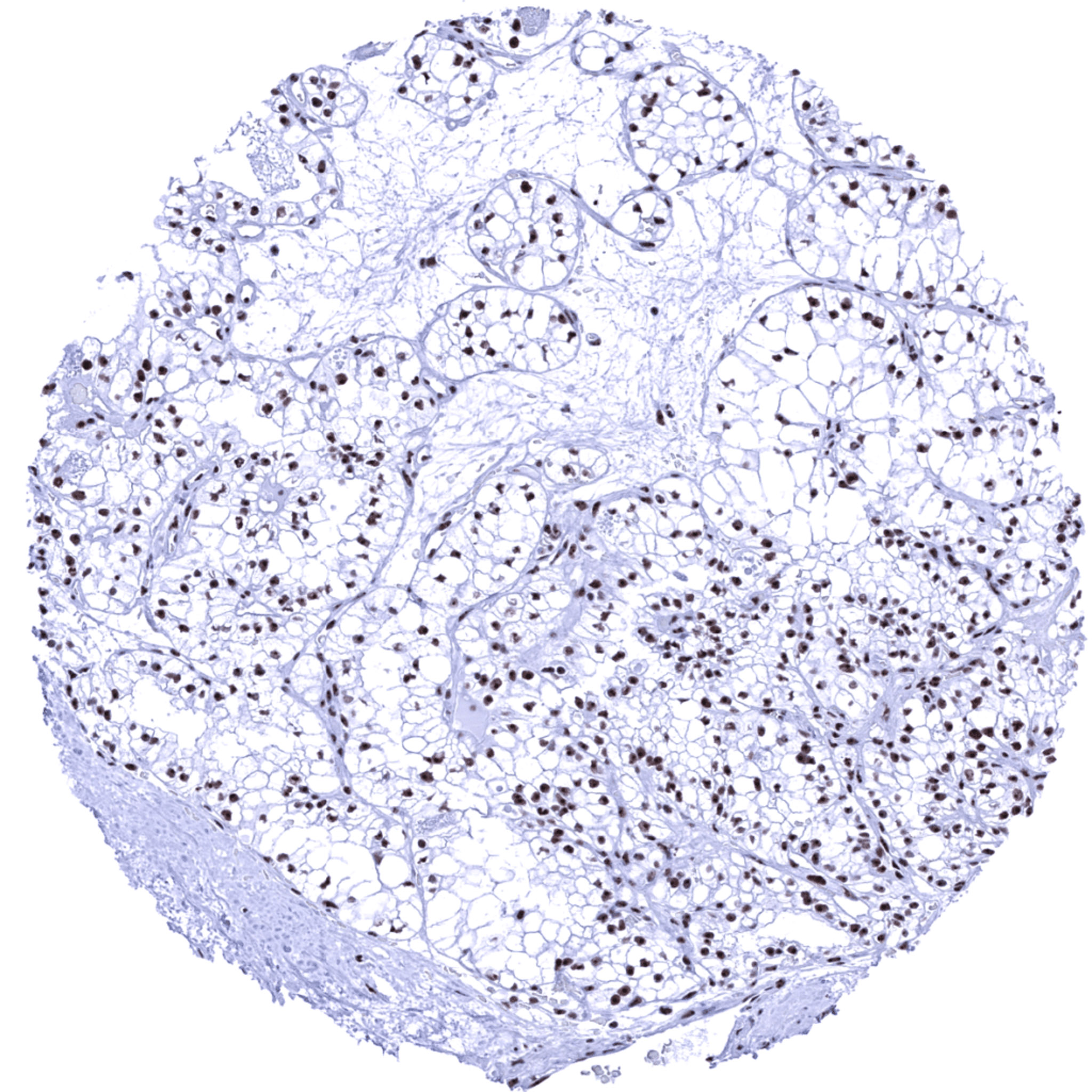
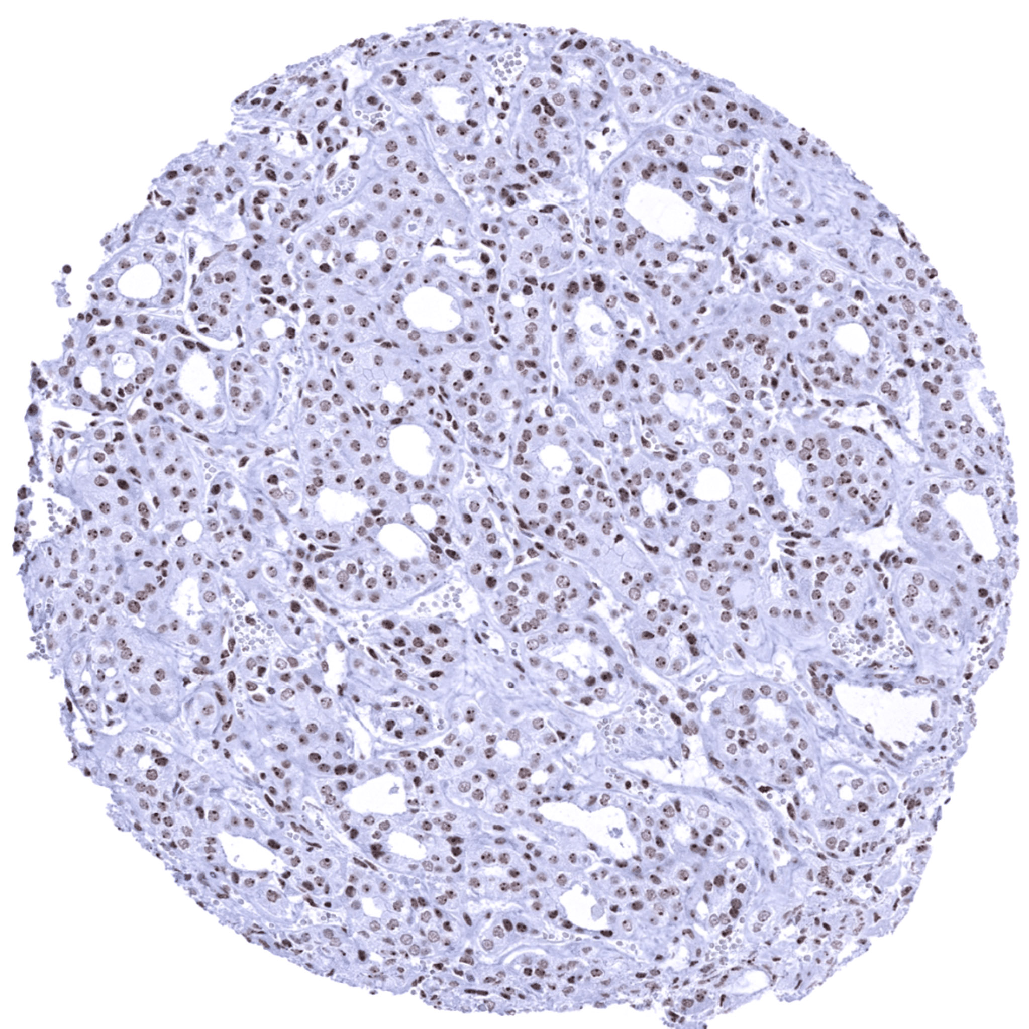
Thyroid- Follicular carcinoma showing a moderate nuclear immunostaining for nucleolin with strong predominance of the staining in nucleoli.
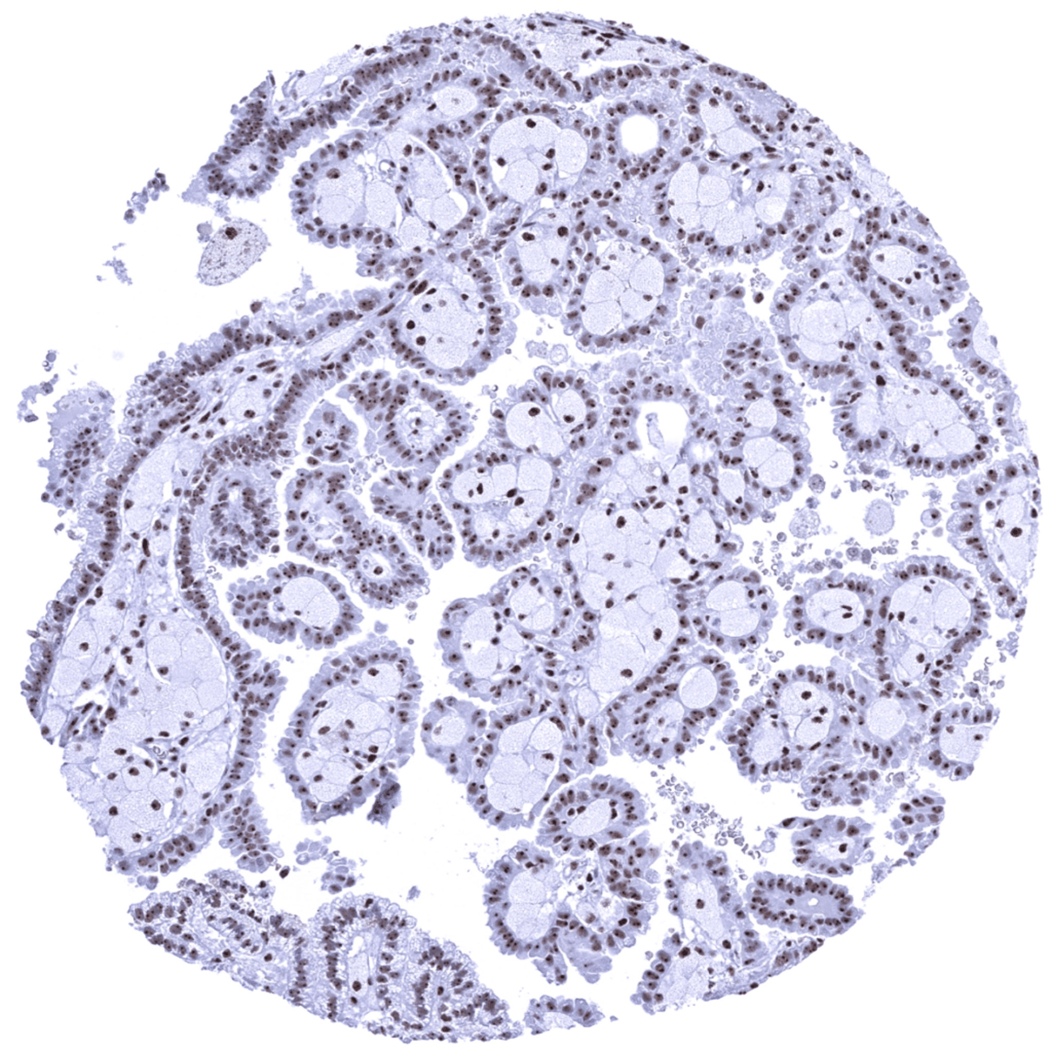
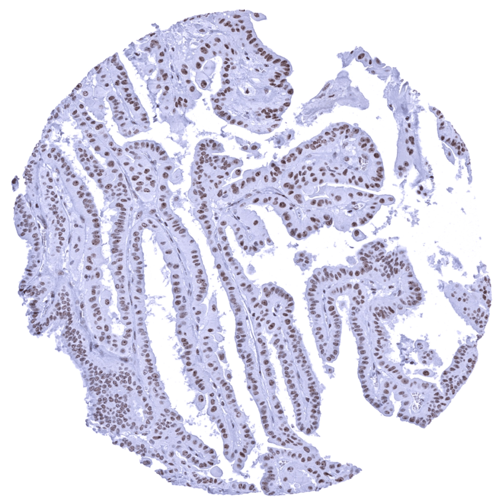
Thyroid- Papillary carcinoma showing a moderate to strong nuclear nucleolin positivity.
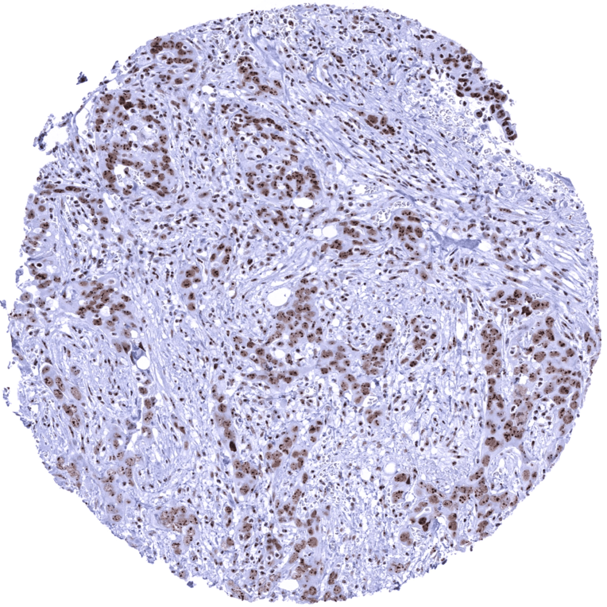
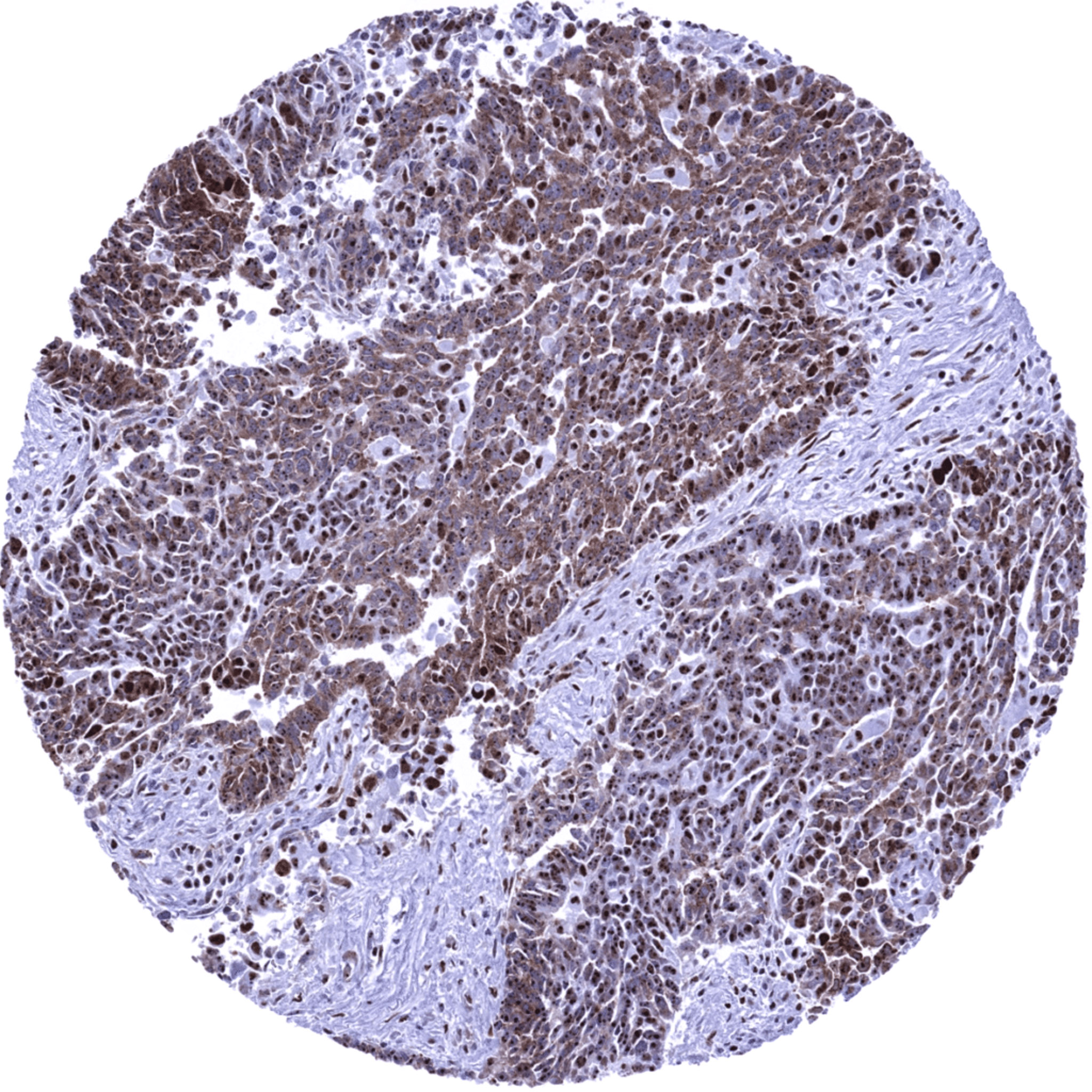
Urinary bladder- Muscle-invasive urothelial carcinoma showing strong nuclear nucleolin positivity and a particularly strong staining intensity in nucleoli.
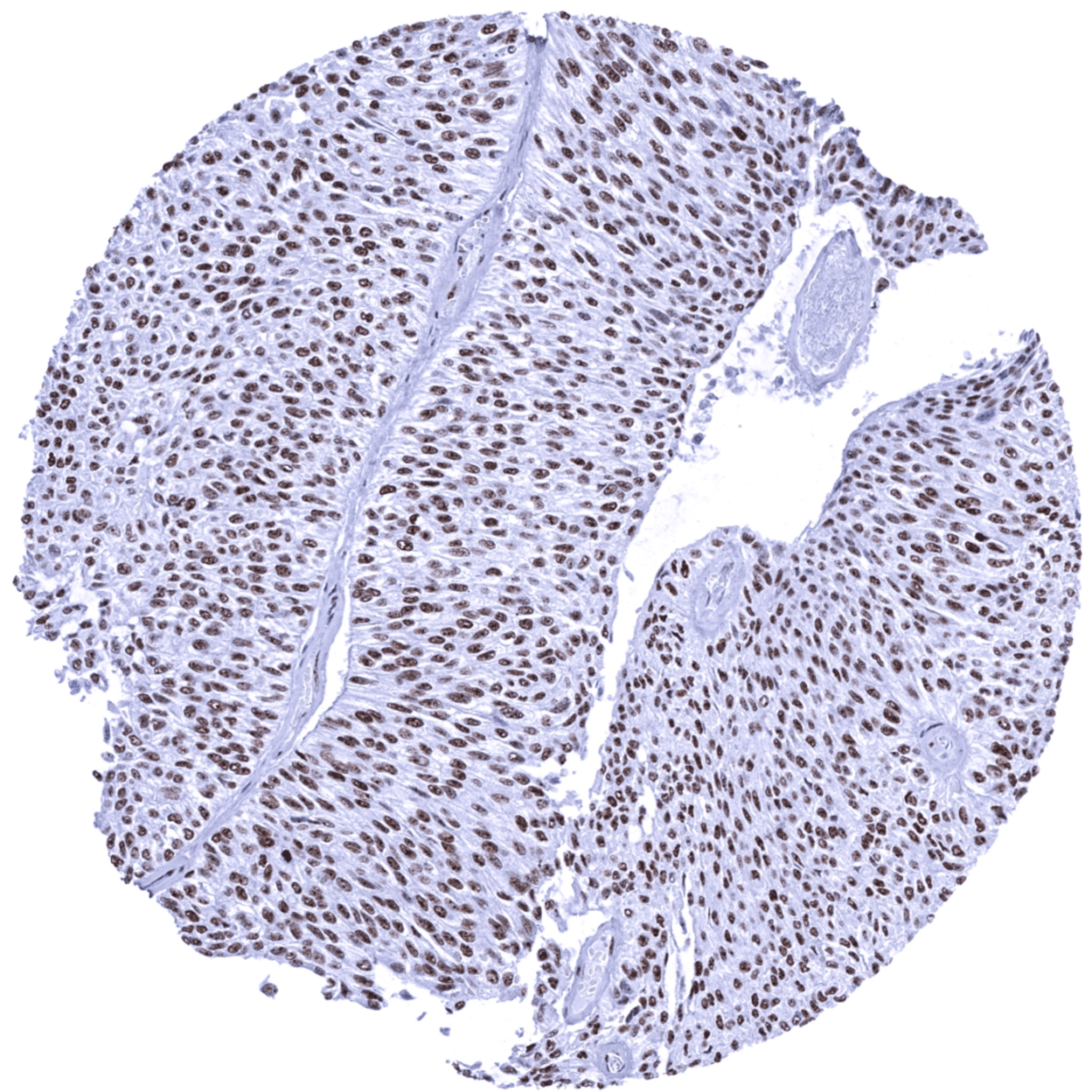
Urinary bladder- Non-invasive papillary urothelial carcinoma (pTa) showing a moderate to strong nucleolin immunostaining of nuclei.
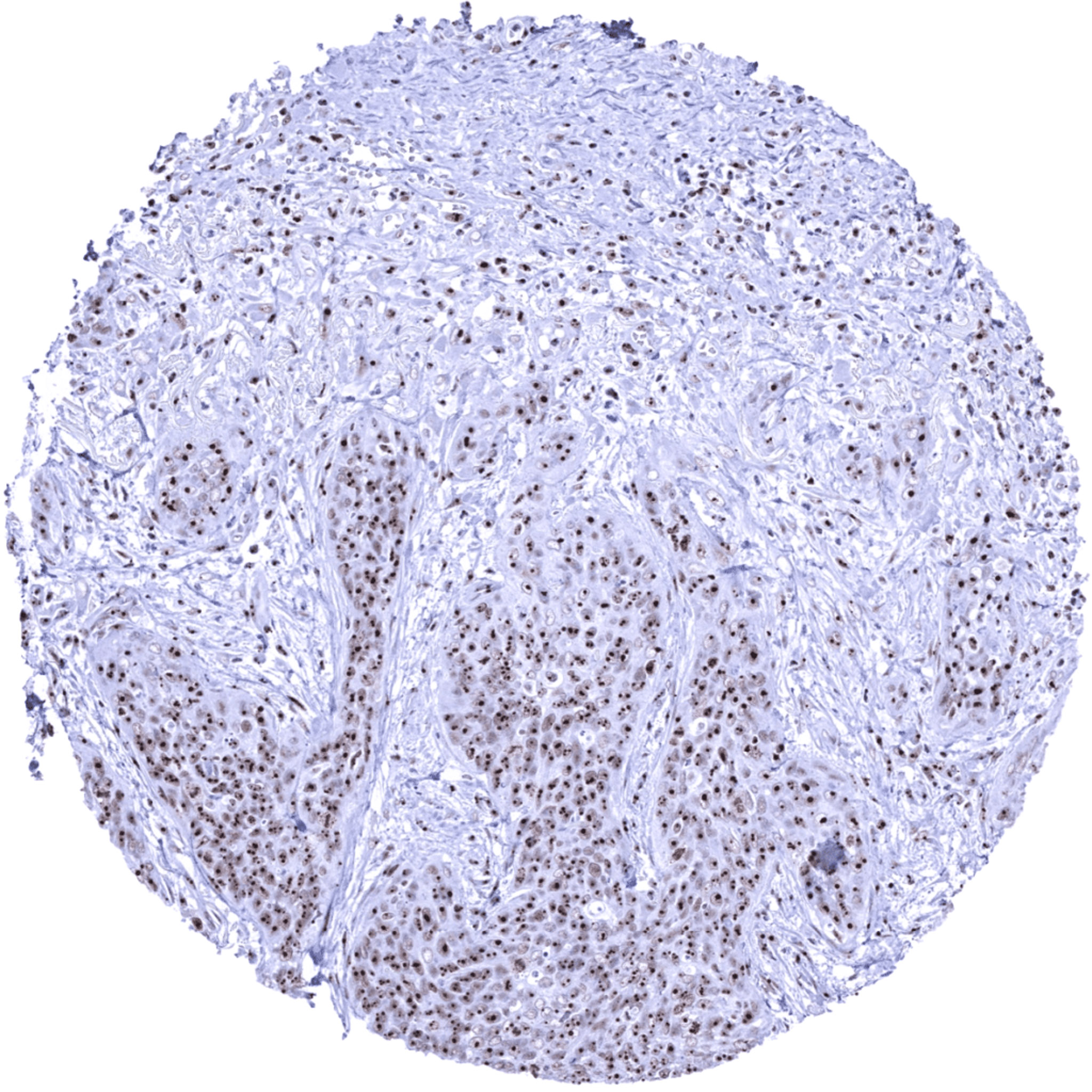
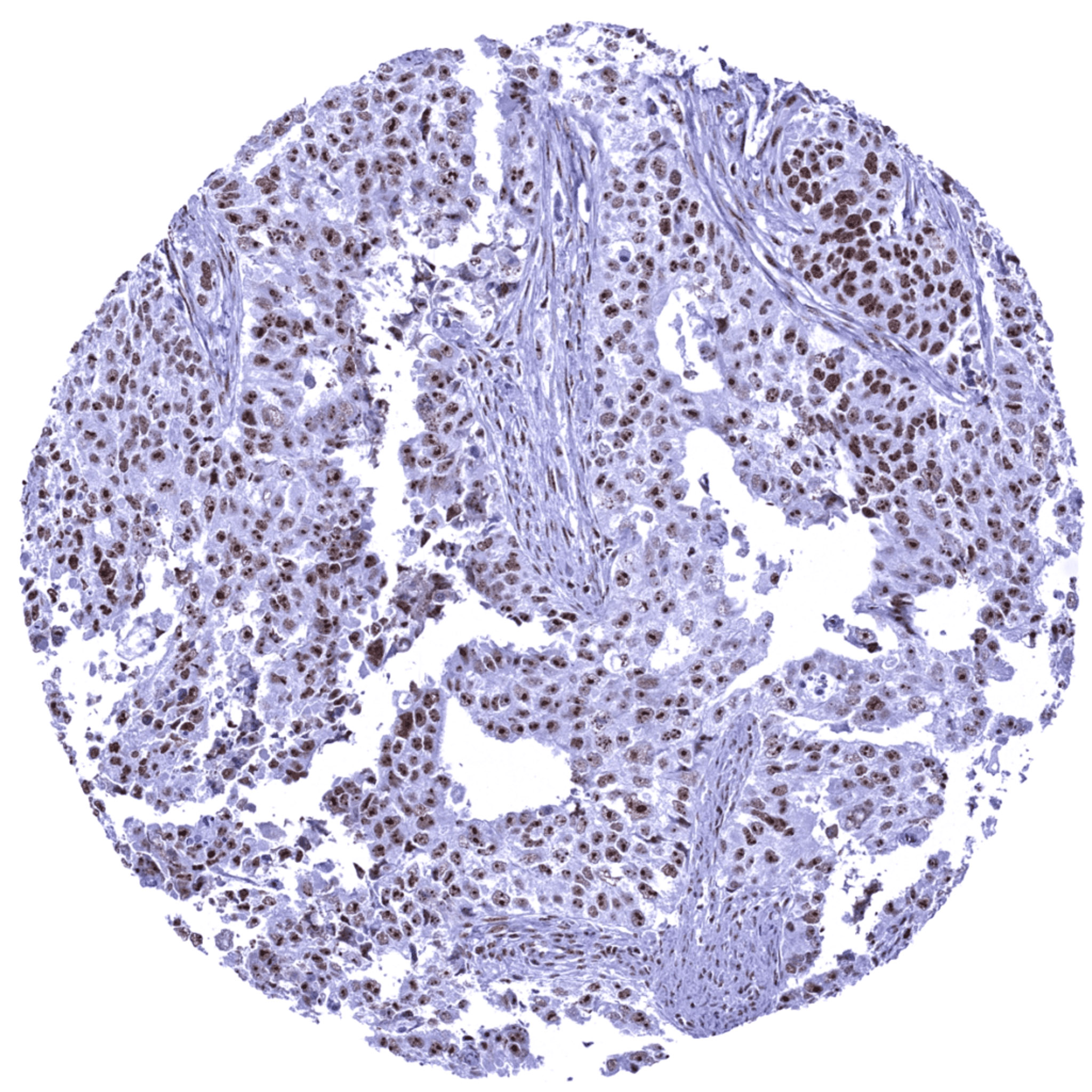
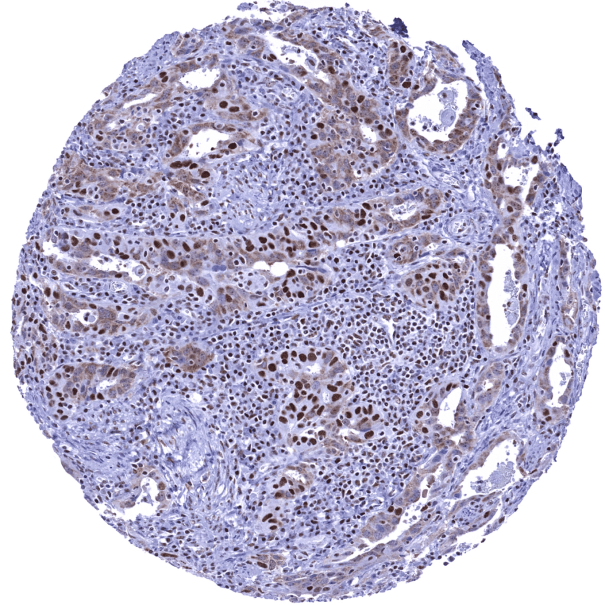
Uterus – Endometroid endometrium carcinoma showing a heterogeneous pattern of nucleolin immunostaining. Cells with predominant cytoplasmic staining make up for the majority. They are intermingled with cells showing purely nuclear staining. Pattern may be biologically relevant or represent an artifact.
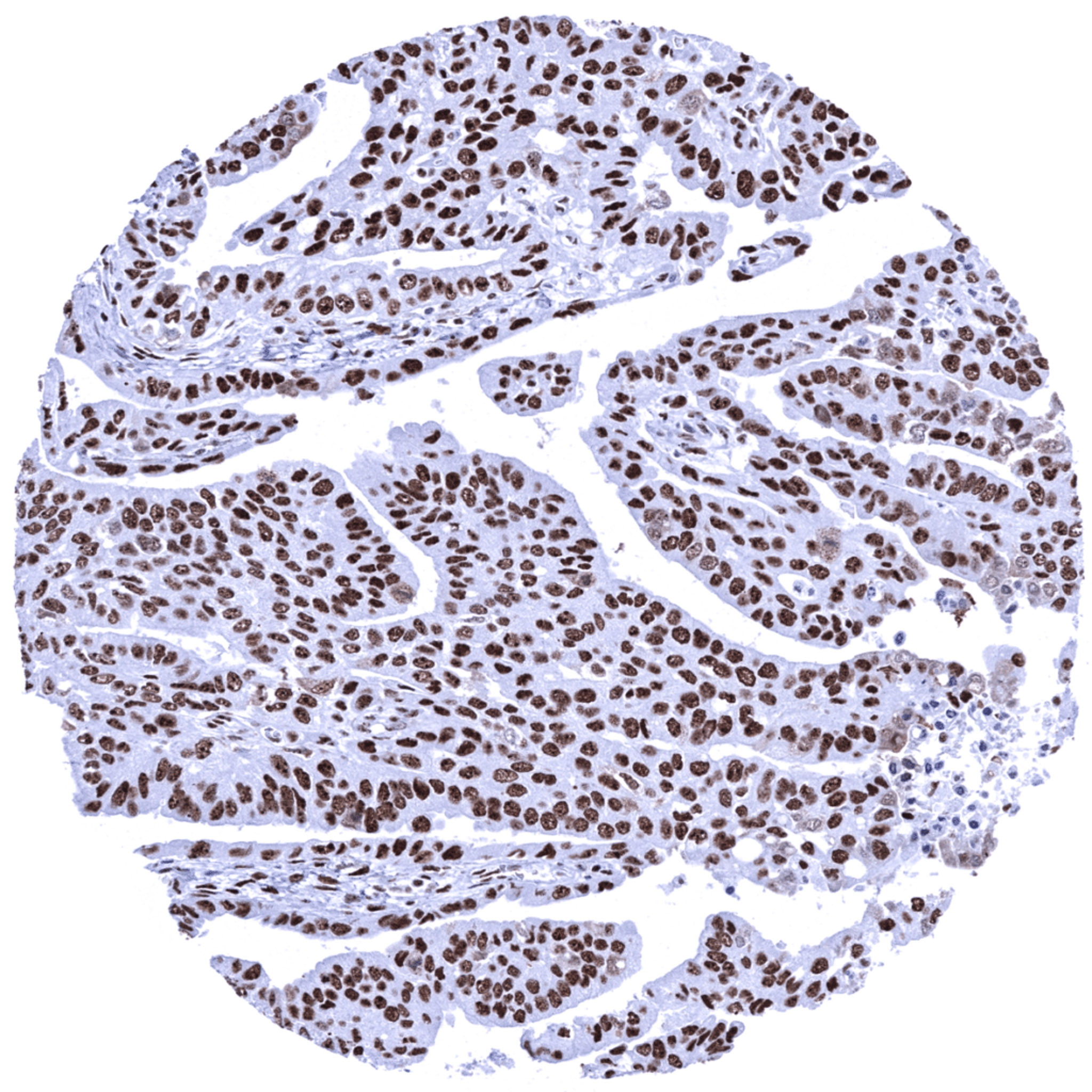
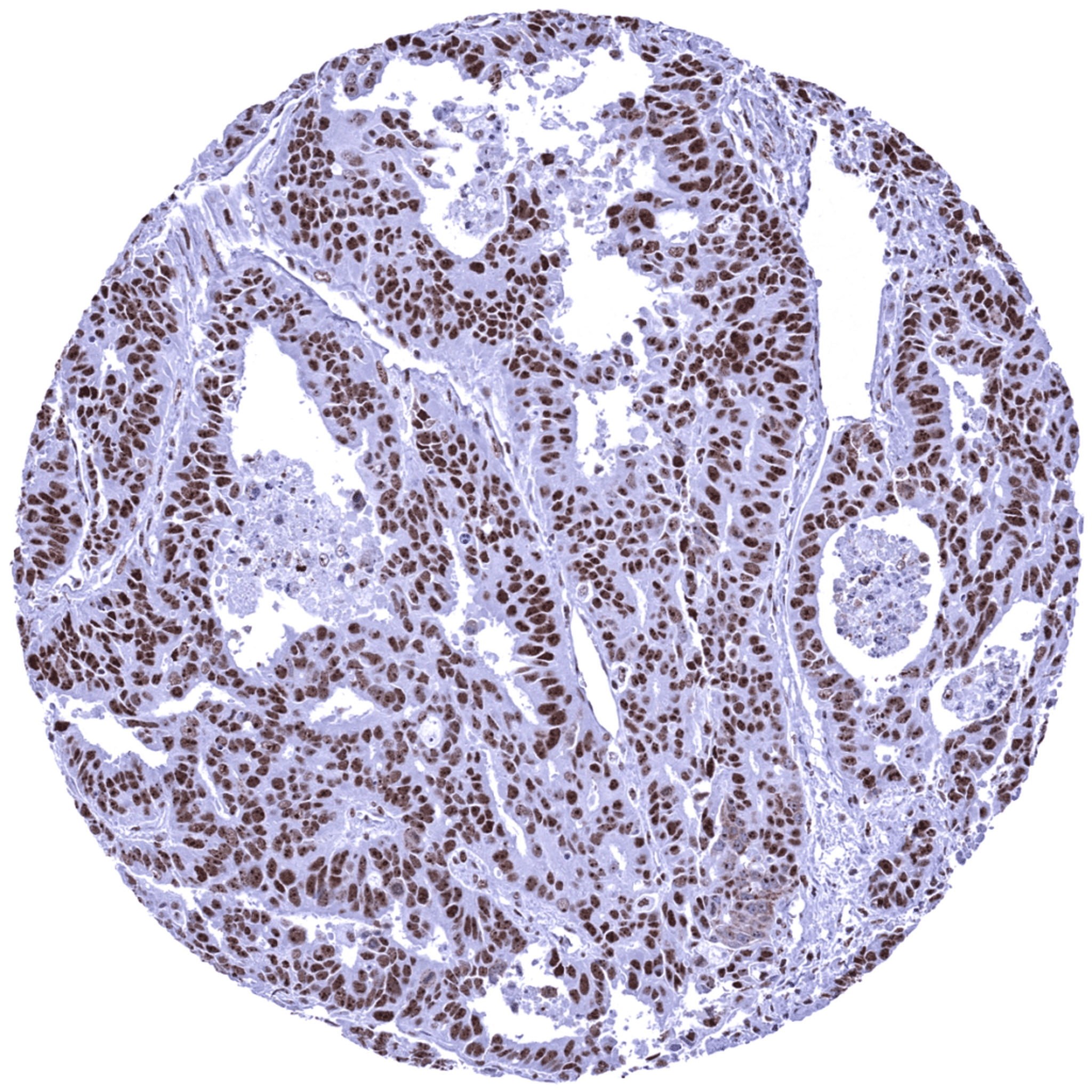
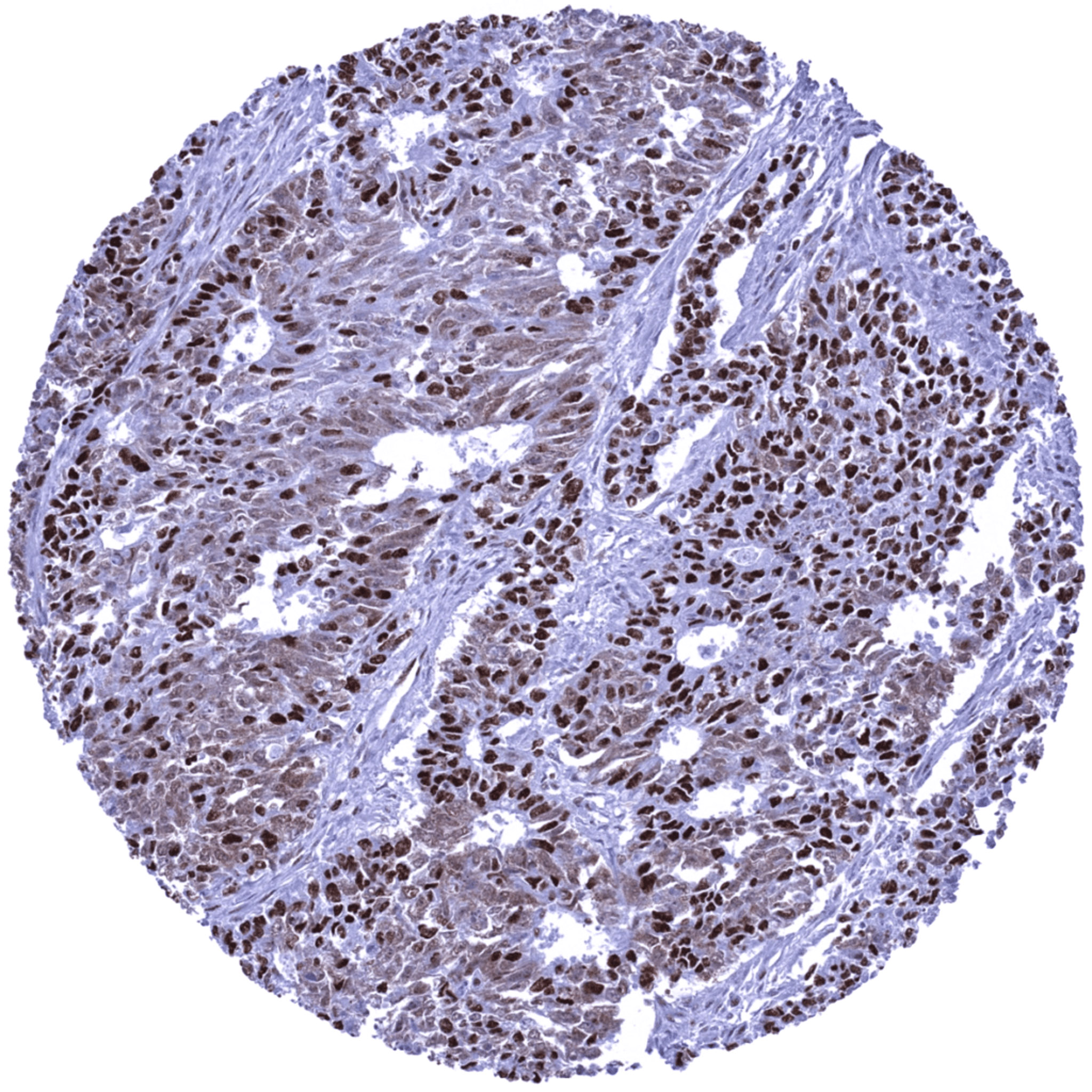
Uterus- Endometroid endometrium carcinoma with strong nuclear nucleolin staining.
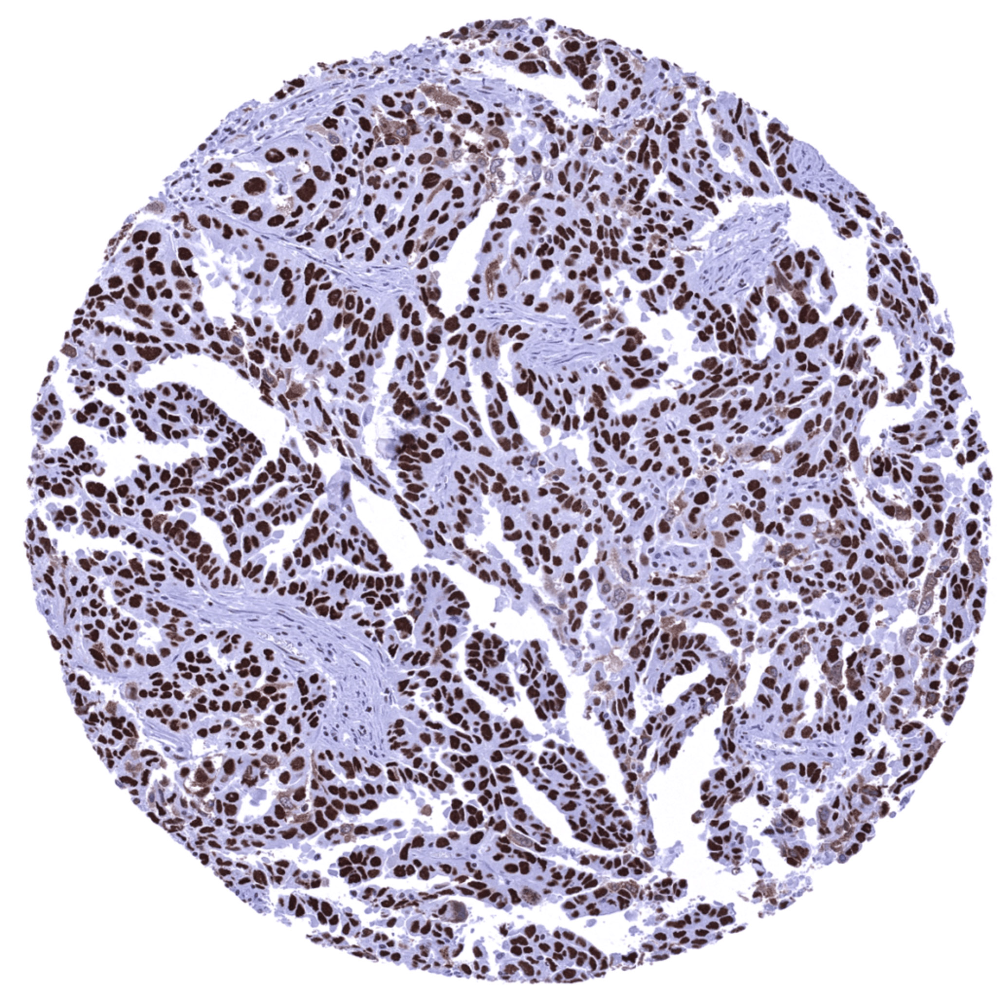
Uterus- Serous endometrium carcinoma showing a strong nuclear nucleolin positivity with a particularly strong staining intensity in nucleoli.
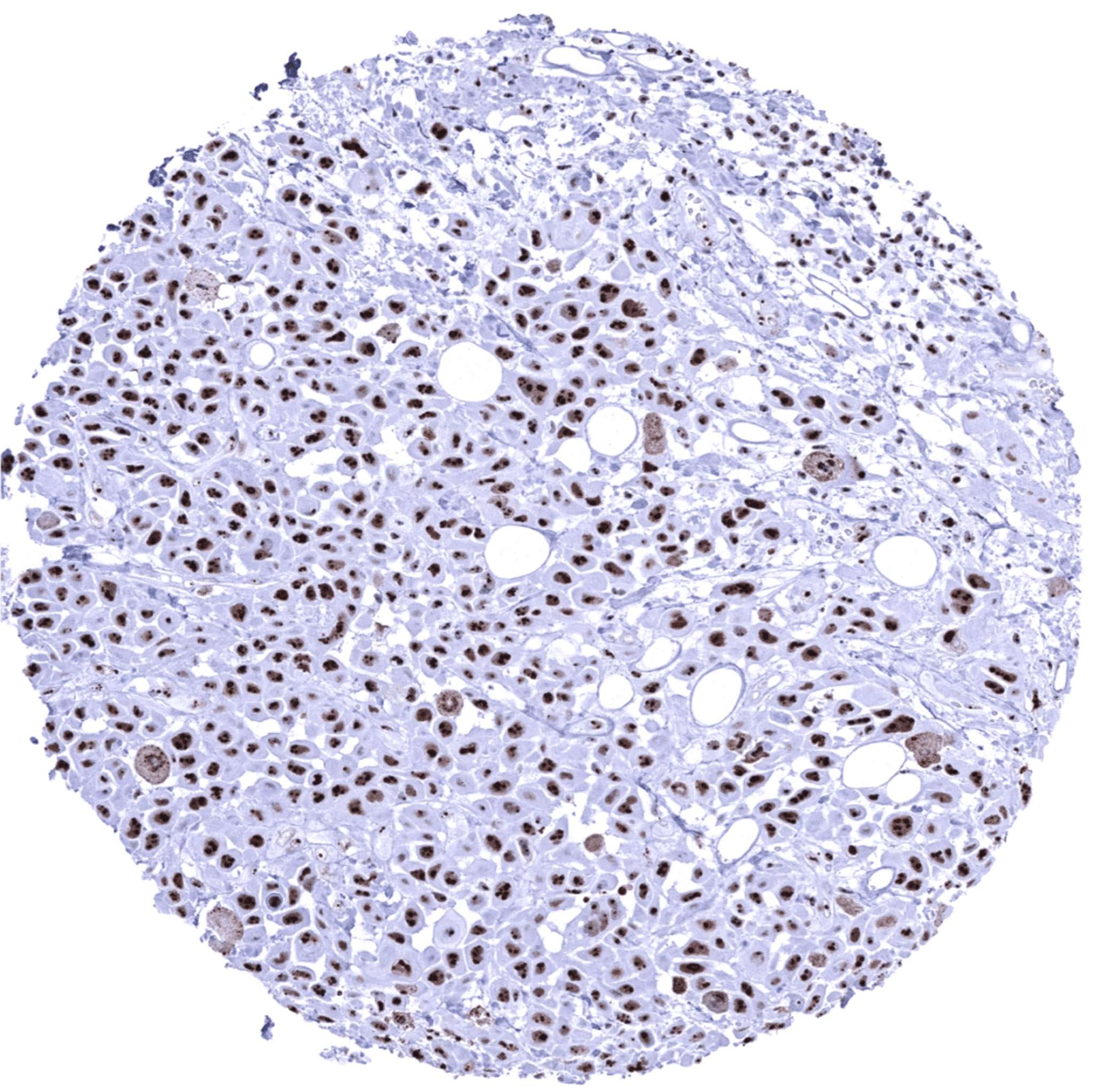
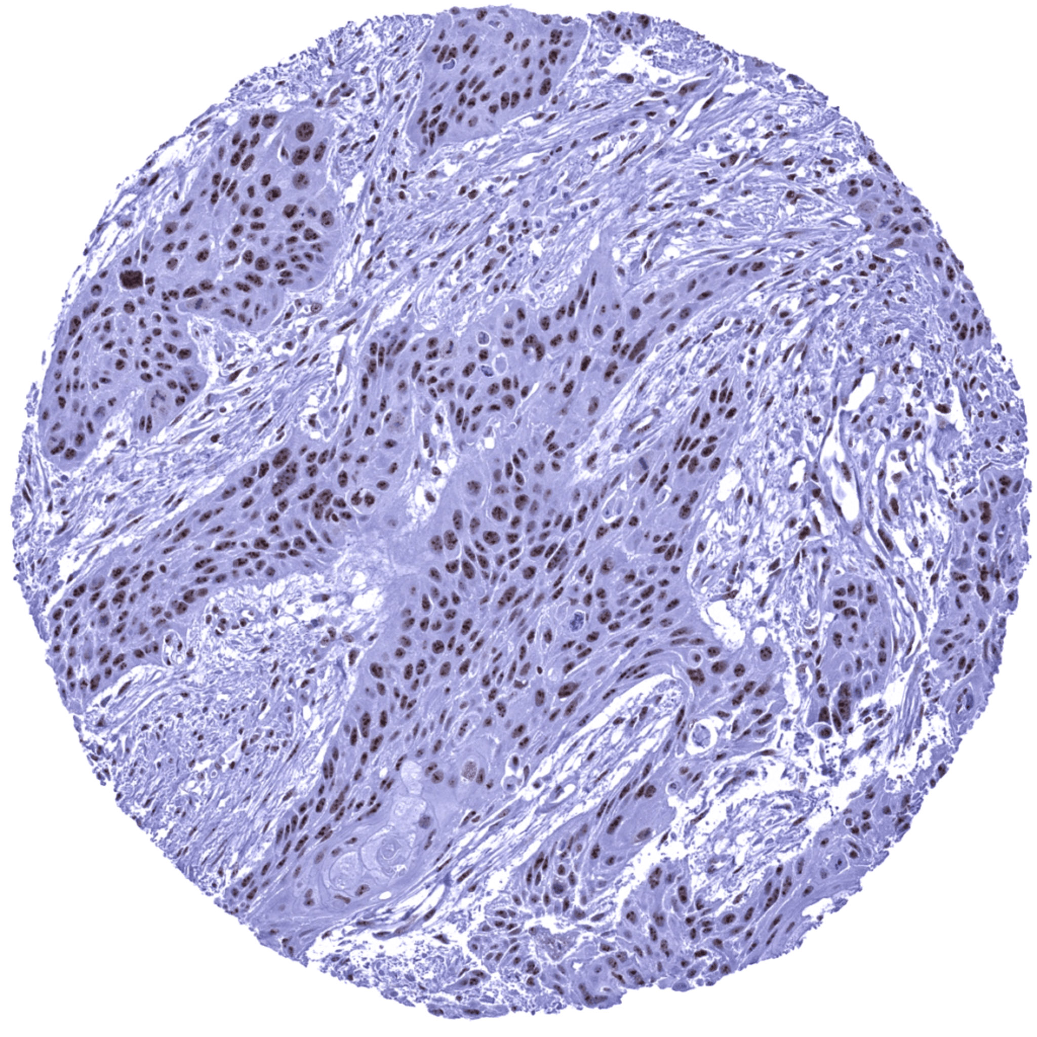
Uterus, cervix – Adenocarcinoma showing a heterogeneous nucleolin immunostaining pattern. Cells with purely nuclear staining are intermingled with cells showing little nuclear but predominant cytoplasmic staining. Pattern may be biologically relevant or represent an artifact.
Uterus, cervix – Adenocarcinoma showing a heterogeneous pattern for nucleolin immunostaining. Cells with purely nuclear staining are intermingled with cells showing little nuclear but predominant cytoplasmic staining. Pattern may be biologically relevant or represent an artifact.
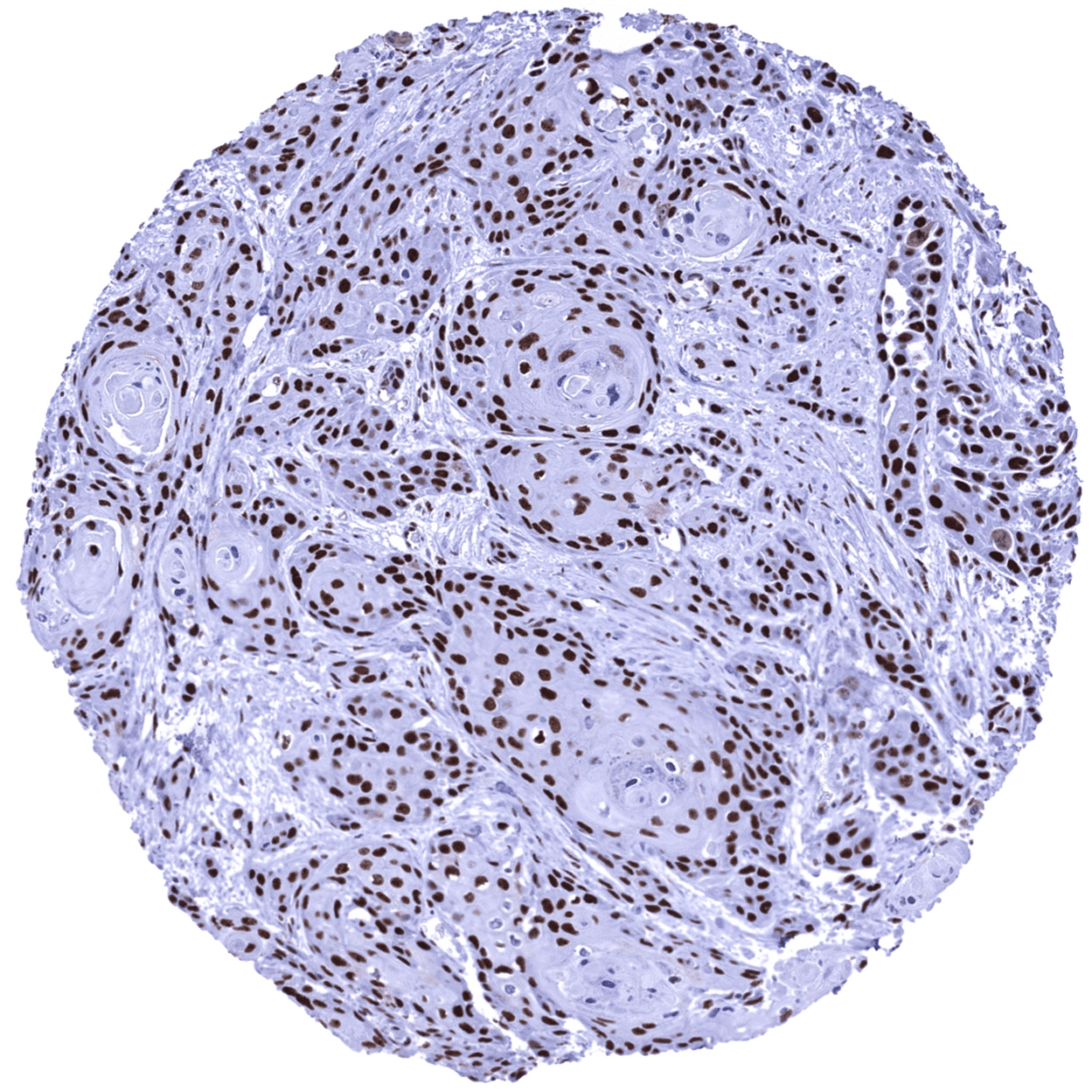
Uterus, cervix- Squamous cell carcinoma showing a strong nuclear positivity for nucleolin.
Vulva- Squamous cell carcinoma showing strong nuclear nucleolin positivity with a particularly strong staining intensity in the nucleoli.