GeneTex
  • Country / Location Selection

United States (US)

Normal Tissue Gallery- GTX04404

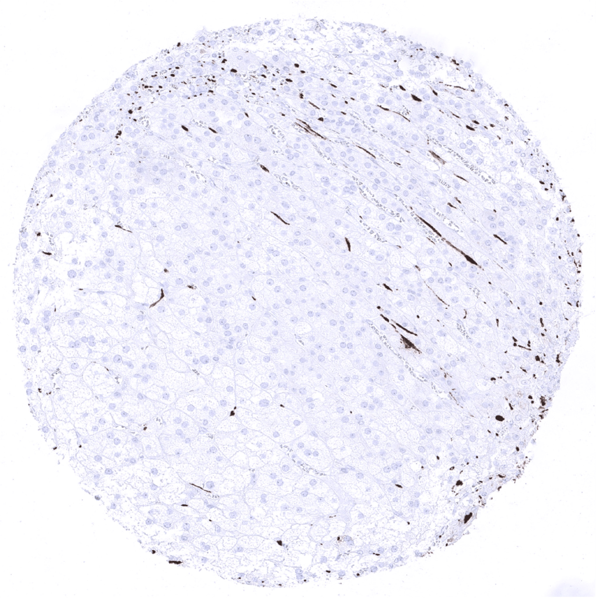
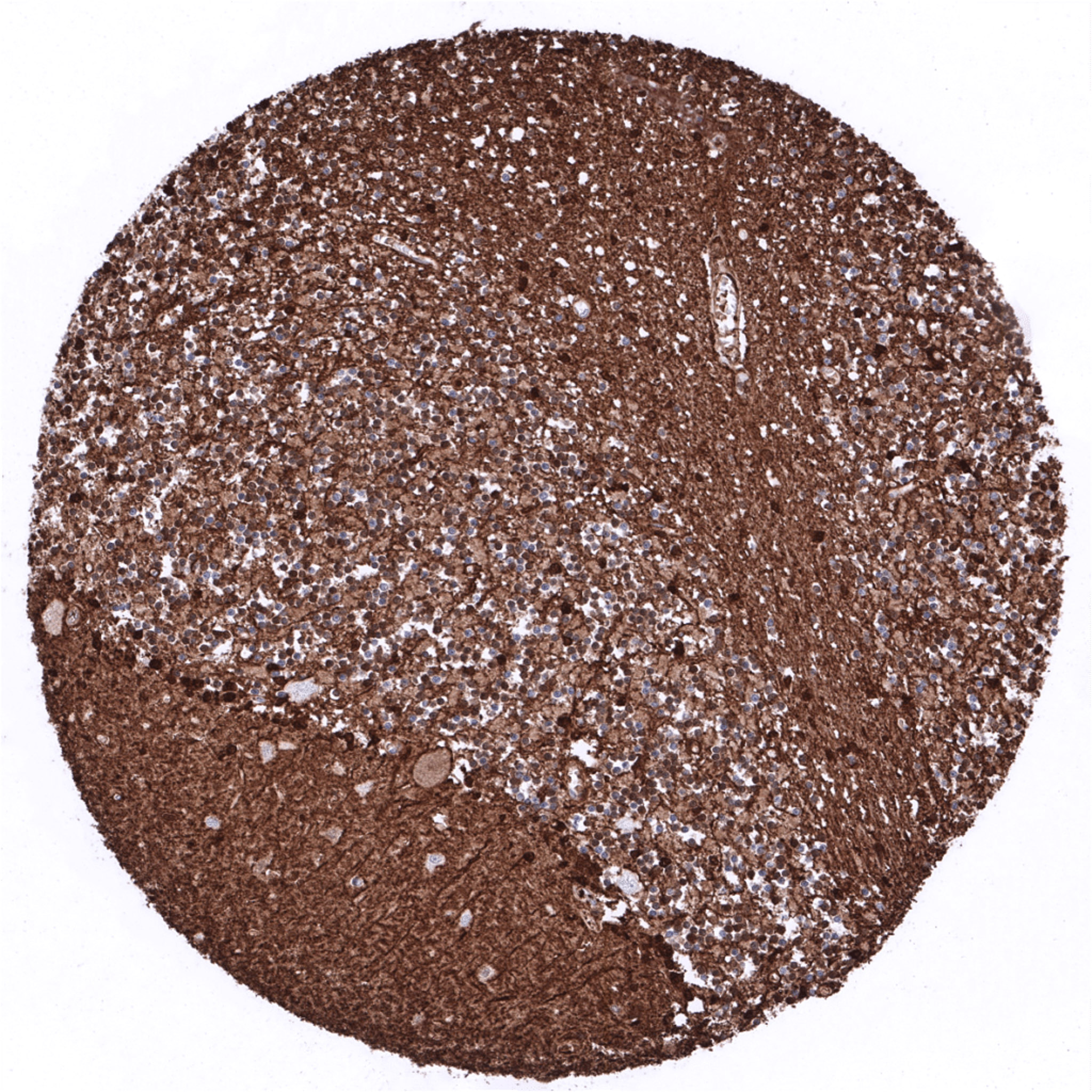
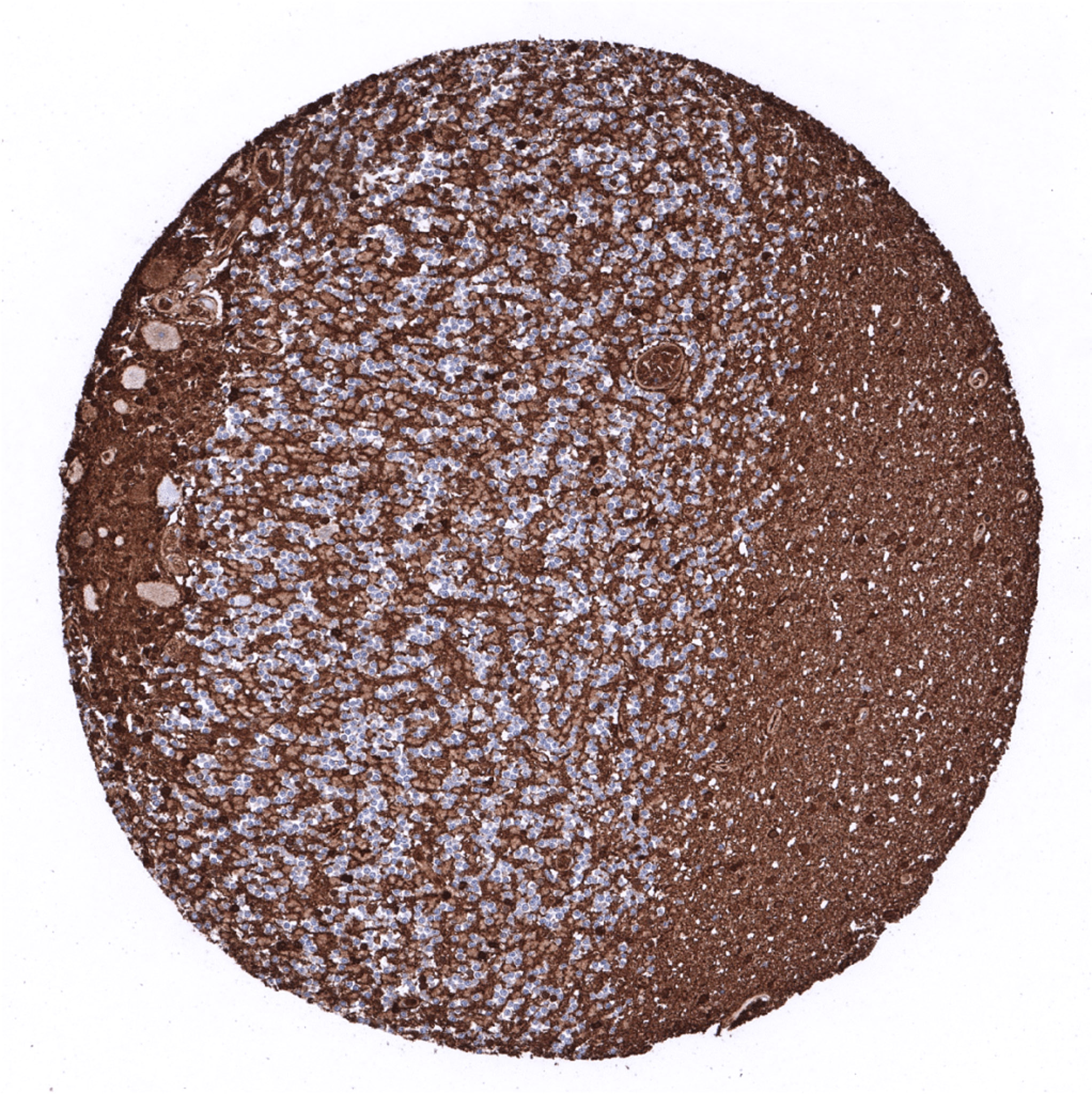
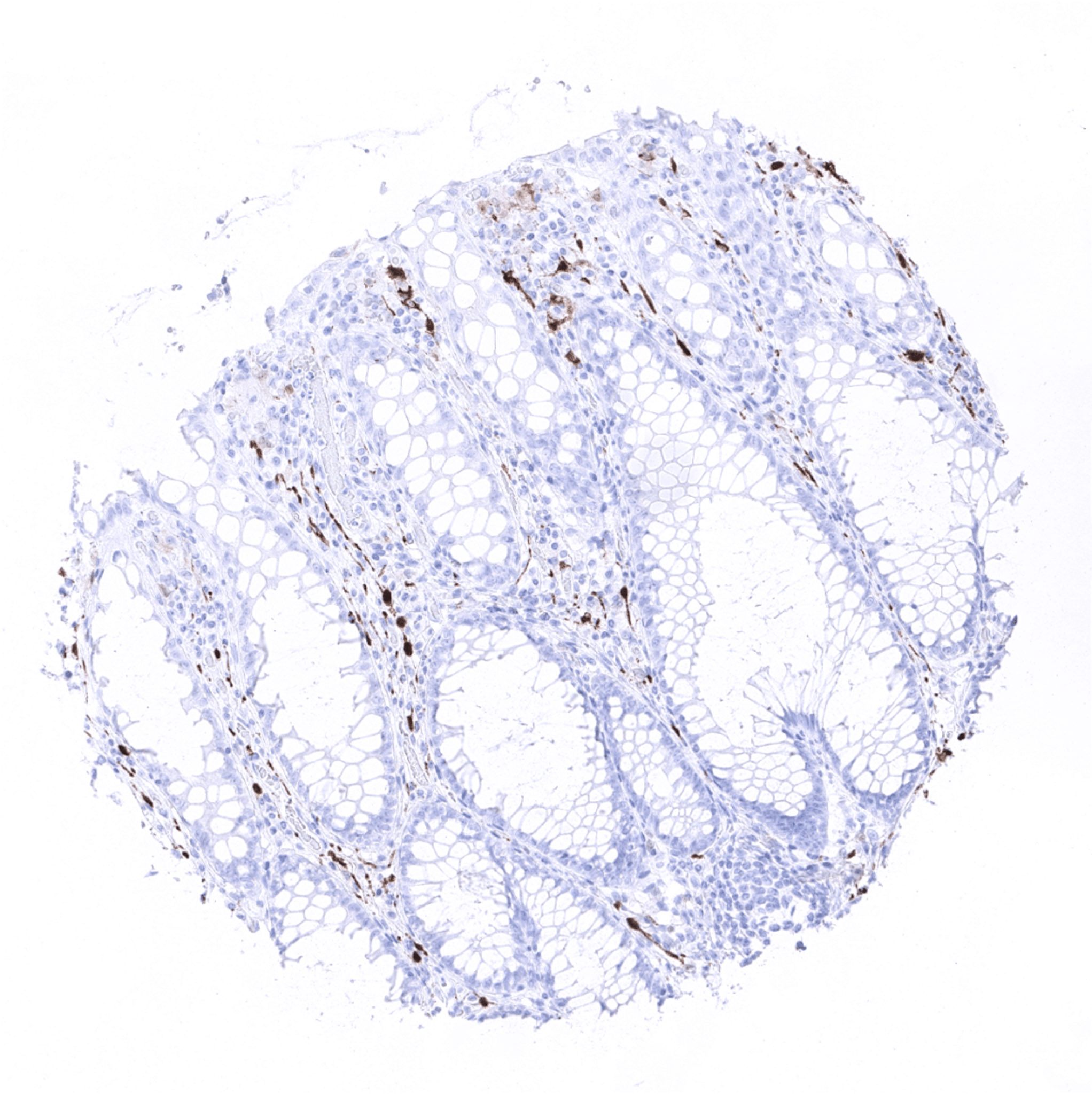
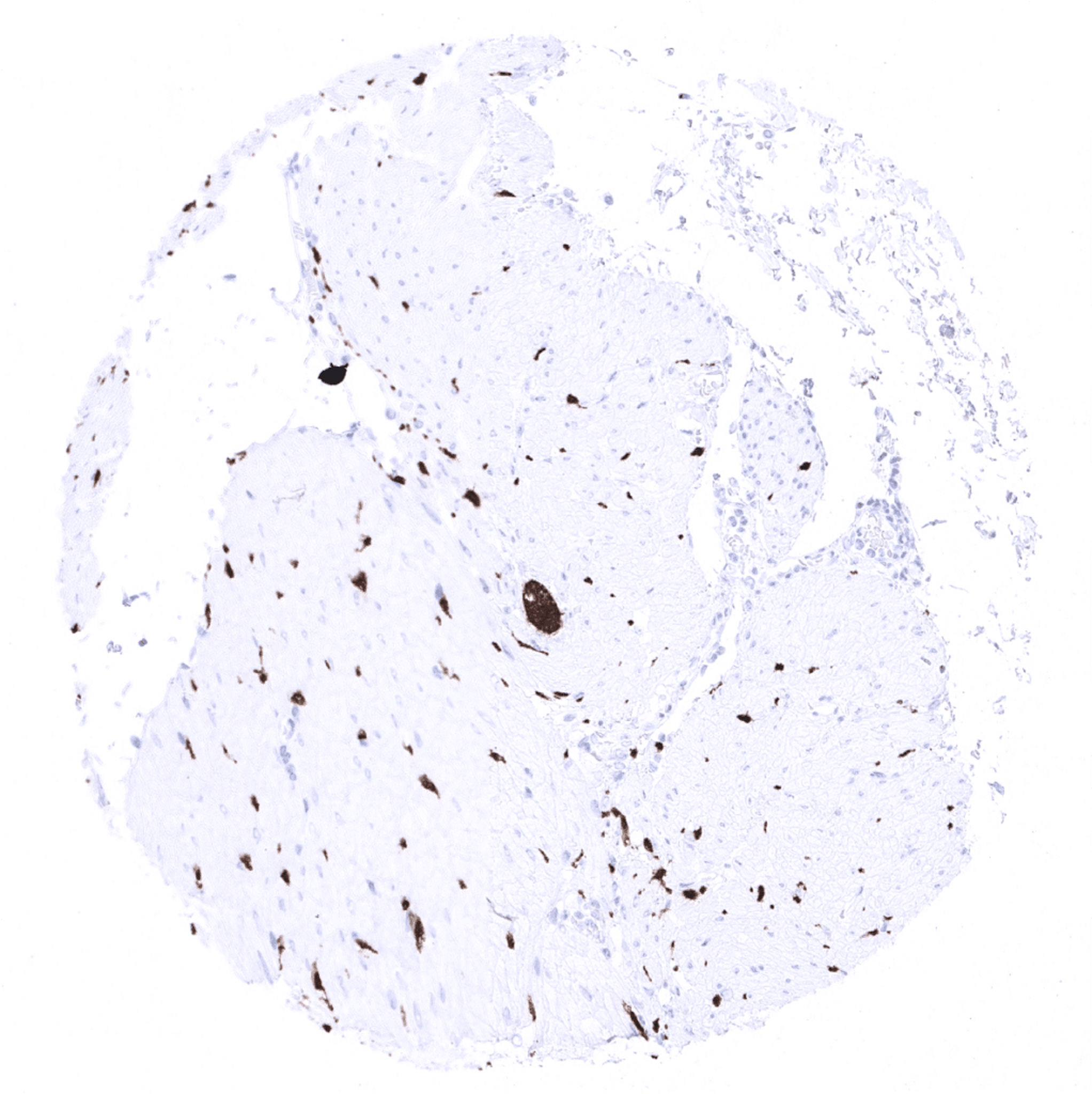
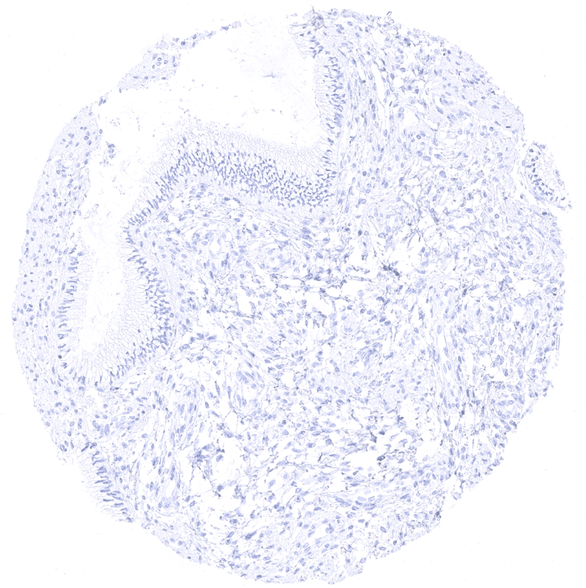
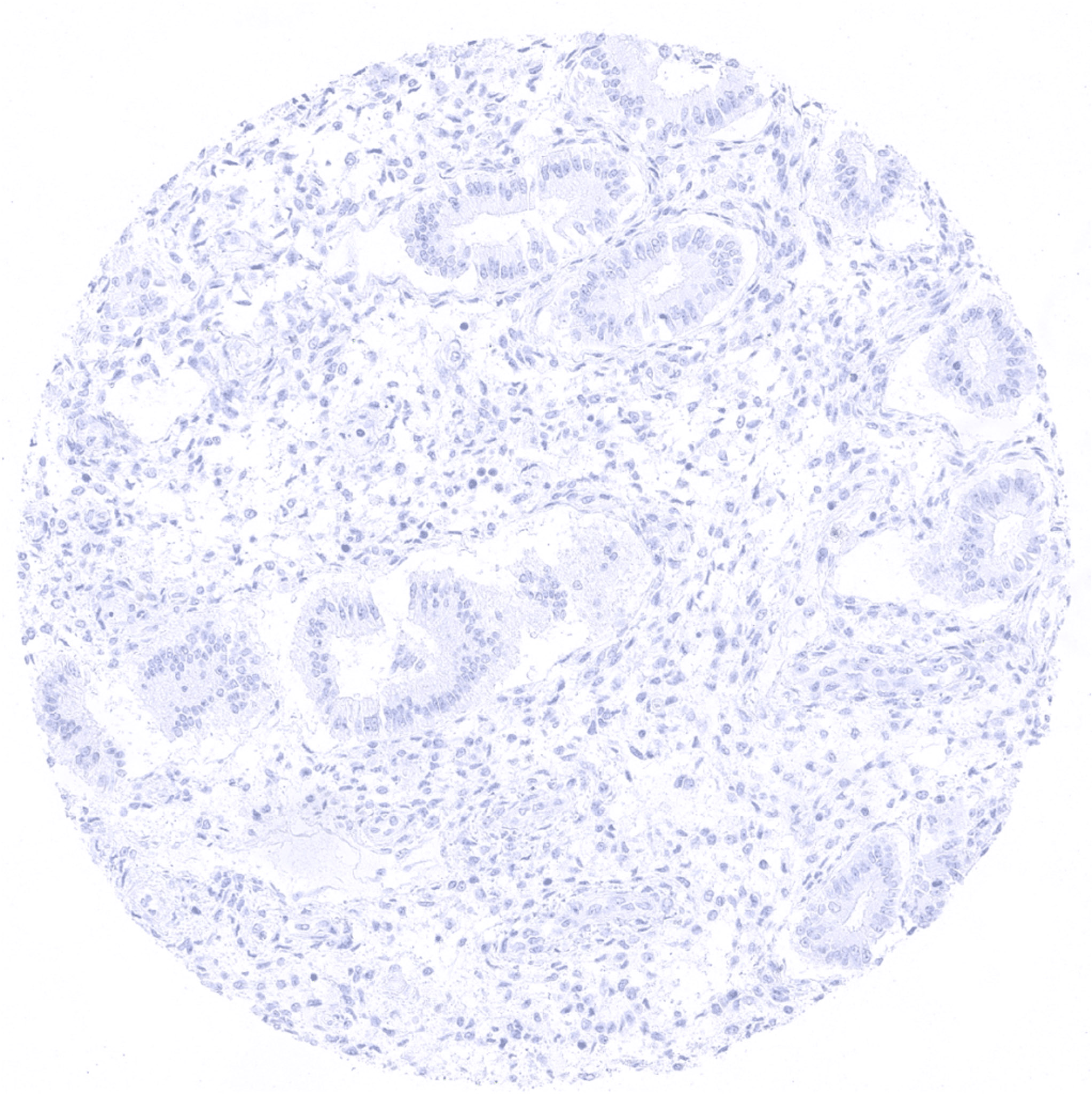
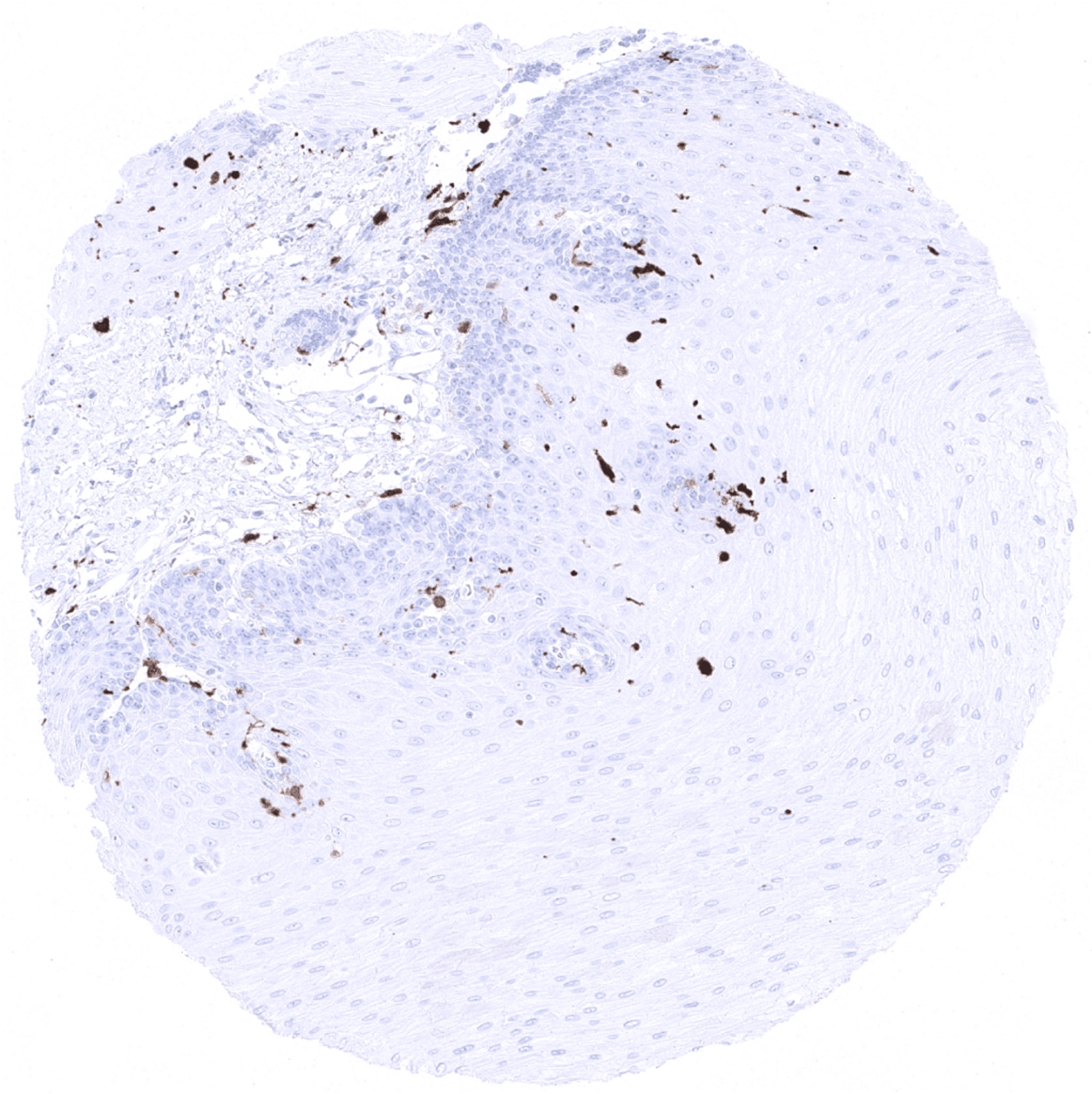
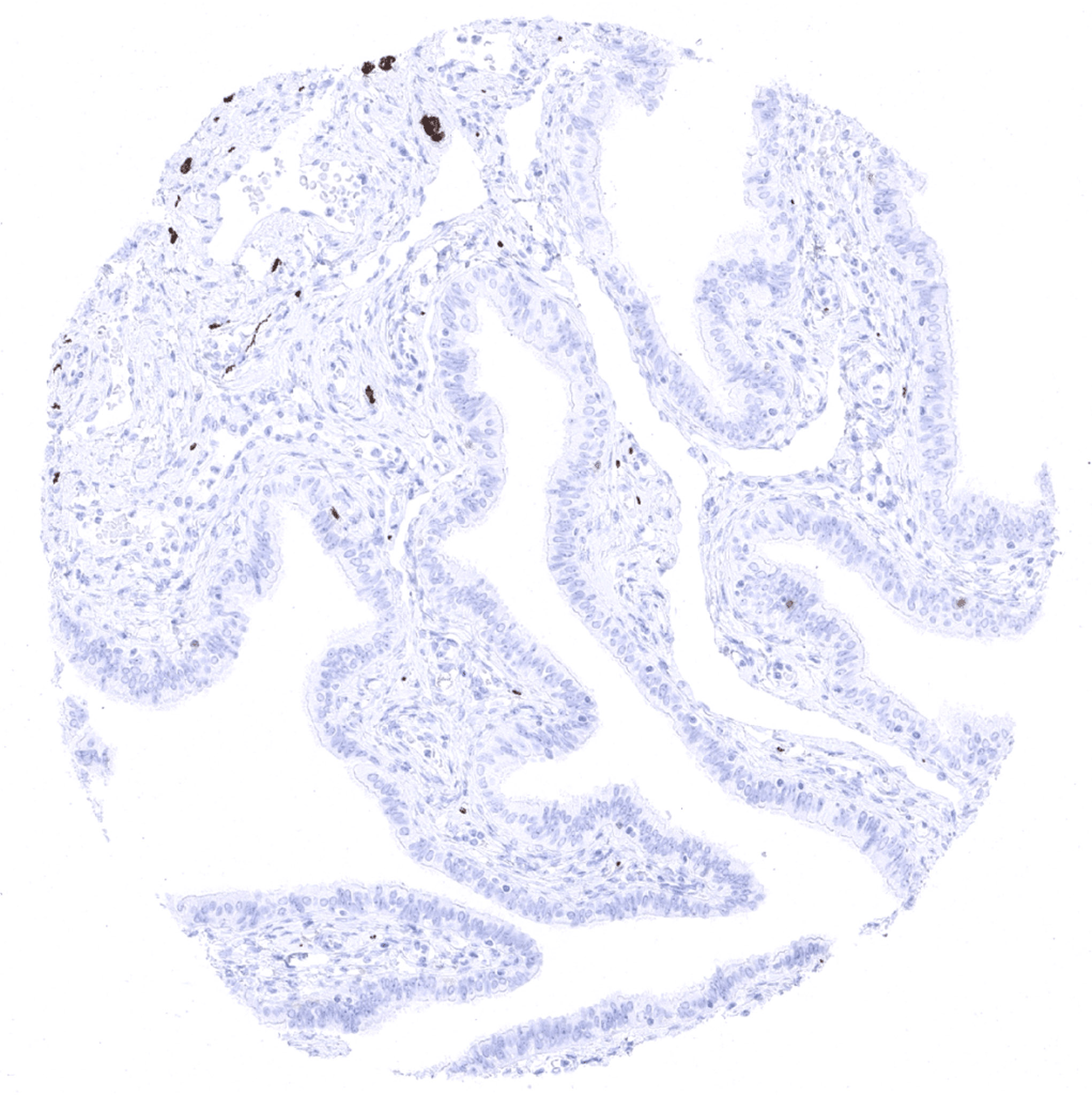
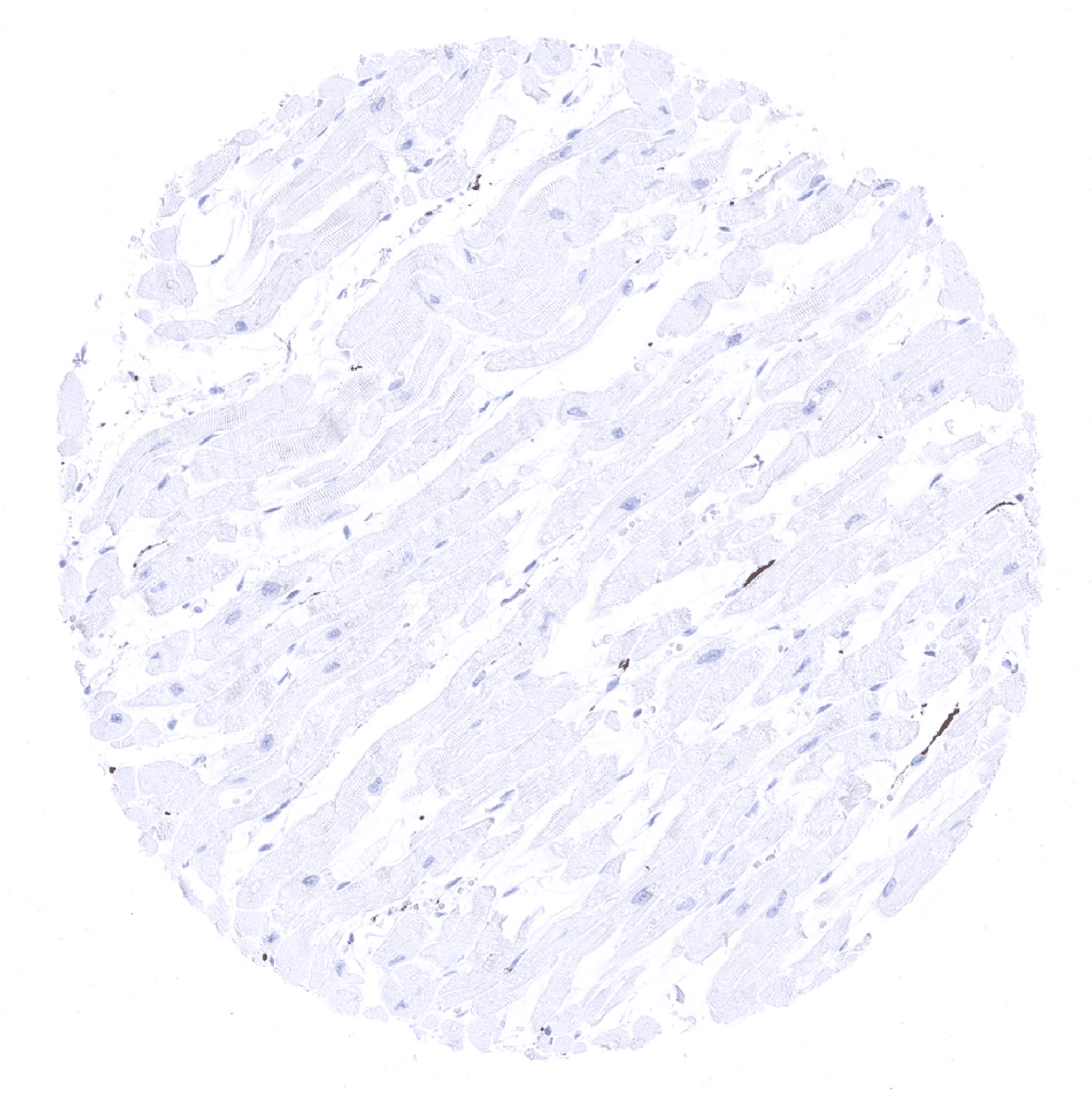
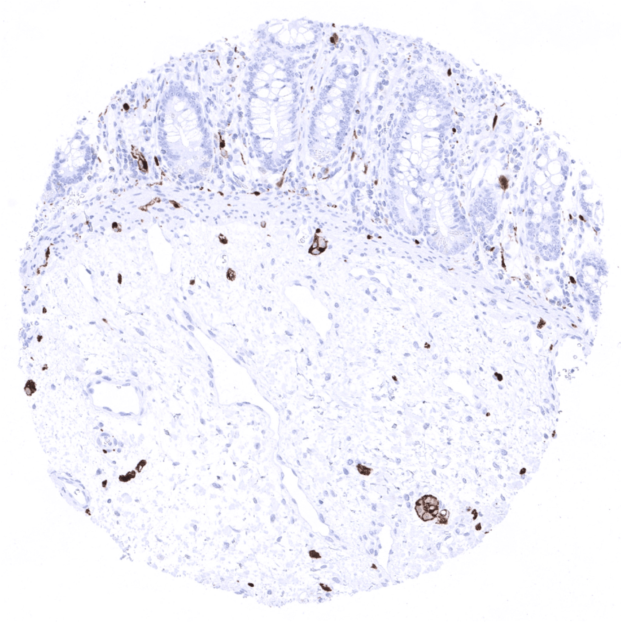
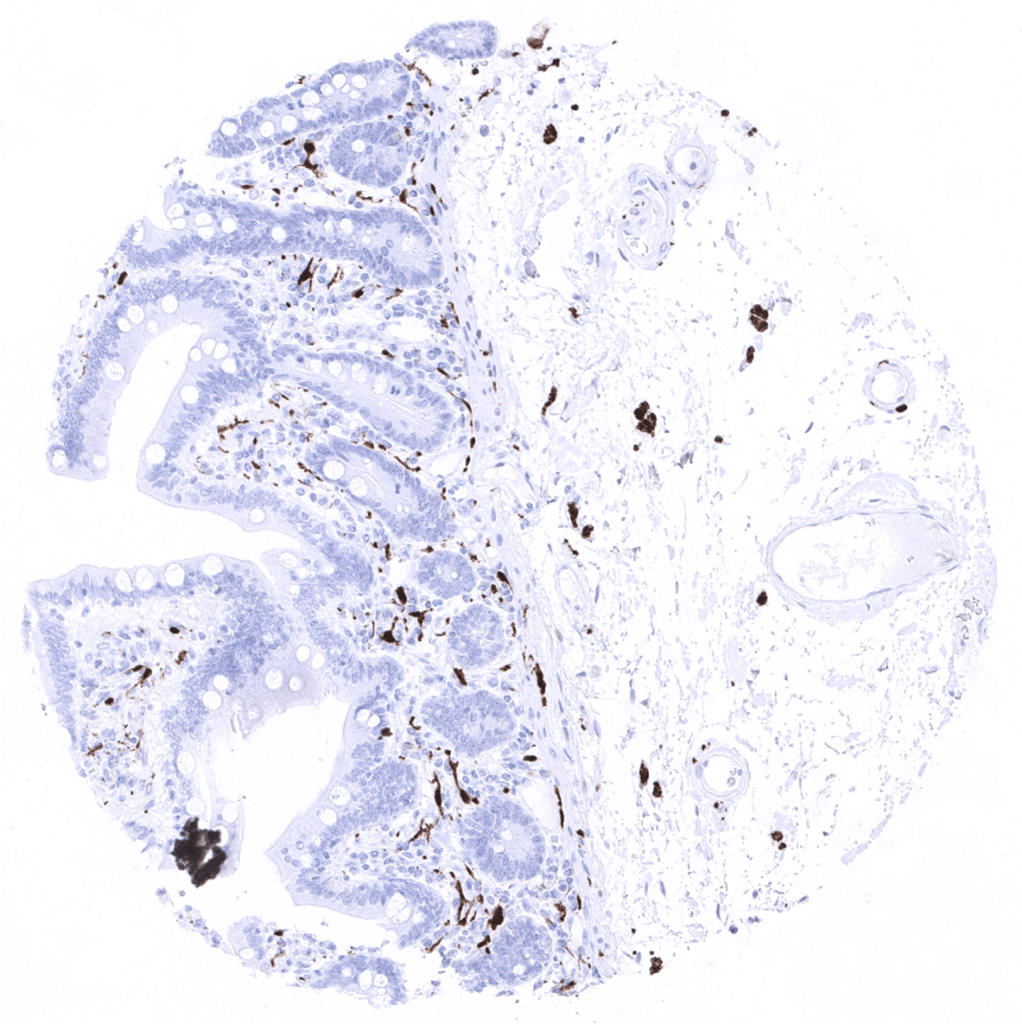
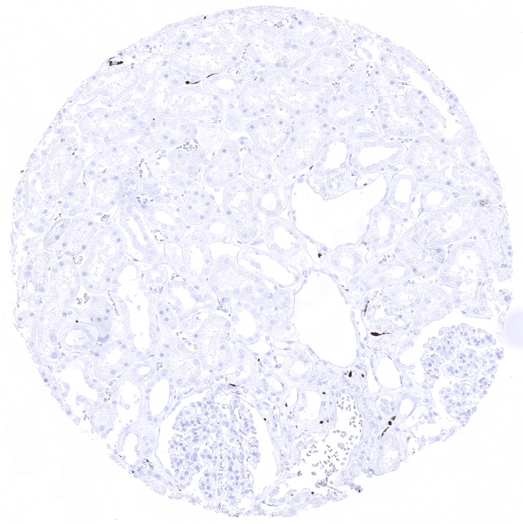
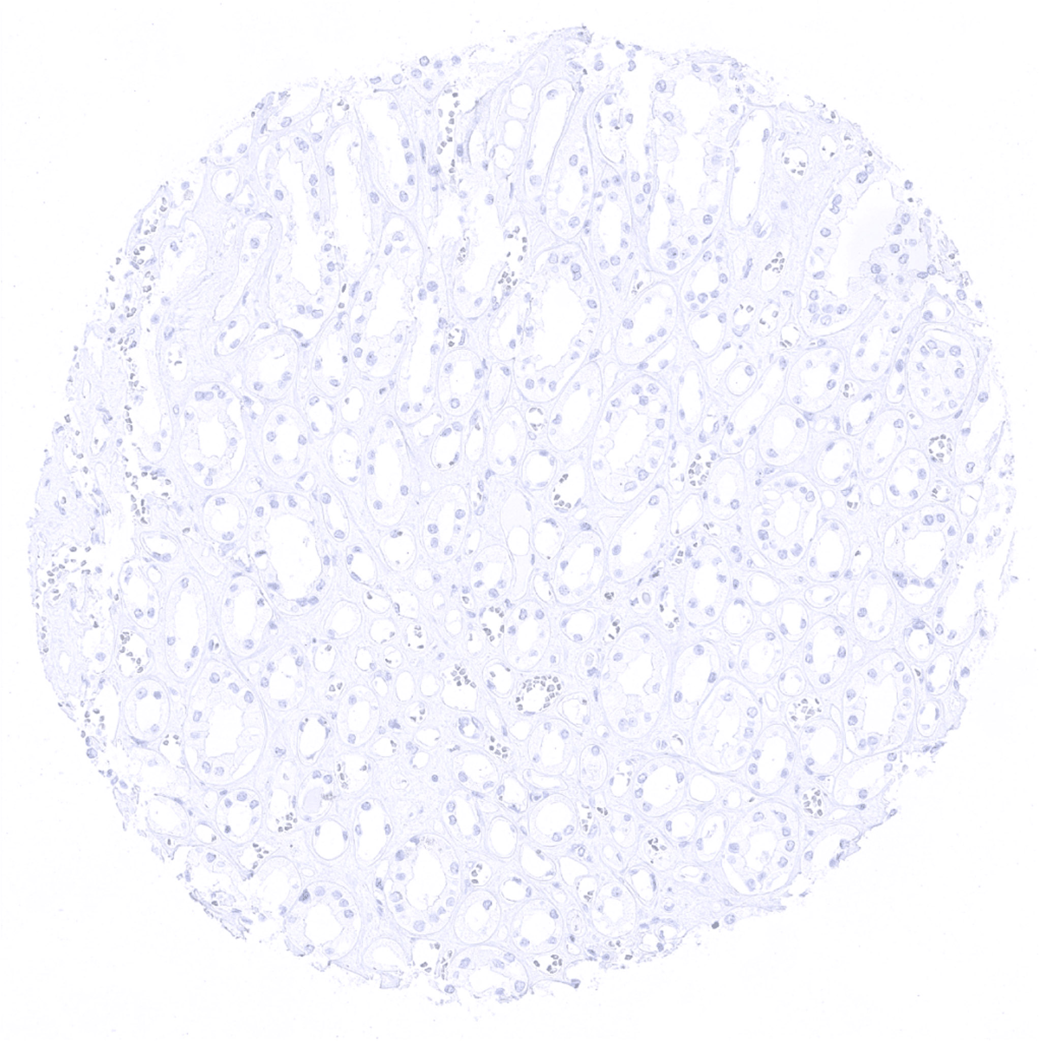
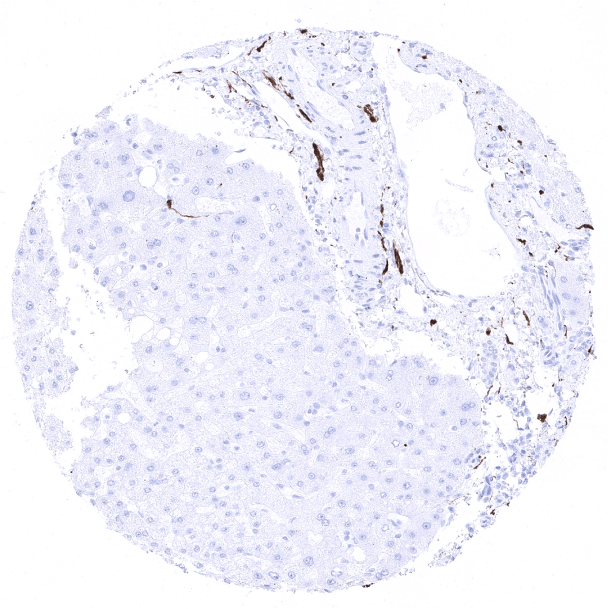
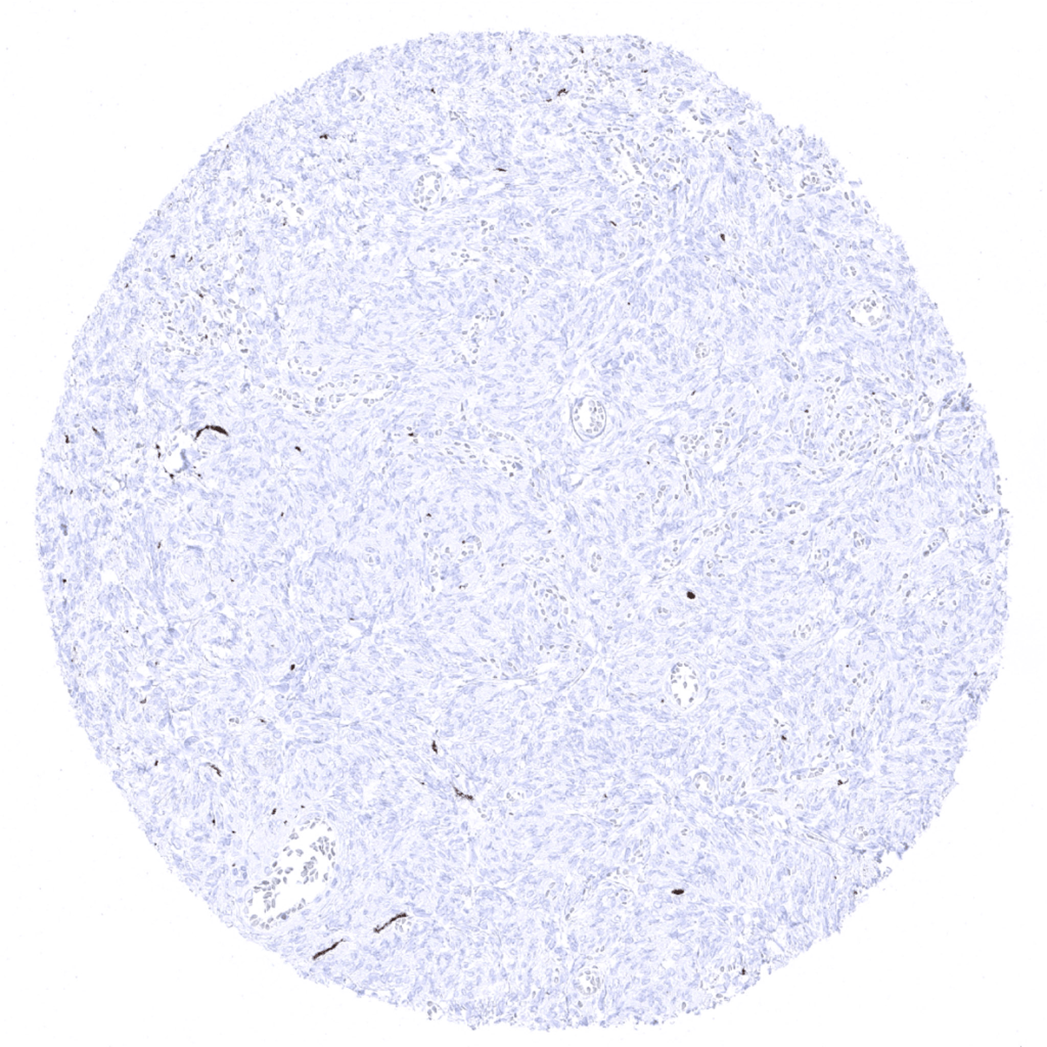
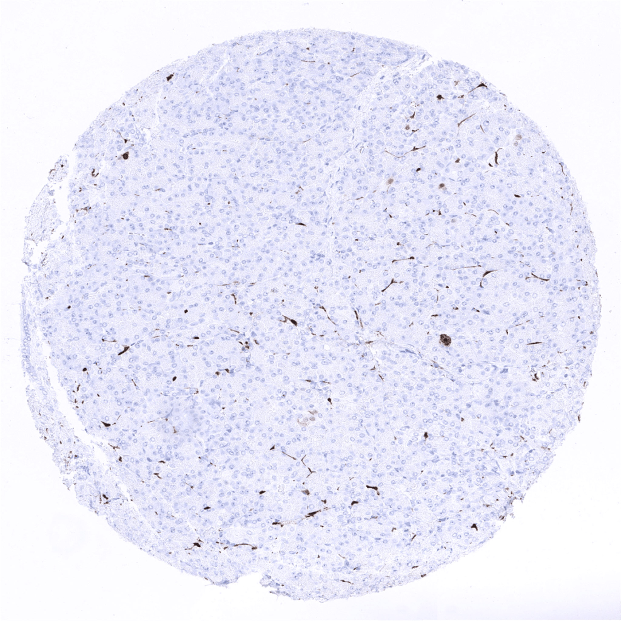
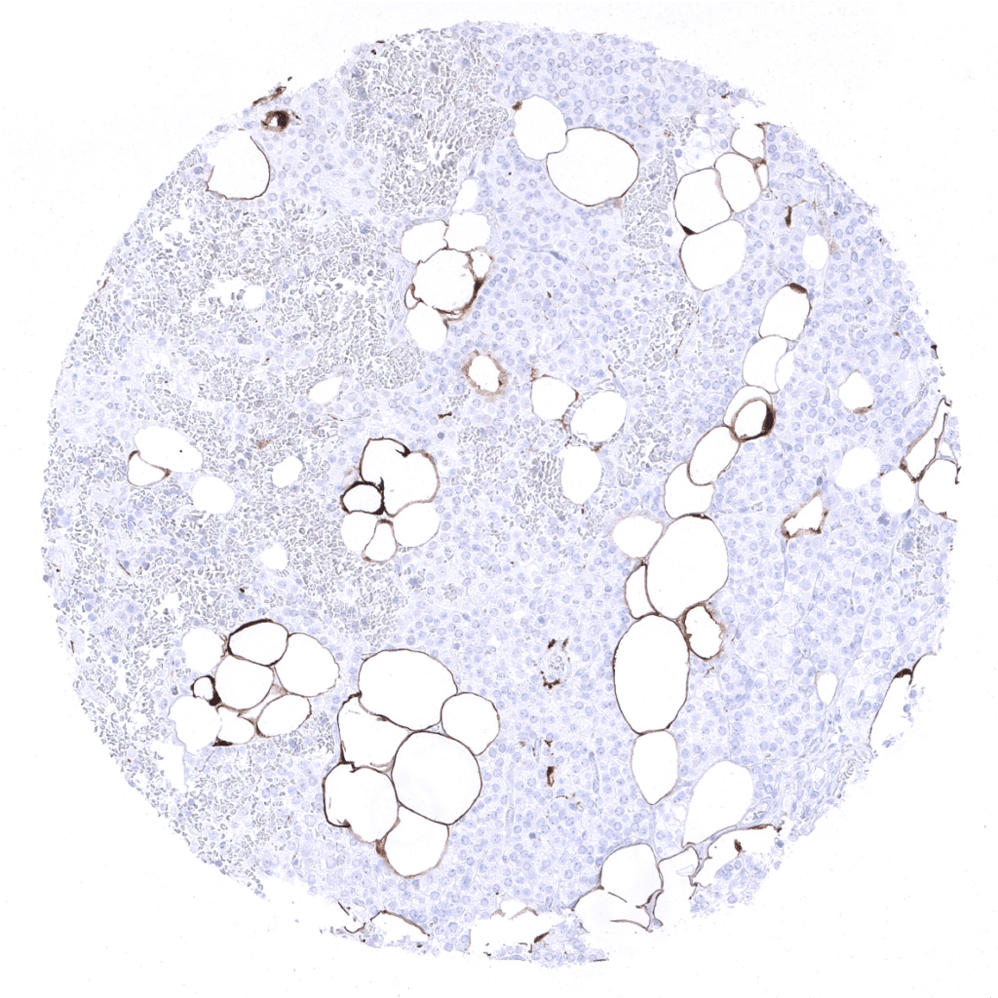
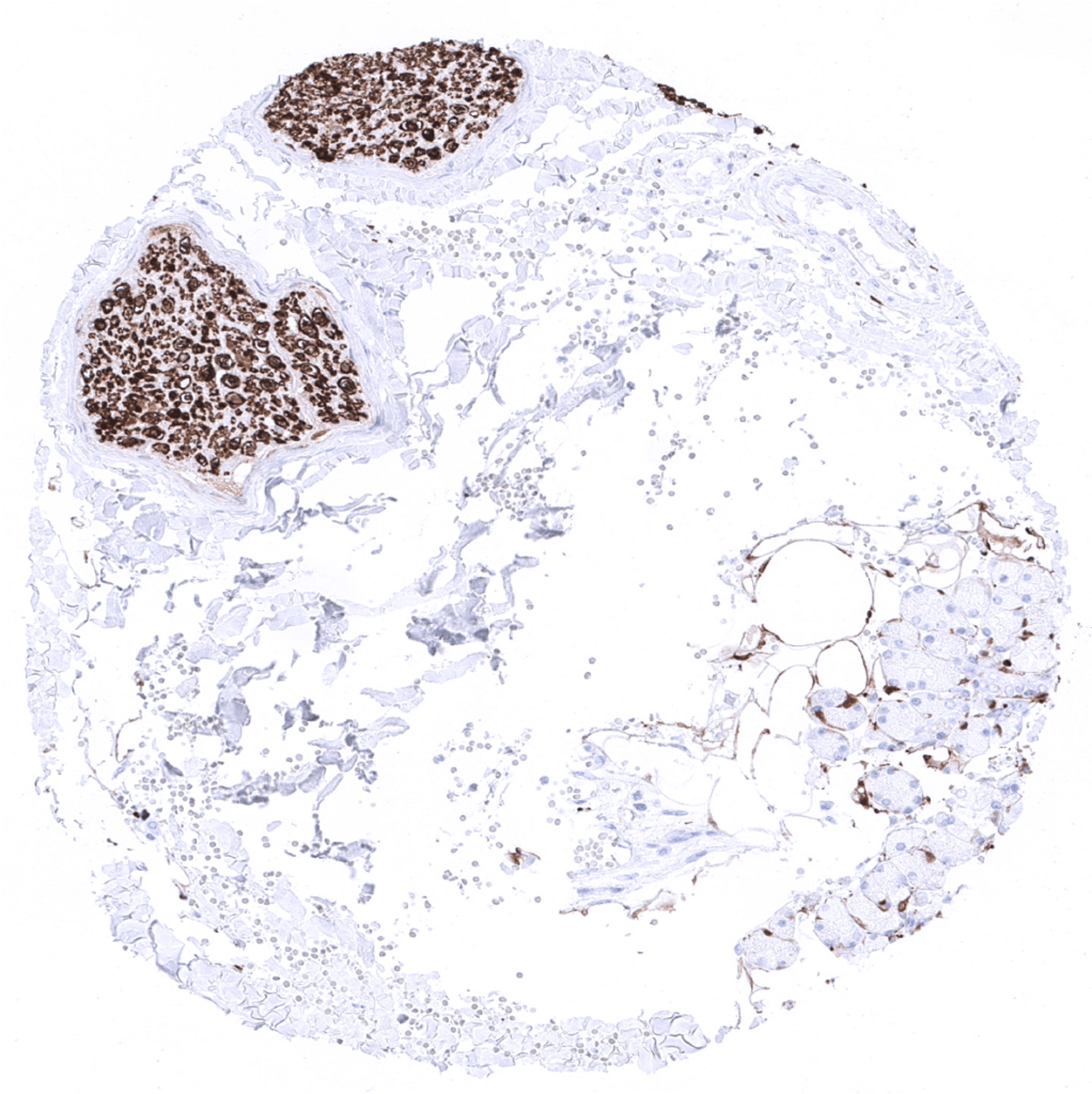
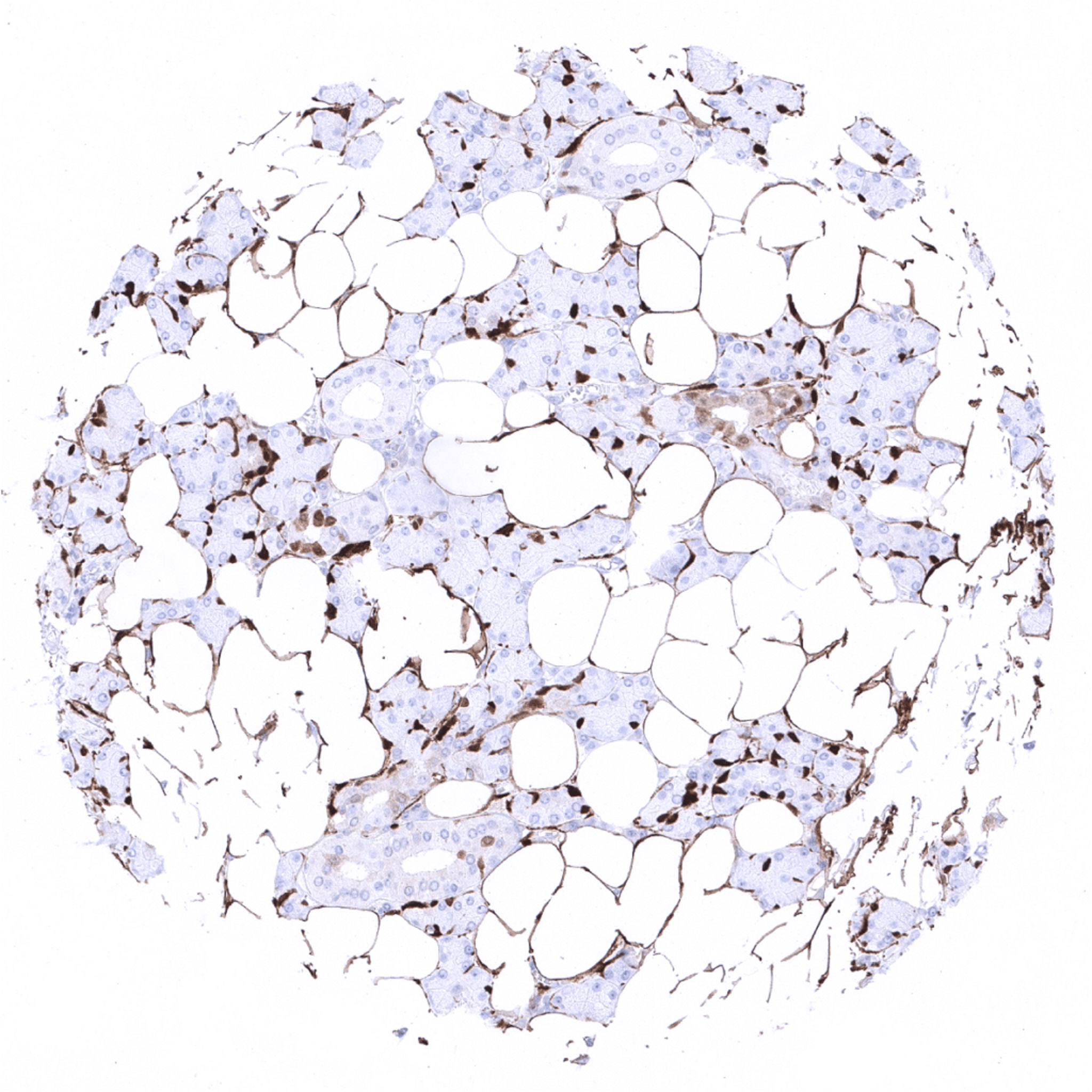
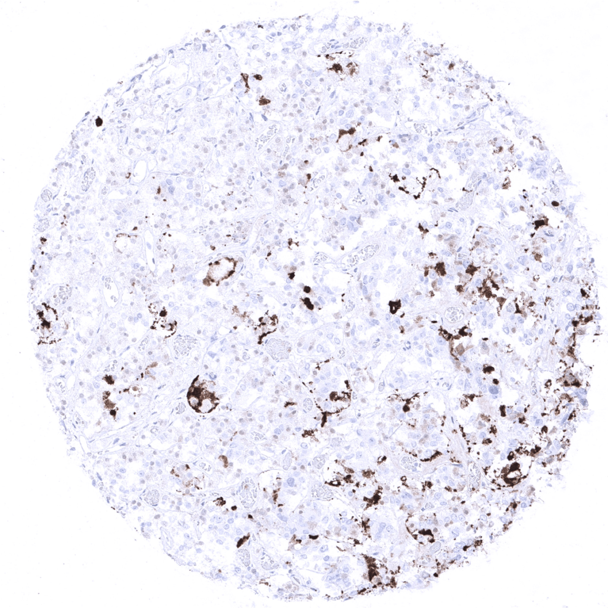
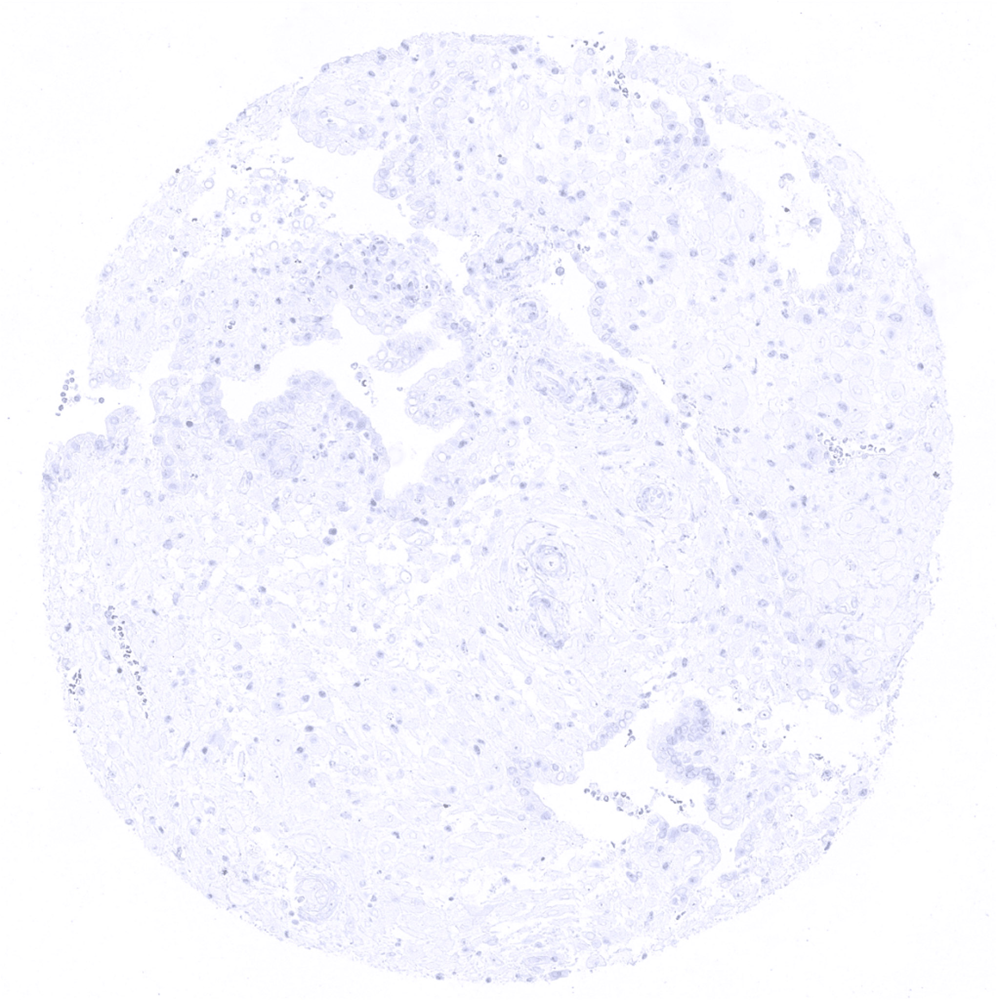
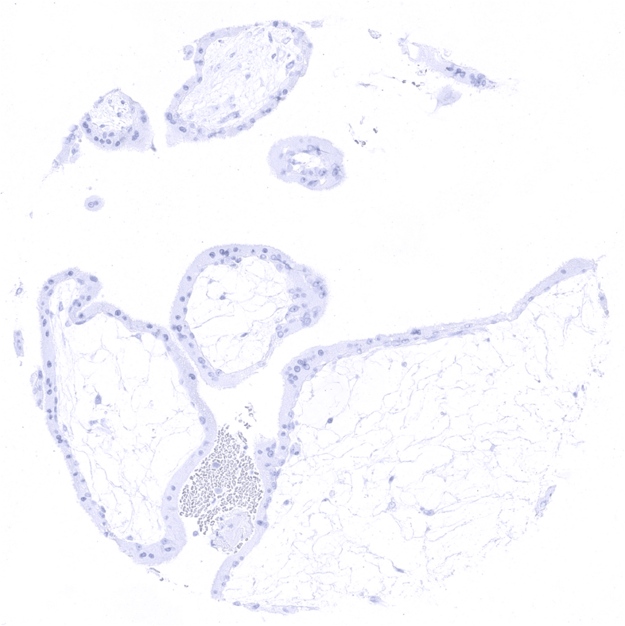
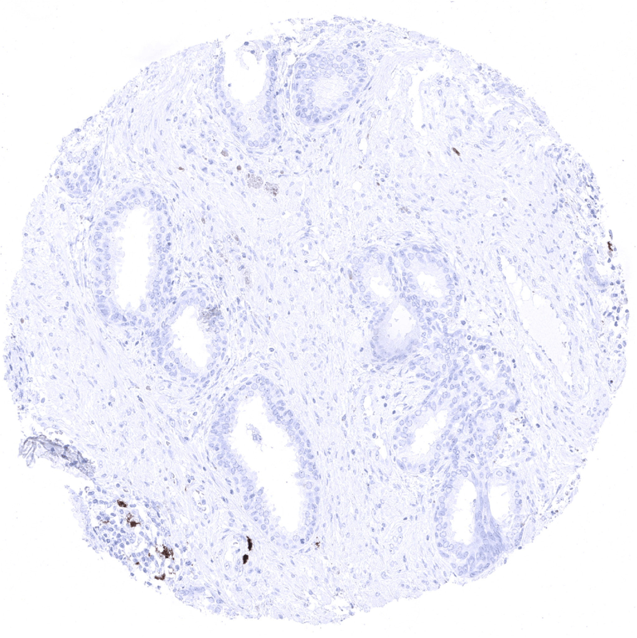
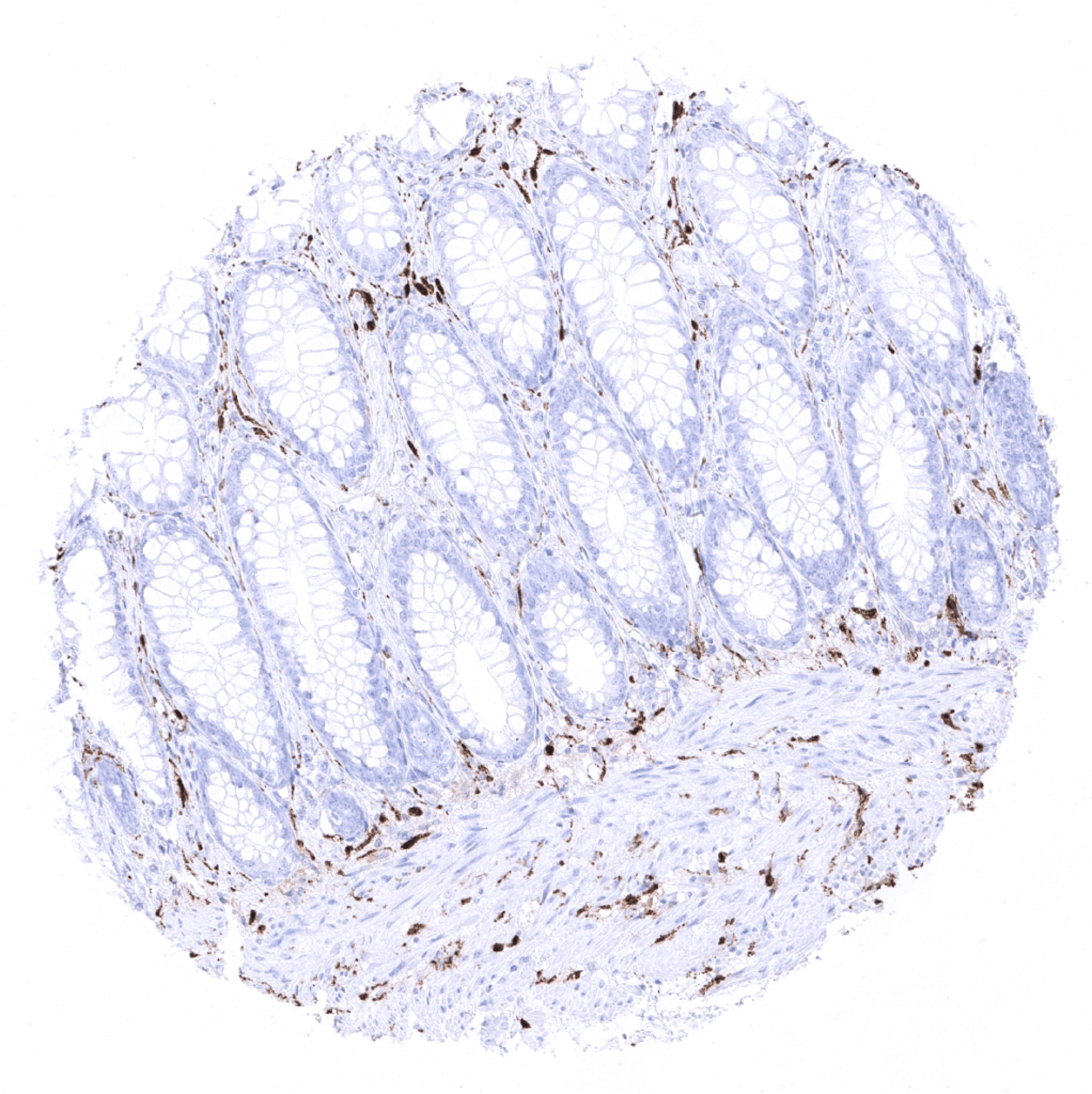
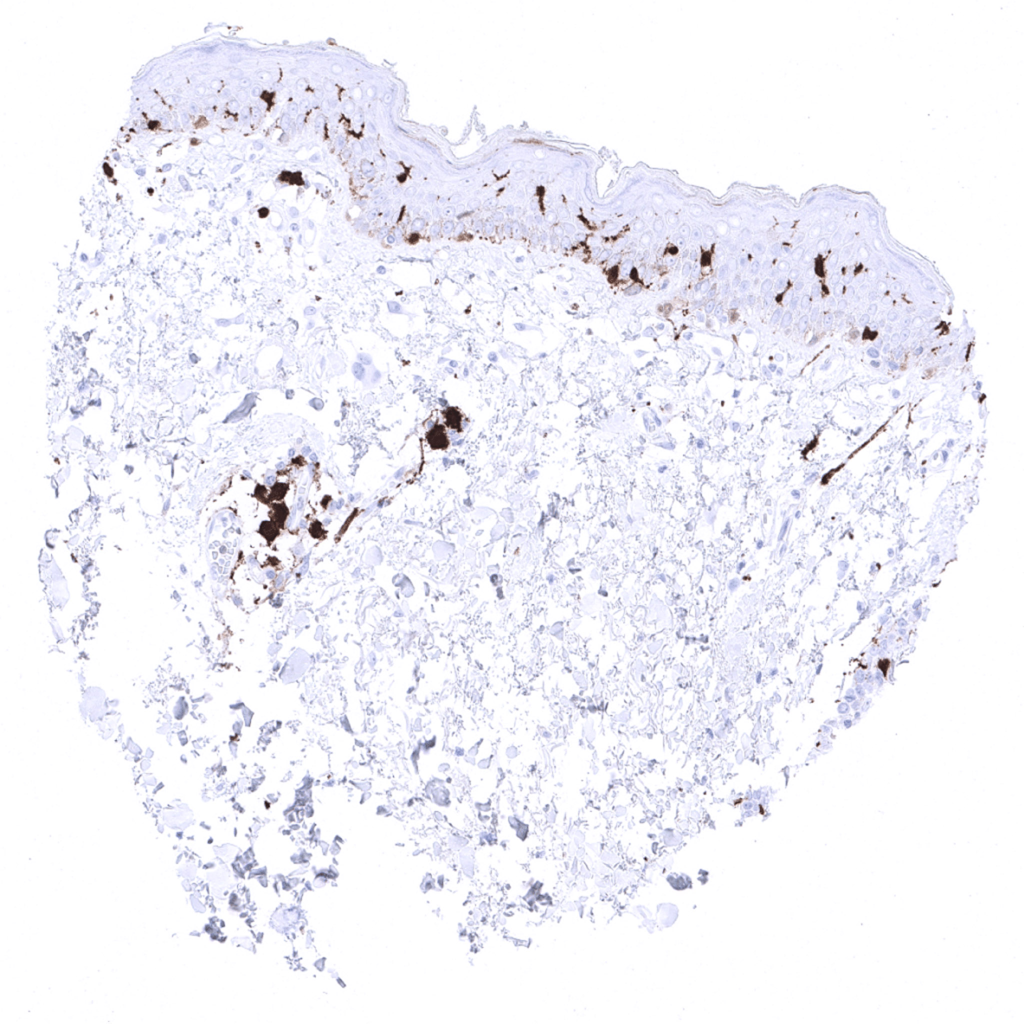
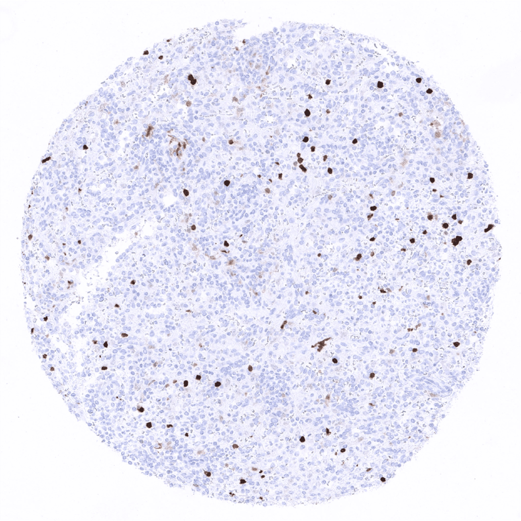
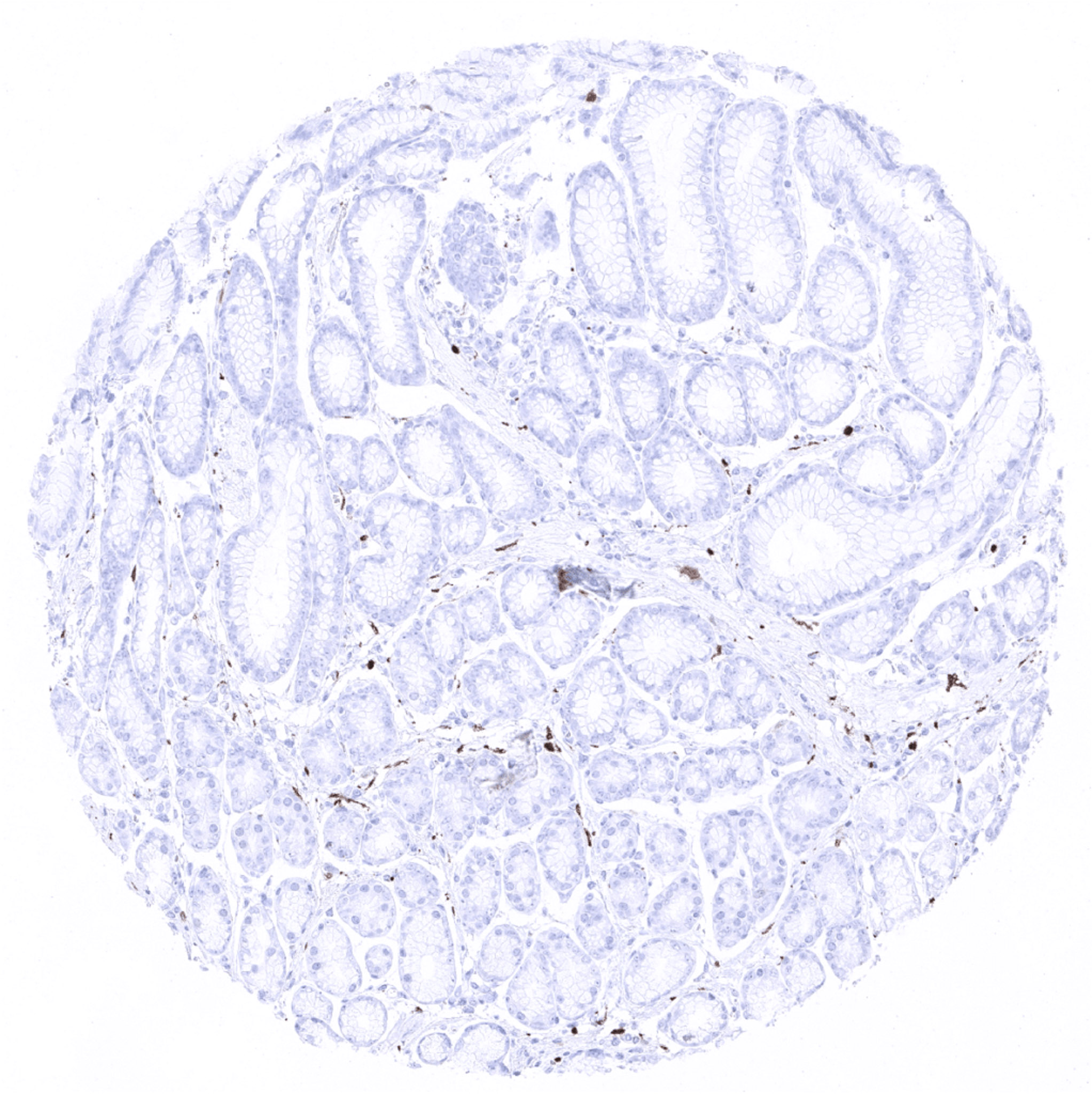
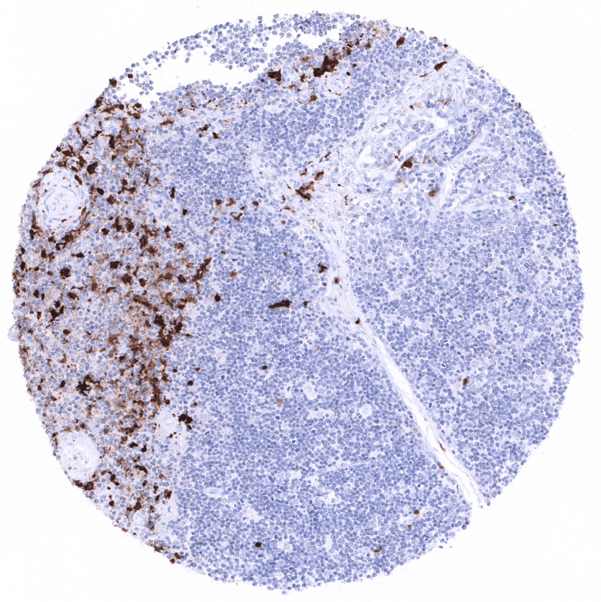
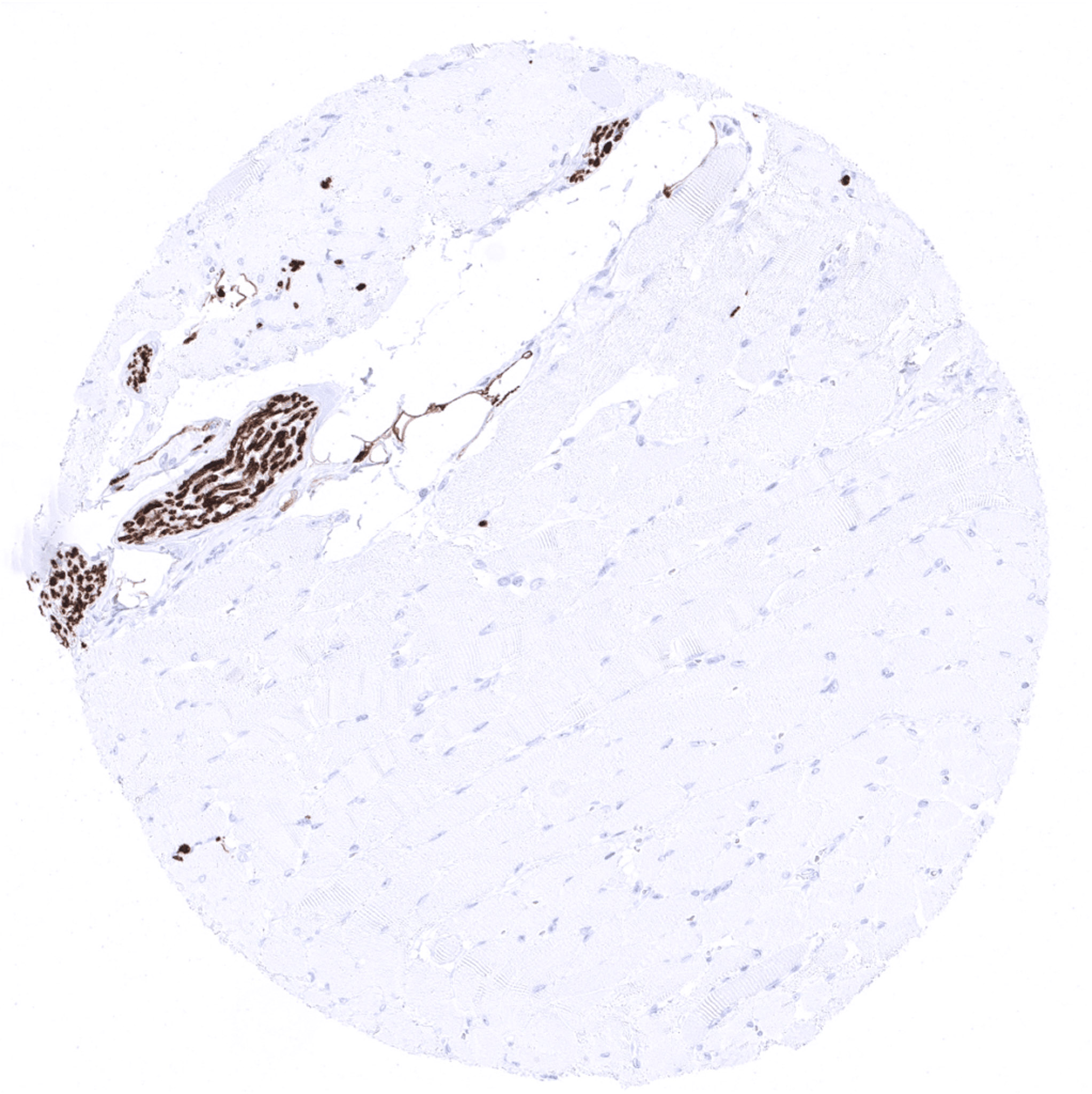
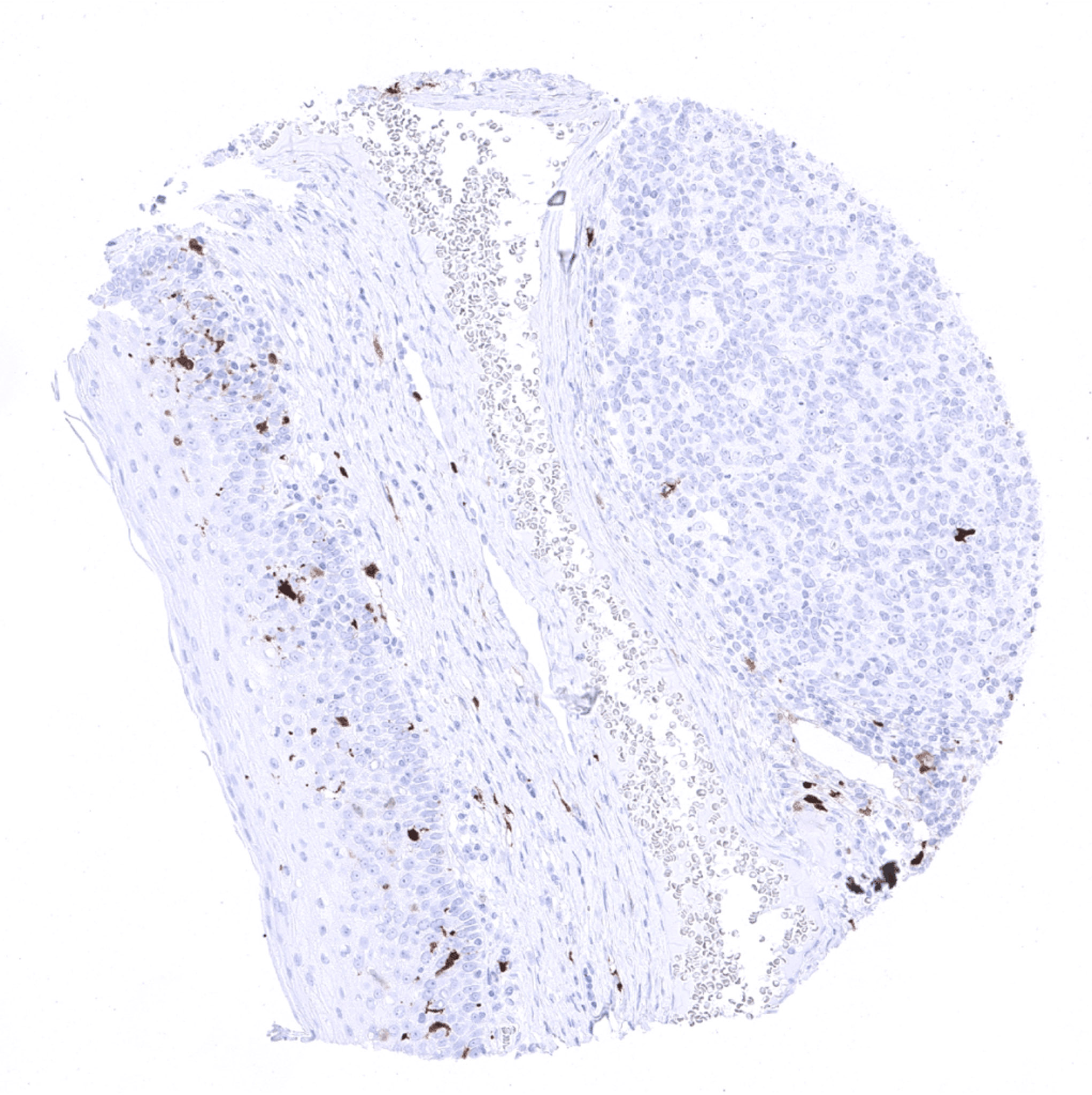
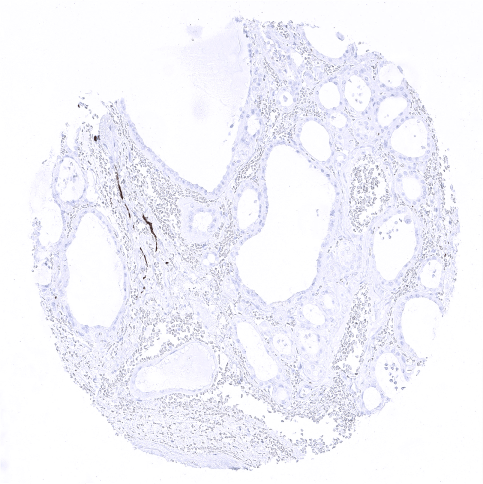
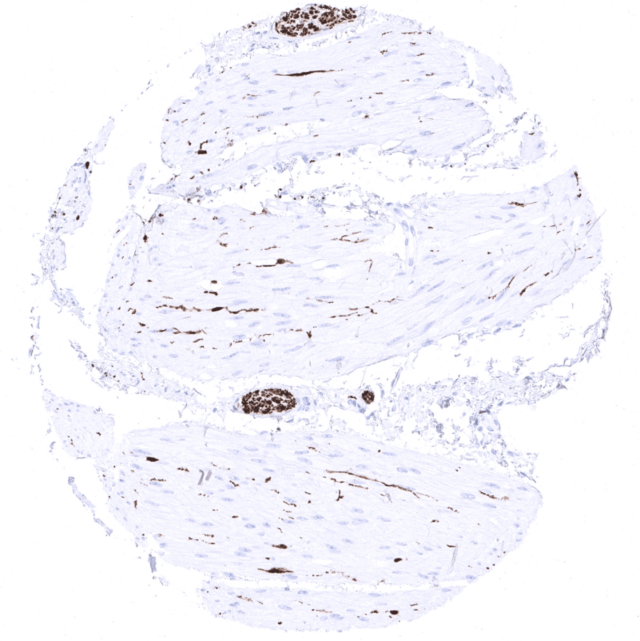
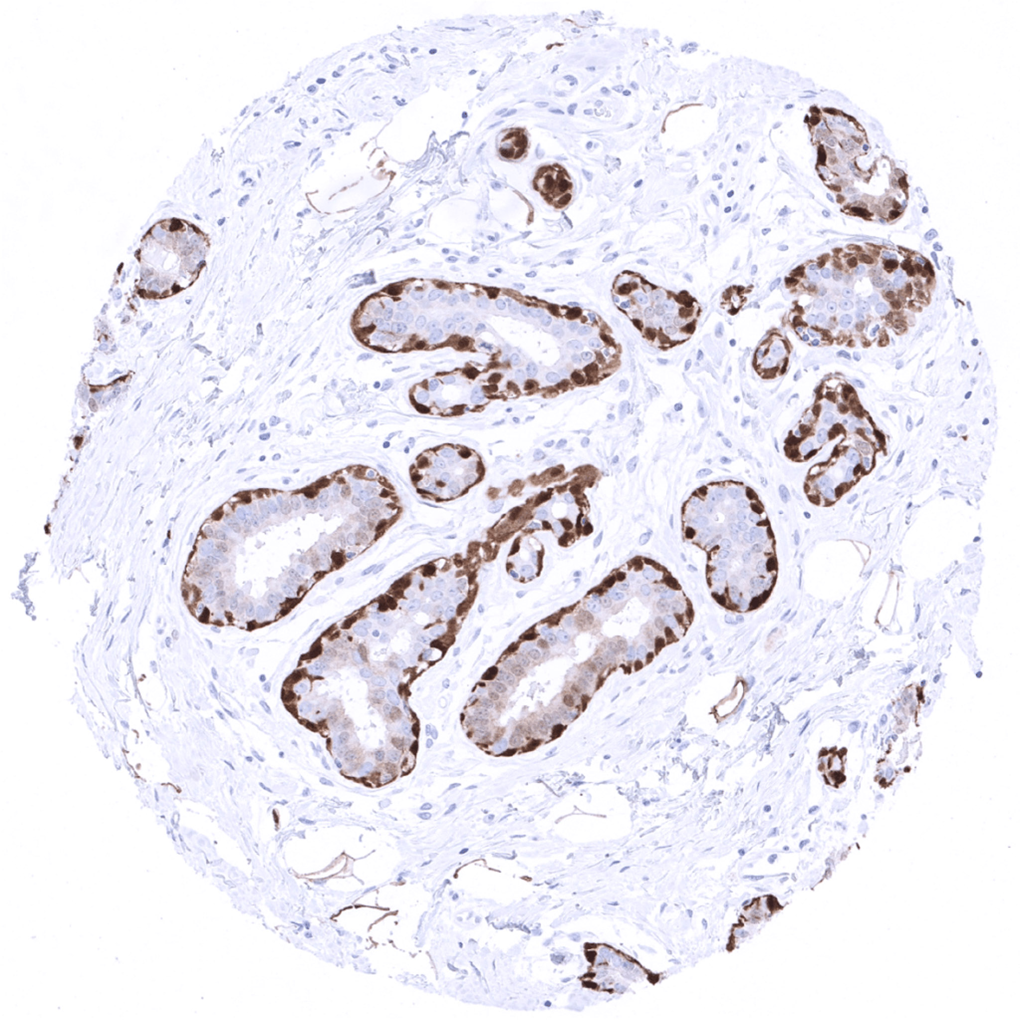
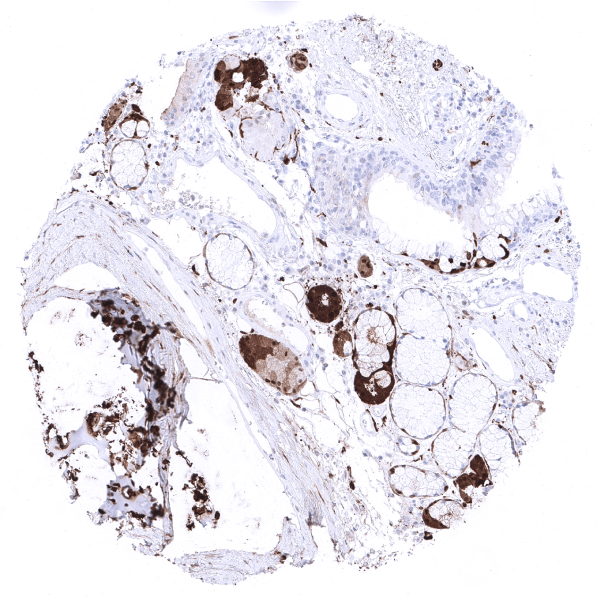
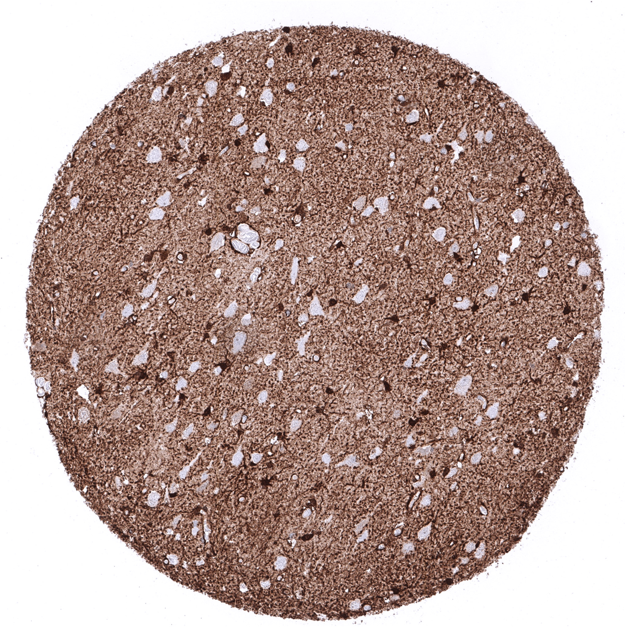
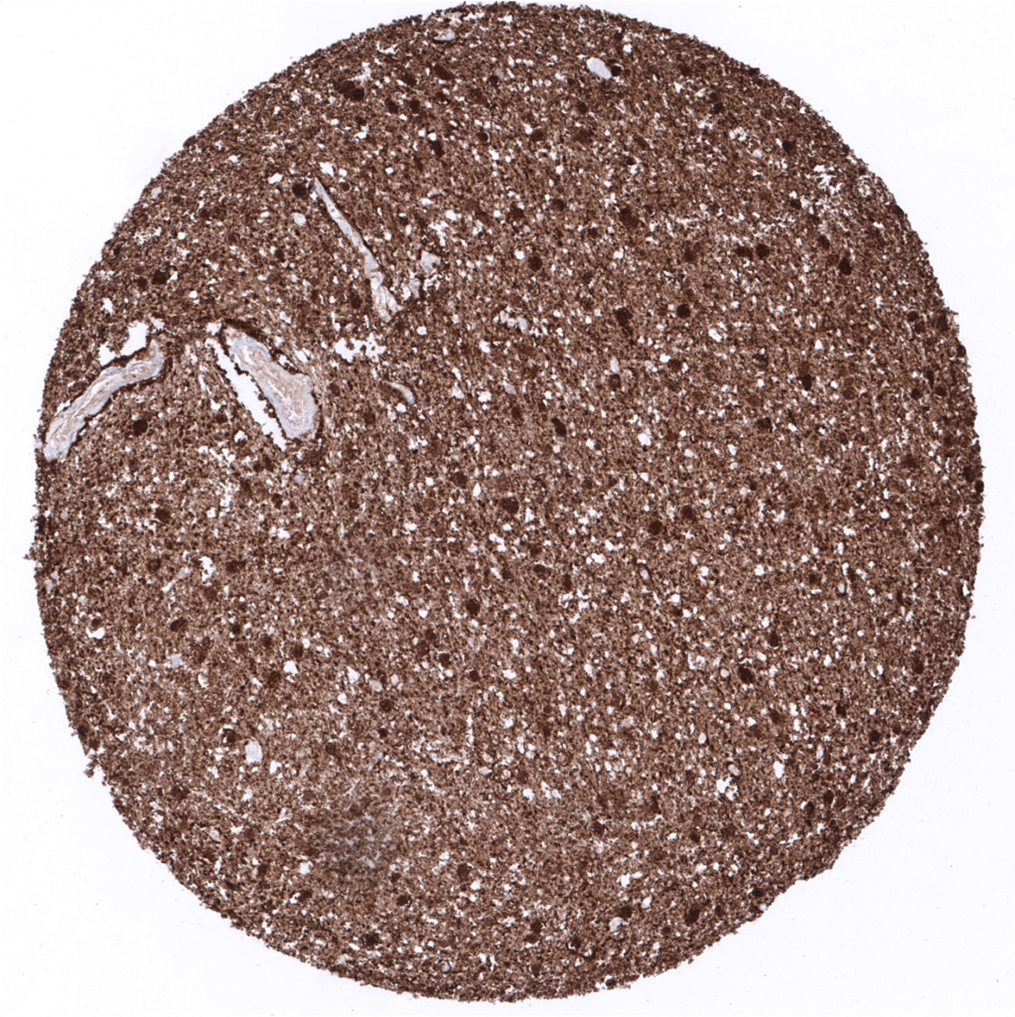
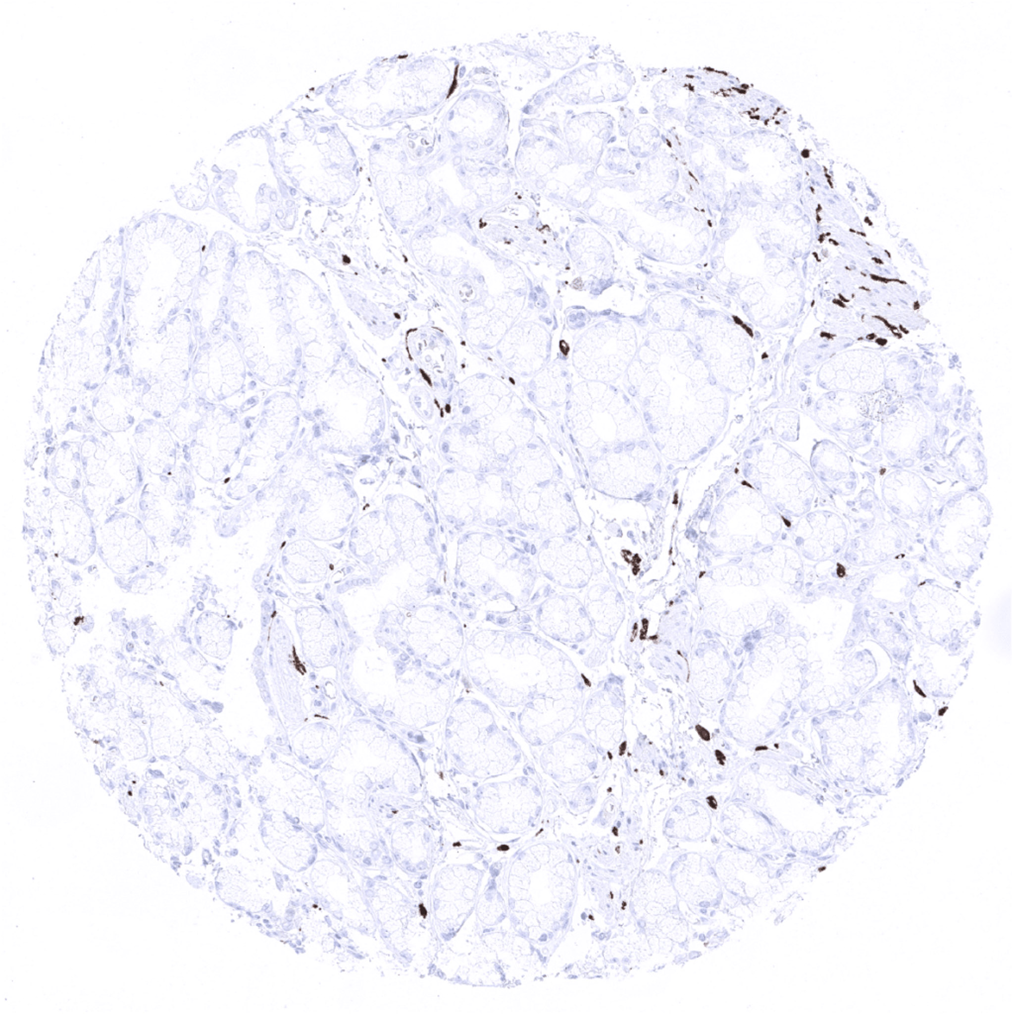
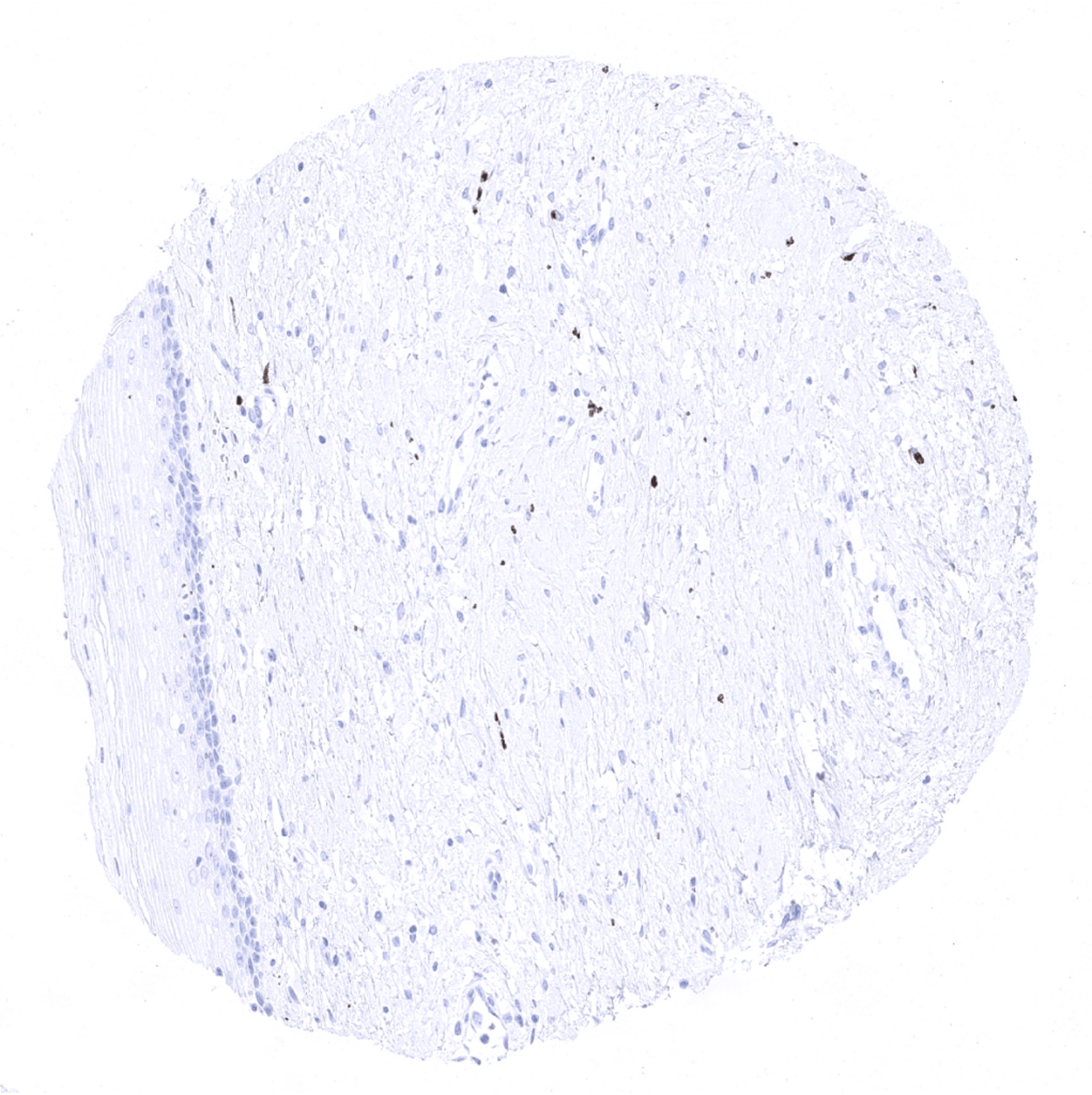
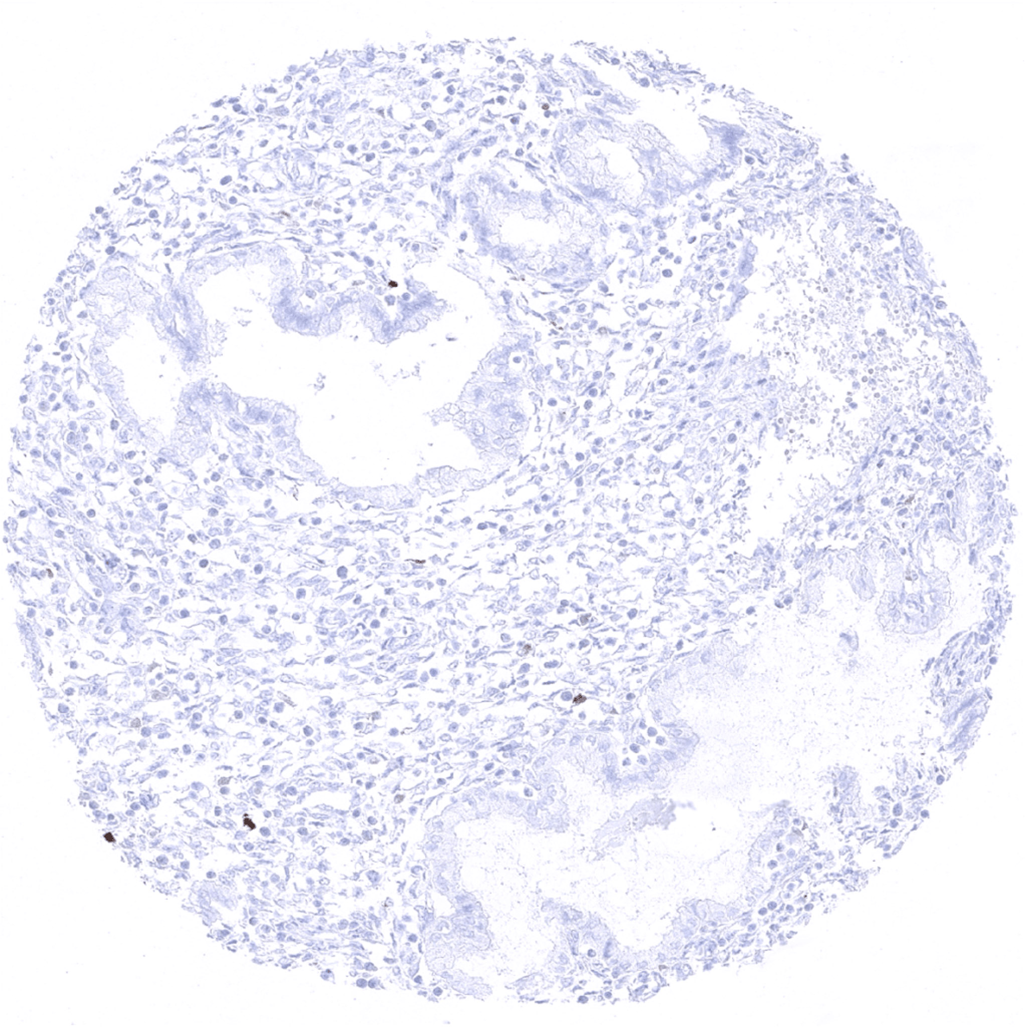
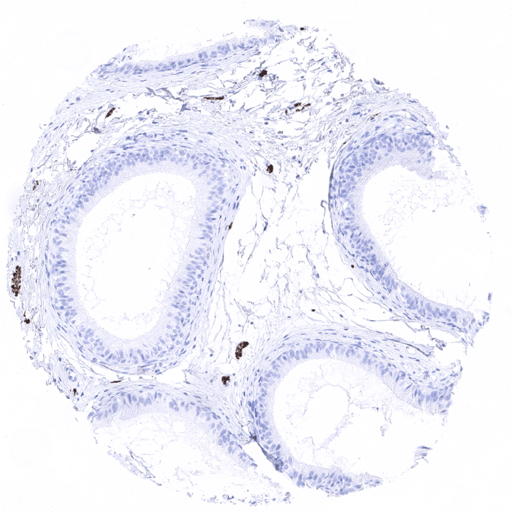
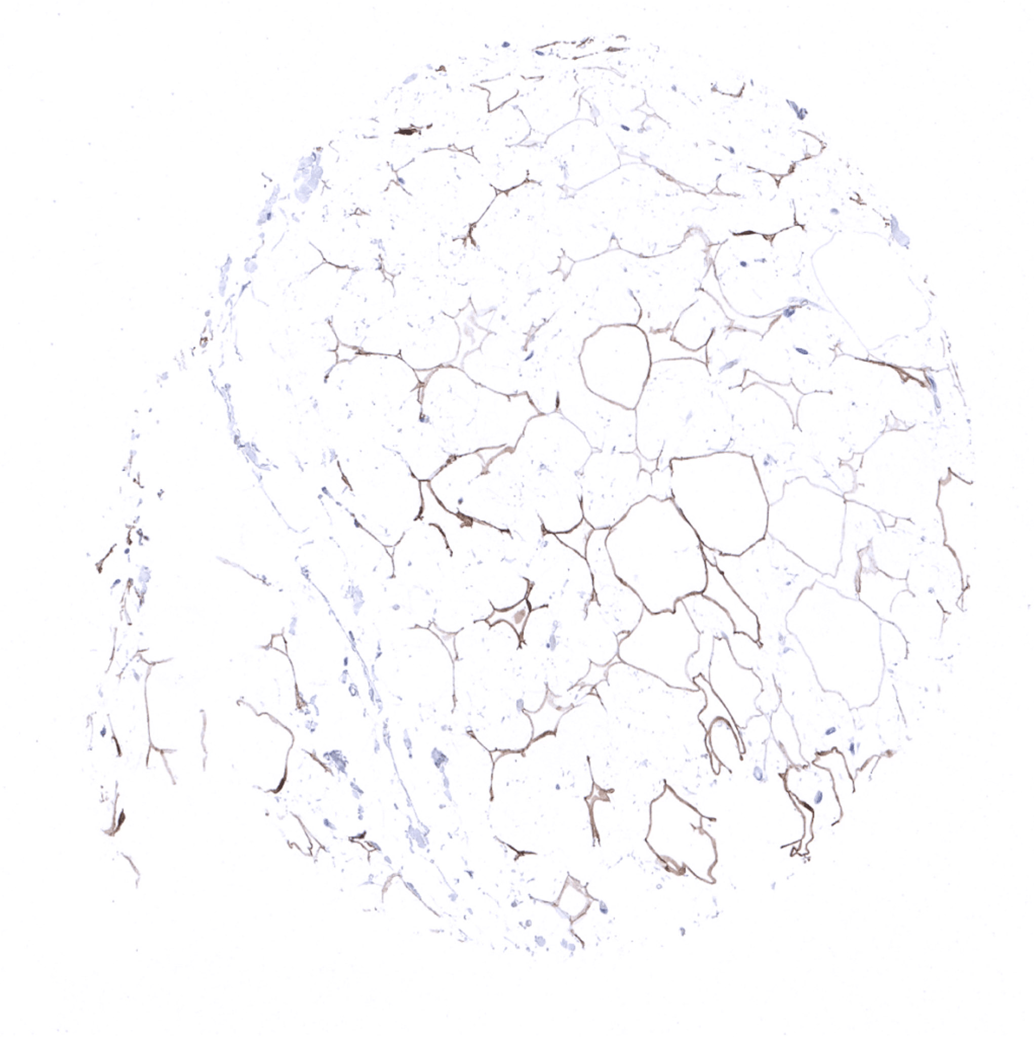
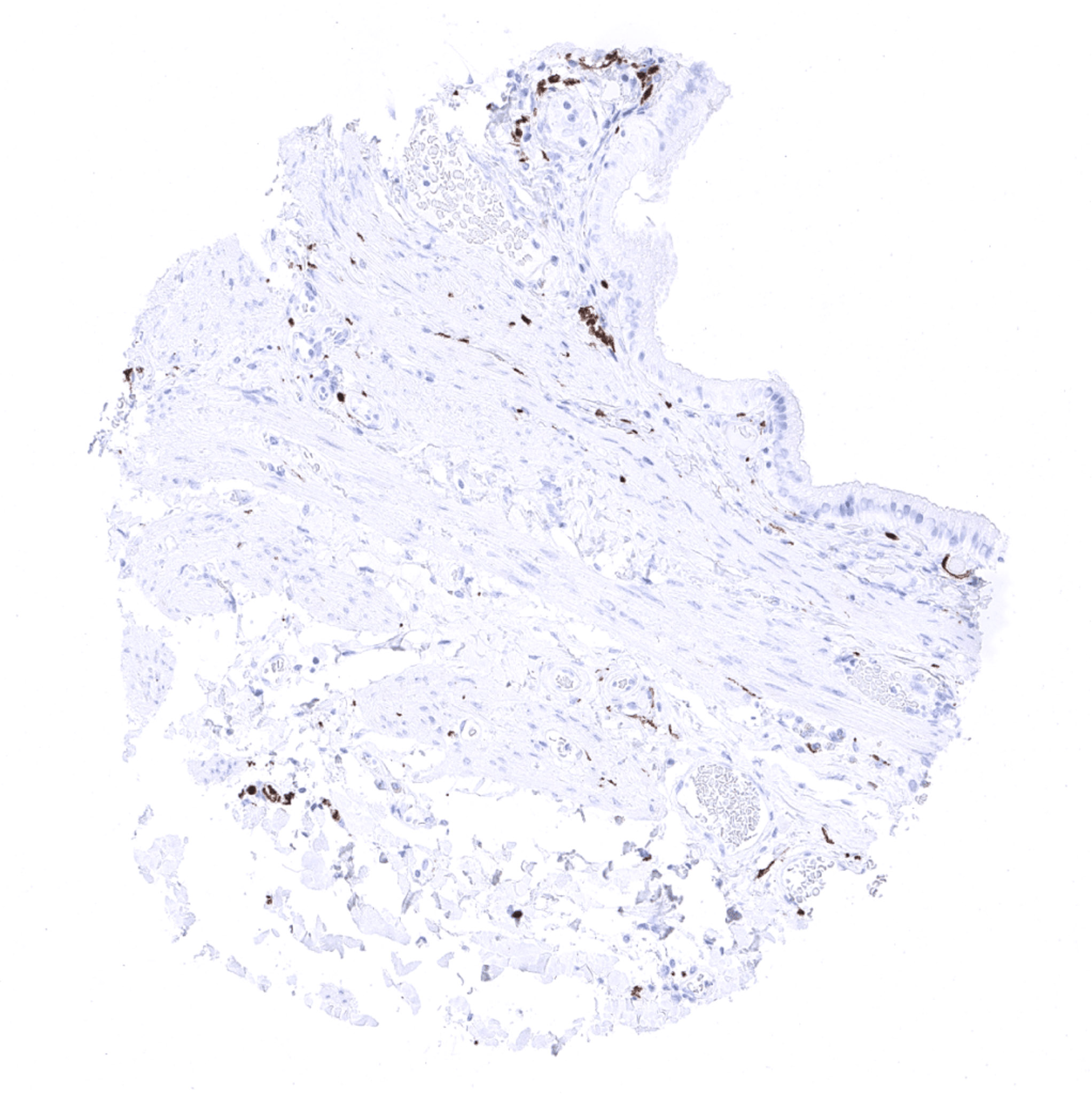
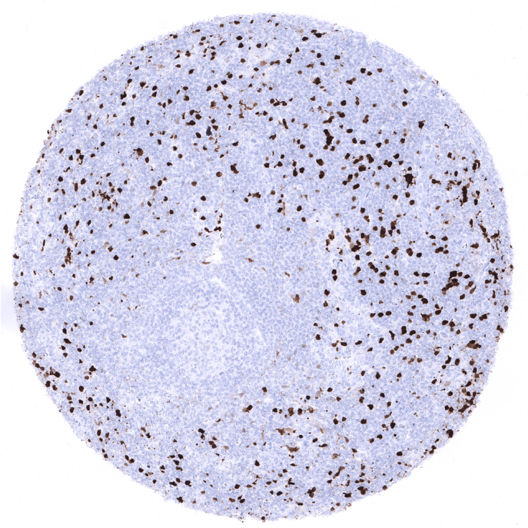
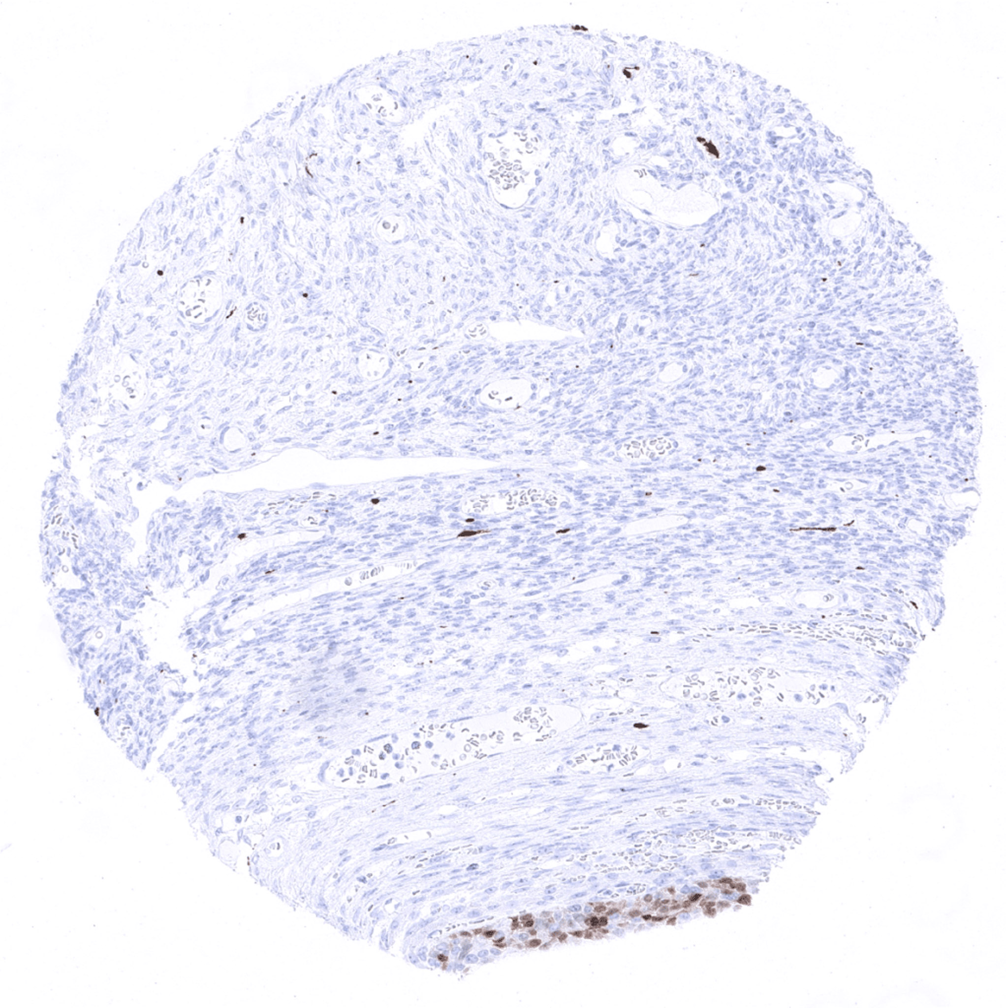
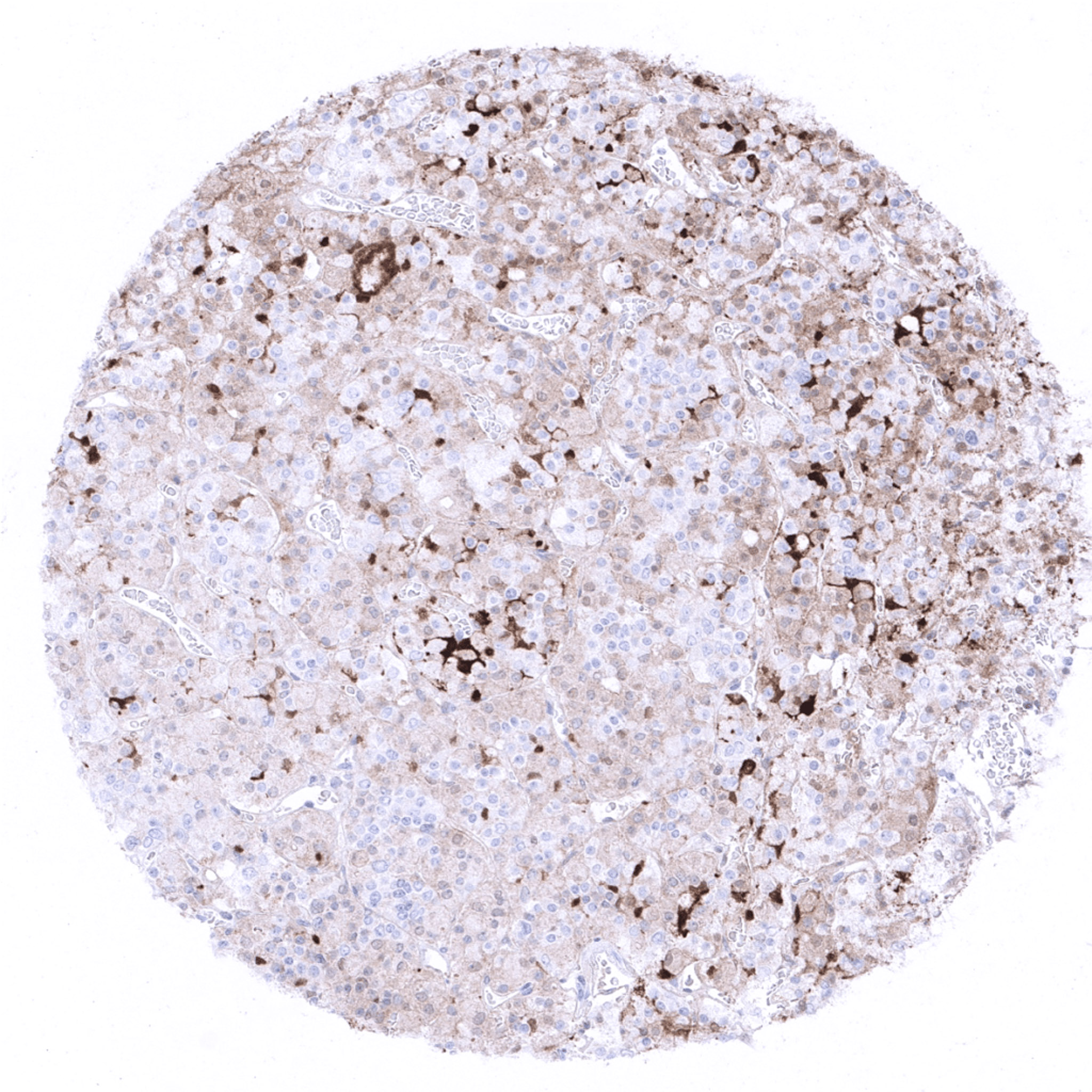
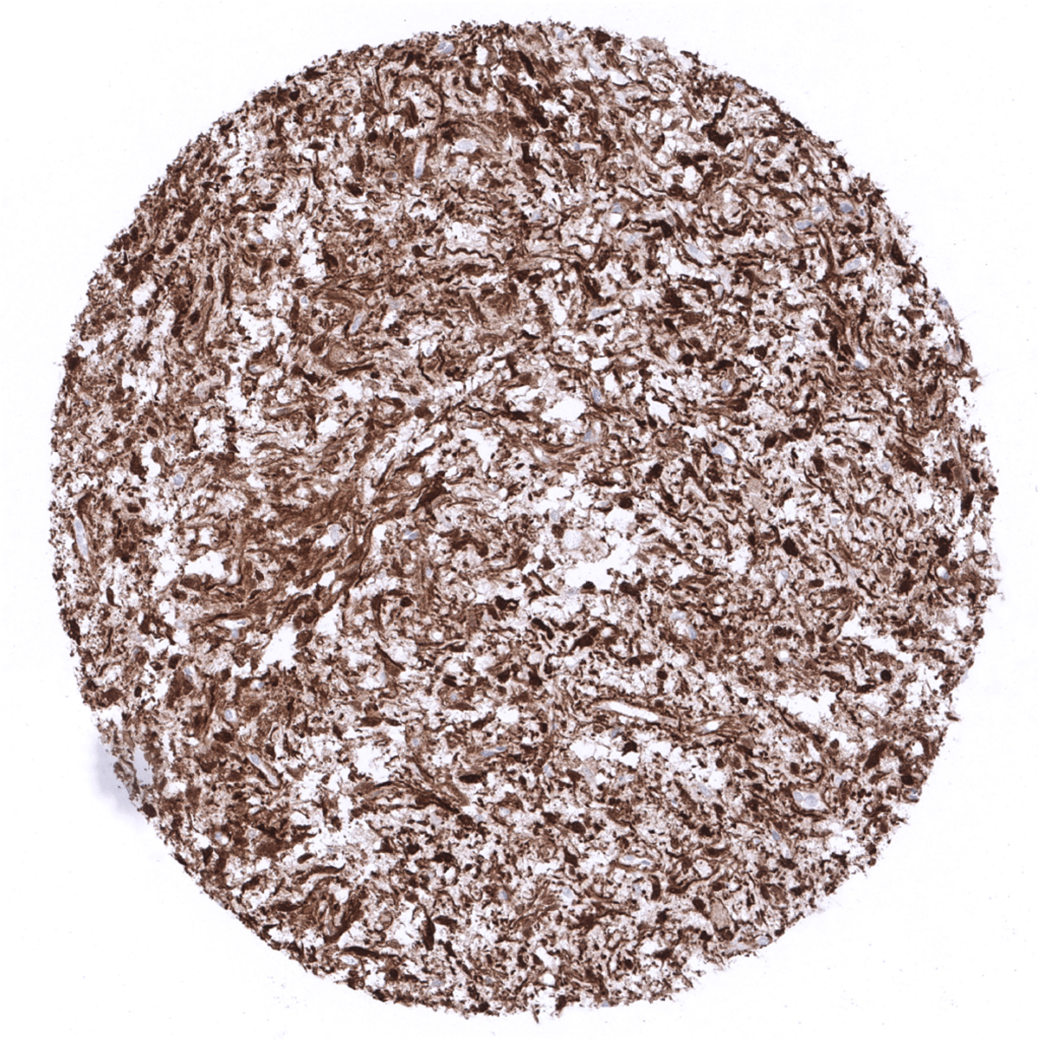
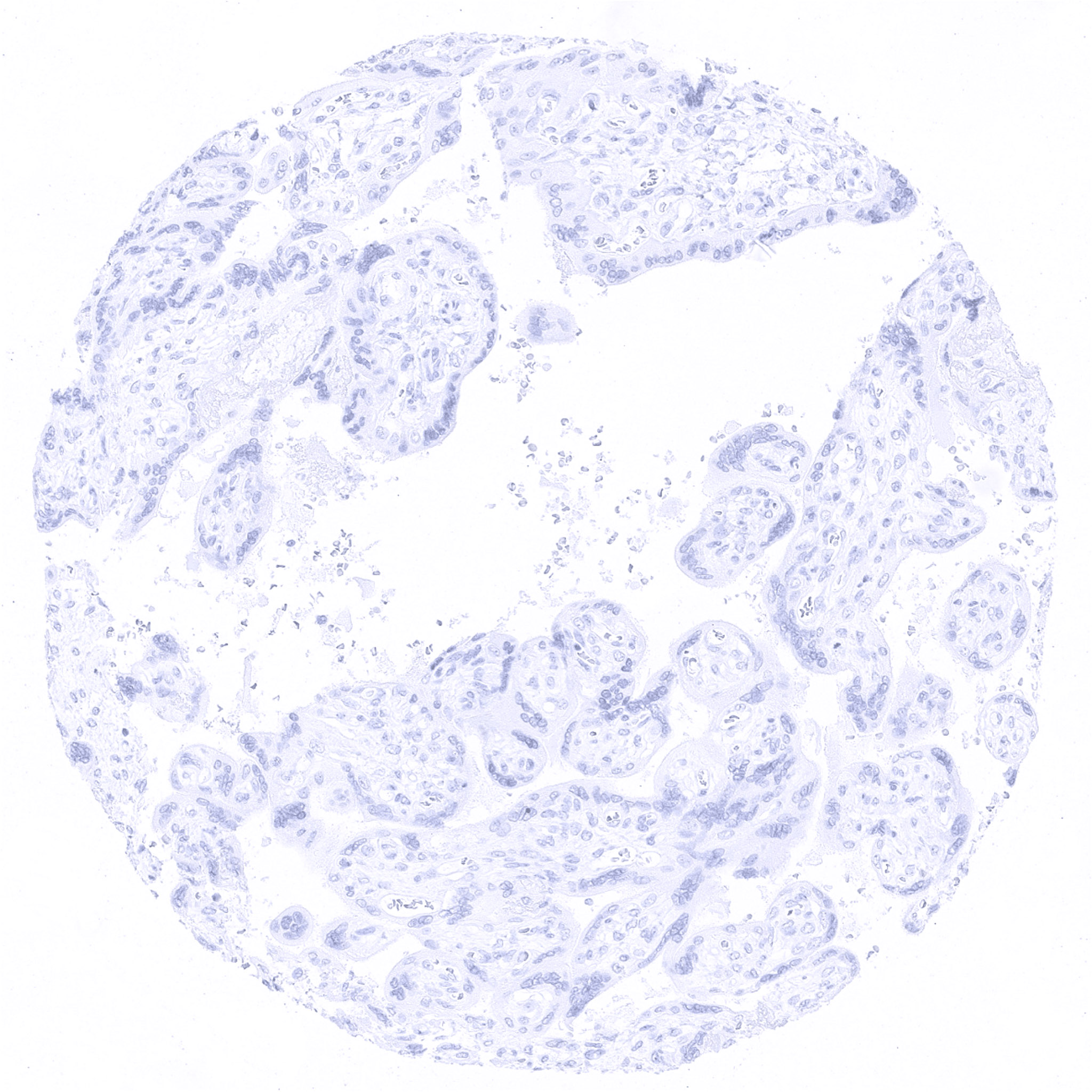
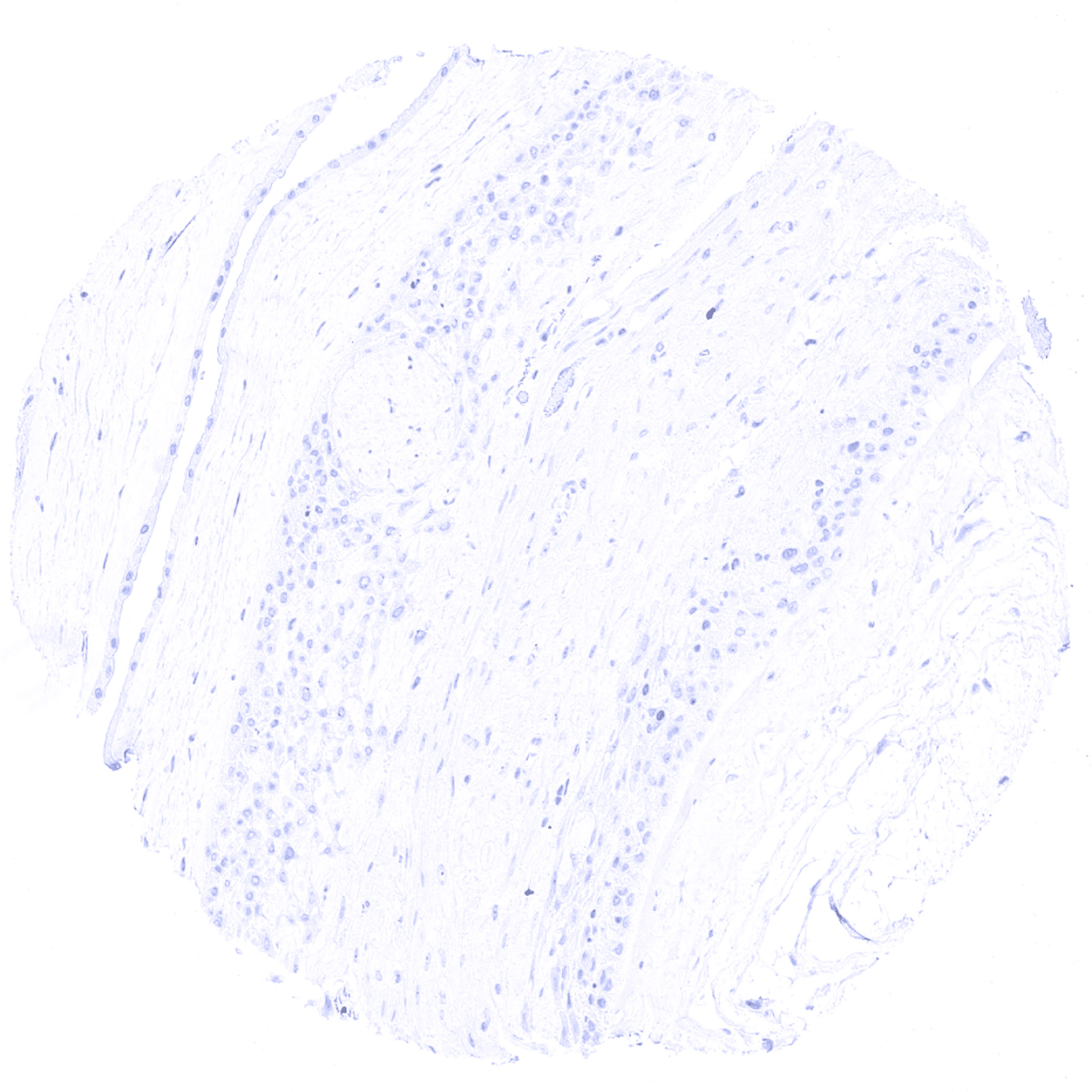
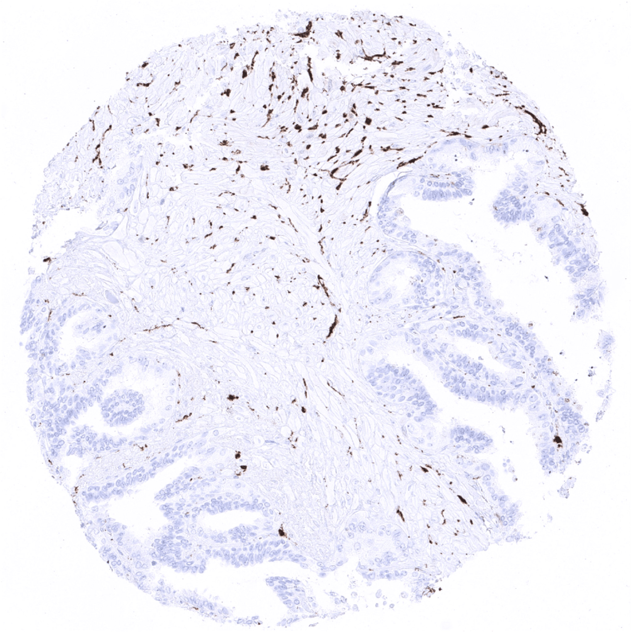
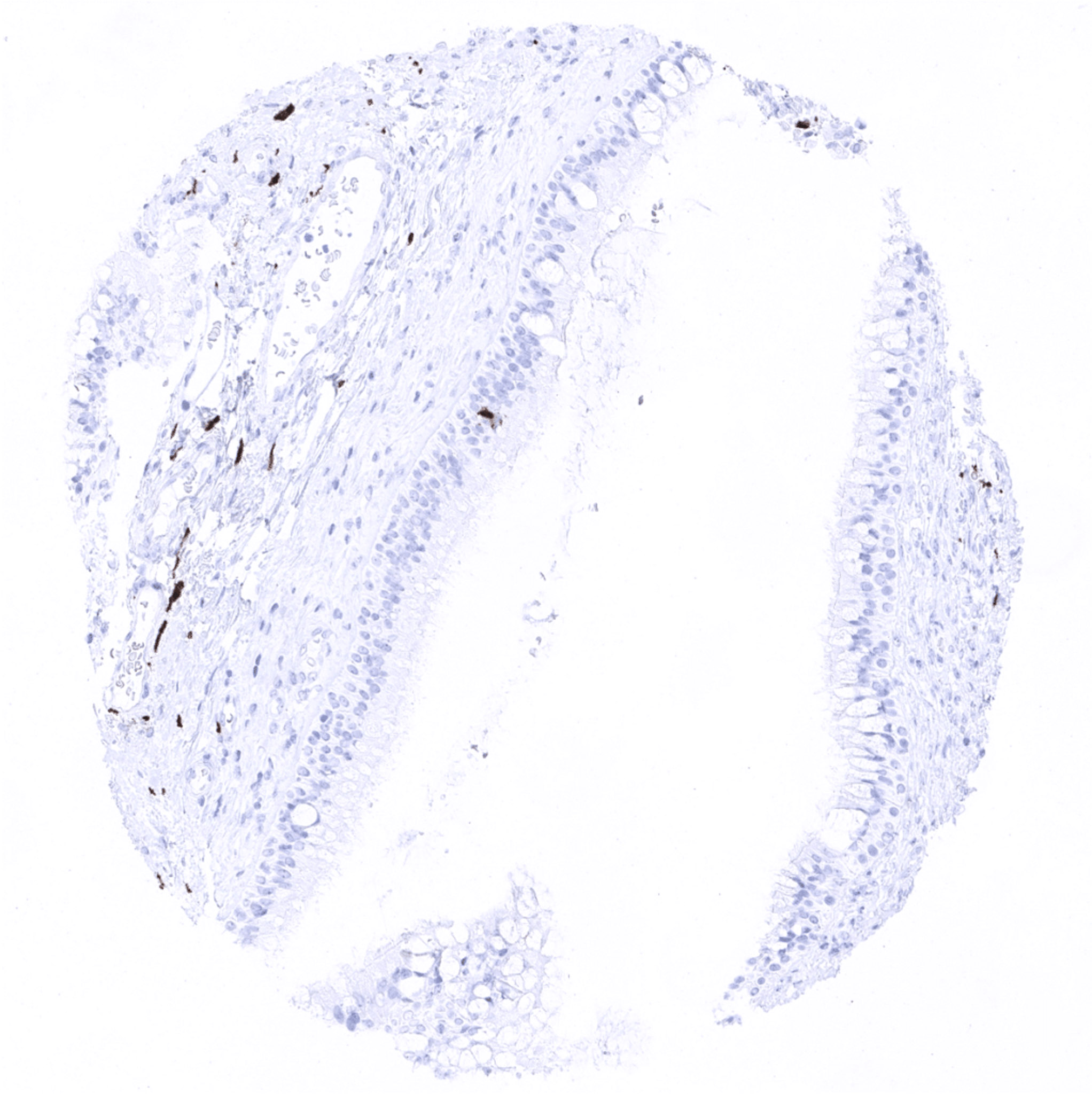
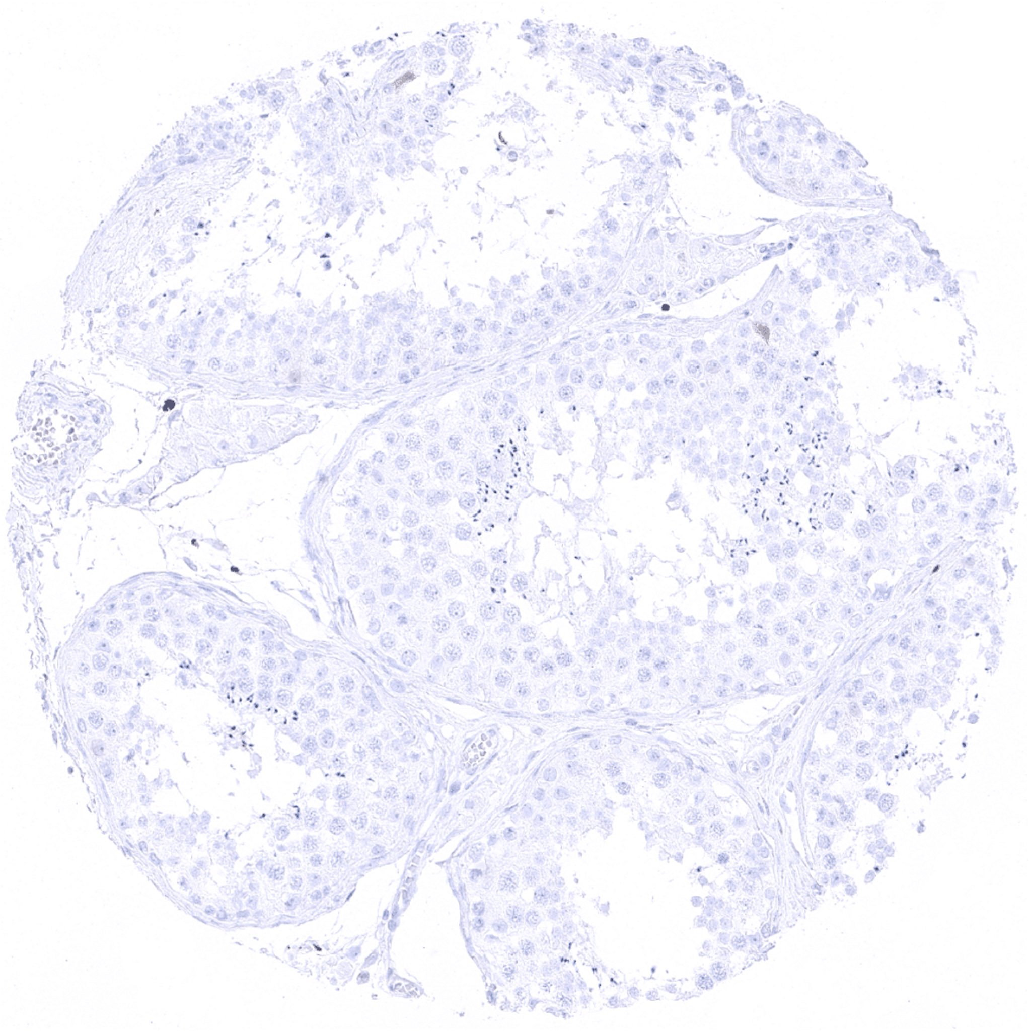
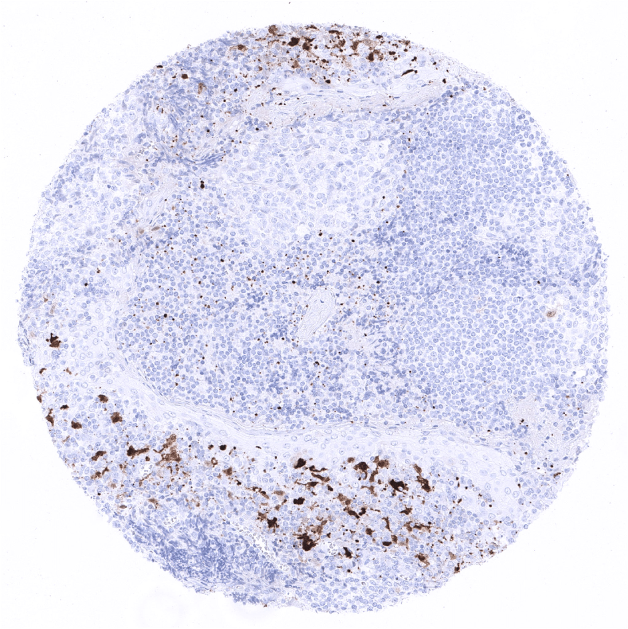
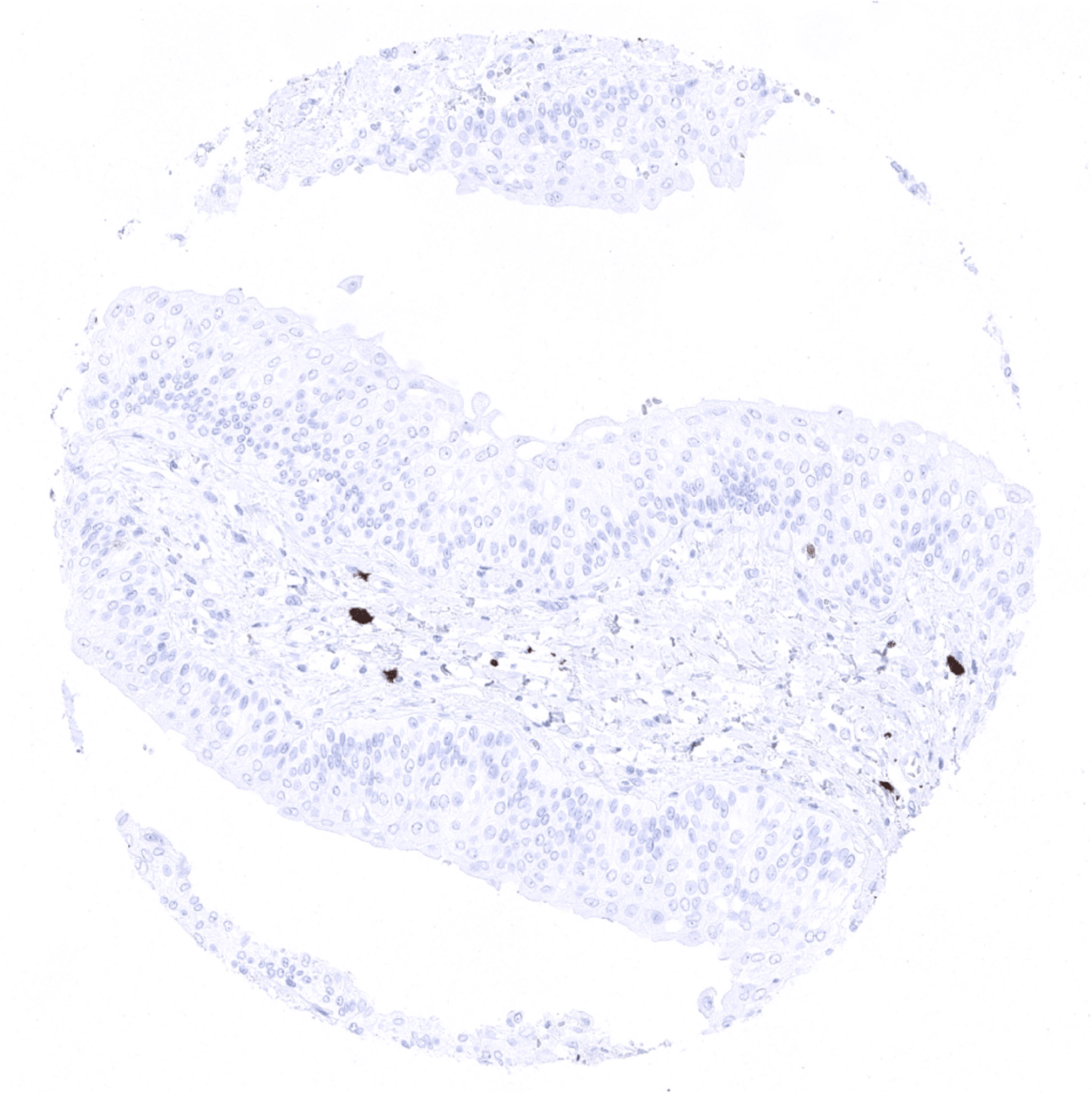
S100 beta antibody [MSVA-490R] HistoMAXTM


Go to Cancer Tissue Gallery Go back to product page Go to HistoMAX page


Adrenal gland
Adrenal gland – S100 beta positivity is seen in sustentacular cells and also in a fraction of medullary cells.
Aorta, media
Appendix, mucosa – Appendix with a multitude of intramucosal S100 beta positive nerve fibres.
Appendix, muscular wall – S100 beta positive nerve fibres are abundant in the muscular wall of the appendix.
Bone marrow – Fat cells are S100 beta positive.
Breast – Strong S100 beta immunostaining in myoepithelial cells while staining is much weaker in luminal breast epithelial cells.
Bronchus, mucosa – Strong S100 beta immunostaining in some serous cells of bronchial glands.
Cerebellum (molecular layer, Purkinje cell layer, granule cell layer, white matter) – Strong ubiquitous S100 beta staining in the cerebellum.
Cerebrum, grey matter – Strong ubiquitous S100 beta staining (except neurons).
Cerebrum, white matter – Strong ubiquitous S100 beta staining.
Colon descendens, mucosa
Colon descendens, muscular wall – Nerve fibres and ganglion cells are S100 beta positive.
Duodenum, Brunner gland
Duodenum, mucosa – A large number of S100 beta positive nerve fibres is seen in this sample from the duodenum.
Ectocervix
Endocervix
Endometrium, proliferation
Endometrium, secretion
Epididymis
Esophagus, squamous epithelium
Fallopian tube, mucosa
Fat – Fat cells are S100 beta positive.
Gallbladder, epithelium
Heart
Ileum, mucosa
Ileum, mucosa – Strong S100 beta staining of nerve fibres.
Kidney, cortex
Kidney, medulla
Liver
Lung
Lymph node – Strong S100 beta immunostaining is seen in a subset of T-lymphocytes.
Ovary, follicular cyst – Moderate S100 beta staining in granulosa cells of a follicular cyst of the ovary.
Ovary, follicular cyst – Follicular cyst of the ovary showing a moderate S100 beta staining of granulosa cells.
Ovary, stroma – Few S100 beta positive nerve fibres are visible in the ovarian stroma.
Pancreas
Parathyroid – S100 beta positivity is seen in fat cells.
Parotid gland – Strong S100 beta staining of nerves and fat cells.
Parotid gland – S100 beta positivity is seen in myoepithelial cells, nerve fibres, and fat cells.
Pituitary gland, anterior lobe
Pituitary gland, anterior lobe – A weak S100 beta staining can be seen in a fraction of cells in the adenohypophysis.
Pituitary gland, posterior lobe
Pregnant uterus (decidua)
Placenta, early
Placenta, mature
Placenta (amnion and chorion)
Prostate
Rectum, mucosa
Seminal vesicle
Sinus paranasales
Skin – Langerhans cells show strong S100 beta positivity.
Spleen – Strong S100 beta immunostaining is seen in a subset of T-lymphocytes.
Stomach, antrum
Stomach, corpus
Testis
Thymus – S100P staining of a fraction of thymus epithelial cells and possibly also of lymphocytes.
Tongue, muscle
Tonsil, surface epithelium
Tonsil
Thyroid gland
Urinary bladder, muscular wall
Urinary bladder, urothelium
Uterus, myometrium

| TOP |