GeneTex
  • Country / Location Selection

United States (US)

Normal Tissue Gallery- GTX04459

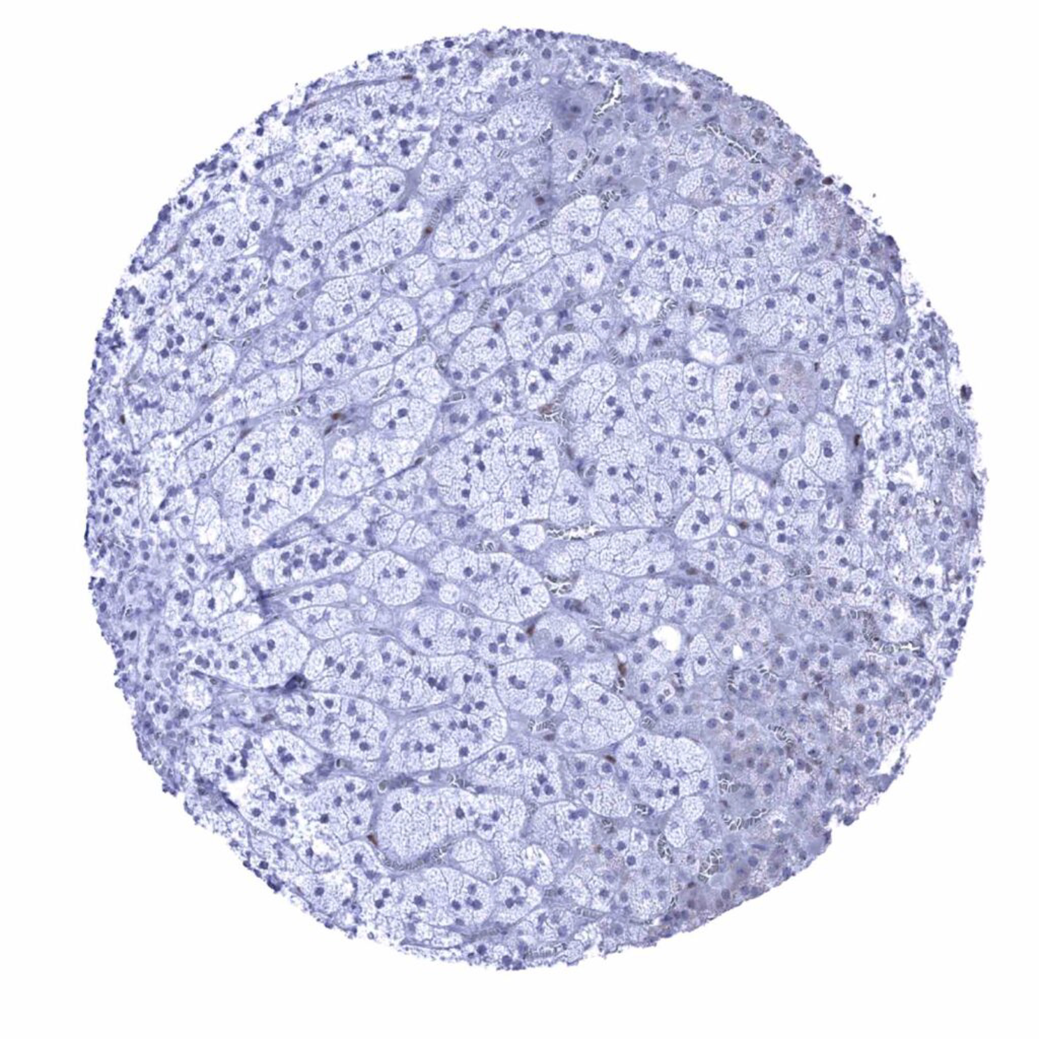
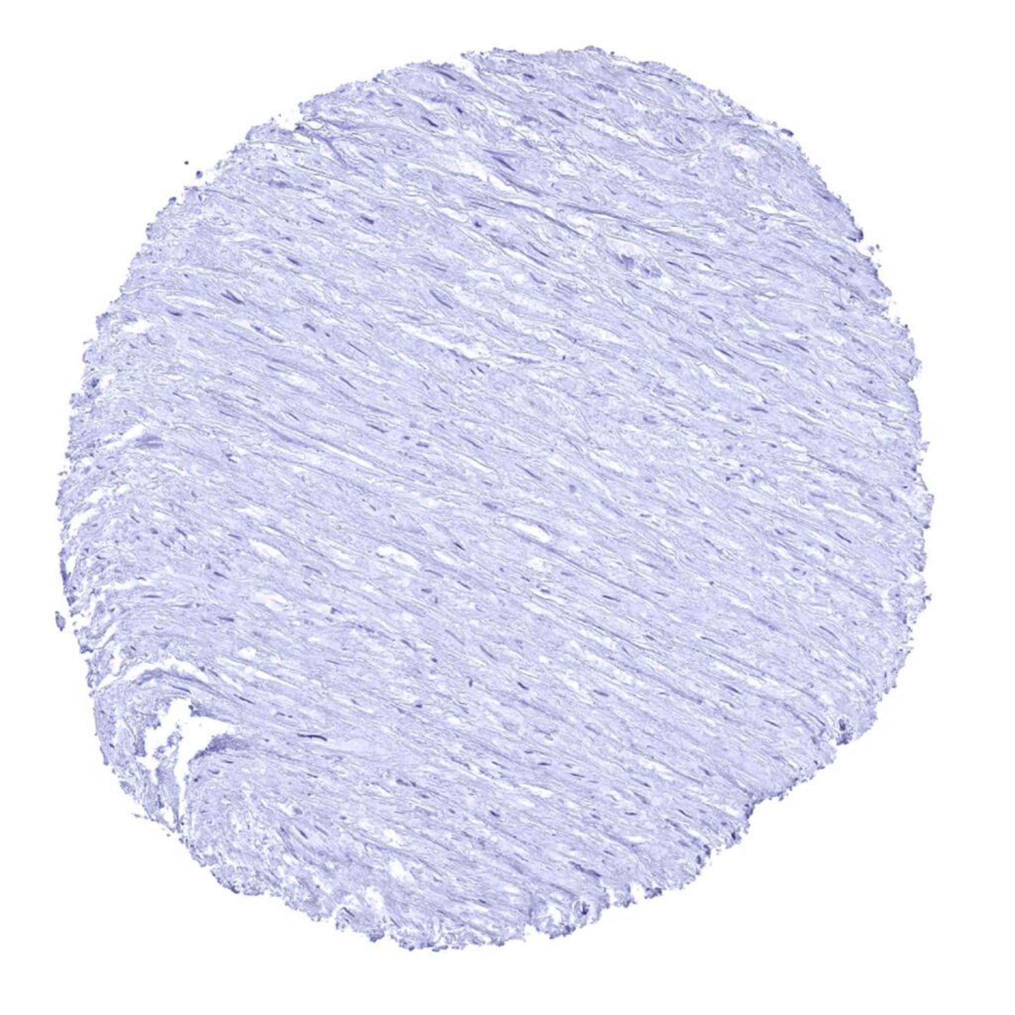
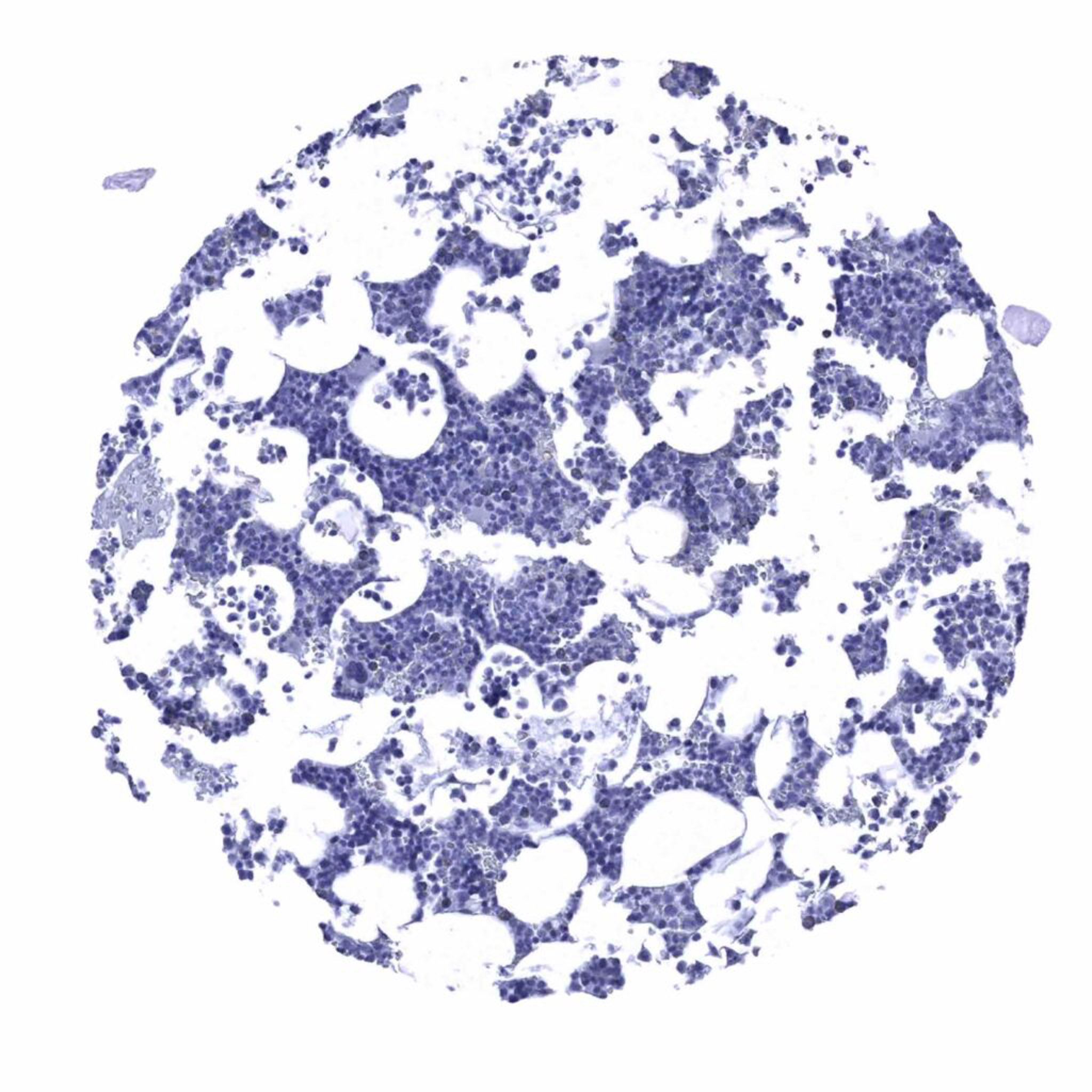
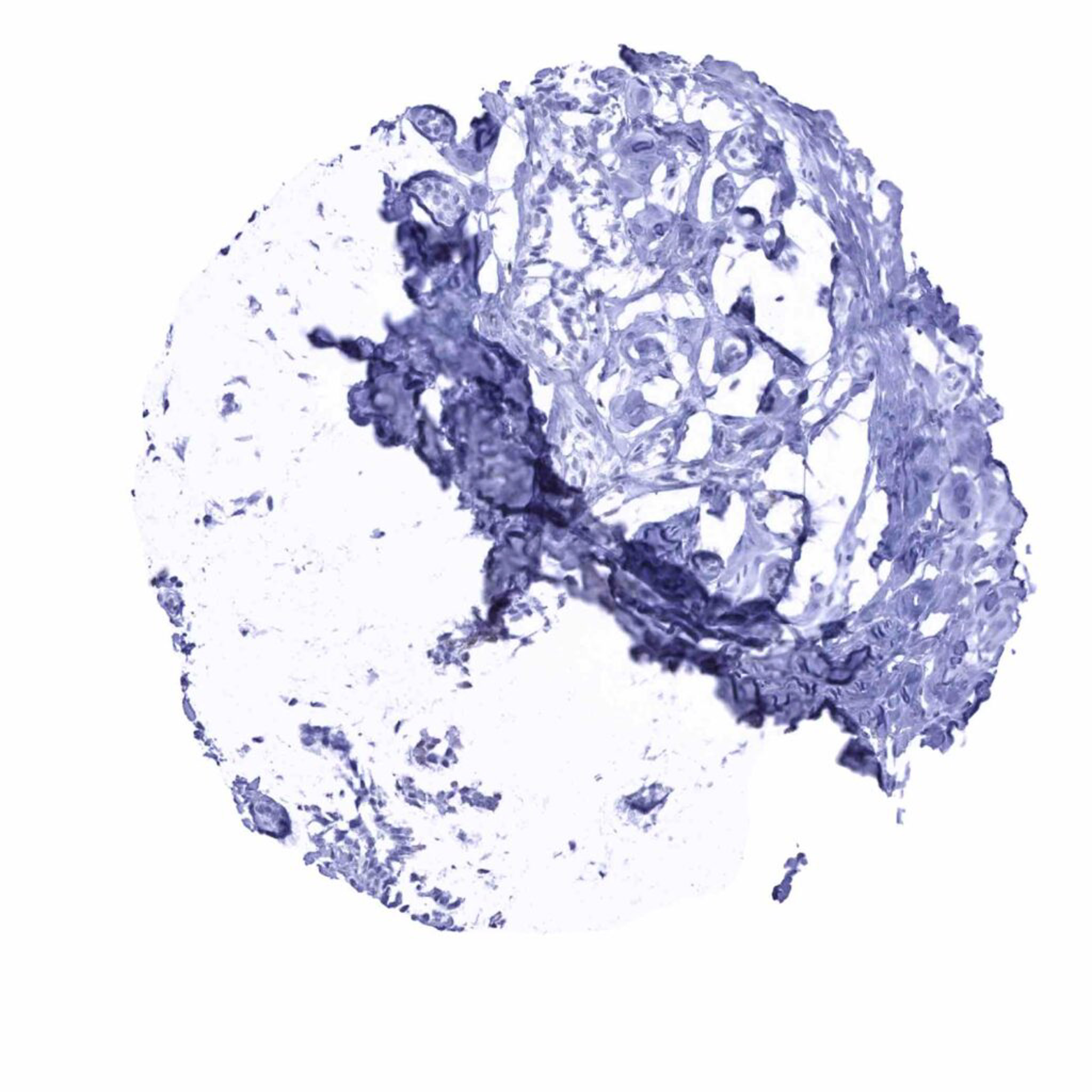
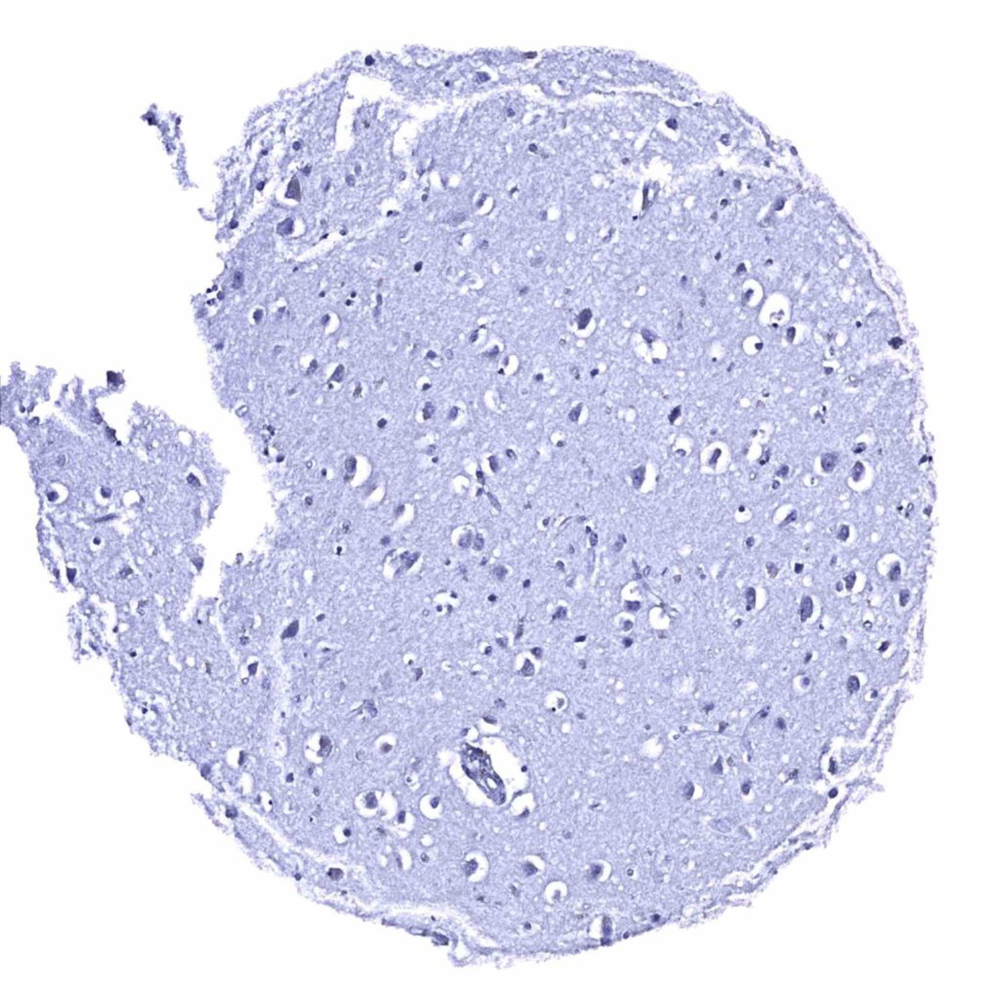
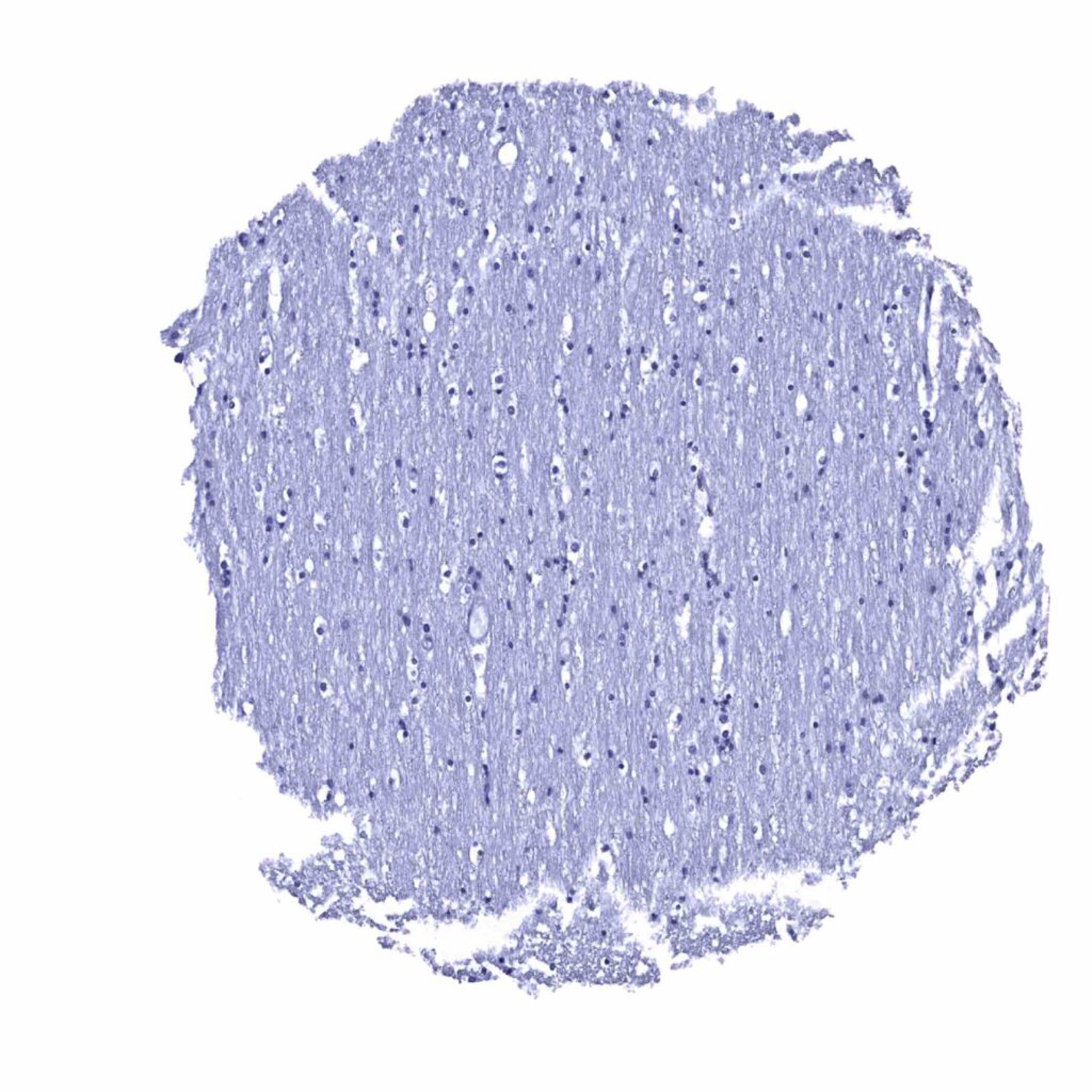
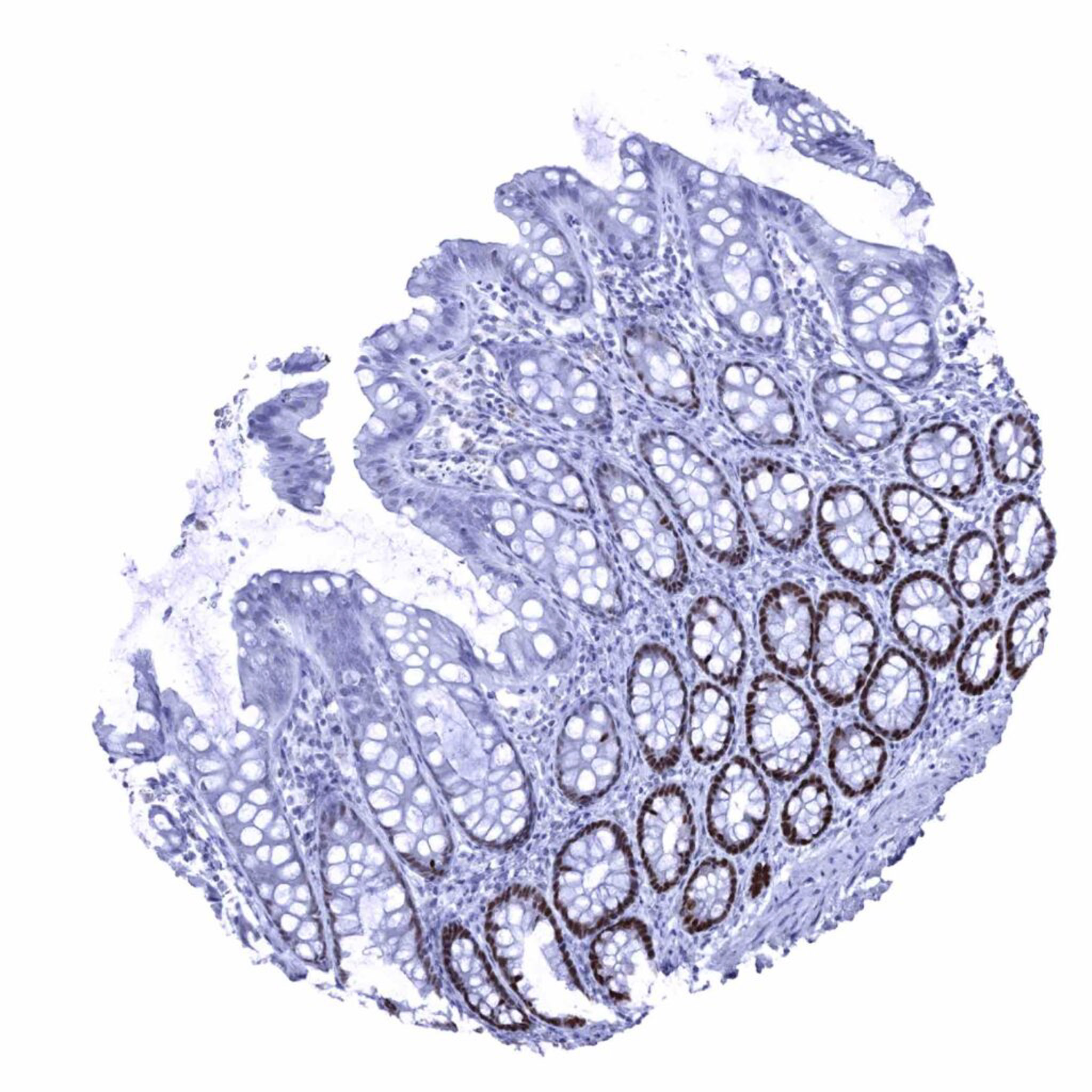
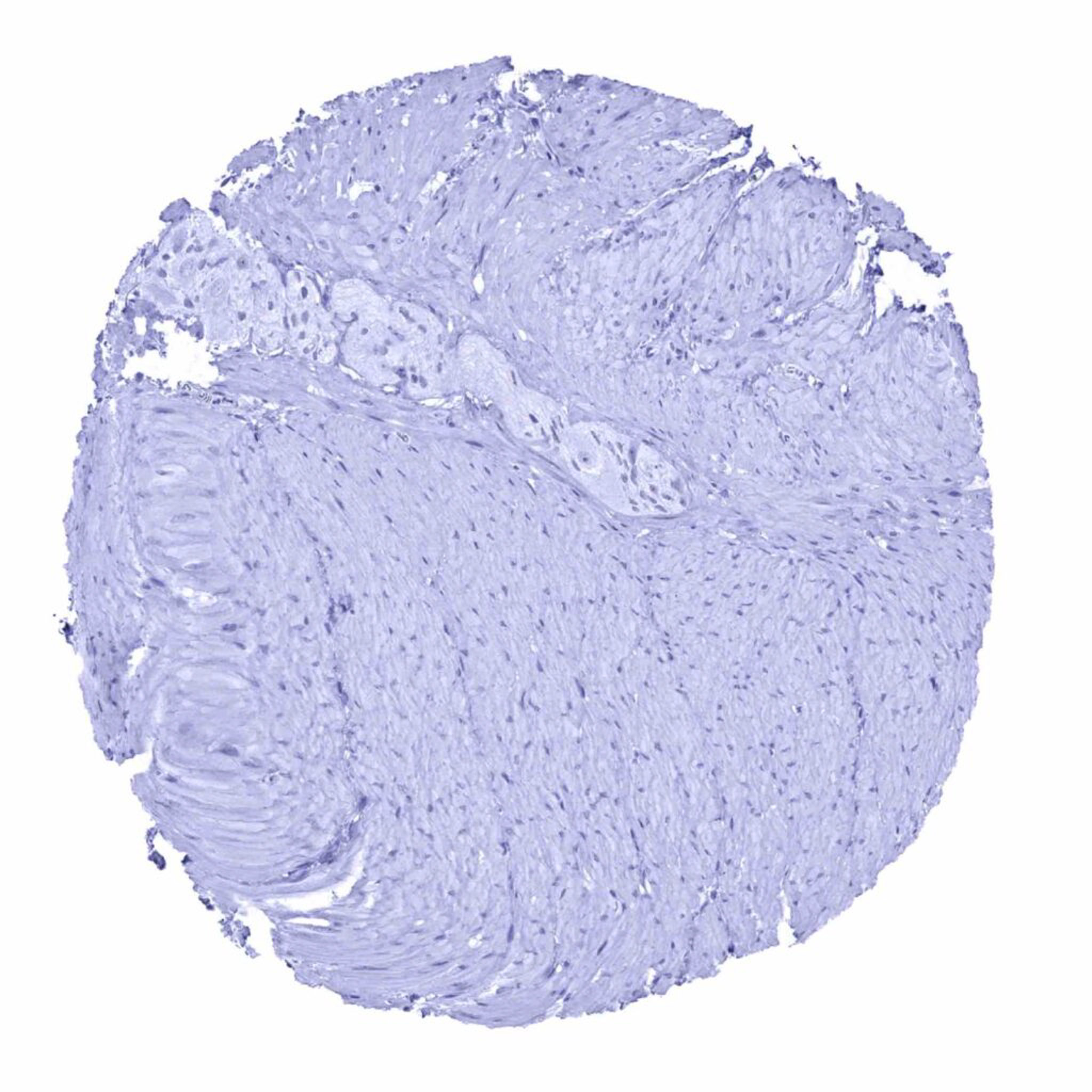
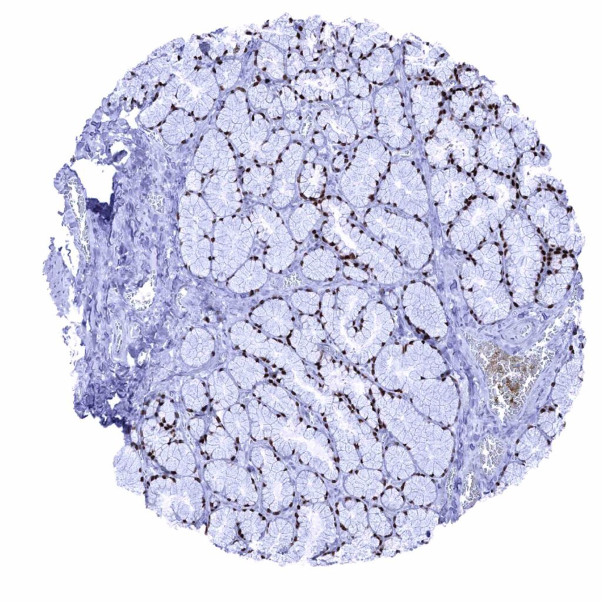
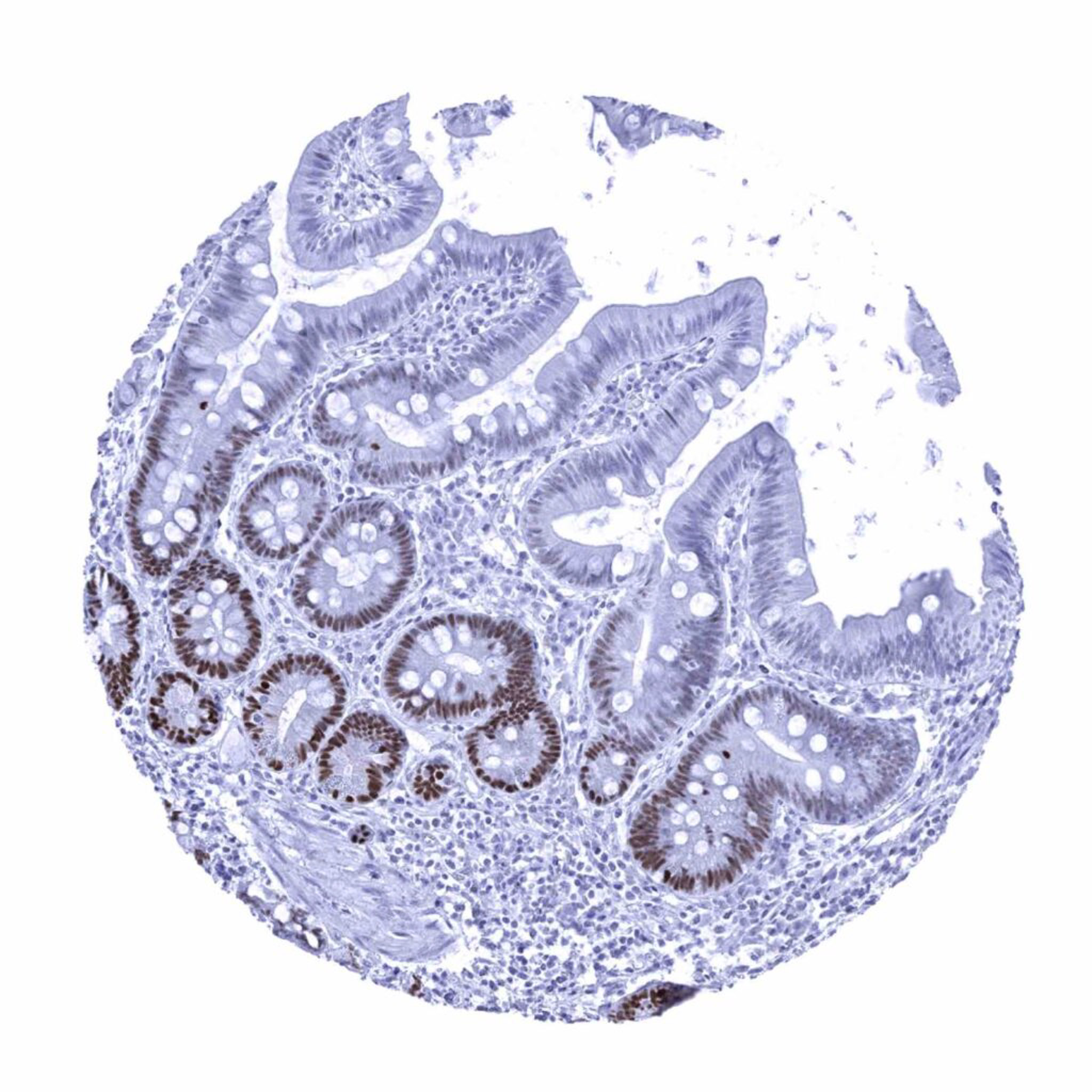
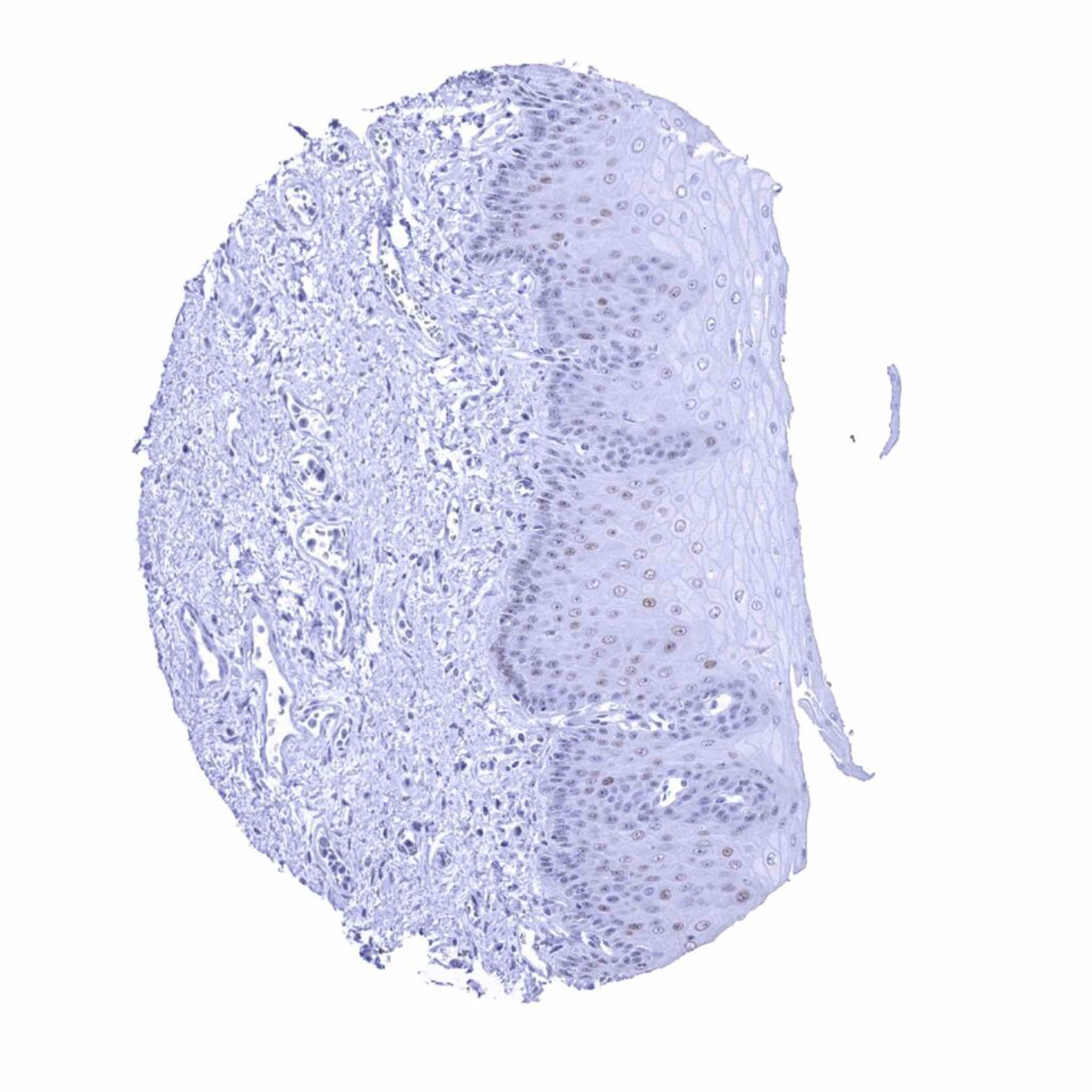
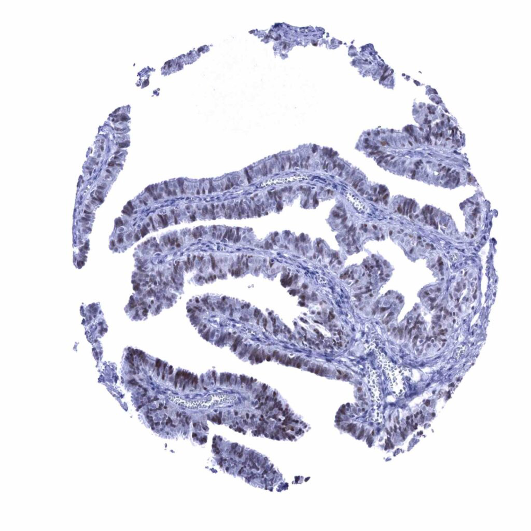
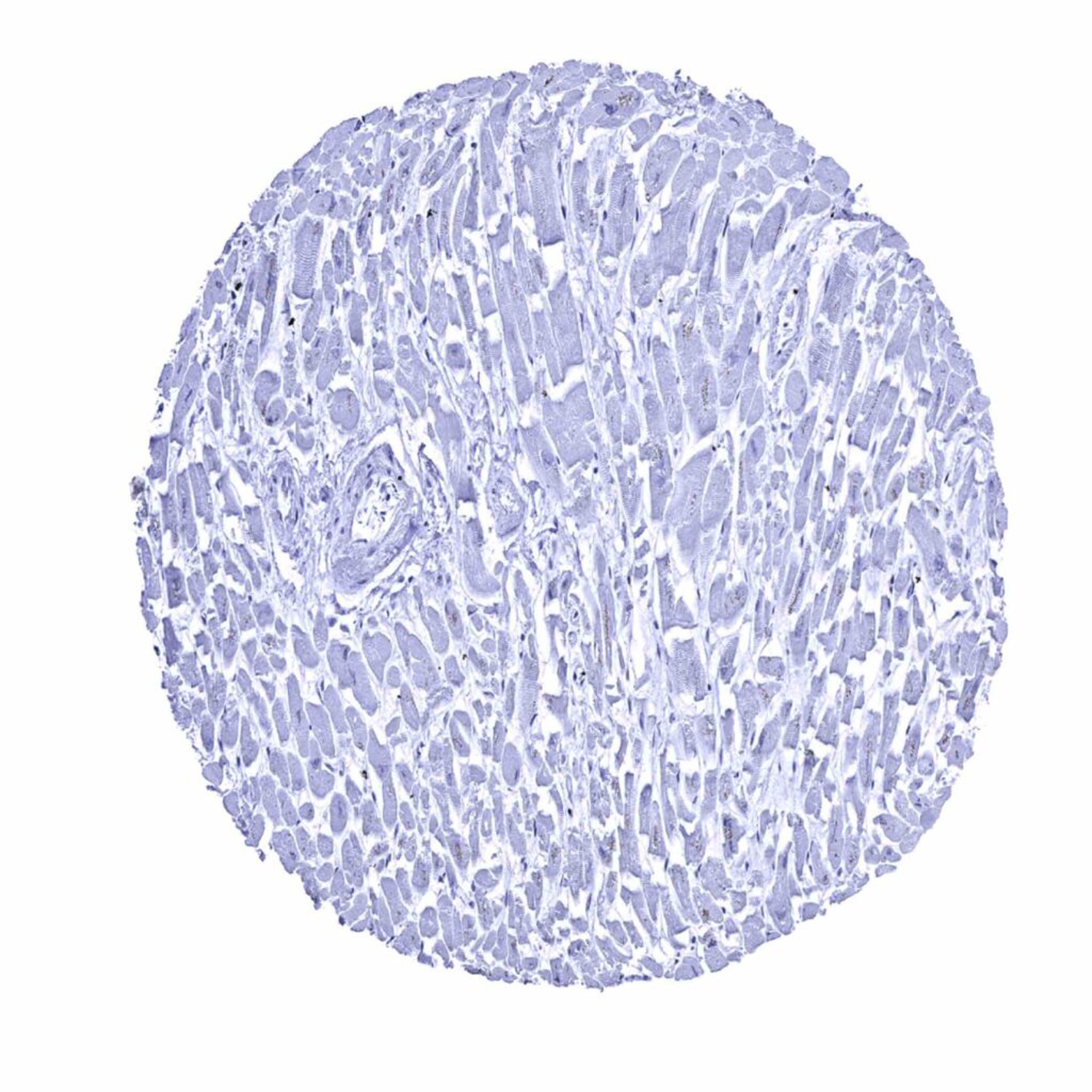
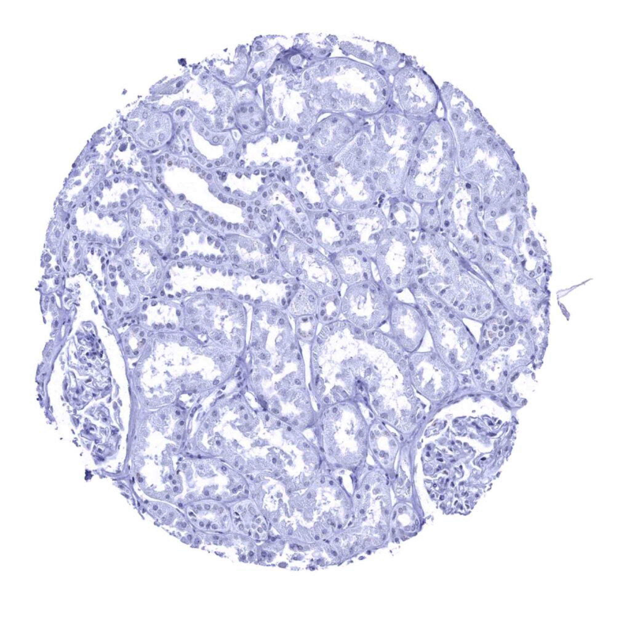
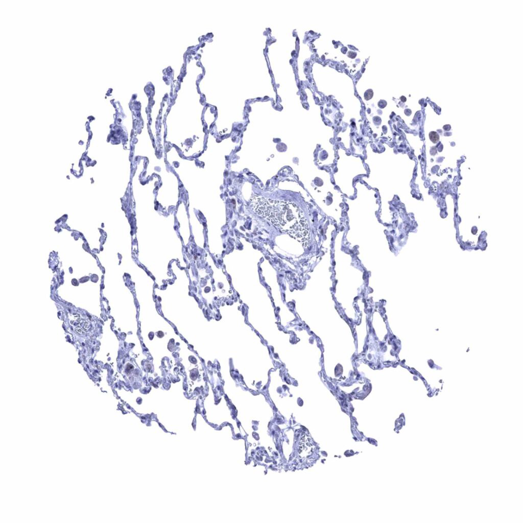
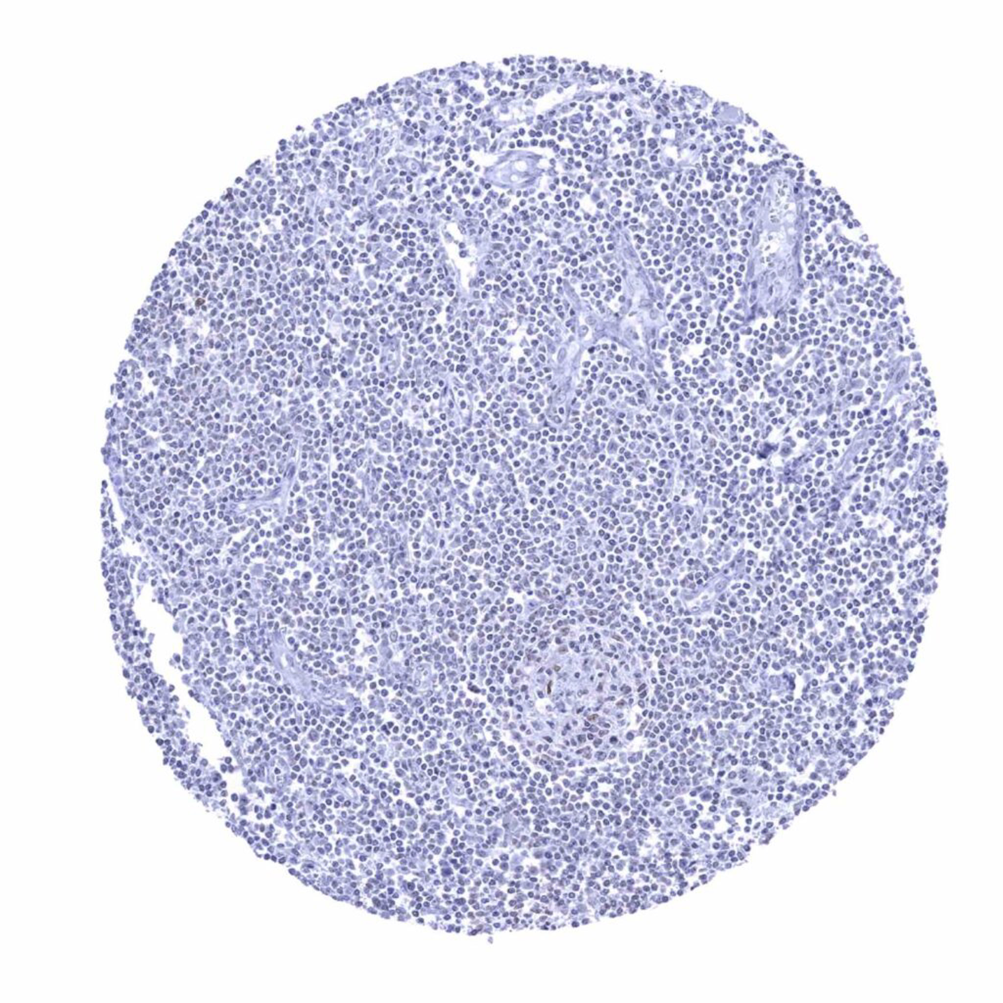
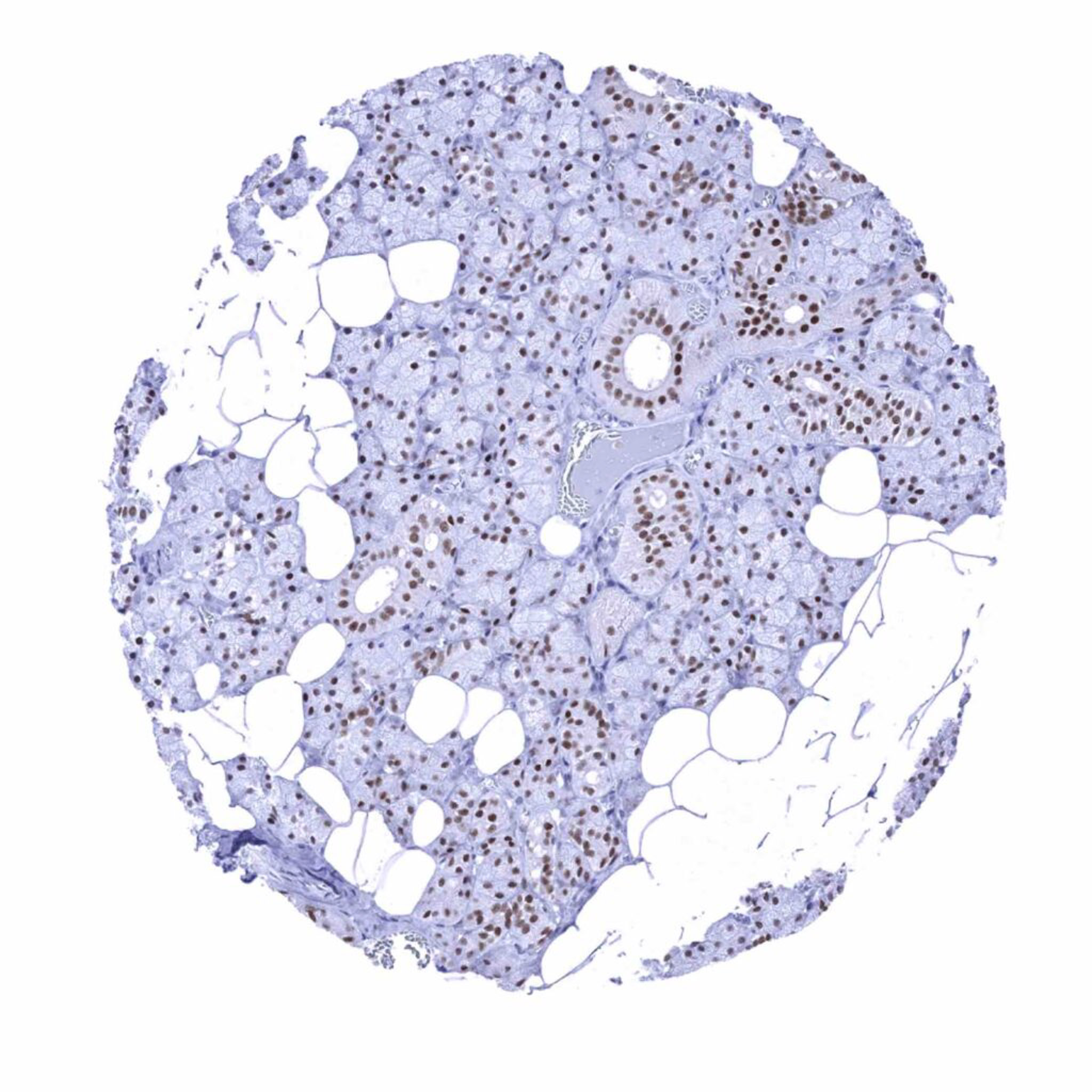
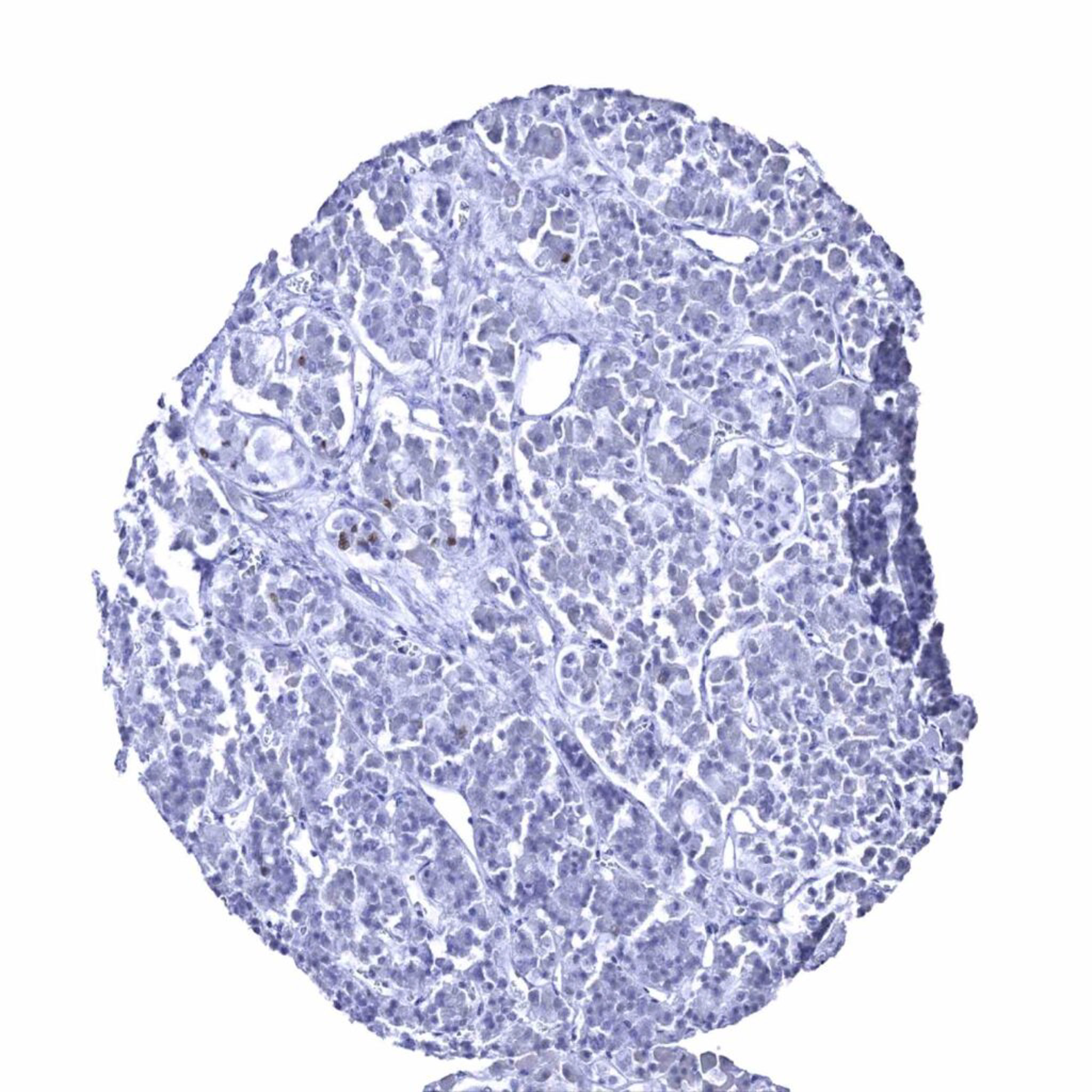
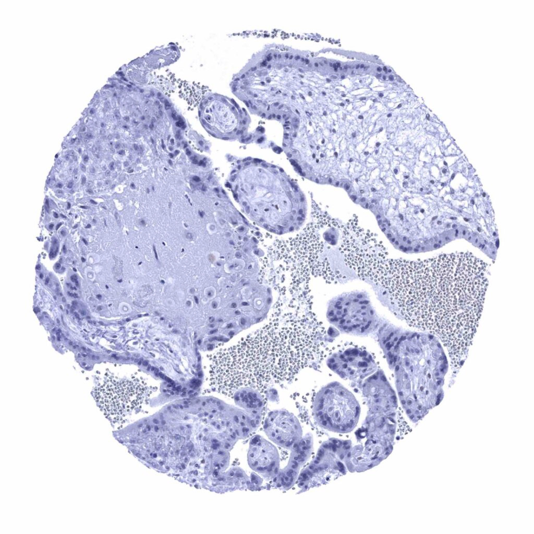
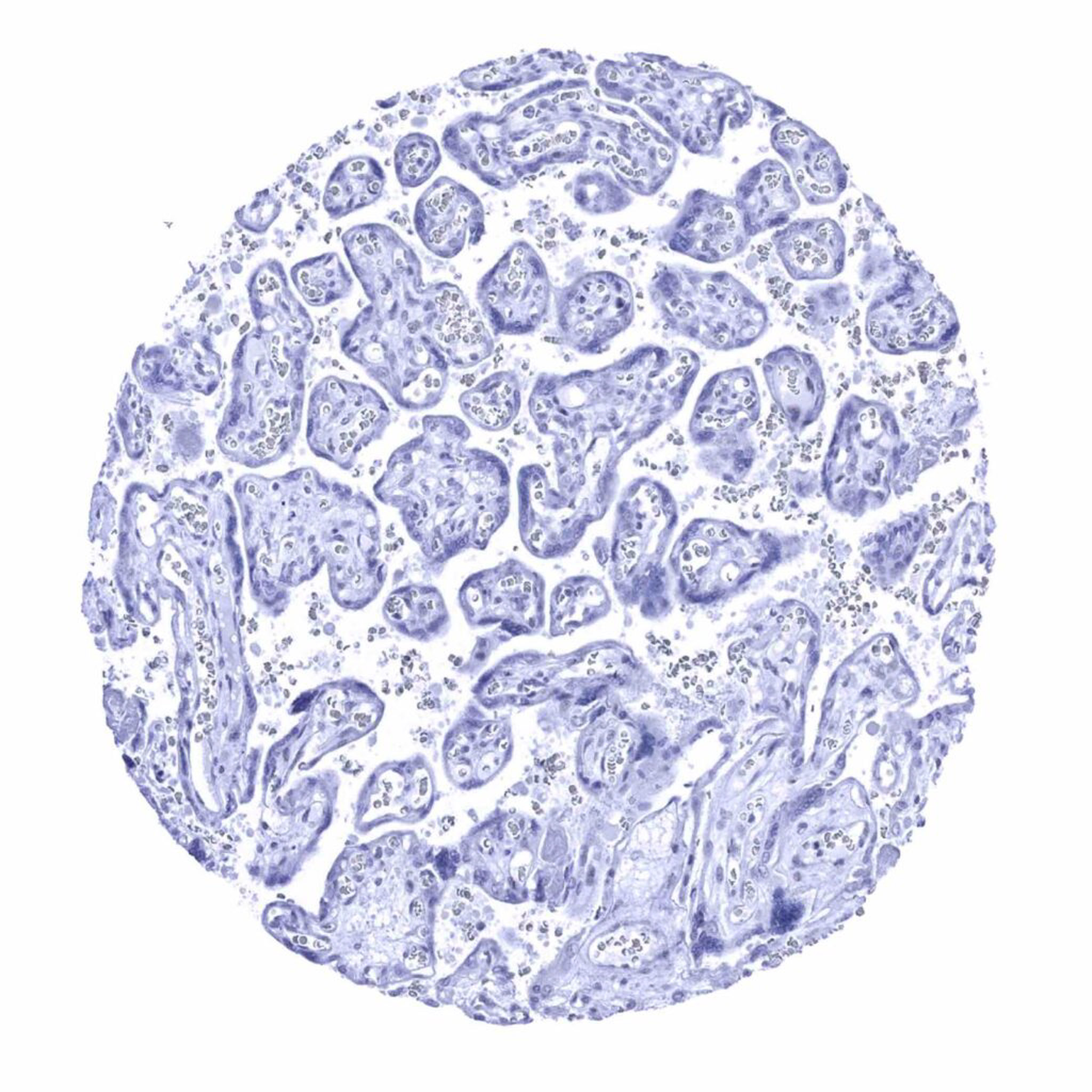
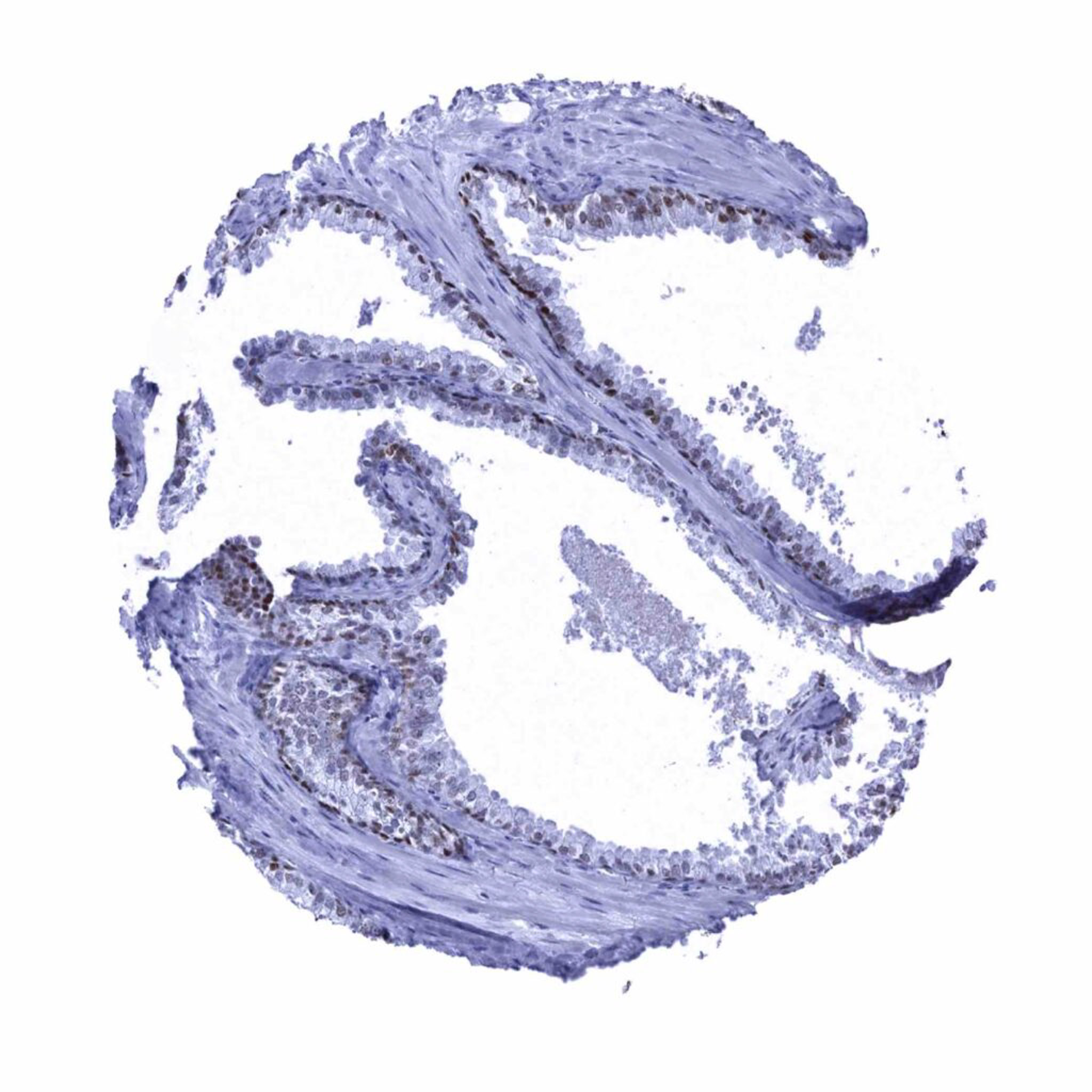
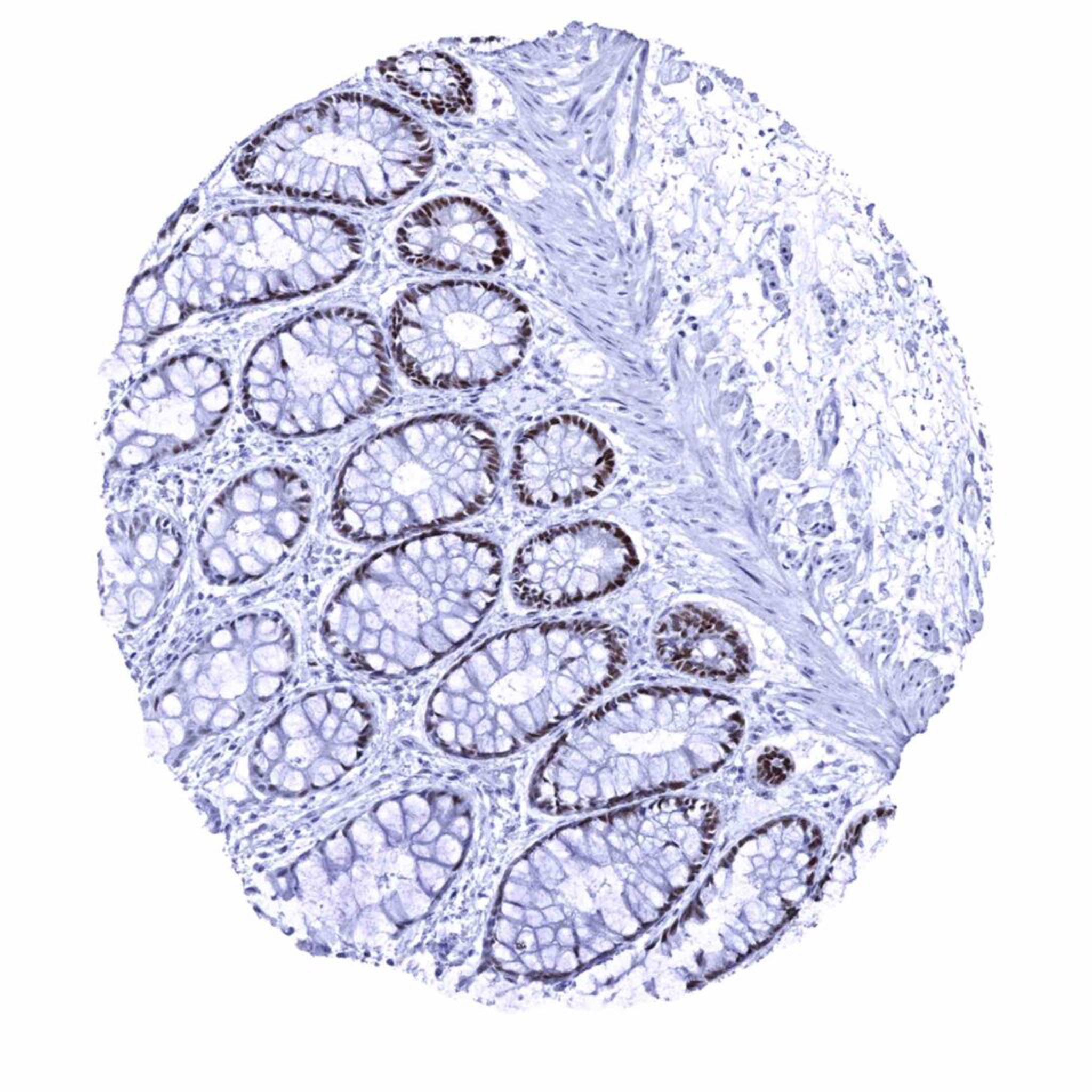
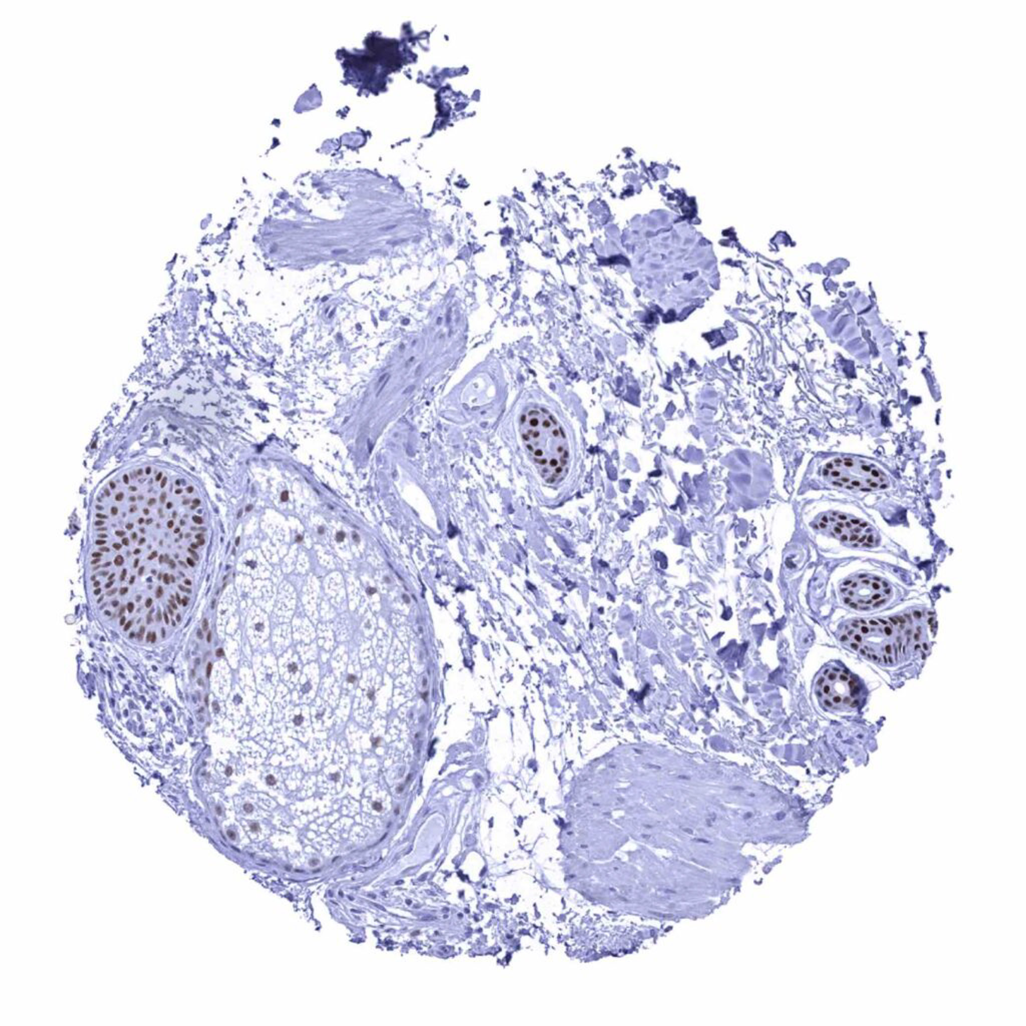
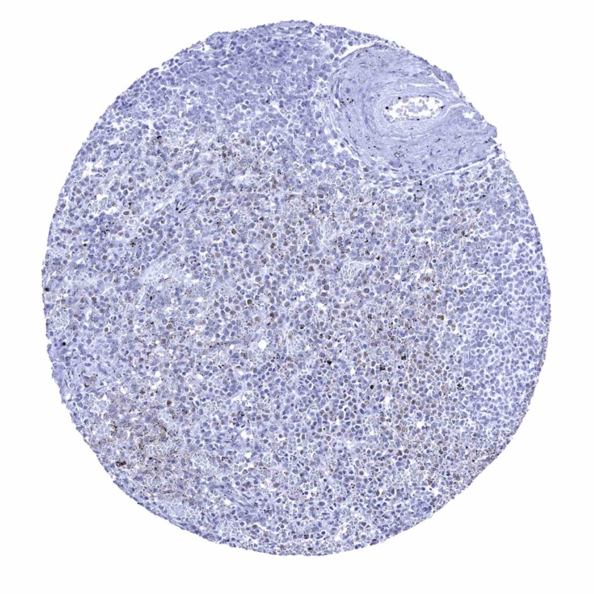
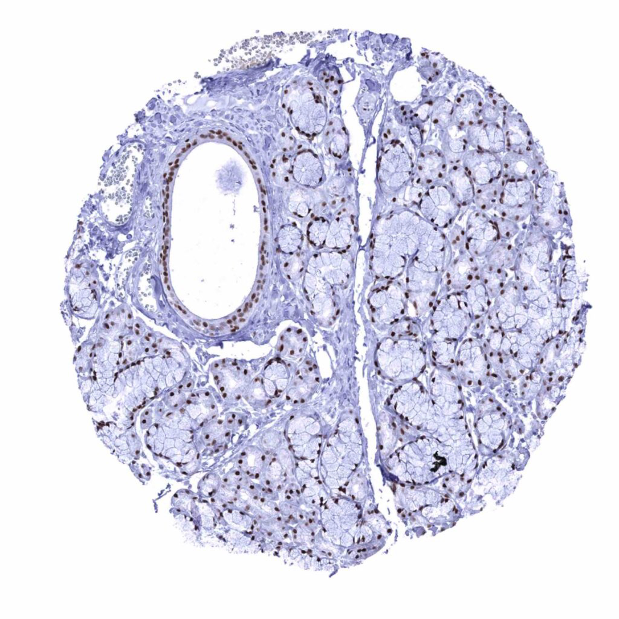
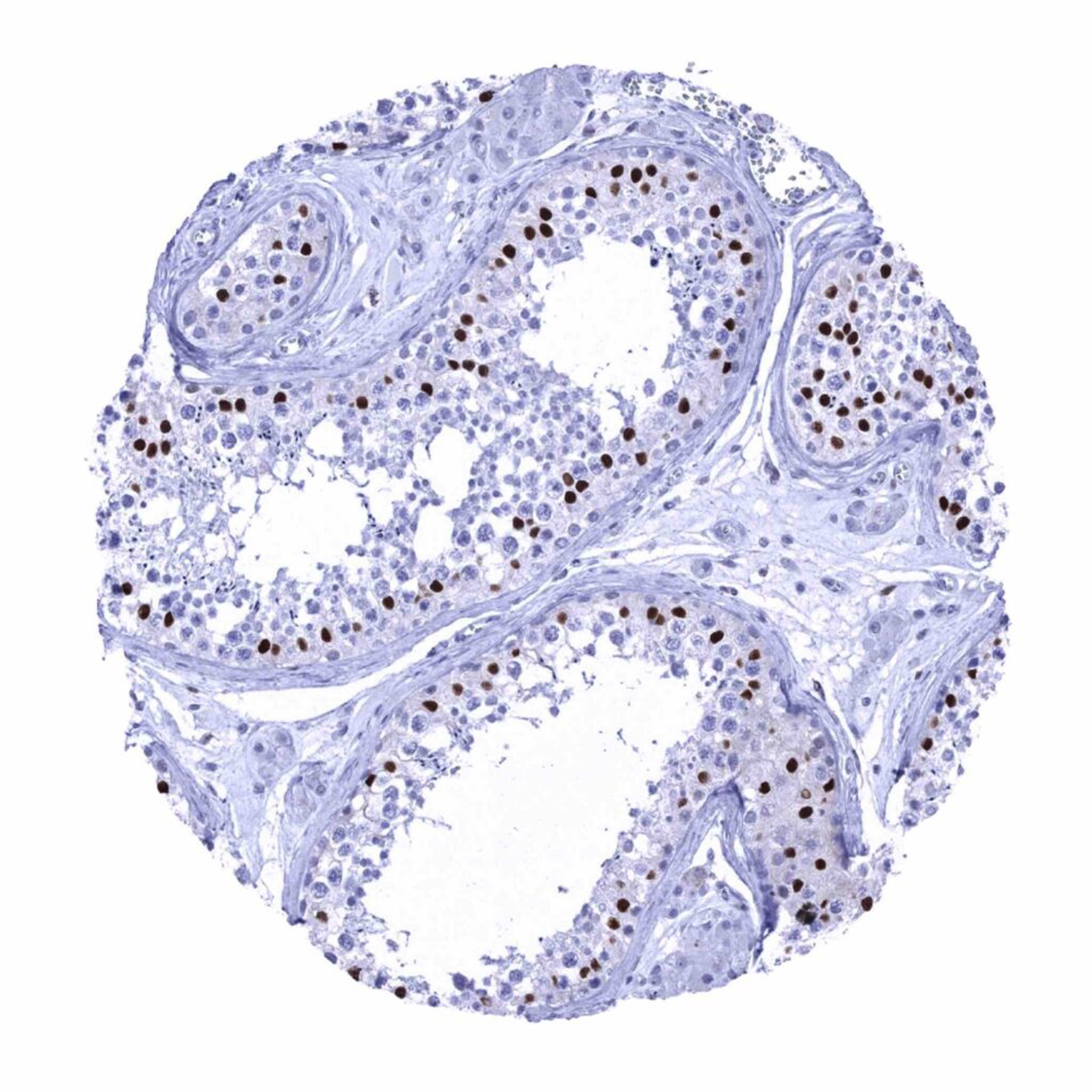
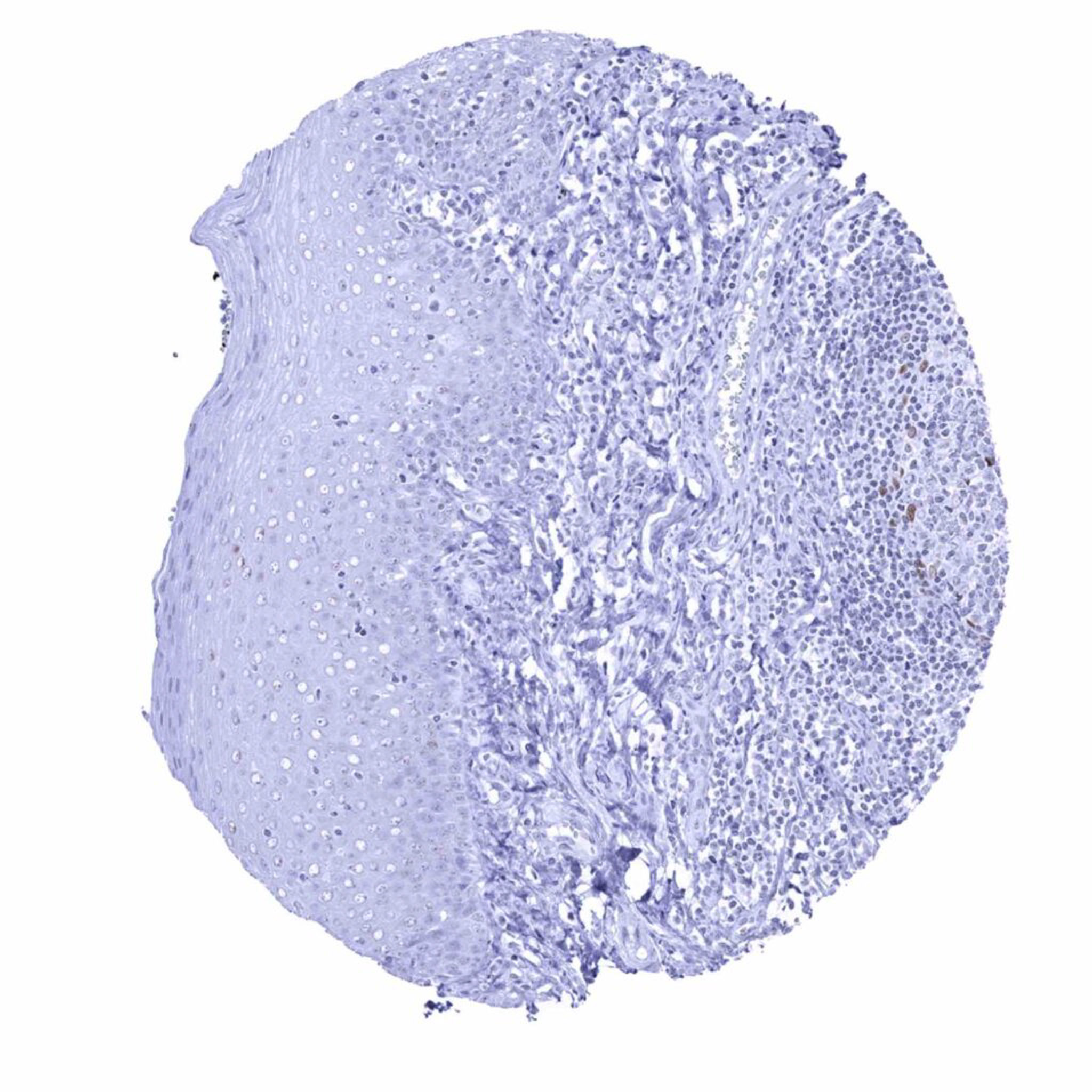
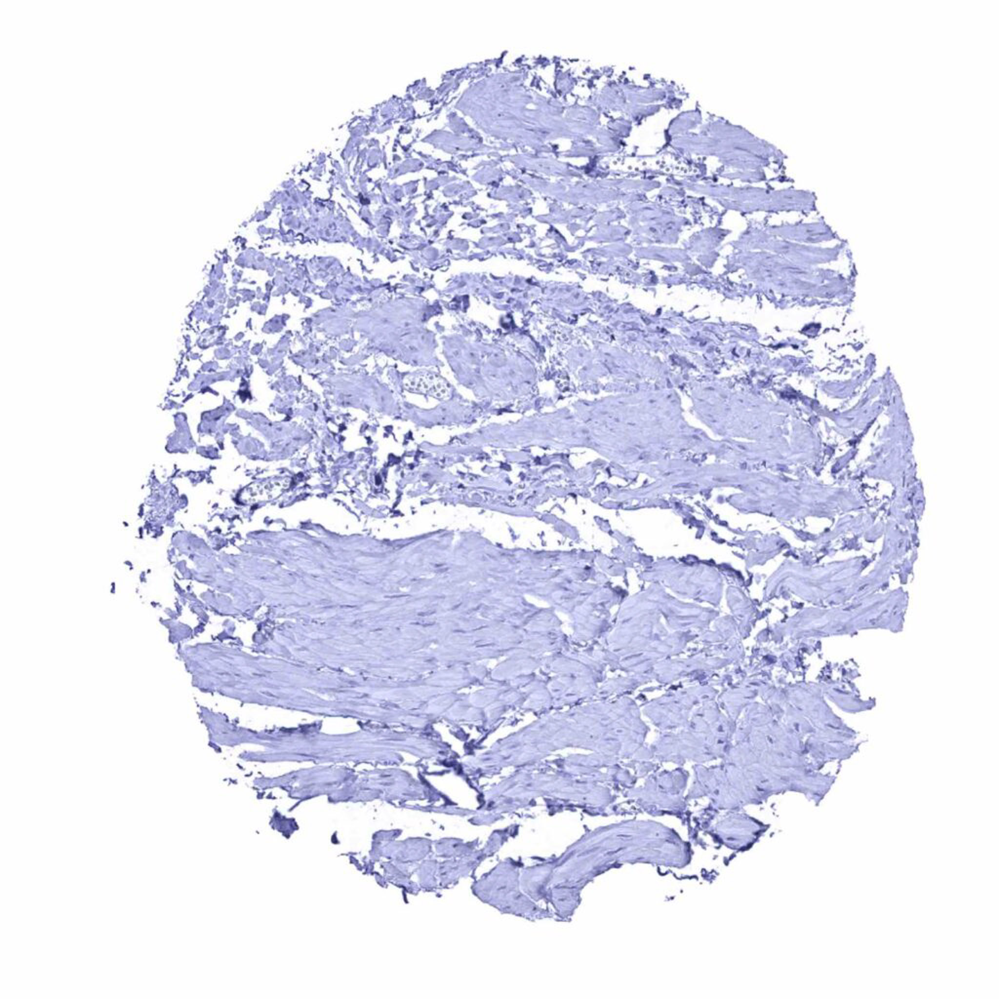
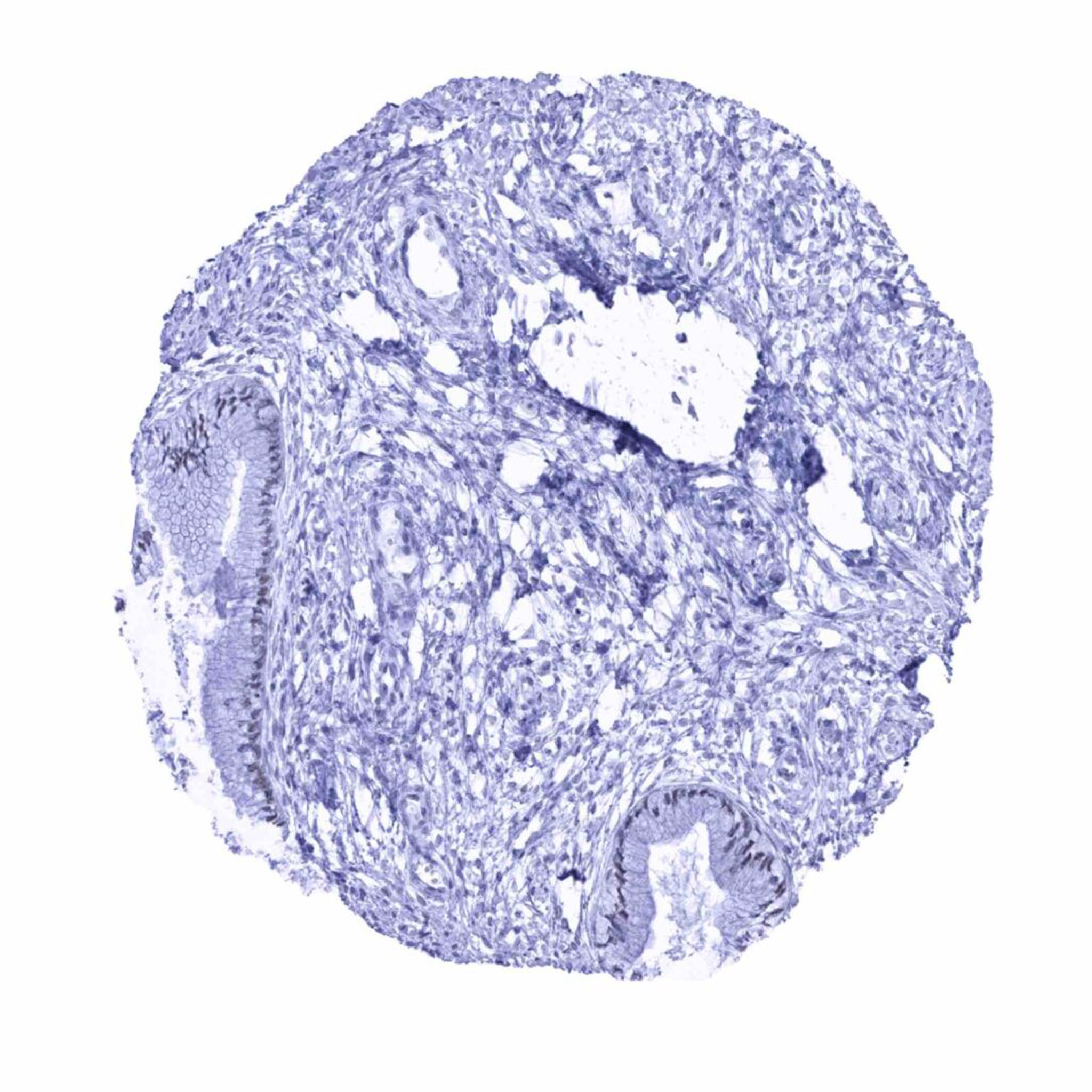
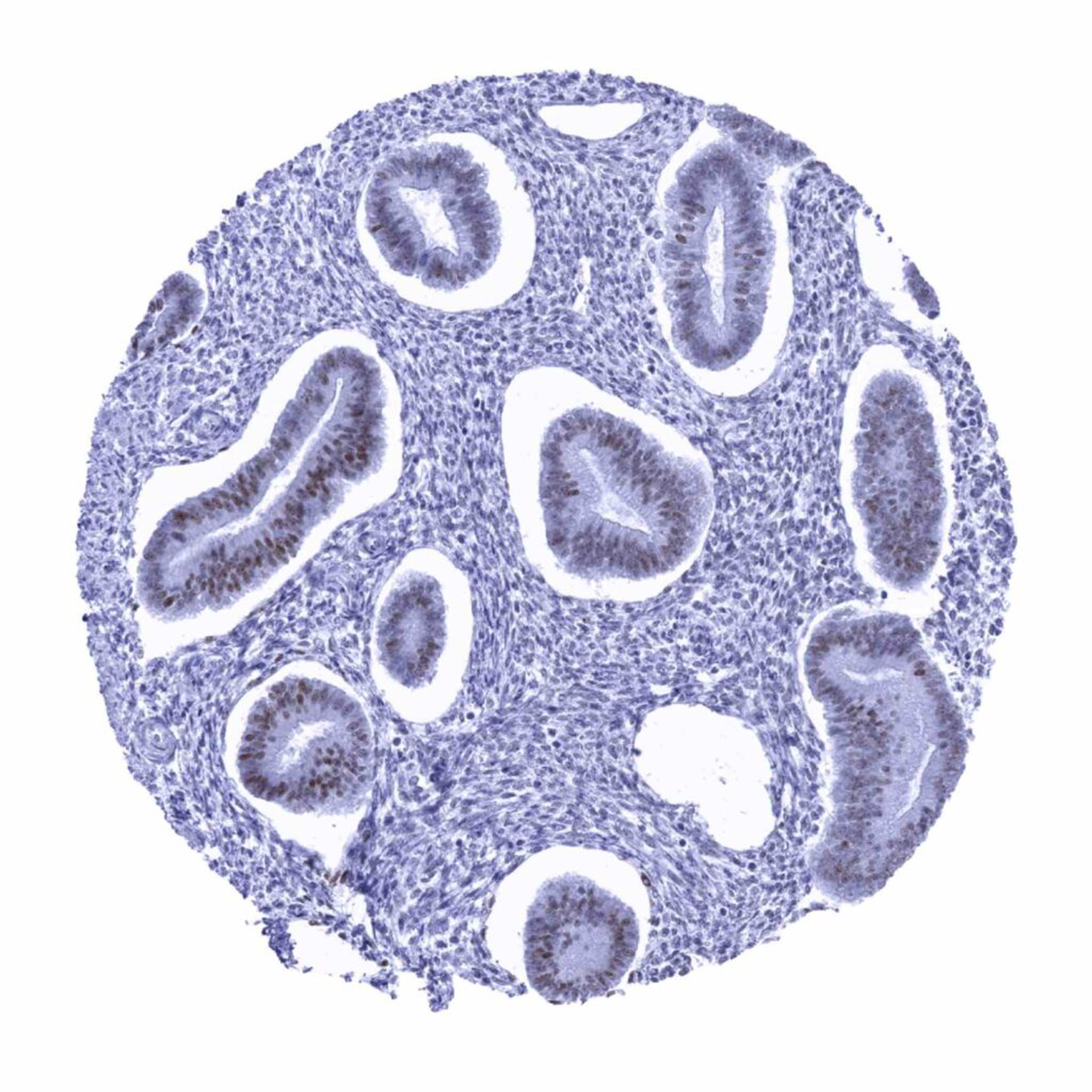
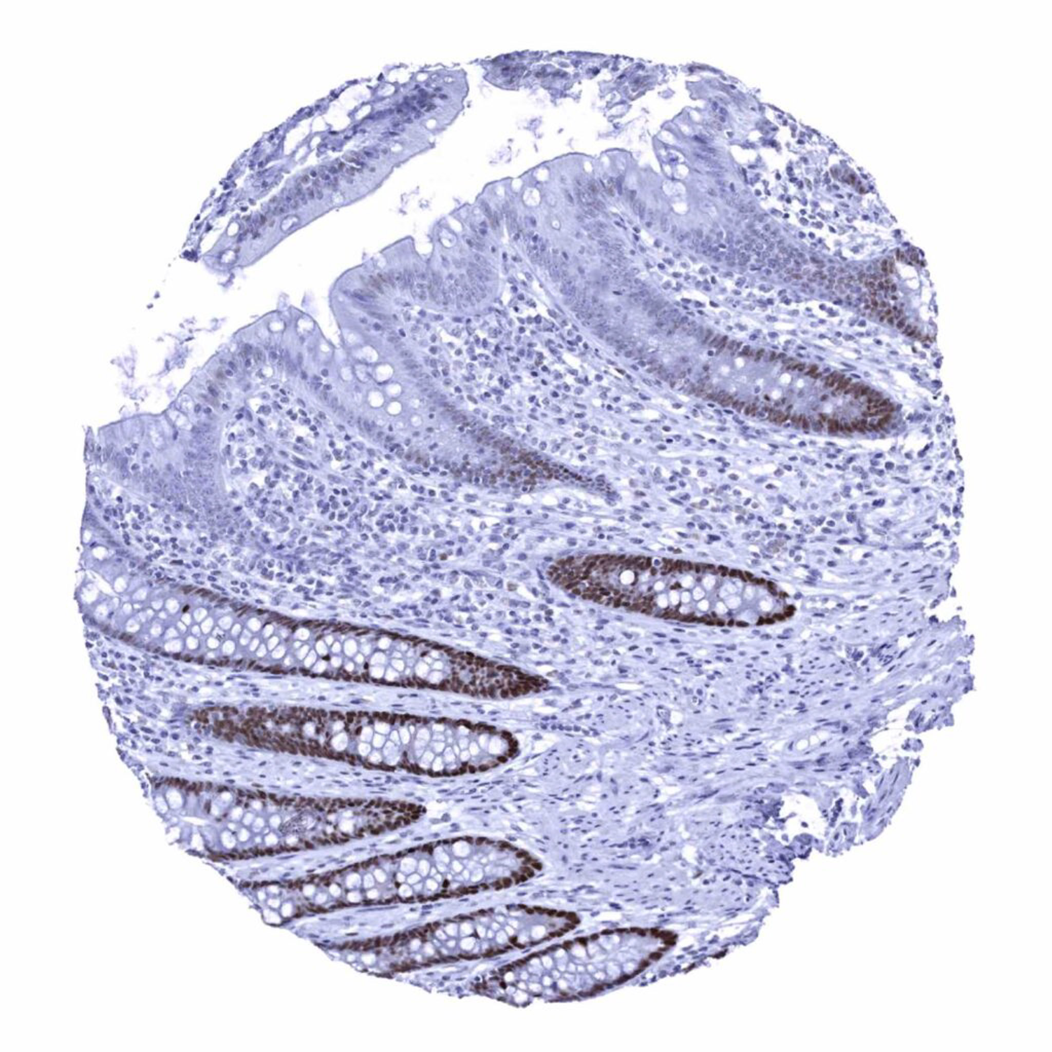
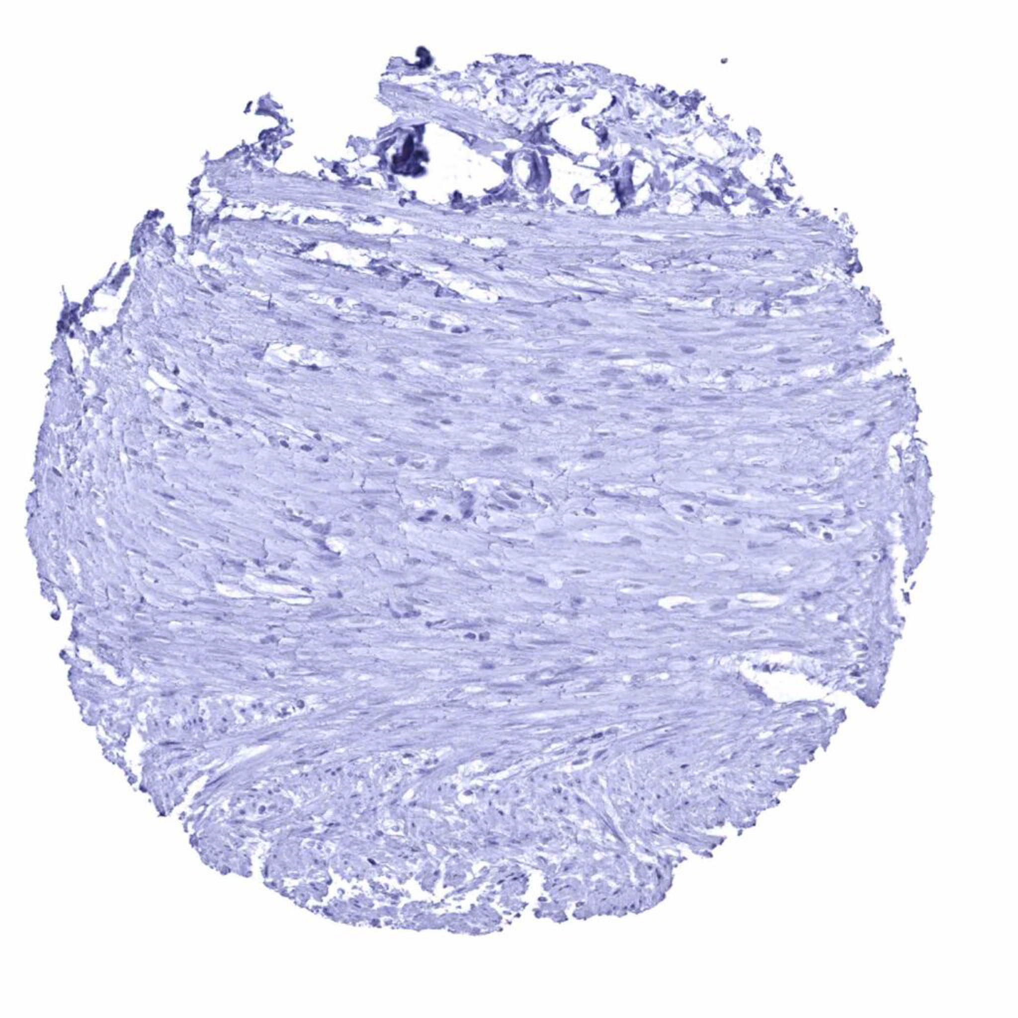
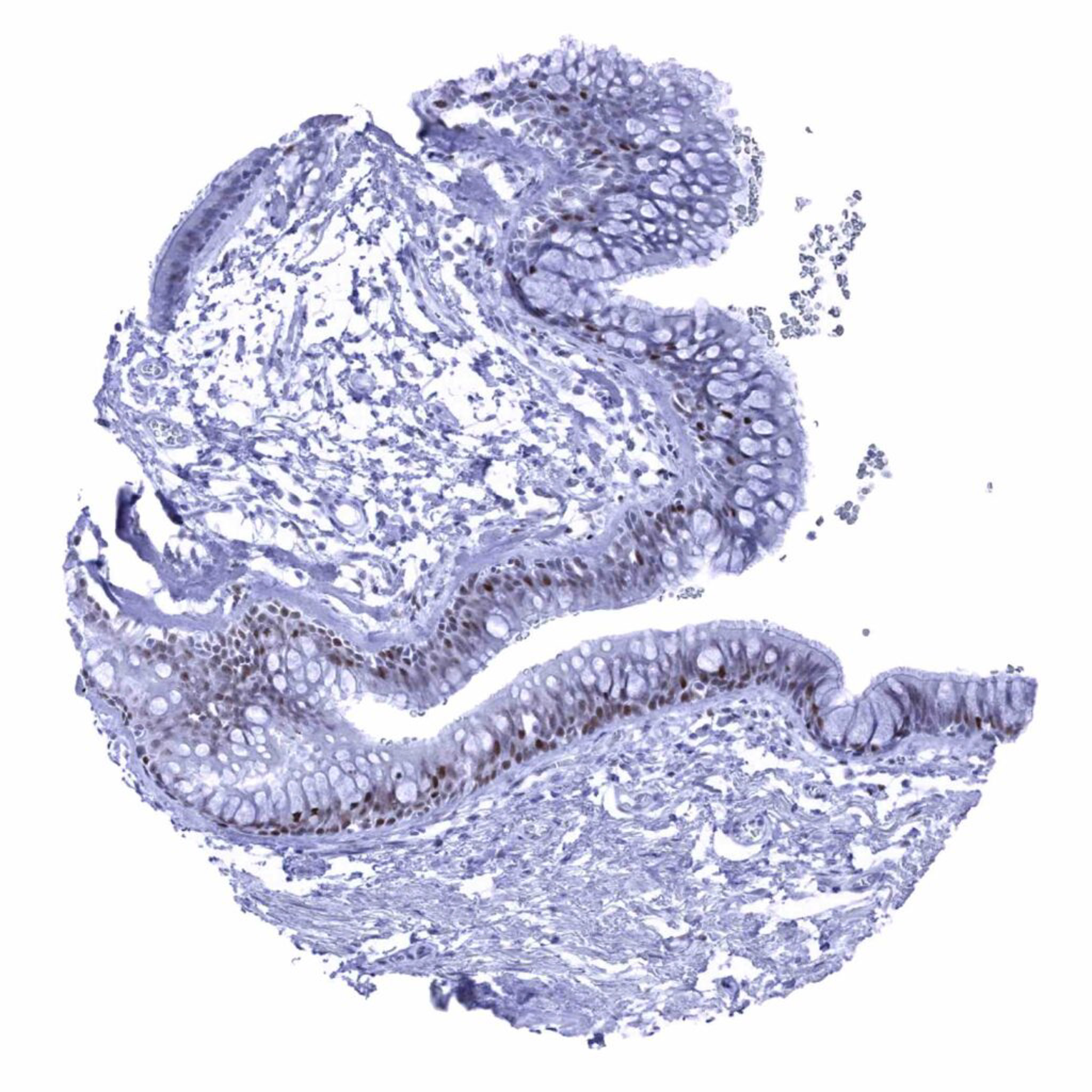
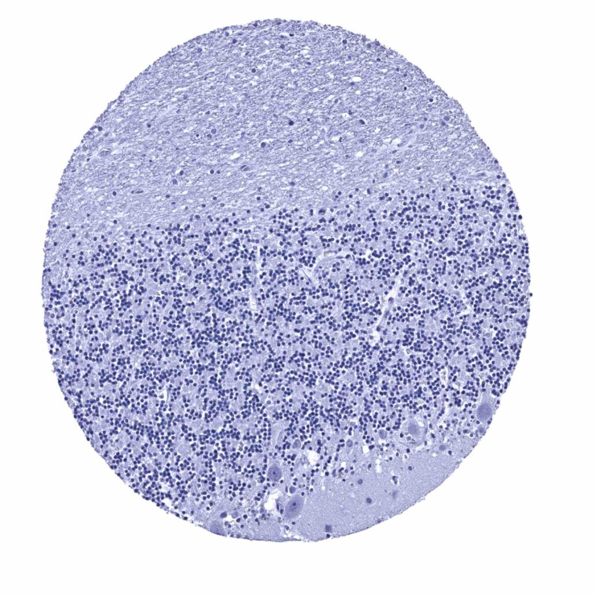
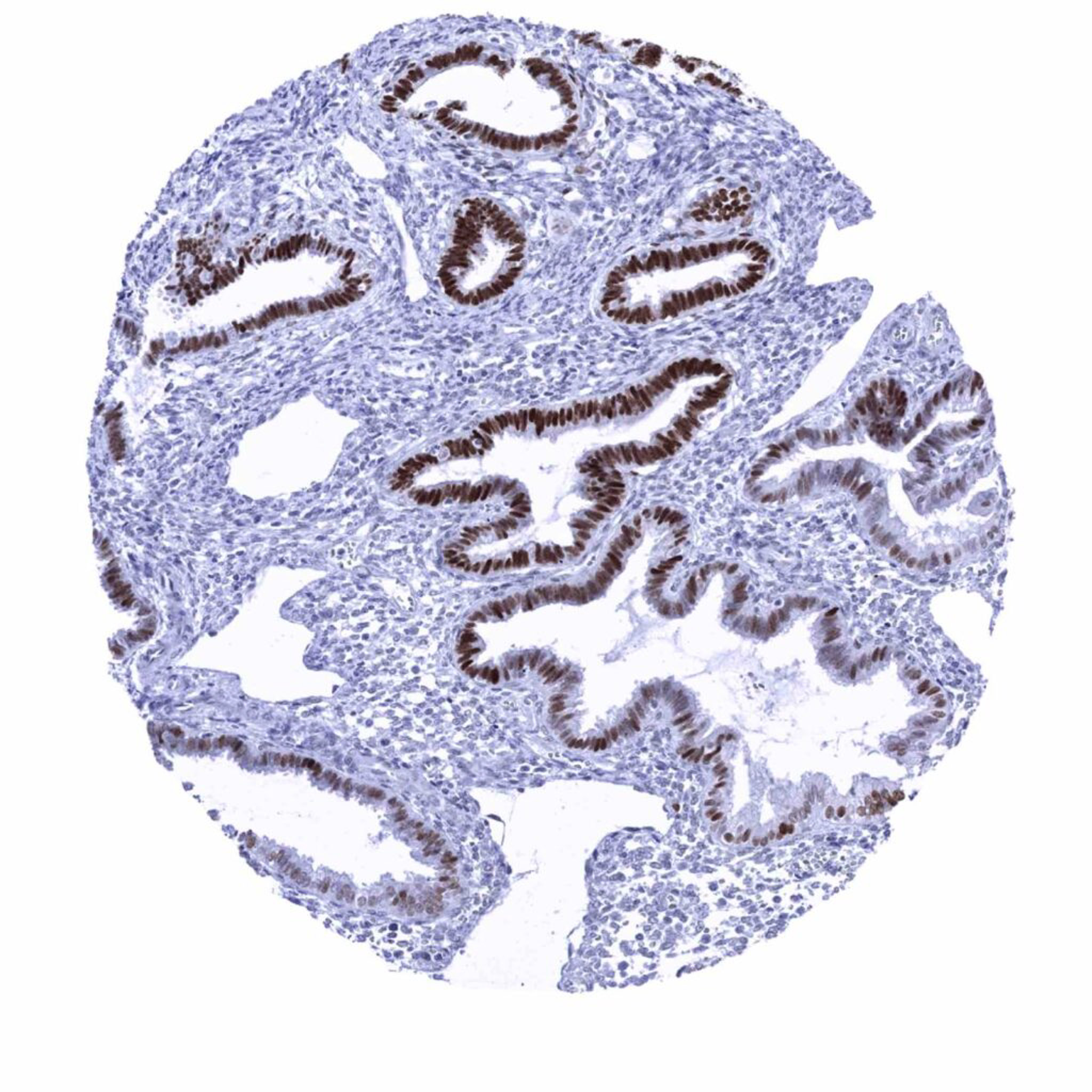
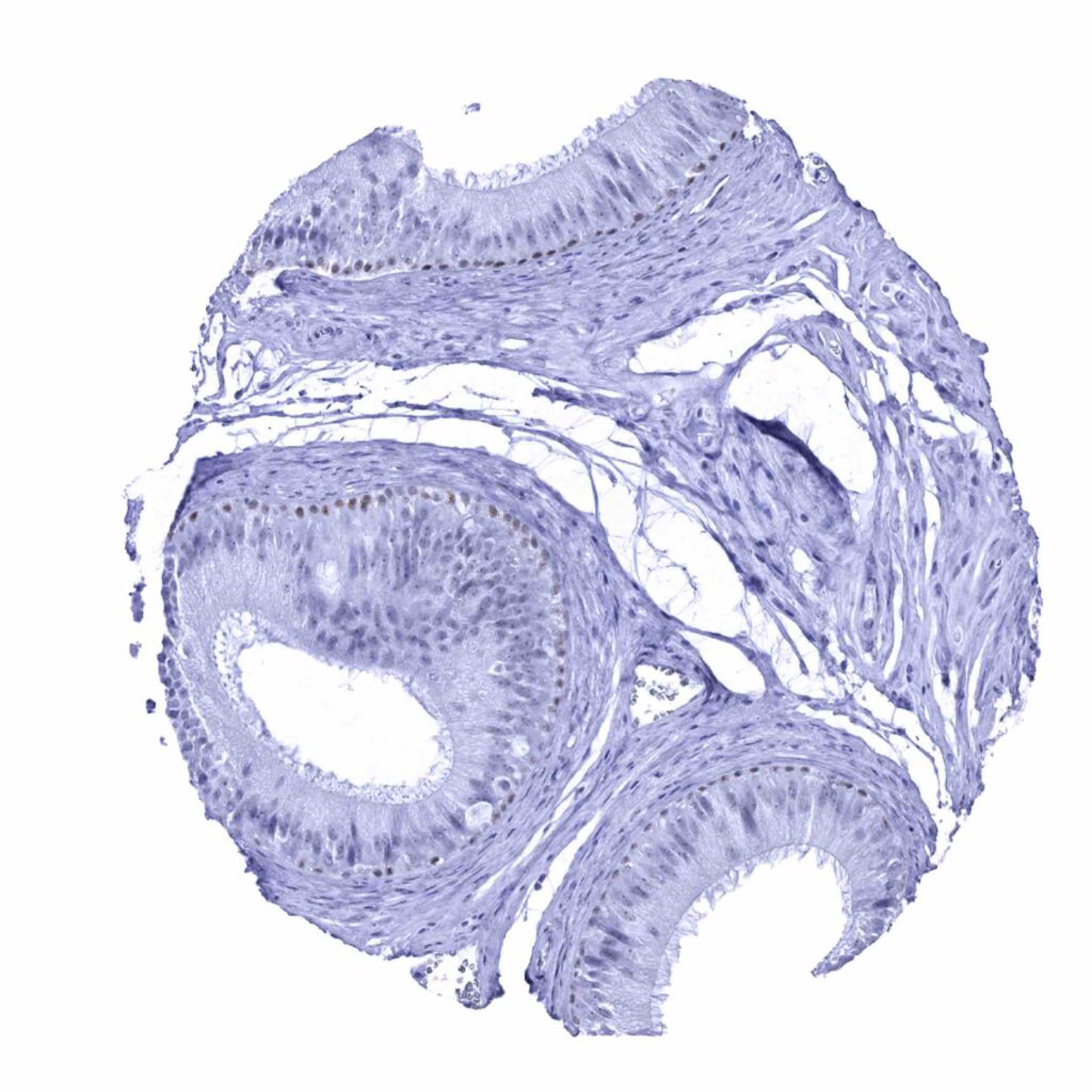
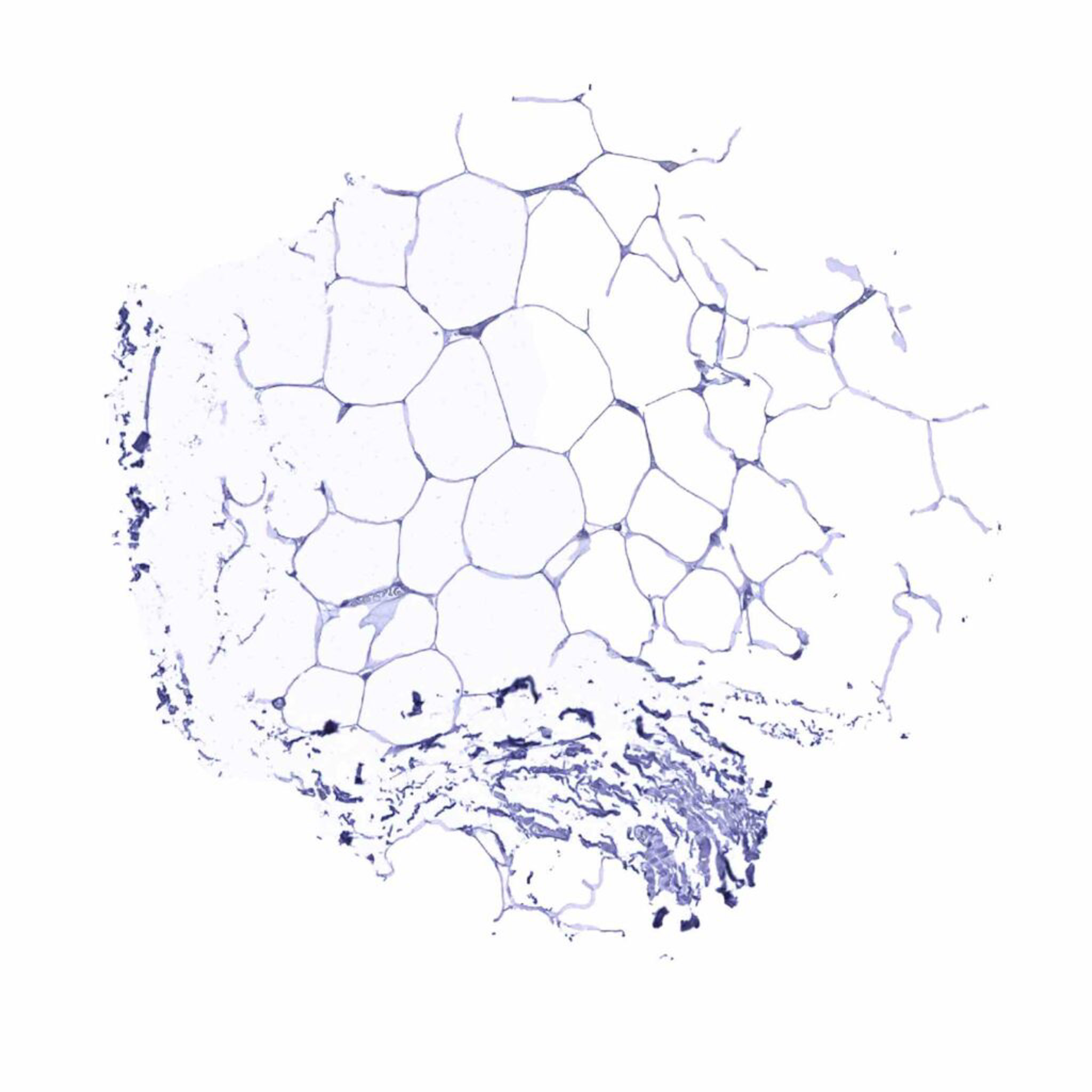
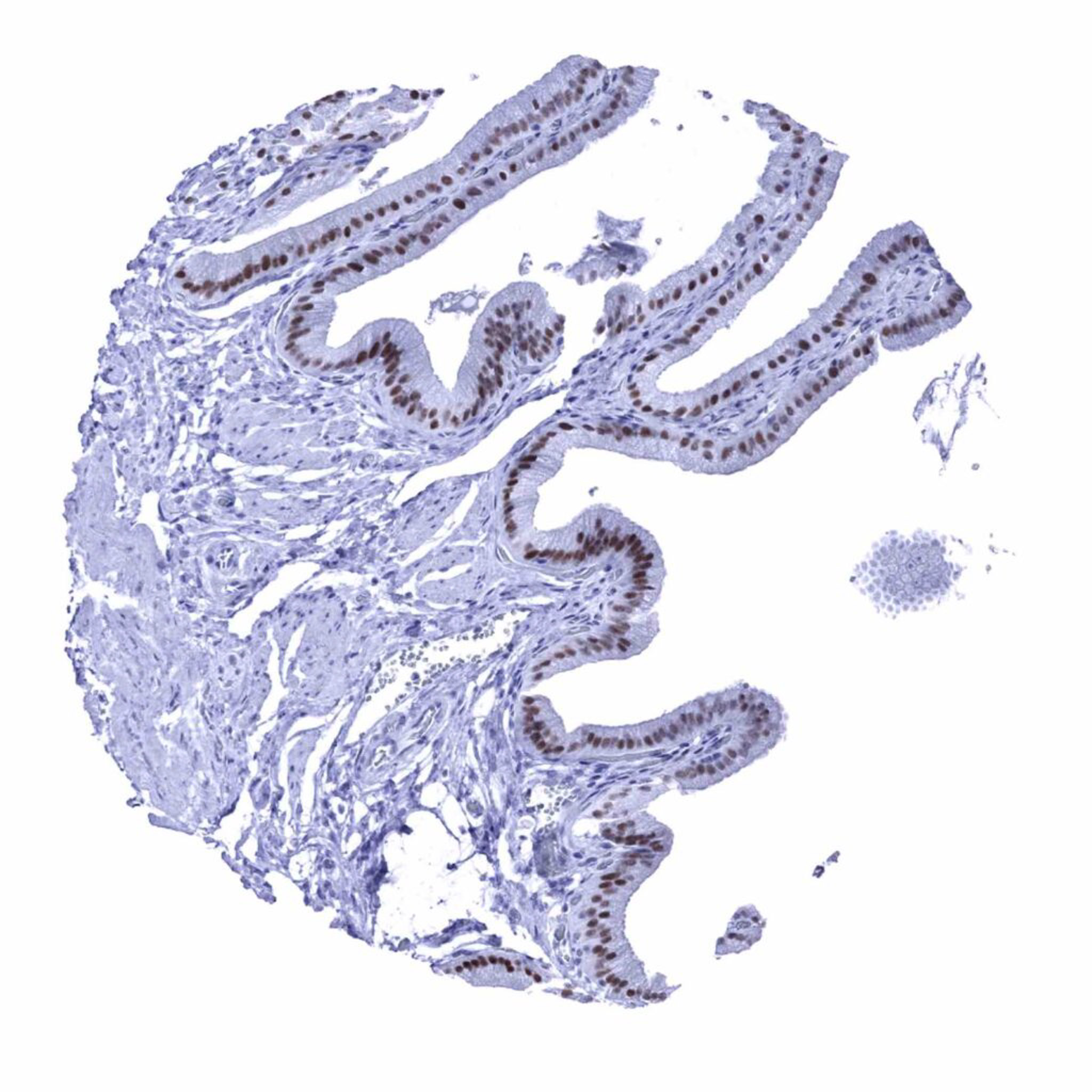
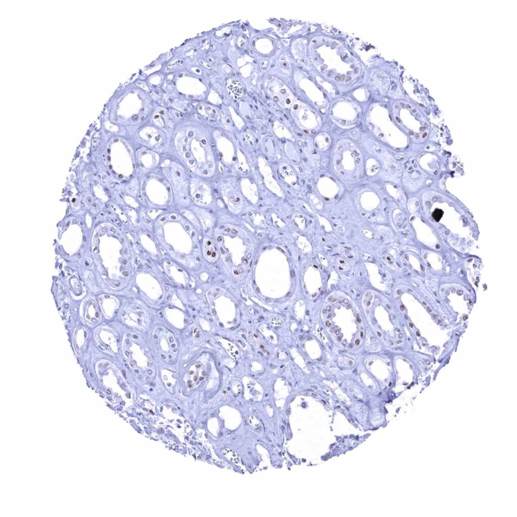
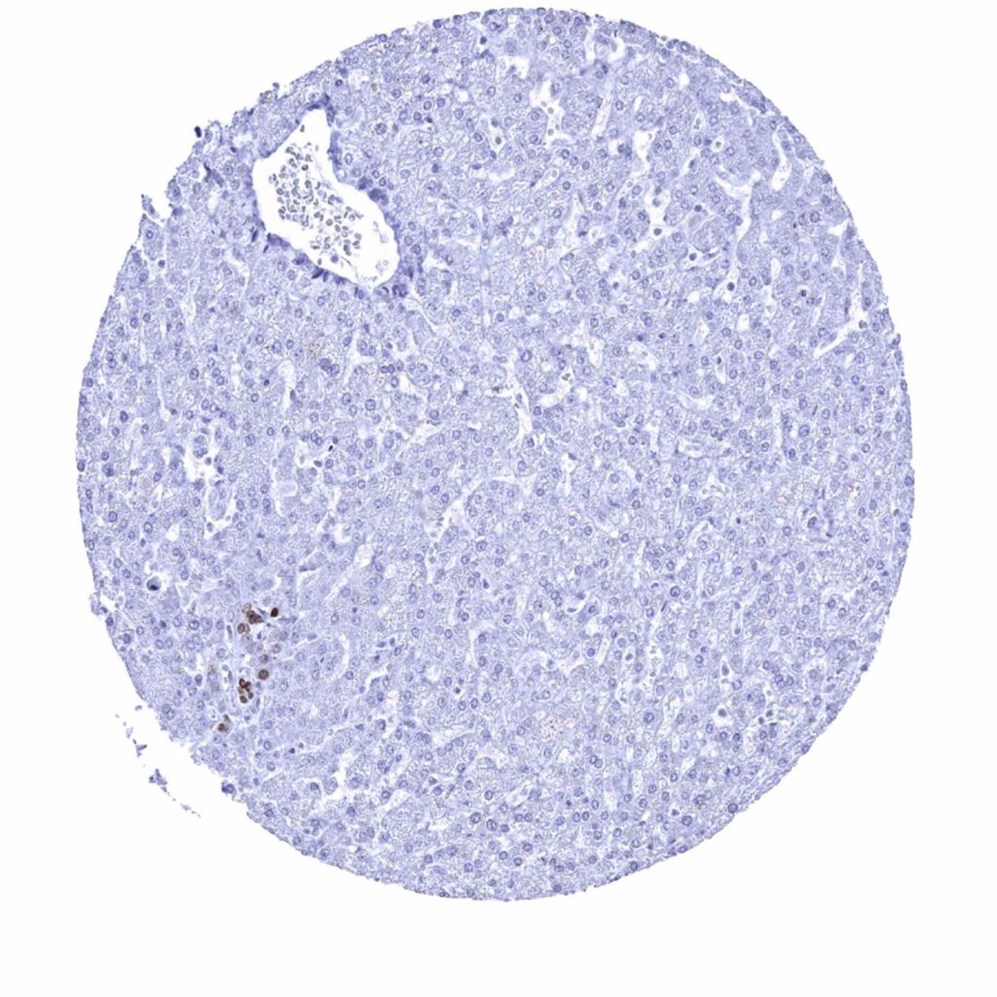
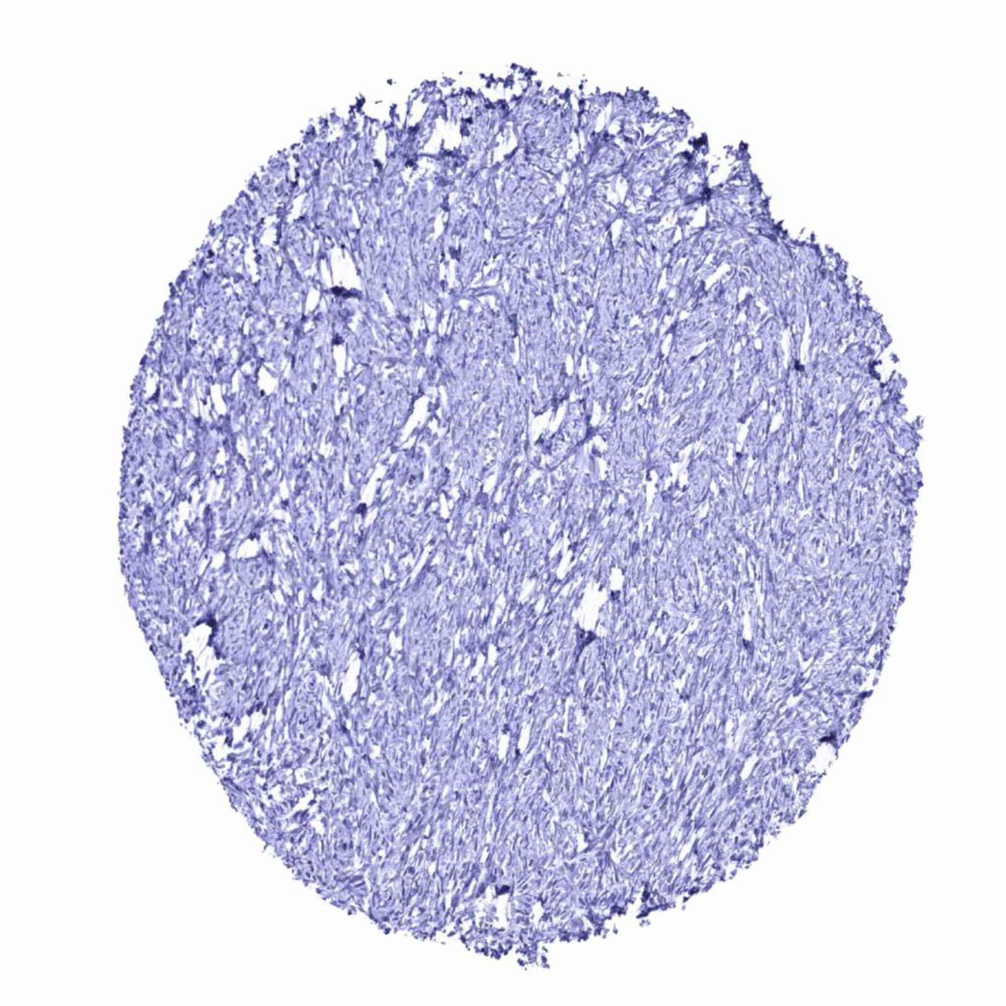
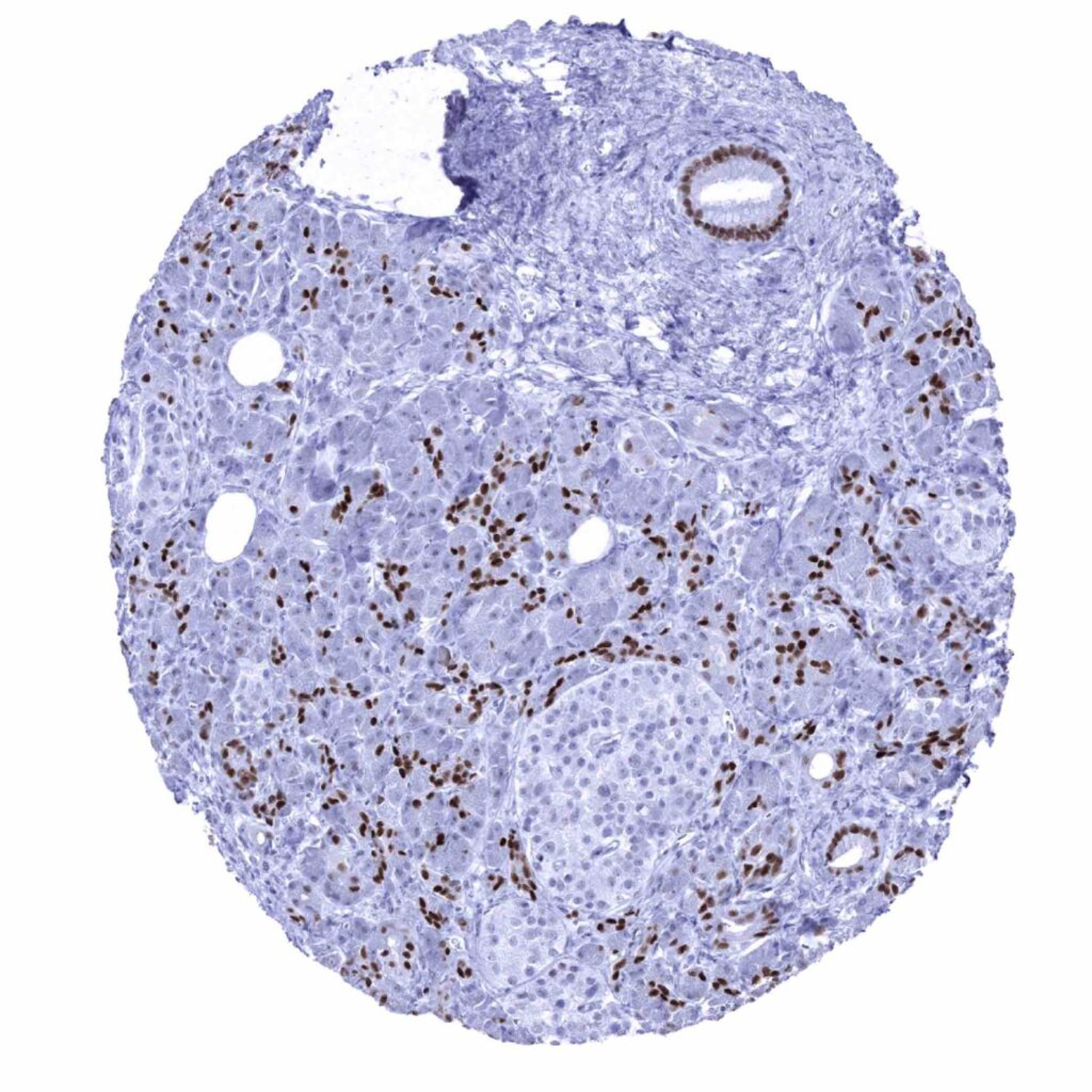
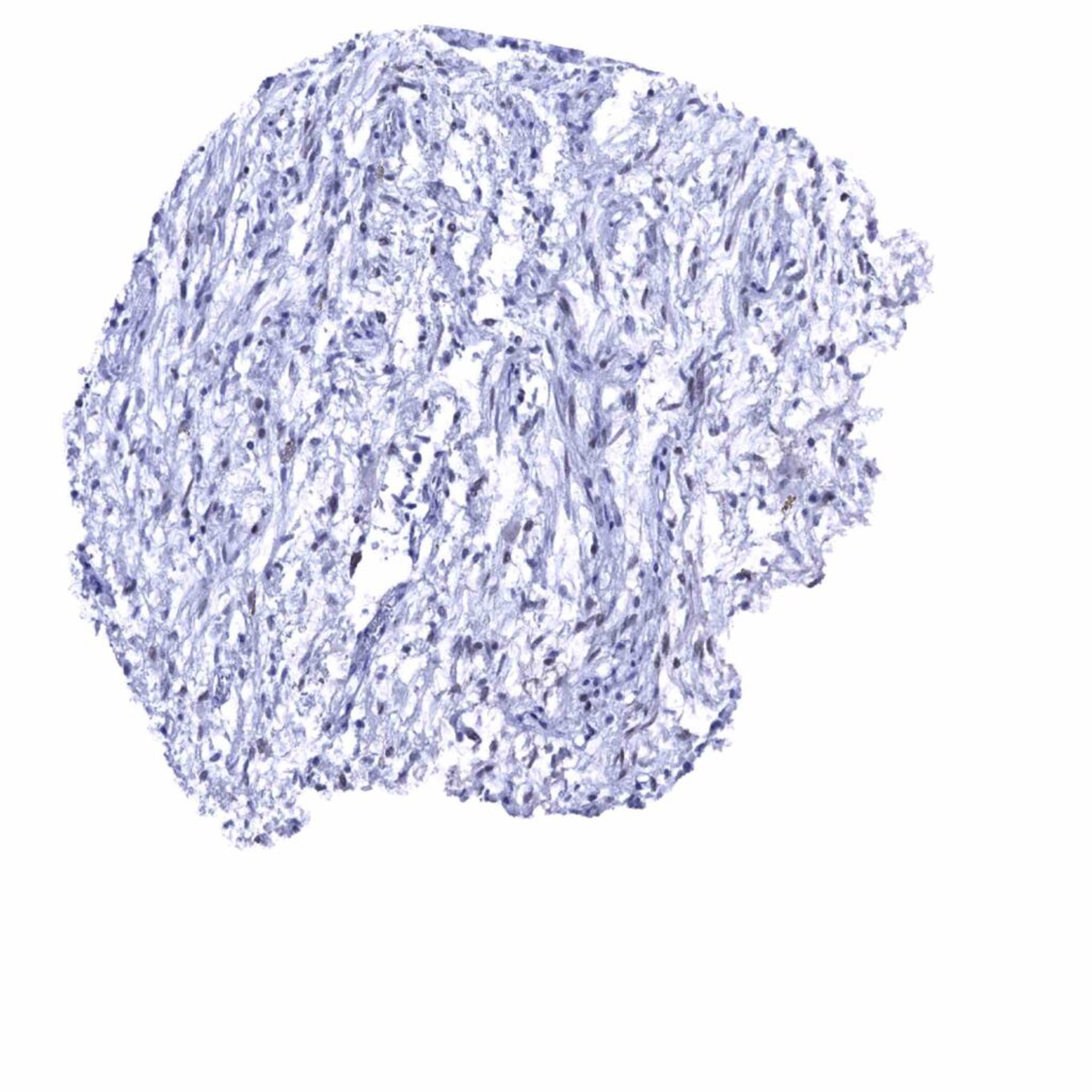
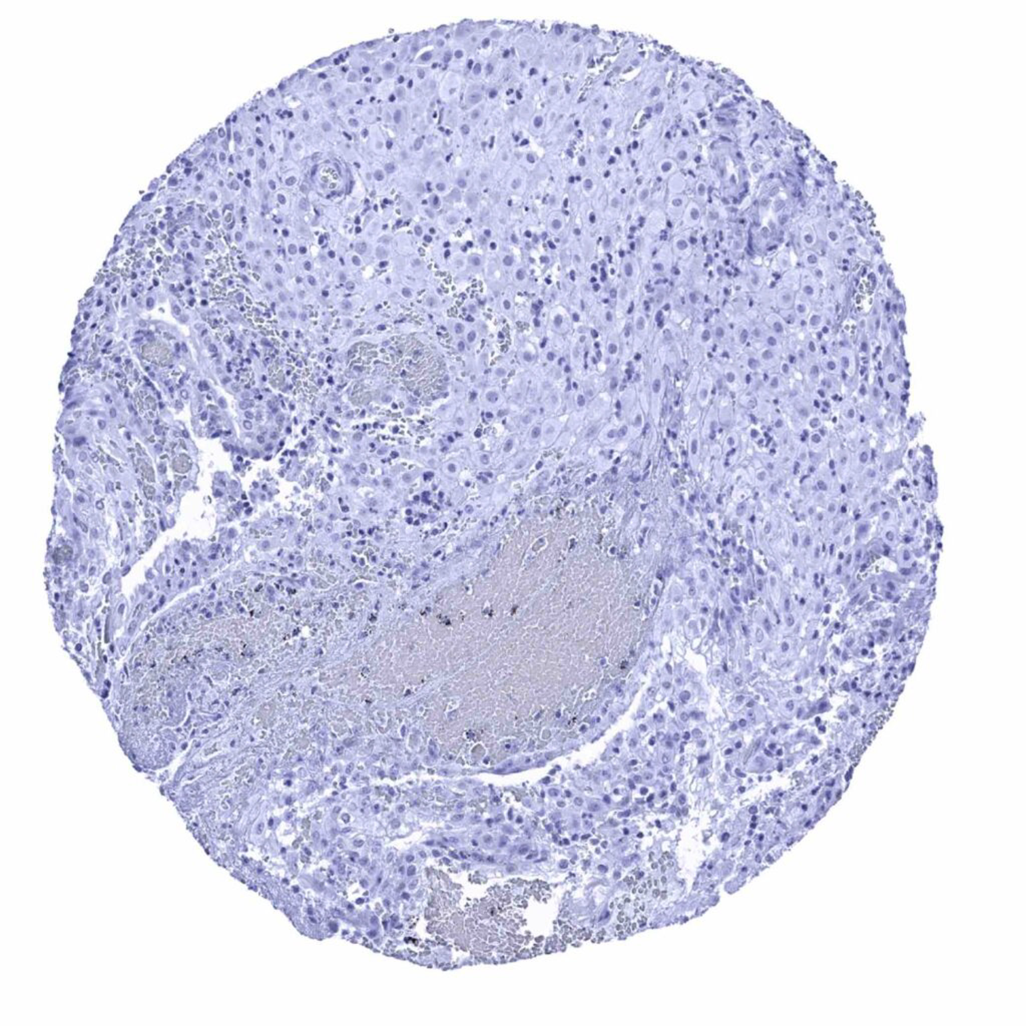
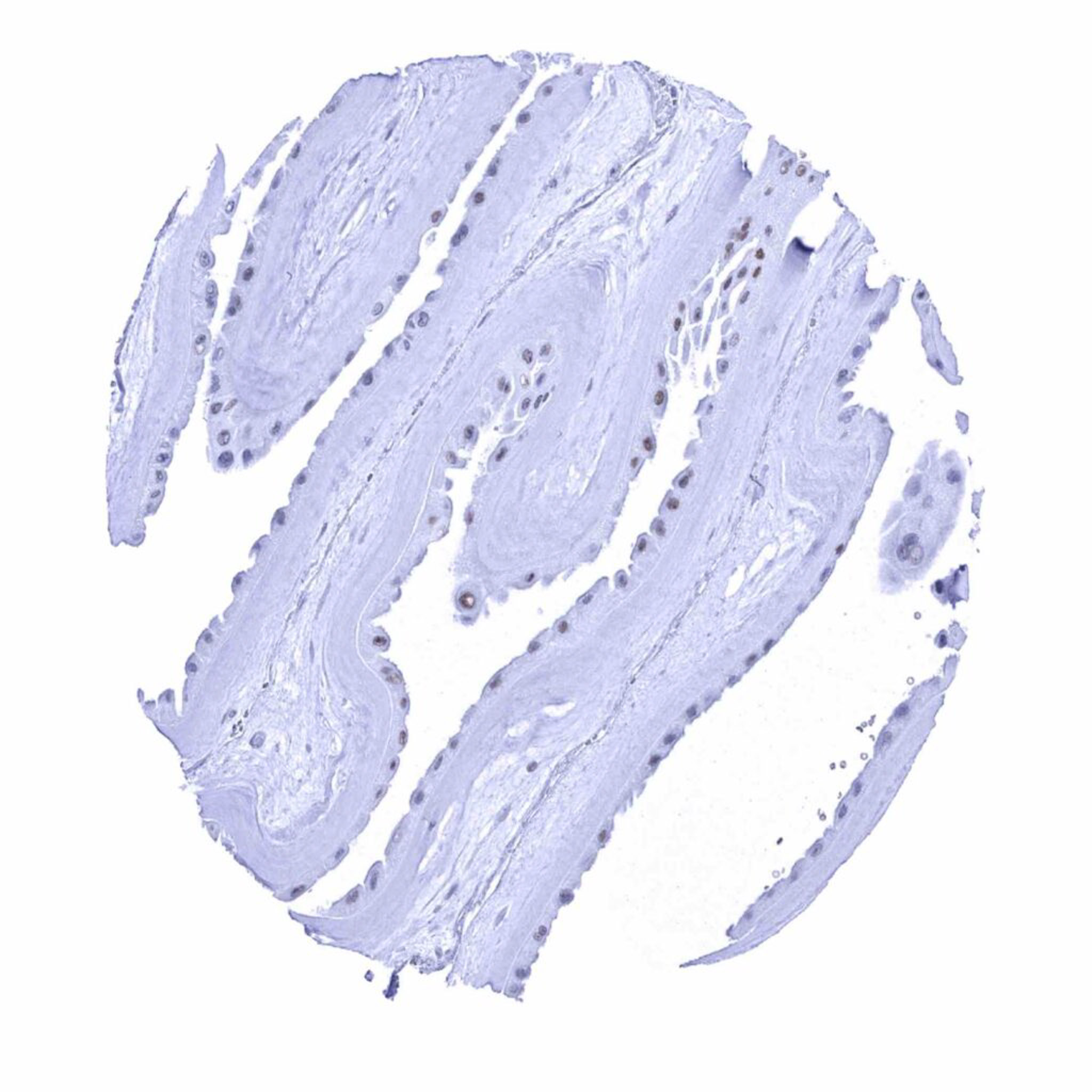
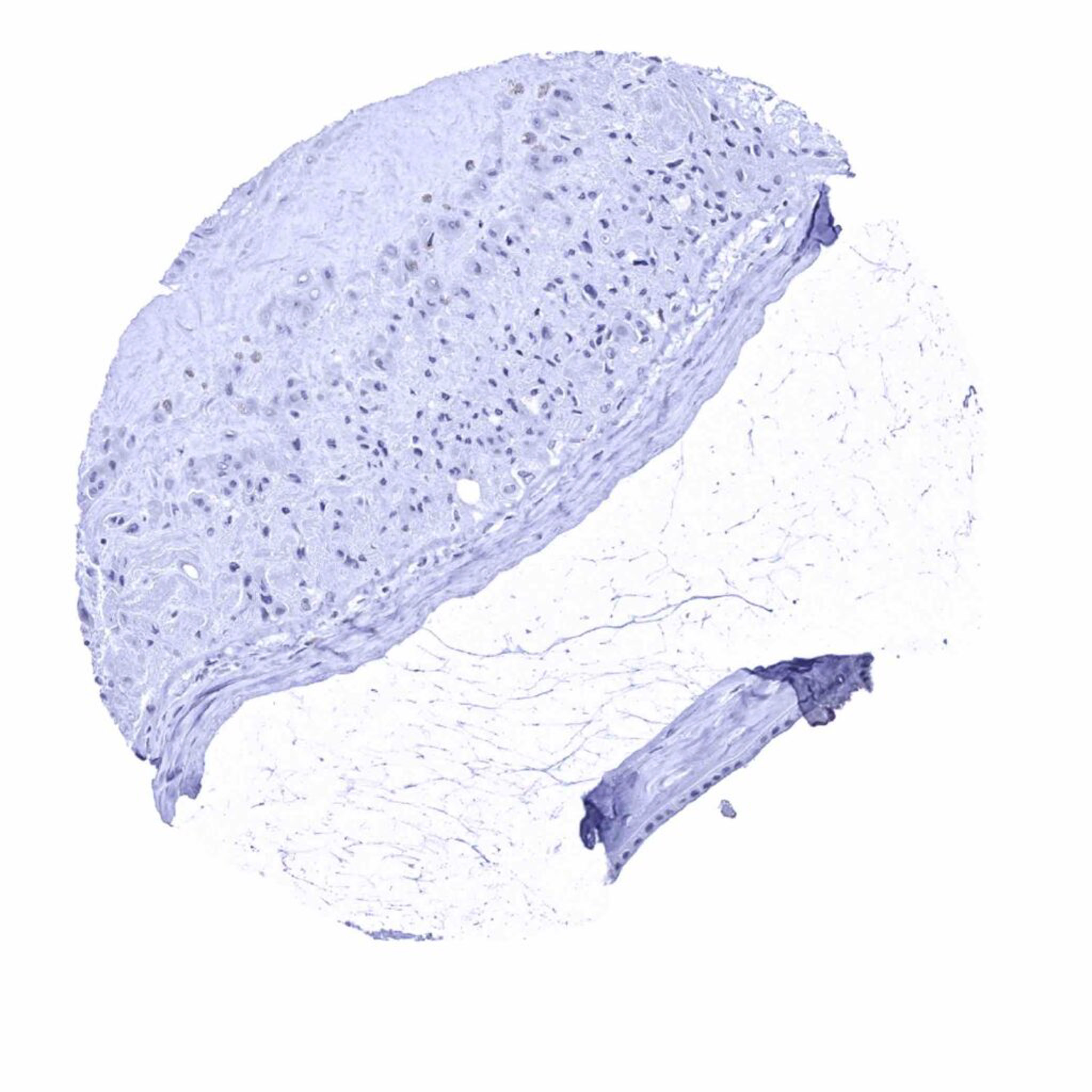
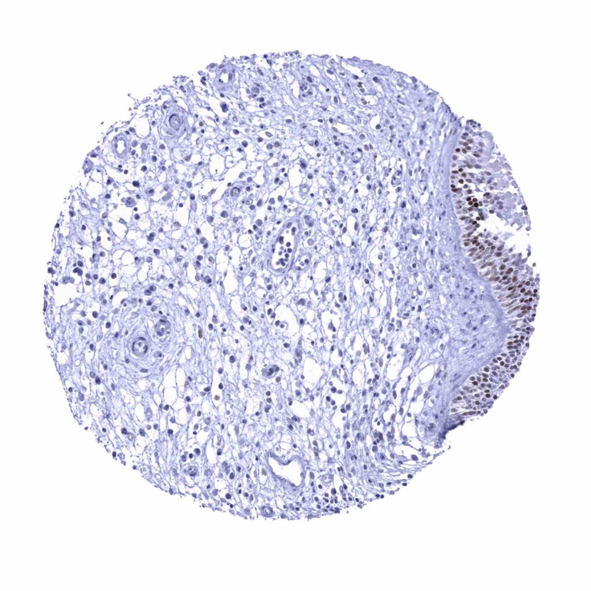
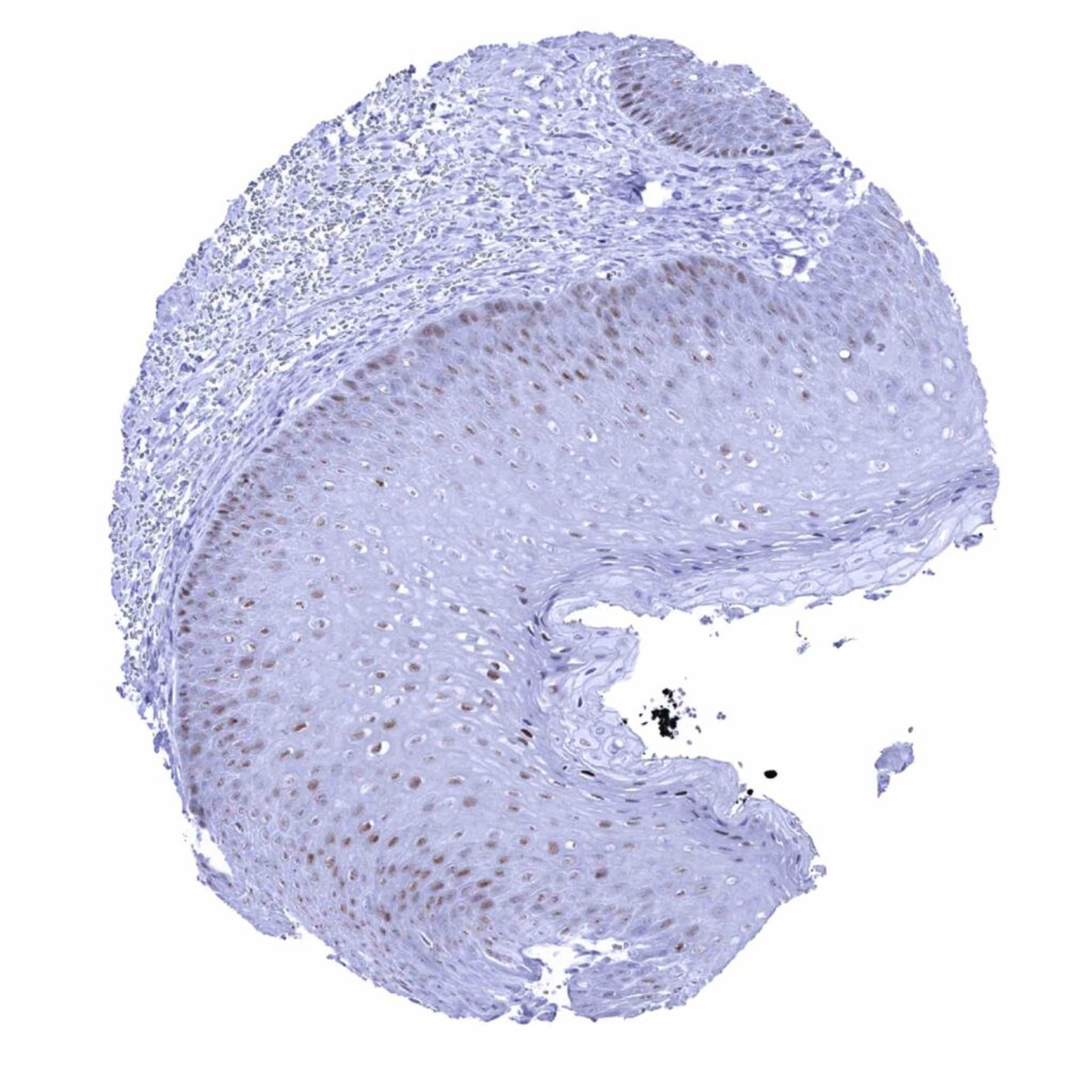
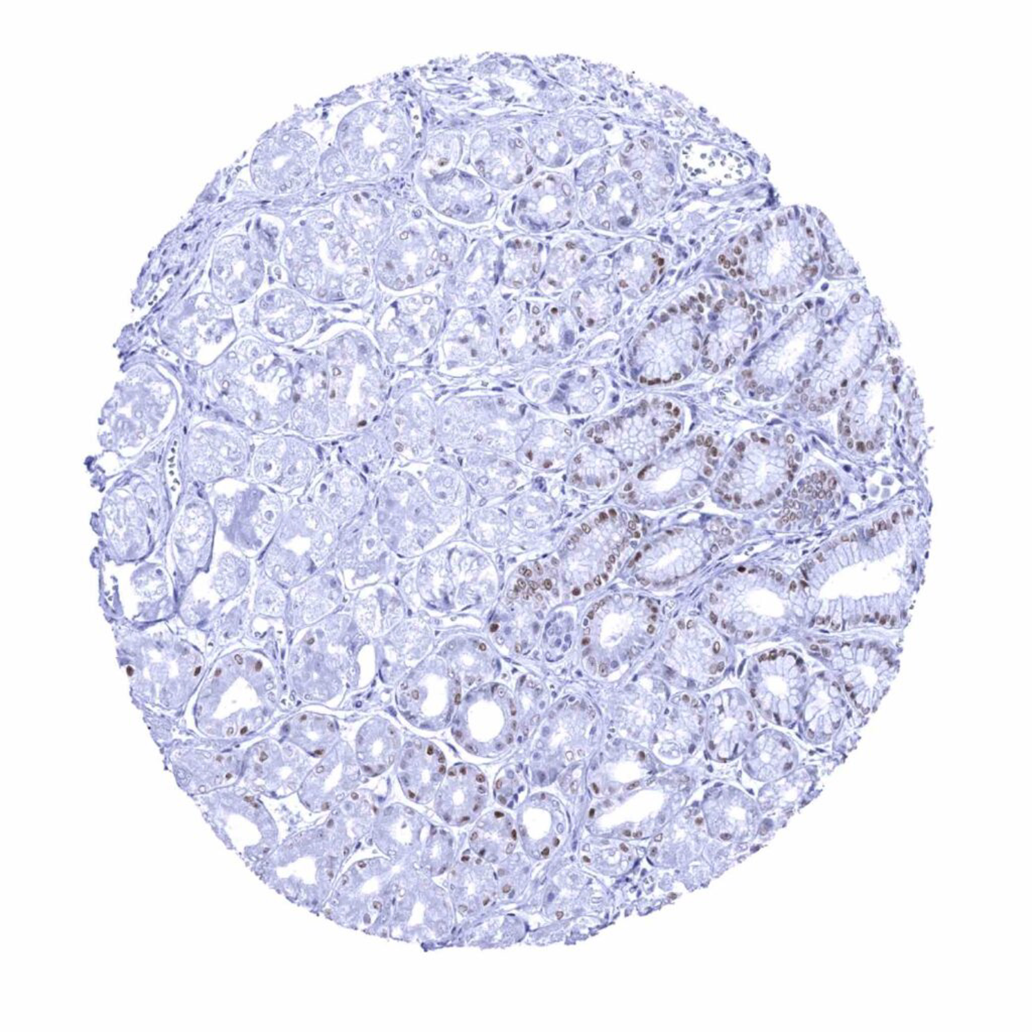
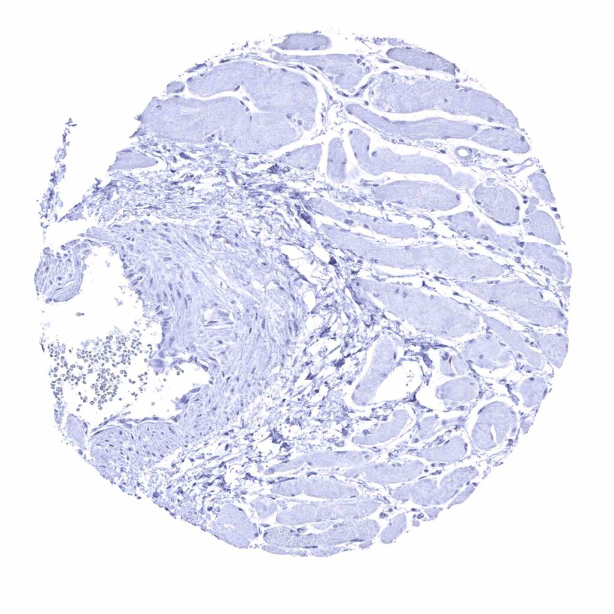
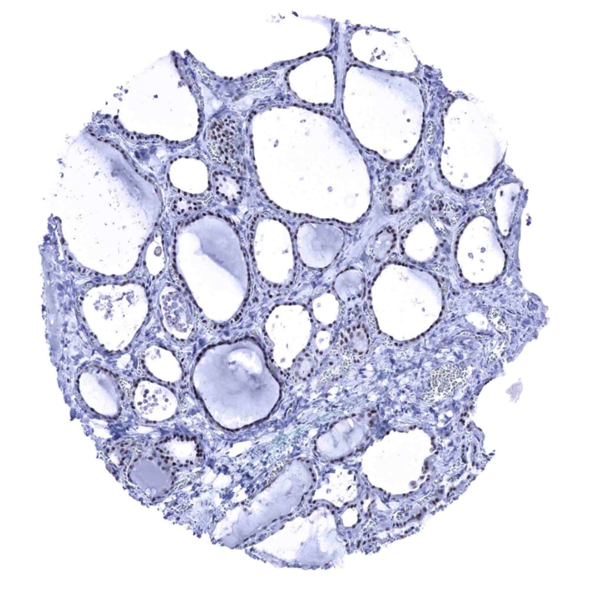
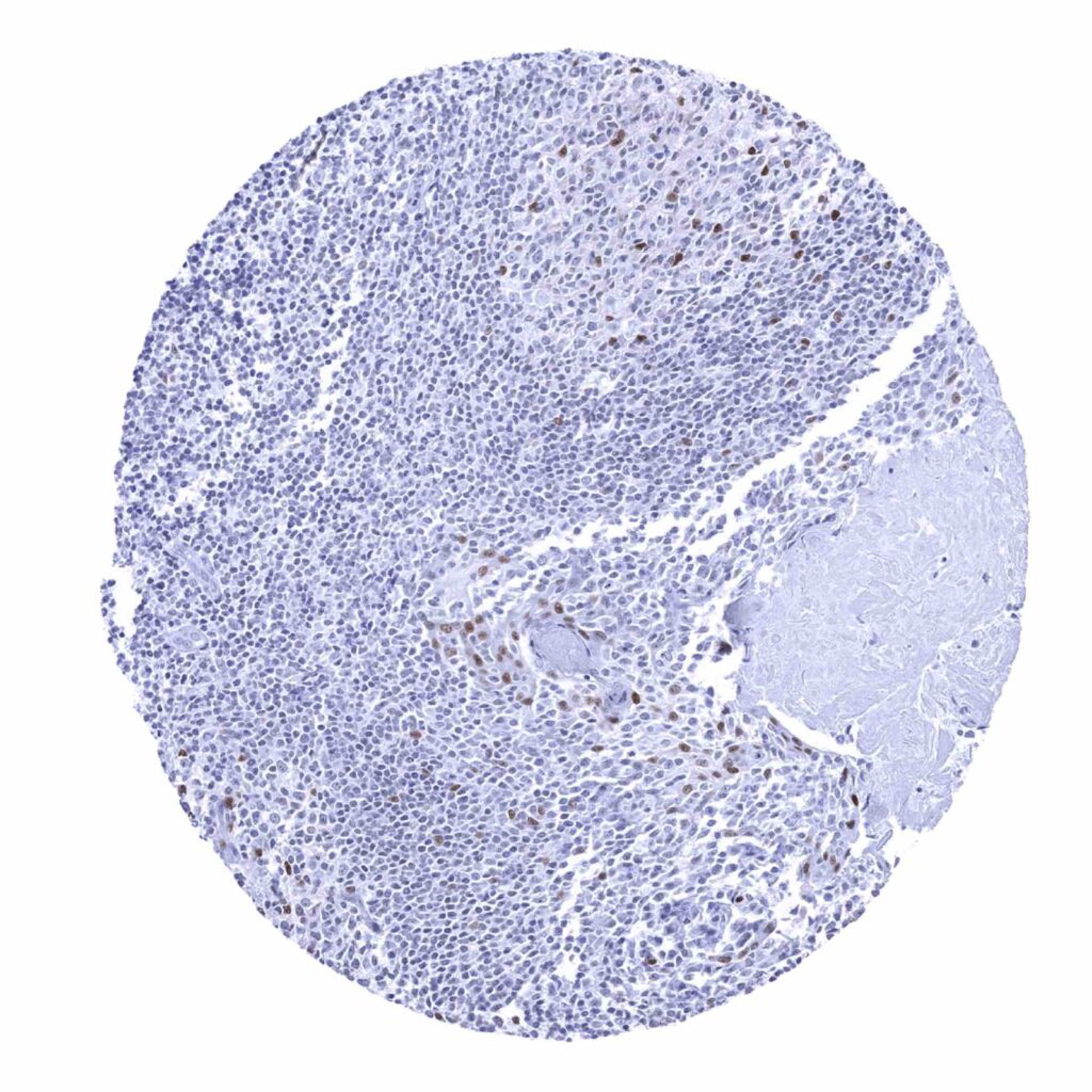
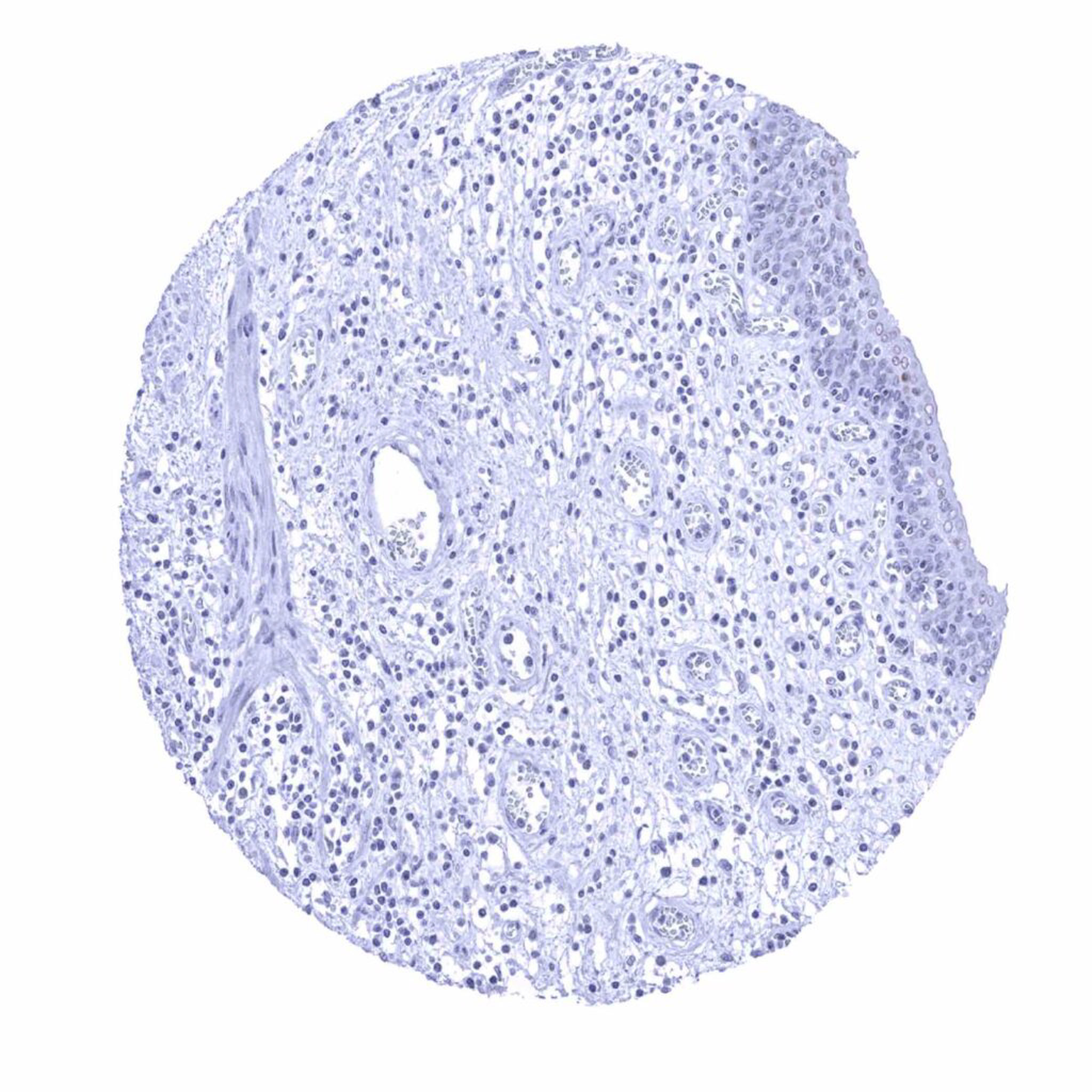
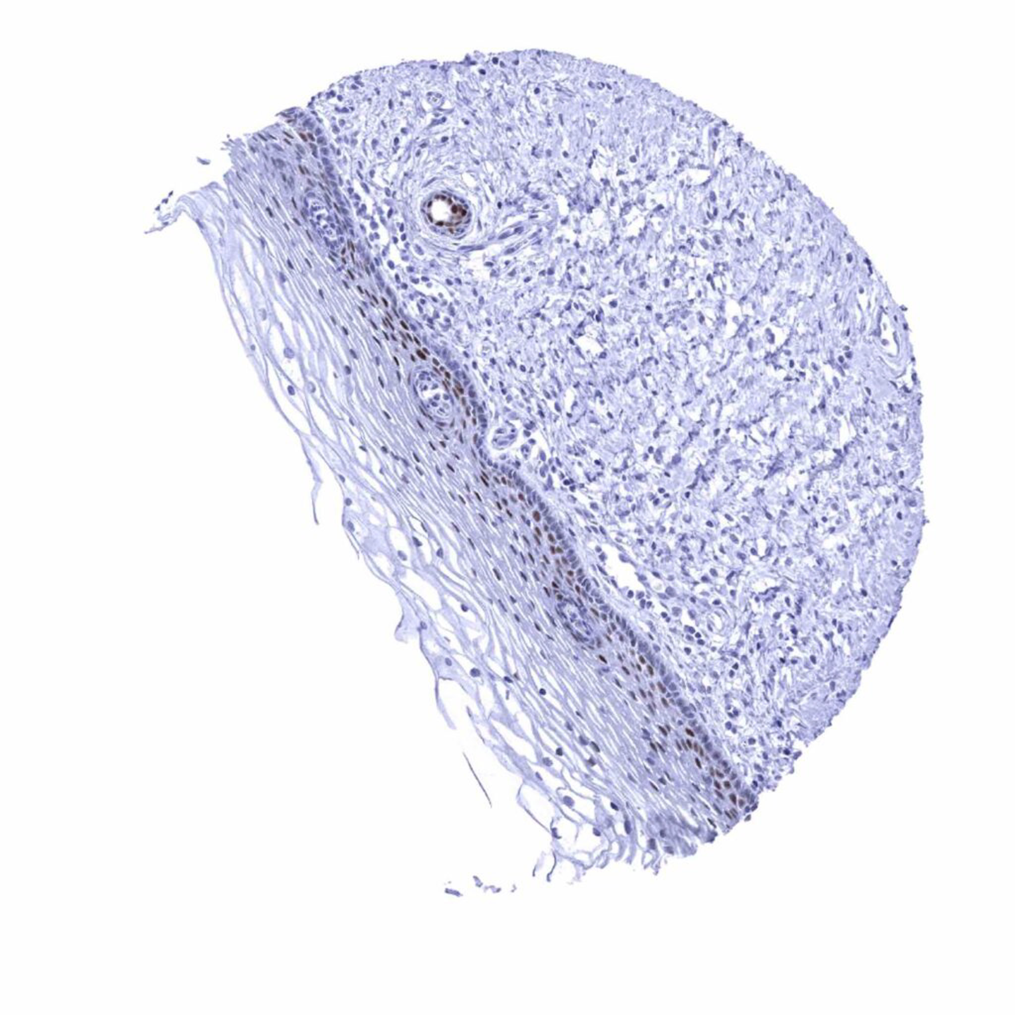
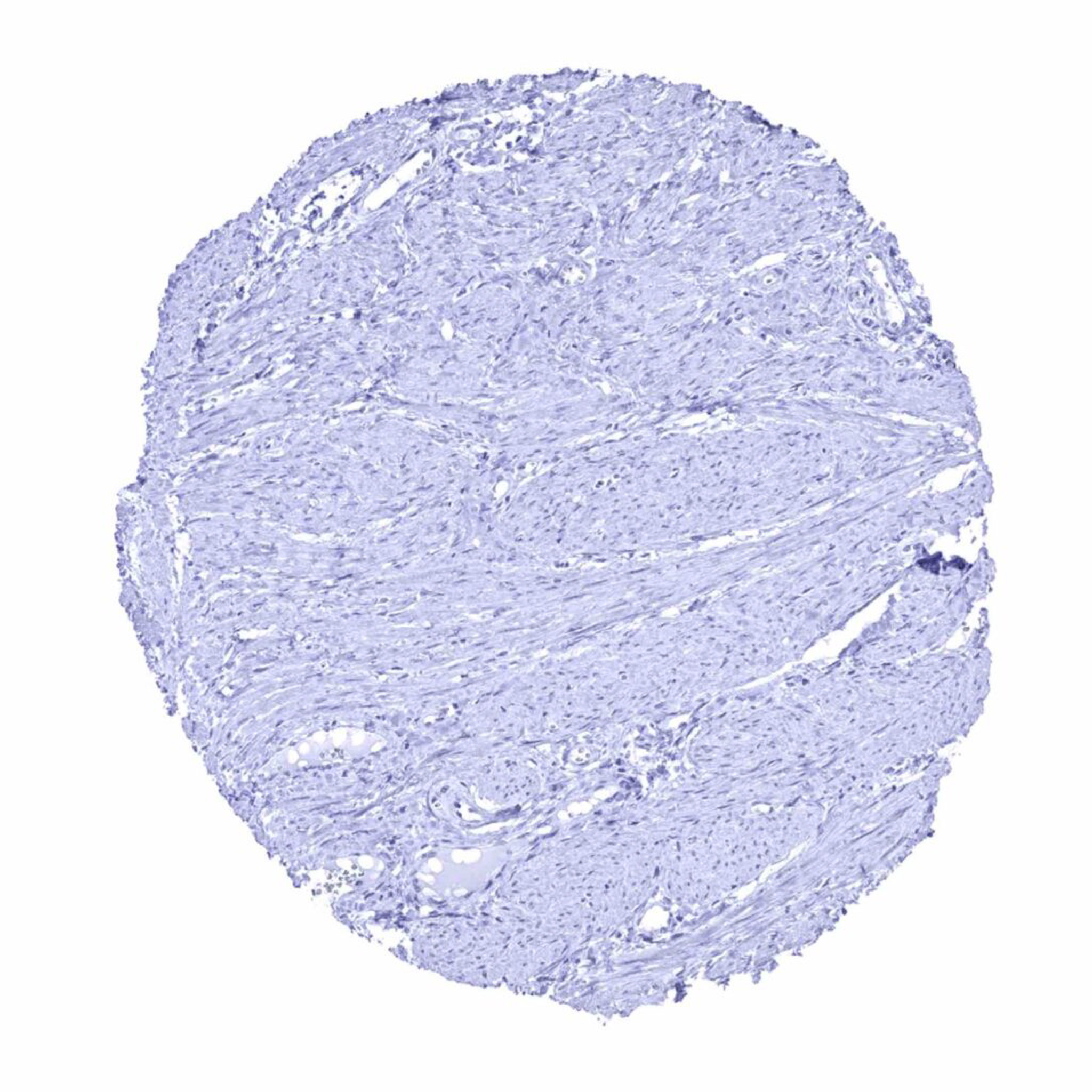
SOX9 antibody [MSVA-709R] HistoMAXTM


Go to Cancer Tissue Gallery Go back to product page Go to HistoMAX page


Adrenal gland – Few interspersed cells in the cortex showing weak to moderate SOX9 positivity
Aorta, media
Appendix, mucosa – Strong nuclear SOX9 staining of crypt epithelial cells, especially at the base. SOX9 staining intensity gradually decreases towards the surface. The surface epithelium remains SOX9 negative (or shows only a very weak staining).
Appendix, muscular wall
Bone marrow
Breast – Epithelial cells are largely SOX9 negative or may show faint staining
Bronchus, mucosa – Moderate to strong SOX9 positivity of most epithelial cells
Cerebellum, cortex (Stratum moleculare)
Cerebrum, grey
Cerebrum, white
Colon descendens, mucosa – Strong nuclear SOX9 staining of crypt epithelial cells, especially at the base. SOX9 staining intensity gradually decreases towards the surface. The surface epithelium remains SOX9 negative (or shows only a very weak staining).
Colon descendens, muscular wall
Duodenum, Brunner gland – Strong SOX9 staining of Brunner gland cells
Duodenum, mucosa – Strong nuclear SOX9 staining of crypt epithelial cells. The SOX9 staining intensity markedly decreases towards the surface epithelium and the tips of the villosities remain SOX9 negative.
Endometrium, secretion – Strong SOX9 positivity of epithelial cells. Stroma cells are SOX9 negative
Epididymis – Weak to moderate nuclear SOX9 staining of basal cells in the caput
Esophagus, squamous epithelium – Weak to moderate nuclear SOX9 staining of the bottom 2-3 of squamous epithelium
Fallopian tube, mucosa – Moderate to strong SOX9 staining of a large subset of epithelial cells
Fat
Gallbladder, epithelium
Heart
Ileum, mucosa – Strong nuclear SOX9 staining of crypt epithelial cells, especially at the base. SOX9 staining intensity gradually decreases towards the surface. The surface epithelium remains SOX9 negative (or shows only a very weak staining).
Kidney, cortex – Few cells of the tubuli may show a weak to moderate SOX9 staining
Kidney, medulla – Moderate to strong SOX9 staining of a subset of collecting duct cells
Liver – Moderate to strong nuclear SOX9 staining of intrahepatic bile ducts
Lung
Lymph node – Weak to moderate nuclear SOX9 positivity of a fraction of cells in germinal centres
Ovary, stroma
Pancreas – Strong nuclear SOX9 staining of intercalated and excretory ducts, while acinar cells are less often positive and islet cells are SOX9 negative
Parathyroid
Parotid gland – Moderate to strong nuclear SOX9 staining of all epithelial cell types
Pituitary, anterior lobe – Moderate to strong SOX9 staining of a small subset of epithelial cells in the adenohypophysis
Pituitary, anterior lobe – Weak to moderate SOX9 positivity of fibrillar cells in the neurohypophysis
Placenta early, decidua
Placenta, early
Placenta, mature
Placenta, mature, amnion and chorion
Prostate – Strong SOX9 positivity of basal cells while luminal cells show only a markedly weaker staining
Rectum, mucosa – Strong nuclear SOX9 staining of crypt epithelial cells, especially at the base. SOX9 staining intensity gradually decreases towards the surface. The surface epithelium remains SOX9 negative (or shows only a very weak staining).
Seminal vesicle – Moderate to strong SOX9 positivity of epithelial cells
Sinus paranasales – Moderate to strong SOX9 positivity of most epithelial cells
Skin – Weak to moderate nuclear SOX9 staining of the bottom 2-3 of squamous epithelium
Skin (sebaceous and ekkrine glands) – Strong SOX9 staining of ekkrine glands. Weak to moderate nuclear SOX9 staining of sebaceous glands
Spleen
Stomach, antrum – Moderate to strong nuclear SOX9 staining of surface epithelial cells, while the glandular cells are SOX9 negative
Stomach, corpus – Moderate to strong nuclear SOX9 staining of surface epithelial cells, while the glandular cells only show weak positivity
Striated muscle
Sublingual gland – Moderate to strong nuclear SOX9 staining of all epithelial cell types
Testis – Strong SOX9 staining of Sertoli cells
Thymus
Thyroid gland – Moderate to strong SOX9 positivity of follicular cells
Tonsil – Weak to moderate nuclear SOX9 positivity of a fraction of cells in germinal centres and in a fraction of krypt epithelial cells
Tonsil, surface epithelium – Faint nuclear SOX9 staining of some epithelial cells, mainly in the bottom 2-3 of the squamous epithelium
Urinary bladder, muscular wall
Urinary bladder, urothelium – A weak SOX9 staining can occur in the superficial cell layers (umbrella cells) of the urothelium
Uterus, ectocervix – Weak to moderate nuclear SOX9 staining of the bottom 2-3 of squamous epithelium
Uterus, endocervix – Weak nuclear SOX9 staining of epithelial cells
Uterus, endometrium (proliferation) – Moderate SOX9 positivity of epithelial cells. Stroma cells are SOX9 negative
Uterus, myometrium
     

| TOP |